effect of camp/pka signaling mechanism on barrier...
TRANSCRIPT

MU
HA
MM
AD
A
SLA
M R
OLE O
F M
YO
SIN
LIG
HT C
HA
IN
P
HO
SP
HA
TA
SE
VVBédition scientifique
VVB LAUFERSWEILER VERLAG
MUHAMMAD ASLAM
VVB LAUFERSWEILER VERLAGédition scientifique
9 7 8 3 8 3 5 9 5 2 2 0 1
ISBN 3-8359-5220-XVVB LAUFERSWEILER VERLAGST AU FEN BER G R I N G 1 5D - 3 5 3 9 6 G I E S S E N
Tel: 0641-5599888 Fax: -5599890redak t ion@dok to rve r lag .dew w w . d o k t o r v e r l a g . d e
EFFECT OF CAMP/PKA SIGNALING MECHANISM
ON BARRIER FUNCTION OF CULTURED
ENDOTHELIAL CELLS:
INAUGURAL-DISSERTATIONzur Erlangung des Doktorgrades der Naturwissenschaften (Dr. rer. nat.)
des Fachbereichs Biologie und Chemieder Justus-Liebig-Universität Giessen
ROLE OF MYOSIN LIGHT CHAIN PHOSPHATASE
Human umbilical vein endothelial cells stained for actin (red) and ß-catenin (green)

Das Werk ist in allen seinen Teilen urheberrechtlich geschützt.
Jede Verwertung ist ohne schriftliche Zustimmung des Autors oder des Verlages unzulässig. Das gilt insbesondere für Vervielfältigungen, Übersetzungen, Mikroverfilmungen
und die Einspeicherung in und Verarbeitung durch elektronische Systeme.
1. Auflage 2007
All rights reserved. No part of this publication may be reproduced, stored in a retrieval system, or transmitted,
in any form or by any means, electronic, mechanical, photocopying, recording, or otherwise, without the prior
written permission of the Author or the Publishers.
st1 Edition 2007
© 2007 by VVB LAUFERSWEILER VERLAG, GiessenPrinted in Germany
VVB LAUFERSWEILER VERLAGédition scientifique
STAUFENBERGRING 15, D-35396 GIESSENTel: 0641-5599888 Fax: 0641-5599890
email: [email protected]
www.doktorverlag.de

Aus dem Physiologischen Institut am Fachbereich Medizin und
Institut für Tierphysiologie am Fachbereich Biologie und Chemie der Justus-Liebig-Universität
Effect of cAMP/PKA signaling mechanism on
barrier function of cultured endothelial cells:
Role of myosin light chain phosphatase
INAUGURAL-DISSERTATION
zur Erlangung des Doktorgrades der Naturwissenschaften (Dr. rer. nat.)
des Fachbereichs Biologie und Chemie der Justus-Liebig-Universität Giessen
vorgelegt von
Muhammad Aslam
(Pharmacist, M.Phil. Molecular Biology) aus Muridke, Pakistan
Gießen 2007

ii
Tag der Disputation:
Friday, September 28, 2007. 14:00 hrs.
Gutachter: Prof. Dr. W. Clauß (Fachbereich Biologie und Chemie)
Gutachter: Priv. Doz. Dr. T. Noll (Fachbereich Humanmedizin)

iii
In the Name of Allah (God),
the Most Gracious, the Most Merciful
Proclaim! (or read!) in the name of thy Lord and Cherisher, Who
created- Created man, out of a (mere) clot of congealed blood:
Proclaim! And thy Lord is Most Bountiful,- He Who taught (the use
of) the pen,- Taught man that which he knew not.
Man We did create from a quintessence (of clay); Then We placed
him as (a drop of) sperm in a place of rest, firmly fixed; Then We
made the sperm into a clot of congealed blood; then of that clot We
made a (foetus) lump; then we made out of that lump bones and
clothed the bones with flesh; then we developed out of it another
creature. So blessed be Allah, the best to create! After that, at length
ye will die. Again, on the Day of Judgment, will ye be raised up.
(Al-Quran)

iv
To
My Mother
v
List of Abbreviations 1
1 Introduction 4
1.1 Barrier function of the vascular endothelium 4
1.2 Endothelial actomyosin cytoskeleton 6
1.3 Endothelial contractile machinery 7
1.3.1 Myosin light chains 7
1.3.2 Mysin light chain kinase 8
1.3.3 Myosin light chain phosphatase 9
1.3.4 Regulation of myosin light chain phosphatase activity
via MYPT1 phosphorylation 10
1.3.5 Regulation of myosin light chain phosphatase activity by
CPI-17 11
1.4 cAMP and endothelial permeability 12
1.5 Aims and objectives of the study 14
2 Methods 16
2.1 Cell culture 16
2.2 General incubation conditions 17
2.3 Protein analysis 18
2.4 Determination of myosin light chain phosphorylation 22
2.5 Co-immunoprecipitation of proteins 23
2.6 Protein phosphatase assay 24
2.7 Detection of activated RhoA 25
2.8 Measurement of RhoA translocation 26
2.9 Measurement of endothelial monolayer permeability 26
2.10 Downregulation of endogenous CPI-17 27
2.11 Statistical analysis 28
3. Results 29
3.1 Effect of forskolin on endothelial permeability 29
3.2 Effect of forskolin on myosin light chain phosphorylation 30
3.3 Effect of forskolin on MLC phosphatase complex formation
and activity 34
3.4 Effect of forskolin on MYPT 1 phosphorylation 38
vi
3.5 Effect of forskolin on RhoA translocation 40
3.6 Effect of forskolin on CPI-17 phosphorylation and
co-immunoprecipitation with PP1 42
3.7 Effect of CPI-17 downregulation on endothelial barrier 48
4 Discussion 50
4.1 Main findings 50
4.2 MLC phosphatase holoenzyme complex formation and activation 51
4.3 Forskolin induced MLC phosphatase complex formation is
independent of RhoA/Rock inhibition 52
4.4 MYPT1 dephosphorylation 54
4.5 cAMP/PKA signaling inactivates CPI-17 55
4.6 CPI-17 as a mediator of thrombin induced barrier failure 57
4.7 Future perspective 59
5 References 60
6 Summary 76
7 Zusammenfassung 77
8 Erklärung 78
9 Appendix 79
9.1 Chemicals and consumables 79
9.2 Antibodies 82
9.3 Laboratory instruments 83
9.4 Solutions 84
10 Acknowledgment 87
11 Curriculum vitae 88
12 Publications 89
13 Published abstracts 90

1
LIST OF ABBREVIATIONS
AC Adenylyl cyclase
app. Approximately
APS Ammonium per sulfate
ATP Adenosine-5-triphosphate
bFGF Basic fibroblast growth factor
BSA Bovine serum albumin
CaCl2 Calcium chloride
CaMKII Ca2+/calmodulin-dependent protein kinase II
cAMP 3'-5'-cyclic adenosine monophosphate
cGMP 3'-5'-cyclic guanosine monophosphate
CPI-17 PKC-potentiated inhibitor 17-kDa protein
DMSO Dimethyl sulfoxide
DTT Dithiothreitol
EC Endothelial cells
ECGS Endothelial cell growth supplement
ECL Enhanced chemiluminescence
EC-MLCK Endothelial cell myosin light chain kinase
EDTA Ethylene diamine tetraacetic acid
EGTA Ethylene glycol-bis(2-aminoethylether)-N,N,N',N'-
tetraacetic acid
F-actin Filamentous actin
FCS Fetal calf serum
FSK Forskolin
GPCR G-protein-coupled receptors
G-actin Globular actin
HBSS Hanks' balanced salt solution
hEGF Human epidermal growth factor
HEPES 4-(2-hydroxyethyl)-1-piperazine ethane sulfonic
acid
HUVEC Human umbilical vein endothelial cells
IU International unit
KCl Potassium chloride

2
KH2PO4 Potassium dihydrogen phosphate
ILK Integrin linked kinase
kDa Kilo dalton
MgCl2 Magnesium chloride
min Minutes
MLC Myosin light chain
MLC~P Monophosphorylated myosin light chain
MLC~PP Diphosphorylated myosin light chain
MLCK Myosin light chain kinase
MLCP Myosin light chain phosphatase
MnCl2 Manganese chloride
MYPT1 Myosin phosphatase targeting subunit 1
NaCl Sodium chloride
NaF Sodium fluoride
Na2HPO4 Di-sodium hydrogen phosphate
NaH2PO4 Sodium dihydrogen phosphate
Na-orthovanadate Sodium orthovanadate
NP-40 Nonidet P-40
PAK p21-activated kinase
PAR1 Protease-activated receptor-1
PBS Phosphate-buffered saline
pH Negative log of H+ concentration
PKA Protein kinase A
PKC Protein kinase C
PKI Protein kinase A inhibitor
PMA Phorbol-12-myristate-13-acetate
PMSF Phenylmethylsulfonyl fluoride
PP1 Protein phosphatase 1
rec. I-2 Recombinant inhibitor 2
Rhotekin-RBD Rho-binding domain of rhotekin
Rock RhoA-dependent kinase
S19 Serine 19
SDS Sodium dodecyl sulfate
SMC Smooth muscle cells

3
SMC-MLCK Smooth muscle cell myosin light chain kinase
Soln. Solution
T18 Threonine 18
T38 Threonine 38
T696 Threonine 696
T850 Threonine 850
TBS Tris-buffered saline
TCA Trichloroacetic acid
Thr Thrombin
TEMED N, N, N’, N’,-tetramethylethylenediamine
Tris Tris(hydroxymethyl)aminomethane
VE-cadherin Vascular endothelial cadherin
% vol/vol Volume by volume percentage
% wt/vol Weight by volume percentage
ZIPK Zipper-interacting protein kinase

4
1 INTRODUCTION
1.1 Barrier function of the vascular endothelium
The vascular endothelium acts as a semi-permeable barrier between the
vascular lumen and the interstitial spaces and extends over a wide surface area. It
controls the passage of ions, solutes, macromolecules and leukocytes across the
vessel wall. It is well known that loss of this barrier function leads to extravasation
of blood components and may finally result in edema formation (Mehta and Malik,
2006; Bazzoni, 2006).
The endothelial barrier function is maintained by an equilibrium of
competing contractile and adhesive forces generated by the actomyosin
cytoskeleton and adhesive molecules located at cell-cell and cell-matrix contacts.
Endothelial cells are tightly connected with each other via interactions of adherens
and tight junctional proteins of adjacent cells. These proteins are linked to the
cortical actin cytoskeleton present directly under the cell membrane (Furuse et al.,
1994; Ben-Ze’ev and Geiger, 1998; Vleminckx and Kemler, 1999). Inflammatory
mediators like thrombin cause activation of the contractile apparatus, derangement
of the actomyosin cytoskeleton, and loss of cell adhesions. This results in barrier
failure, increased macromolecule extravasation, and edema formation in the
inflamed tissue (Lum and Malik, 1996; van Hinsbergh 1997; Wojciak-Stothard et
al., 1998).
The present study focuses on the role of the endothelial contractile
machinery in regulating endothelial barrier function under pathophysiological
inflammatory conditions, during which activation of endothelial cells leads to
edema formation and organ failure. It is well documented that agents which
counteract contraction in smooth muscle cells can also reduce endothelial
permeability (van Hinsbergh and van Nieuw Amerongen, 2002). In accordance,
maneuvers increasing the intracellular levels of cyclic adenosine monophosphate
(cAMP) can counteract imminent barrier failure induced by inflammatory mediators
like thrombin (Qiao et al., 2003). Presently, the molecular mechanisms of this
barrier protection are not completely understood. Thrombin activates the
contractile machinery of endothelial cells mainly via inhibition of the myosin light
chain phosphatase (MLCP) and activation of myosin light chain kinase (MLCK)

5
leading to phosphorylation of the small regulatory myosin light chains (MLC) (Zhao
and Davis, 1999; Gündüz et al., 2003), the key regulatory element of the
contractile machinery. Activation of the cAMP-dependent protein kinase A (PKA)
pathway leads to activation of MLCP in smooth muscle cells (Azam et al., 2007) as
well as in endothelial cells (Bindewald et al., 2004) which leads to
dephosphorylation of MLC. In endothelial cells, dephosphorylation of MLC goes
along with inactivation of the contractile machinery and this causes stabilization of
the endothelial barrier (Tinsley et al., 2004).
Fig. 1.1 Schematic presentation of the mechanism of endothelial barrier failure induced by inflammtory mediators. Mediators like thrombin cause failure of endothelial barrier function mediated by intracellular signal transduction mechanism. Effectors of these mechanisms are: the contractile machinery, cell-cell and cell-matrix adhesion structures and the actin cytoskeleton.
Disintegration of cell
adhesion structures
Barrier failure
Fragmentation of
cytoskeleton
Activation of contractile
machinery
Inflammatory mediators
Signal transduction Signal transduction

6
The main objective of the present study was to elucidate the mechanism of
cAMP/PKA-mediated activation of MLCP as an important molecular target for
endothelial barrier protection. The study was performed with a well-established in
vitro cell culture model, using monolayers of human umbilical vein endothelial cells
(HUVEC). Thrombin was used as an inflammatory mediator to simulate the in vivo
state of hyperpermeability while forskolin (FSK), a direct activator of adenylyl
cyclase, was used to activate the cAMP/PKA pathway.
1.2 Endothelial actomyosin cytoskeleton
Like other eukaryotic cells, endothelial cells possess a functional
cytoskeleton consisting of actin and myosin filaments. The first direct evidence of
endothelial cell contraction, in response to permeability-increasing agonists, was
given by Majno and co-workers (1961a; 1961b). Using electron microscopy, they
demonstrated endothelial cell contraction and gap formation in intact capillaries
exposed to inflammatory mediators. A primary function of the contractile apparatus
in endothelial cells is to regulate endothelial barrier integrity. Endothelial cell-cell
adhesion and thus barrier integrity is mainly dependent on the actin cytoskeleton.
Disruption of actin cytoskeleton by C2 toxin and cytochalasin D led to loss of
endothelial cell-cell and cell-matrix contacts and detachment of cells from the
substratum (Schnittler et al., 2001), whereas phallacidin, an actin stabilizer,
prevented agonist-mediated barrier dysfunction (Phillips et al., 1989).
In endothelial cells actin and myosin filaments represent ~16% of total
cellular protein (Wong and Gotlieb, 1990). Actin comprises about 5% of the total
protein and exists in two different forms: in a filamentous form, called F-actin, and
in a monomeric form, called G-actin (Tobacman and Korn, 1983). Actin filaments
are dynamic structures, and the shift between the monomeric and the polymeric
form of this protein plays a central role in several cell functions, especially in cell
contraction and migration. In endothelial cells, about half of the actin is present in
F-actin form, and half in G-actin form. Stress fibers are composed of bundles of F-
actin and myosin filaments and are the primary elements of the contractile
machinery of endothelial cells (Dudek and Garcia, 2001). Inhibition of actin
polymerization by cytochalasin D or latrunculin leads to actin depolymerisation and
abrogates the contractile response to inflammatory mediators (Goeckeler and

7
Wysolmerski, 1995; Moy et al., 1996, Mehta et al., 2002). Stimulation of
endothelial cells with thrombin increases polymerization of actin filaments, as
determined by the conversion of G-actin to F-actin (Thurston and Turner, 1994;
Ehringer et al., 1999) reduces cortical actin content and leads to reorganization of
actin to form stress fibers (Goeckeler and Wysolmerski, 1995; Ehringer 1999, van
Nieuw Amerongen et al., 2000a; 2000b). This increase in stress fiber formation
leads to a change in endothelial cell shape (Vouret-Craviari et al., 1998), which is
an important factor in increased endothelial gap formation.These results provide a
concept that actin polymerization plays a key role in induction of endothelial
contraction.
1.3 Endothelial contractile machinery
Endothelial contractile machinery consists of actin and myosin filaments, the
activation of which is mainly regulated by the phosphorylation state of the
regulatory MLC. The phosphorylation state of MLC is precisely regulated by
balanced activities of MLCK and MLCP. Thus, the major components of
endothelial contractile machinery are actin-myosin filaments, MLC, MLCK and
MLCP.
1.3.1 Myosin light chains
MLC is a 20 kDa small protein and an important determinant of the state of
contractile activation in endothelial cells. It was shown that phosphorylation of
serine 19 (S19) (monophosphorylation) and/or threonine 18 (T18)
(diphosphorylation) of the regulatory MLC not only increases actomyosin ATPase
activity, but also shifts the equilibrium from the folded to unfolded myosin forms
(Kamisoyama et al., 1994), thus providing the assembly and function of the
contractile apparatus of the cells. After thrombin treatment, MLC phosphorylation
is accompanied by an increase in the F-actin and decrease in G-actin contents in
endothelial cells (Goeckler and Wysolmerski, 1995). Phosphorylation of MLC has
been shown to be involved in the regulation of permeability of cultured endothelial
cells as well as intact isolated postcapillary venules and pulmonary microvessels in
response to histamine and thrombin (Yuan et al., 1997; Vogel et al., 2000). The

8
relationship between MLC phosphorylation and interendothelial gap formation is
only partly understood. It is clear, however, that activation of the contractile
machinery leads to an increase in paracellular permeability. The maximal increase
in MLC phosphorylation in response to an agonist such as thrombin precedes
tension development and the increase in endothelial barrier permeability
(Goeckeler and Wysolmerski, 1995; Moy et al., 1996; Moy et al., 2002).
1.3.2 Myosin light chain kinase
MLCK is a Ca2+/calmodulin-dependent enzyme that phosphorylates MLC at
S19 and/or T18 (Goeckeler and Wysolmerski, 1995). In contrast to smooth muscle
cell MLCK (SMC-MLCK), which is 110-130 kDa, endothelial cell MLCK (EC-
MLCK) is a 210-kDa protein (Dudek and Garcia, 2001). Structurally, EC-MLCK
contains all the domains present in the smooth muscle form, but in addition, has a
unique 922 amino acid NH2-terminal domain containing consensus sites that may
be phosphorylated by diverse protein kinases, including PKA (de Lanerolle et al.,
1984; Garcia et al., 1997), PKC (Bogatcheva et al., 2003), p21-activated kinase
(PAK) (Goeckeler et al., 2000), Src (Birukov et al., 2001), and Ca2+/calmodulin-
dependent protein kinase II (CaMKII) (Verin et al., 1998). It is well established that
activation of EC-MLCK leads to endothelial cell contraction and barrier dysfunction
in response to inflammatory mediators like thrombin and histamine (Dudek and
Garcia, 2001), while inhibition of EC-MLCK, using pharmacological inhibitors like
KT5926 or ML-7, abrogates the increase in vascular permeability (Yuan et al.,
1997; Tinsley et al., 2000). Recently, it has been shown that expression of a
constitutive active form of EC-MLCK resulted in an elevated basal permeability of
venules as well as venular endothelial cells in culture (Tinsley et al., 2000). The
selective knockout of EC-MLCK in a mouse model resulted in protection against
barrier failure induced by lipopolysaccharides (Wainwright et al., 2003) and burn
injury (Reynoso et al., 2007). These studies indicate that EC-MLCK plays a critical
role in agonist-induced endothelial barrier failure via activation of the contractile
machinery.

9
1.3.3 Myosin light chain phosphatase
MLCP is a holoenzyme consisting of a catalytic subunit (PP1), a regulatory
subunit (MYPT1), which targets PP1 to myosin and a smaller subunit of 20 kDa
(M20) of unknown function. In endothelial cells the primary function of MLCP is to
dephosphorylate MLC at S19 and T18.
PP1 catalytic subunit. The PP1 catalytic subunit in mammalian cells is encoded
by three genes, α, γ, and δ (also called β). Alternative splicing generates α1, α2, γ1,
and γ2 variants (Sasaki et al., 1990; Durfee et al., 1993). The resulting five
isoforms have high sequence homology and are expressed in many different cell
types. They share over 95% identity in the core catalytic domain. With the
exception of α2, which has an N-terminal insert, the other isoforms differ mostly in
their C-terminal sequence, sharing less than 50% identity. Substrate specificity is
modulated by association with a large number of regulatory and/or inhibitory
subunits. In endothelial cells it is well established that PP1δ is the predominant
isoform of PP1 existing in the MLCP holoenzyme (Verin et al., 2000; Härtel et al.,
2007).
Myosin phosphatase targeting subunit. The specific activity of PP1 of MLCP to
dephosphorylate the MLC is dependent upon its binding to the regulatory subunit
MYPT1. MYPT1 is a 130 kDa protein and plays a key role in determining the
physical and functional integrity of the trimeric myosin phosphatase (Khatri et al.,
2001). PP1δ binds to the N-terminus and M20 to the C-terminus of MYPT1.
Myosin binds to both C- and N-terminal of MYPT1. The N-terminal of MYPT1
shows ~15 fold increased activity and ~10-fold higher affinity for phosphorylated
MLC than the isolated catalytic subunit (Hartshorne, 1998; Hartshorne et al., 2004;
Ito et al., 2004).

10
Fig. 1.2 Regulation of endothelial cell contractility: Phosphorylation of myosin light chain (MLC~P) is a key step in the regulation of the activation of the contractile machinery. Calcium/calmodulin (Ca2+/CM)-dependent myosin light chain kinase (MLCK) phosphorylates, while myosin light chain phosphatase (MLCP) dephosphorylates MLC. Activation of the contractile machinery leads to endothelial cell contraction and barrier failure, while inactivation to relaxation and barrier stabilization. 1.3.4 Regulation of myosin light chain phosphatase activity via MYPT1
phosphorylation
The activity of MLCP can be regulated either through its regulatory subunit
MYPT1 or through direct inhibition of the catalytic subunit PP1 by low molecular
weight endogenous inhibitors. Stimulation of endothelial and nonendothelial cells
with thrombin leads to activation of RhoA-dependent kinase (Rock), which induces
phosphorylation of MYPT1 and inhibition of MLCP activity (Goeckeler and
Wysolmerski, 2005; Pandey et al., 2006).
Most of our understanding about the regulation of the contractile machinery
by MYPT1 is based on data obtained primarily from smooth muscle cells and little
from endothelial cells. Several studies have well documented that MLCP activity is
regulated by phosphorylation of its targeting subunit (MacDonald et al., 2001;
Wooldridge et al., 2004). The two main inhibitory phosphorylation sites identified in
smooth muscle cells as well as in endothelial cells are threonine 696 (T696) and
threonine 850 (T850). However, the mechanism of MLCP inhibition remains
unclear. Several kinases have been reported to phosphorylate MYPT1 at one or
MLC MLC~P Contraction Barrier failure
Relaxation Barrier stabilization
Ca2+/CMMLCK
MLCP
MLC MLC~P Contraction Barrier failure
Relaxation Barrier stabilization
Ca2+/CMMLCK
MLCP

11
both of these sites. The major well-known kinase is Rock, which phosphorylates
MYPT1 at both sites and inhibits its catalytic activity (Hartshorne, 1998; Fukata et
al., 2001). Other kinases phosphorylating MYPT1 in a Ca2+-independent manner
include zipper-interacting protein kinase (ZIPK) and the so called MYPT1-
associated kinase, which is highly homologous to ZIPK ( Hartshorne, 1998;
Borman et al., 2002). Phosphorylation at T696 by Rock or ZIPK inhibits the activity
of MLCP (Fukata et al., 2001; MacDonald et al., 2001; Borman et al., 2002).
Accordingly, phosphorylation of T850 by Rock was shown to induce dissociation of
MYPT1 from myosin and thus inactivation of MLCP (Velasco et al., 2002). These
data confirm that the phosphorylation of MYPT1 plays an important role in the
regulation of MLCP activity.
1.3.5 Regulation of myosin light chain phosphatase activity by CPI-17
Recent data, mainly from smooth muscle cells suggests an alternative
mechanism for the regulation of the activity of MLCP holoenzyme. A small 17 kDa
protein, the PKC-potentiated inhibitor 17-kDa protein (CPI-17), can directly interact
with, and inactivate the catalytic subunit of MLCP (Murthy et al., 2003; Somlyo and
Somlyo, 2003). CPI-17 is a soluble globular protein and was purified as a myosin
phosphatase specific inhibitor from pig aorta (Eto et al., 1995). Phosphorylation of
CPI-17 at threonine 38 (T38) was shown to enhance its inhibitory potency by more
than 1000-fold (Eto et al., 2004). Several kinases such as PKCα/δ, PAK, ZIPK,
integrin linked kinase (ILK), and Rock were shown to activate CPI-17 by
phosphorylation at T38 (Koyama et al., 2000; MacDonald et al., 2001; Erdödi et
al., 2003). Initially, CPI-17 was assumed to be present only in smooth muscle
cells, but recently it was found in platelets (Watanabe et al., 2001), brain (Dubois
et al., 2003) and also in endothelial cells of different origins (Kolosova et al., 2004).
The phosphatase activity of purified MLCP towards phosphorylated MLC was
completely inhibited in the presence of phosphorylated CPI-17 (Senba et al.,
1999). Agonists like histamine and thrombin, cause phosphorylation of CPI-17 in
smooth muscle cells (Kitazawa et al., 2003), as well as in platelets (Watanabe et
al., 2001) and endothelial cells (Kolosova et al., 2004).

12
1.4 cAMP and endothelial permeability
cAMP is a well-known intracellular second messenger mediating
stabilization of barrier function in isolated vessels as well as in cultured endothelial
monolayers (Adamson et al., 1998; Stelzner et al., 1989; He et al., 2000).
Consistently, activation of adenylyl cyclase via G-protein-coupled receptor (GPCR)
agonists like, adenosine, prostacyclin, prostaglandin E2, and β-adrenergic
agonists, direct activation of adenylyl cyclase (by FSK) as well as elevation of
cytosolic cAMP concentration by blocking its degradation via phosphodiesterases,
reduce endothelial hyperpermeability induced by inflammatory stimuli both in vitro
and in in vivo (Carson et al., 1989; Langeler and van Hinsbergh, 1991; Suttorp et
al., 1993; He and Curry, 1993; Fu et al., 2006). The effect of cAMP is fast and
occurs in endothelial cells both under basal conditions as well as after exposure to
inflammatory mediators. Its efficacy is independent on whether cAMP is elevated
by activation of adenylyl cyclase or by inhibition of cAMP-degrading
phosphodiesterases (Van Hinsbergh, 2001).
cAMP exerts its effect primarily through direct activation of PKA (Yuan,
2002). It has been shown in endothelial cells that inhibition of PKA by
overexpression of a specific peptide inhibitor of PKA (PKI) as well as by
pharmacological inhibitors (e.g. Rp-cAMP), resulted in increased endothelial
permeability (Lum et al., 1999; Liu et al., 2001). The barrier protective effects of
cAMP towards inflammatory mediators were abolished in these cells. In an in situ
study in intact frog and rat microvessels it was shown that inhibition of PKA leads
to increased basal permeability (He et al., 2000). Liu and co-workers (2005)
showed in cultured endothelial cells that inhibition of PKA leads to an increase in
stress fiber formation and basal endothelial permeability. These studies
demonstrate clearly that cAMP exerts its effects on endothelial barrier function
mainly by activating PKA.
Recently, it has been shown that cAMP can also strengthen barrier function
by PKA-independent mechanism. cAMP can activate the small GTPase Rap1 via
activation of the exchange factor Epac1/2 (Cullere et al., 2005; Fukuhare et al.,
2005) leading to stabilization of adherens junctions. However, there is no evidence
that activation of Epac causes dephosphorylation of MLC and inactivation of the
contractile machinery.

13
The present study focuses on the molecular mechanism of the cAMP/PKA
pathway which has been related to barrier stabilisation and protection via inhibition
of the contractile machinery. A number of studies have shown that activation of
PKA causes dephosphorylation of MLC (Moy et al., 1993), dissociation of F-actin
from myosin (Langeler et al., 1991) and stabilization of cytoskeleton filaments
(Hastie et al., 1997). Several studies have tried to elucidate the molecular
mechanisms by which cAMP/PKA stabilizes the endothelial cytoskeleton. It has
been proposed that cAMP/PKA inhibits the small GTPase RhoA and this in turn
results in inhibition of phosphorylation of the regulatory MLC and thus activation
state of the contractile machinery (Essler et al., 2000; Qiao et al., 2003). In an
early study, Garcia and co-workers (1995) showed that cAMP/PKA activation
causes MLCK phosphorylation and proposed that this might lead to MLCK
inhibition. In a recent study Goeckeler and Wysolmerski (2005) clearly excluded
that activation of cAMP/PKA pathway can lead to MLCK phosphorylation and
inactivation.
A number of studies in smooth muscle cells and few in endothelial cells
(Birukov, 2003; Goeckeler and Wysolmerski, 2005) have shown that activation of
cAMP/PKA pathway leads to activation of MLCP but a detailed molecular analysis
has not yet been performed.

14
Fig 1.3 Regulation of myosin light chain phosphatase (MLCP). Thrombin, via protease-activated receptor-1 (PAR1), activates RhoA/Rock pathway. Rock inactivates myosin light chain phosphatase (MLCP) leading to increased myosin light chain (MLC) phosphorylation and contractile activation. Agonists like adenosine, via activation of adenosine A2 receptors activate adenylyl cyclase (AC) leading to increased production of cAMP and activation of protein kinase A (PKA). Activation of cAMP/PKA pathway can activate MLCP, which dephosphorylates MLC causing inactivation of the contractile machinery. CPI-17 is an endogenous inhibitor of MLCP, activated by several kinases including PKC and Rock.
1.5 Aims and objectives of the study
The present study was conducted to elucidate the molecular mechanisms
by which activation of the cAMP/PKA pathway leads to inactivation of the
endothelial contractile machinery and protection of endothelial barrier function.
Since previous studies in endothelial as well as smooth muscle cells demonstrated
that cAMP/PKA exerts its main effect on contractile machinery via activation of
MLCP, the present study was focused to analyze the molecular mechanism of
MLCP activation via the cAMP/PKA signaling pathway.
MLC MLC~P
Ca2+/CMMLCK
MLCP
CPI-17
cAMP
PKA
AC
AgonistThrombin
RhoA/Rock
MLC MLC~P
Ca2+/CMMLCK
MLCP
CPI-17
MLC MLC~P
Ca2+/CMMLCK
MLCP
CPI-17
cAMP
PKA
AC
AgonistThrombin
RhoA/Rock
Thrombin
RhoA/Rock

15
The study was performed using an established model of cultured
monolayers of human umbilical vein endothelial cells (HUVEC). The following
questions were addressed:
• Does cAMP/PKA induce complex formation of the MLCP holoenzyme?
Special emphasis was laid on analysis of recruitment of PP1 to MYPT1 and
and the translocation of both to myosin.
• Is this cAMP/PKA-induced MLCP holenzyme complex formation dependent
on inhibition of RhoA/Rock pathway?
• Does cAMP/PKA pathway cause inactivation of CPI-17, a specific inhibitor
of PP1?
• Does the RhoA/Rock pathway play a role in the cAMP/PKA-mediated effect
on CPI-17?
• Does inhibition of CPI-17 have any functional role in endothelial barrier
failure caused by inflammatory mediators (e.g. thrombin)?
The following experimental strategies were used to answer these questions.
• Recruitment of MYPT1 and PP1 to myosin was analyzed by co-
immunoprecipitation analysis.
• Activation of MLCP was analyzed by direct determination of phosphatase
activity in co-immunoprecipitated complexes.
• Activation of RhoA/Rock pathway was analyzed by pulldown assay and
RhoA translocation to membranes by cell fractionation.
• Interaction of CPI-17 with PP1 was analyzed by co-immunoprecipitation
analysis and CPI-17 phosphorylation (i.e. activation) was analyzed by
western blot analysis.
• Macromolecule permeability across endothelial monolayers was used as a
functional assay to evaluate the impact of CPI-17. in these experiments
CPI-17 was downregulated by siRNA technique.

16
2 Methods
2.1 Cell culture
Isolation of human umbilical vein endothelial cells (HUVEC)
Collagenase solution
HBSS x ml
Collagenase II, 293 IU/mg (wt/vol) 0.025 %
MgCl2 0.5 mM
CaCl2 1.5 mM
Endothelial cell culture medium
Endothelial cell basal medium (PromoCell® ) supplemented with
FCS (vol/vol) 10 %
Endothelial cell growth supplement/Heparin (wt/vol) 0.4 %
Hydrocortisone (wt/vol) 0.1 %
bFGF (wt/vol) 1 ng/ml
hEGF (wt/vol) 0.1 ng/ml
Penicillin/streptomycin (vol/vol) 2 %
Procedure. The study conforms with the principles outlined in the ‘’Declaration of
Helsinki’’ (Cardiovascular Research 1997;35:2–3). Human umbilical vein
endothelial cells (HUVEC) were isolated from freshly collected umbilical cords
(from Gynecology Department, University Hospital Giessen) according to Jaffe et
al., (1973) with some modifications. After cleaning, the umbilical vein was
canulated and perfused with HBSS to remove traces of blood. Afterwards, the
lumen of the vein was filled with collagenase solution and incubated for 20-30
minutes at 37 °C. Afterwards, the collagenase solution, containing endothelial
cells, was gently flushed from the vein by perfusion with 30 ml of HBSS containing
3% (vol/vol) FCS, to inactivate the collagenase. The effluent was collected in a 50
ml Falcon tube and centrifuged at 250 x g for 5 minutes at room temperature. The
supernatant was removed and the cell pellet was resuspended in endothelial cell
culture medium containing 0.1% gentamycin. The cell suspension was seeded on

17
3-4 primary cell-culture dishes and incubated at 37 °C with 5% CO2 for 2 hours.
Thereafter, cells were washed with HBSS to remove erythrocytes, non-adherent
cells, and cell debris and were incubated with cell culture medium containing 0.1%
(vol/vol) gentamycin at 37 °C with 5% CO2. After 24 hours the medium was
replaced with normal endothelial cell culture medium.
Sub-culturing of HUVEC. Confluent monolayers of primary endothelial cells were
trypsinized (5-7 days after isolation) in phosphate-buffered saline [PBS;
composition in mM: 137 NaCl, 2.7 KCl, 1.7 KH2PO4, and 10 Na2HPO4; pH 7.4,
supplemented with 0.05% (wt/vol) trypsin, and 0.02% (wt/vol) EDTA] and seeded
at a density of 7 x 10⁴ cells/cm2 on Transwell® filters (for permeability) or on cell
culture dishes (for western blot analysis, immunoprecipitation and pulldown
assay). Experiments were performed with confluent endothelial monolayers of
either primary or passage 1, 3 days after seeding.
2.2 General incubation conditions
The basal medium used in incubations was HBSS supplemented with 1.3
mM CaCl2, 1.2 mM MgCl2, and 2 % (vol/vol) FCS. After an initial equilibration
period of 20 minutes, agents were added as indicated. Stock solutions of thrombin,
Y27632 and PKI were prepared immediately before use with basal medium. Stock
solutions of forskolin were prepared with dimethyl sulfoxide (DMSO). Appropriate
volumes of these solutions were added to the cells yielding final solvent
concentrations < 0.1% (vol/vol). The same final concentration of DMSO was
included in all respective control experiments. Stock solutions of all other
substances were prepared in basal medium (composition as described above).
Appropriate volumes of these solutions were added to the cells. Identical additions
of basal medium were included in all respective control experiments.
In a set of pilot experiments, concentration-response relationships were
determined to find the optimum effective concentration of the agents to be used in
this study. The agents were applied in their optimum effective concentrations as
follows: forskolin (5 µM), thrombin (0.2 IU/ml), PKI (100 µM), Y-27632 (10 µM).

18
2.3 Protein analysis
Sample preparation
Endothelial cells were washed with HBSS and subsequently lysed in 150 µl
2x SDS sample buffer (250 mM Tris/HCl; pH 6.8, 20% (vol/vol) glycerol, 4%
(wt/vol) SDS, 1% (vol/vol) β-mercaptoethanol, 10 µM cantharidin, 0.001% (wt/vol)
bromphenol blue, and 10 mM DTT added freshly before use). Subsequently, 50
IU/ml Benzonase® and 2 mM MgCl2 was added and lysate was collected in a 1.5
ml Eppendorf tube. Samples were denatured for 3 minutes at 95 °C and used
immediately or stored at –20 °C.
SDS-polyacrylamide gel electrophoresis (SDS-PAGE)
Resolving gel buffer: Tris/HCl; pH 8.8 120 mM
Stacking gel buffer: Tris/HCl; pH 6.8 120 mM
10x Gel running buffer
Tris 250 mM
Glycine 2.0 M
SDS (wt/vol) 10 %
SDS gels
The composition of gels of different percentages is given below:
Gels Resolving gels Stacking
gel Solutions 7.5 % 10 % 12.5 % 15 % 6 %
Acrylamide 40% (wt/vol) 7.7 ml 10.2 ml 12.7 ml 15.3 ml 3.8 ml
Bisacrylamide 2% (wt/vol) 4.2 ml 5.6 ml 7.0 ml 8.4 ml 2.0 ml
Millipore water 17.7 ml 13.8 ml 9.8 ml 5.8 ml 17.5 ml
Resolving gel buffer 9.5 ml 9.5 ml 9.5 ml 9.5 ml ------
Stacking gel buffer ------ ------ ------ ------ 6.0 ml
SDS 10% (wt/vol) 0.4 ml 0.4 ml 0.4 ml 0.4 ml 0.25 ml
TEMED 30 µl 30 µl 30 µl 30 µl 20 µl
APS 10% (wt/vol) 0.4 ml 0.4 ml 0.4 ml 0.4 ml 0.25 ml

19
Procedure. After cleaning the glass plates and spacers with water and ethanol,
the gel apparatus was assembled and the resolving gel solution was poured (app.
10 cm height), and layered with water. The gel was let to polymerize for 3-4 hours
or overnight at room temperature.
The layer of water was removed, the stacking gel solution was poured on
top of the resolving gel, the comb was inserted and the stacking gel was let to
polymerize for 1 hour at room temperature. After removing the comb 1x running
gel buffer was added to the chamber and the wells were washed with a syringe.
Protein samples were loaded into the wells and the gel was run overnight at 45
volts. The run was stopped when bromophenol blue had passed through the gel.
Electroblotting and immunodetection of proteins (Western Blot)
Proteins separated by SDS-PAGE were transferred onto a nitrocellulose
membrane by semi-dry blotting. Afterwards specific proteins were
immunodetected using specific antibodies.
Materials and solutions
• Nitrocellulose transfer membrane, cut to the same dimensions as the gel
• Six pieces of Whatman® 3 MM filter paper, cut to the same dimensions as
the gel
• Blotting chamber
• Anode buffer 1: 0.3 M Tris/HCl; pH 10.4, 20% (vol/vol) methanol
• Anode buffer 2: 30 mM Tris/HCl; pH 10.4, 20% (vol/vol) methanol
• Cathode buffer: 25 mM Tris/HCl; pH 9.4, 40 mM 6-amino-n-hexanoic acid,
20% (vol/vol) methanol
• Millipore water
Procedure. The blotting chamber was assembled as follows: Two sheets of filter
paper (Whatman® 3MM) soaked in anode buffer 1, were placed in the centre of the
graphite anode of the blotting chamber. On top of these sheets, two sheets of filter
paper, soaked in anode buffer 2, were placed followed by nitrocellulose membrane
equilibrated in anode buffer 2 for 10-15 minutes. After briefly equilibrating with

20
cathode buffer, the SDS-gel (devoid of stacking gel) was layered on top of the
nitrocellulose membrane, avoiding air bubbles. Two sheets of filter paper, pre-
soaked in cathode buffer, were placed on top of the gel followed by the graphite
cathode of the blotting chamber. Transfer was achieved by application of 0.8-0.9
mA/cm2 current for approximately 2-2.5 hours.
Ponceau staining of proteins
To estimate the efficiency of protein transfer after blotting, the membrane
was stained with ponceau S. This stain is reversible and produces pink bands on a
light background. The nitrocellulose membrane was washed with Millipore water
for 1 minute, incubated in Ponceau-S solution for 2-3 minutes with constant
shaking at room temperature. Subsequently the membrane was destained by
washing in Millipore water to the desired contrast and photographed. To remove
the stain completely, the membrane was washed with TBST (1x TBS plus 0.1%
tween 20) under constant shaking.
Immunodetection of proteins
Solutions10x Tris-buffered saline (TBS)
Tris/HCl (pH 7.4) 100 mM
NaCl 1.6 M
TBS Tween (TBST)
1x TBS
0.1% (vol/vol) Tween 20
Blocking-buffer and antibody-dilution buffer
3% (wt/vol) BSA in 1x TBST (BSA) or
5% (wt/vol) non-fat dried milk powder in 1x TBST (Milk)

21
Primary Antibodies
Antibody Dilution Dilution buffer CPI-17 (Rabbit IgG, polyclonal) 1:1000 Milk
Phospho CPI-17 (Rabbit IgG, polyclonal) 1:1000 BSA
MLC (Clone MY-21, mouse IgM, monoclonal) 1:2000 Milk
MYPT1 (Sheep IgG, polyclonal) 1:1000 Milk
Phospho MYPT696 (Rabbit IgG, polyclonal) 1:1000 BSA
Phospho MYPT850 (Rabbit IgG, polyclonal) 1:1000 BSA
PP1δ (Rabbit IgG, polyclonal) 1:1000 BSA
RhoA (Mouse IgG, monoclonal) 1:1000 BSA
Vinculin (Clone hVIN-1, mouse IgG,
monoclonal) 1:1000 BSA
Secondary antibodies, horseradish peroxidase (HRP)-labeled
Antibody Dilution Dilution buffer
Anti-rabbit IgG 1:1000 BSA or Milk
Anti-mouse IgG 1:1000 BSA or Milk
Anti-mouse IgM 1:2000 BSA or Milk
Anti-sheep IgG 1:1000 BSA or Milk
Procedure. After a brief washing with Millipore water and TBST, the membrane
was blocked with either 5% (wt/vol) non-fat milk powder or 3% (wt/vol) BSA in
TBST for 2 hours at room temperature. After blocking, the membrane was
incubated with primary antibody overnight at 4°C. The membrane was then
washed with TBST 3-4 times for 5-10 minutes each at room temperature and
incubated with secondary antibody for 1 hour at room temperature. The membrane
was then washed with TBST 3-4 times for 10-15 minutes (each) and was then
incubated with enhanced chemiluminescence (ECL) solution (30 seconds to 1
minute) and the luminescence was detected and recorded with Bio-Rad Quantity
One gel documentation system.

22
2.4 Determination of MLC phosphorylation
The phosphorylation of MLC was determined by glycerol-urea polyacrylamide gel
electrophoresis and Western blot analysis (Persechini et al., 1986). This procedure
allows separation of non-phosphorylated from phosphorylated MLC protein, the
latter of which migrates more rapidly.
Urea-glycerol gel
Following solutions were used to make 6 small gels:
Glycerol (87%) 20.70 ml
Acrylamide solution 40% (wt/vol) 11.25 ml
Bisacylamide solution 2% (wt/vol) 9.50 ml
Urea-gel buffer 3.80 ml
To remove air bubbles, the solution was degassed for 10 minutes with a water
vacuum pump.
TEMED 7.10 µl
APS 10% (wt/vol) 200 µl
Urea-gel buffer
Tris/HCl 240 mM
Glycine 276 mM
Adjust pH to 8.8
Anode buffer
Urea-gel buffer 83 ml
Millipore water 917 ml
Cathode buffer
Anode buffer 450 ml
DTT 2.3 mM
Sodium thioglycolate 2.4 mM
Procedure. Experimental incubations of cultures were terminated by a rapid
removal of the medium and addition of 10% (wt/vol) ice-cold trichloroacetic acid
and incubation on ice for 30-60 minutes. Precipitated proteins were transferred

23
into 1.5 ml Eppendorf tubes and centrifugated at 10,000 x g for 10 minutes at 4 °C.
Sediments were washed 2-3 times with diethylether. After evaporation of
diethylether, sediments were suspended in 30 µl lysis buffer (8.8 M urea, 60 mM
imidazole, 23 mM glycine, 20 mM Tris/HCl; pH 8.8, 10 mM DTT, 5 mM sodium
thioglycolate, 10 µM cantheridin, 0.001 % (wt/vol) bromophenol blue). Before
loading the lysates, the gels were pre-run at 400 V for 1 h. Approximately 20-40 of
µg protein per lane was loaded on 10% urea-glycerol polyacrylamide gels and
allowed to run at 400 V and 18 °C for 80 minutes. Separated proteins were blotted
on nitrocellulose membranes (0.2 µm) and incubated as described under section
2.3, with an anti-MLC antibody (1:2000) over night, followed by incubation of HRP-
labeled anti-mouse IgM antibody (1:2000) for 1 hour at room temperature.
Luminescence was detected and recorded with Bio-Rad Quantity One gel
documentation system. The percentage of MLC phosphorylation (expressed as %
of total MLC) was calculated from densitometrical values of non- (MLC), mono-
(MLC~P), and diphosphorylated MLC (MLC~PP) as follows:
(2 x MLC ~ PP) + MLC ~ P MLC phosphorylation (%) = -------------------------------------------- x 100
Total MLC
As all MLC can become diphosphorylated, MLC phosphorylation (%) varies
between 0 and 200 %.
2.5 Co-immunoprecipitation of proteins
Preparation of beads. Protein G-coated magnetic beads (6 µl beads suspension
for approximately 1 mg of total cell lysate) were washed 3-4 times with 0.1 M
sodium phosphate buffer (composition: 80 mM Na2HPO4, 20 mM NaH2PO4; pH
7.4) and incubated with the respective antibody (4-5 µg for 1 mg total cell lysate)
overnight at 4 °C with end-over-end rotation. Afterwards the beads were washed
3-4 times with 0.1 M sodium phosphate buffer containing 0.1 % (vol/vol) Tween 20
and stored in 50 µl of PBS.
Immunoprecipitation. Confluent endothelial monolayers cultured on a 10-cm cell
culture dish were stimulated as indicated in the text. Cells were incubated with 600

24
µl lysis buffer containing 50 mM Tris/HCl; pH 7.4, 150 mM NaCl, 1% (vol/vol)
Triton X-100, 0.5% (vol/vol) NP-40, 1 mM EDTA, 1 mM EGTA, 20 mM NaF, 1.5
mM Na-orthovanadate, 10 mM DTT, 0.5 mM PMSF, and Complete® protease
inhibitor cocktail, for 10 minutes on ice and subsequently harvested by scraping
with a rubber policeman and lysed by passing through a 26G needle (4-5 times).
Lysates were cleared by centrifugation at 1,000 x g for 5 minutes at 4 °C. The
supernatant was transferred to another tube and incubated with the respective
antibodies pre-immobilized on protein G-coated magnetic beads for 1.5 hours at 4
°C with end-over-end rotation. After incubation, beads were washed three times
with PBS containing 0.1 % (vol/vol) Tween 20. The beads were collected and the
bound proteins were eluted in 2x SDS sample buffer and analyzed by western blot
analysis.
2.6 Protein phosphatase assay
Preparation of [32P]-labeled substrate. [32P]-labeled phosphorylase-a was
prepared according to Essler et al., (1998) with some modifications. Briefly,
phosphorylase-b (5 mg/ml) and phosphorylase-kinase (200 IU/ml) were incubated
in a 2 ml incubation mixture (composition: 20 mM MgCl2, 31 mM β-
mercaptoethanol, 0.5 mg/ml BSA, 1 mM CaCl2, 1 mM ATP, 1 mCi γ-[32P]-ATP, and
50 mM Tris/HCl; pH 7.4) for 2.5 hours at 30 °C. The radioactive-labeled
phosphorylase-a was precipitated by addition of 2 volumes of ice-cold saturated
ammonium sulfate solution. The tube was incubated for 20 minutes on ice and
centrifuged for 30 minutes at 12,000 x g at 4 °C. The precipitate was solubilized in
2 ml dialysis buffer (10 mM Tris/HCl; pH 7.4, 1 mM EDTA) dialyzed two times at
room temperature against 2 liter dialysis buffer and finally stored at 4 °C.
Radioactive labeling was verified by measuring the product in a liquid scintillation
counter (Tri-Carb 1600 TR liquid scintillation counter).
Determination of protein phosphatase activity. Protein phosphatase activity
was determined according to Neumann et al. (1991). For determination of protein
phosphatase activity of MLCP, the holoenzyme was immunoprecipitated using an
anti-MYPT1 specific antibody pre-immobilized on protein G-coated magnetic
beads (see section 2.5). Aliquots were preincubated in a total volume of 30 µl for

25
10 minutes at 30 °C in the presence or absence of 5 nM okadaic acid, a
concentration inhibiting protein phosphatase 2A, or 0.5 µM human recombinant
inhibitor 2 (rec. I-2), a specific inhibitor of PP1. The reaction was started by
addition of 20 µl [32P]-labeled phosphorylase-a in an incubation mixture containing
50 mM Tris/HCl; pH 7.4, 12.5 mM caffeine, 0.25 mM EDTA, 1.25 mM MnCl2,
0.25% (vol/vol) β-mercaptoethanol. After incubation for 20 minutes at 30 °C, the
reaction was terminated on ice by addition of 20 µl of 50% (wt/vol) ice-cold
trichloroacetic acid (TCA) and 30 µl of 2% (wt/vol) bovine serum albumin (BSA).
After 15 minutes on ice, the suspension was centrifuged at 12,000 x g for 5
minutes at 4 °C. 70 µl of the supernatant was measured in a liquid scintillation
counter. Reactions were carried out in duplicate or triplicate. To ensure linear rates
of dephosphorylation, the extent of dephosphorylation of [32P]-labeled
phosphorylase-a was restricted to <25%.
2.7 Detection of activated RhoA
The assay was performed according to the manufacturer’s instructions
using the Rho binding domain of rhotekin (Rhotekin-RBD) to specifically bind and
isolate activated GTP-bound RhoA.
Procedure. Endothelial cells were stimulated with FSK, thrombin, or combination
of both. Afterwards the cells were washed with ice-cold PBS and incubated with
600 µl of lysis buffer (25 mM Hepes; pH 7.4, 150 mM NaCl, 5 mM MgCl2, 1 mM
EDTA, 10 mM NaF, 2 mM Na-orthovanadate, 5 mM DTT, 0.5 mM PMSF, 2%
(vol/vol) glycerol, 0.5% (vol/vol) Triton X-100, and Complete® protease inhibitor
cocktail. The cells harvested by scraping with a rubber policeman and lysed by
passing through a 26G needle (4-5 times). Lysates were cleared by centrifugation
at 14,000 x g for 5 minutes at 4 °C. The supernatant was transferred into another
tube and incubated with 10 µg of Rhotekin-RBD beads at 4 °C for 40 minutes. The
beads were washed four times with wash buffer (25 mM Tris/HCl; pH 7.4, 150 mM
NaCl, 10 mM MgCl2, 1% (vol/vol) Triton X-100, 0.5 mM PMSF and Complete®
protease inhibitor cocktail, heated to 95 °C for 5 minutes with 40µl of 2x SDS
sample buffer (see section 2.3) and loaded on a 12.5% SDS-PAGE. RhoA protein
was detected by western blot analysis using anti-RhoA mouse monoclonal

26
antibody. RhoA activation was estimated by correlation of isolated GTP-bound
RhoA to total amount of RhoA in cell lysates.
2.8 Determination of RhoA translocation
Activation of RhoA leads to its translocation to the cell membrane (Takaishi
et al., 1996), so RhoA translocation to membrane was also determined by cell
fractionation. After stimulation the cells were washed briefly with ice-cold PBS and
then incubated with lysis buffer (5 mM Tris/HCl; pH 7.4, 250 mM sucrose, 5 mM
NaCl, 1 mM MgCl2, 5 mM EDTA, 10 mM DTT, 0.5 mM PMSF, and Complete®
protease inhibitor cocktail, for 10 minutes on ice. Subsequently, cells were
collected with a rubber policeman and lysed by passing through a 26G needle (4-5
times). Cell debris and nuclei were removed by centrifugation at 1000 x g for 5
minutes at 4 °C to clear the lysate. Afterwards, the supernatant was centrifuged at
100,000 x g for 30 minutes at 4 °C. The pellet was washed three times with lysis
buffer, dissolved in 2xSDS-sample buffer and analyzed by SDS-PAGE and
western blot analysis.
2.9 Measurement of endothelial monolayer permeability
The endothelial permeability was measured according to Noll et al. (1999)
using a two compartment system. The luminal and abluminal compartments were
separated by a porous membrane (pore size 0.4 µm). The cells were cultured on
the membrane (Transwell®) filters until confluent. HBSS [supplemented with 1.3
mM CaCl2, 1.2 mM MgCl2, and 2% (vol/vol) fetal calf serum (FCS)] was added in
both compartments as basal medium. The luminal compartment contained 2.5 ml
the while the abluminal compartment contained 9.5 ml of this medium. There was
no difference in hydrostatic pressure between the luminal and abluminal
compartment. In the luminal compartment, trypan blue-labeled albumin was added
in a final concentration of 60 µM. The diffusion of trypan blue-labeled albumin from
the luminal to the abluminal compartment was measured with a spectrophotometer
(Specord 10, Zeiss Jena, Germany) continuously every minute. To avoid
measurement artefacts, two-wavelength measurement mode was used (trypan
blue 600 nm versus control 720 nm).

27
The albumin flux (F, measured in mol/(sec x cm2) through endothelial monolayer
area (S) was calculated as the increase in albumin concentration (d[A]2) during the
time interval (dt) in the abluminal compartment with the volume (V) as follows:
d[A]2 / dt x V F = ------------------------ (1)
S
The combined permeability coefficient (P [cm/sec]) of both endothelial cell
monolayer and filter membrane was calculated as:
F P = ------------------------ (2)
([A]1 – [A]2)
Where [A]1 and [A]2 are the albumin concentrations in luminal and abluminal
compartments, respectively.
2.10 Downregulation of endogenous CPI-17
To reduce the content of CPI-17, endothelial cells were treated with CPI-17-
specific siRNA duplex. siRNA was ordered from QIAGEN in purified, desalted, 2'-
deprotected duplex form. Duplex of sense 5'-ACCUGUCGAGGACUUCAUCdTdT-
3' and antisense 5'-GAUGAAGUCCUCGACAGGUdTdT-3' siRNA was used as
described by Kolosova et al. (2004). Nonspecific RNA duplex was used as a
control treatment. Endothelial cells were seeded on 35-mm cell culture dishes (for
western blotting), and on Transwell® filters (for permeability experiments). When
70% confluence was reached siRNA (100 nM) was transfected with FuGENE® 6
transfection reagent. Experiments were performed 48 hours after the incubations.
Downregulation of CPI-17 was determined by western blot analysis.

28
Transfection
Transfection was carried out with 100 nM of siRNA. Calculations for 7 wells (6
well culture dish) are given below:
Two mixtures (siRNA and FuGENE® 6) were prepared in two different tubes.
Tube 1. 35 µl of 40 µM siRNA was mixed with 315 µl of serum-free medium.
Tube 2. 6 µl of FuGENE® 6 was mixed with 344 µl of serum-free medium.
The content of Tube 1 were added to Tube 2 and mixed by vortexing for 10
seconds and were incubated for 25 minutes at room temperature. Afterwards, the
mixture was added to 6.3 ml of serum free medium and mixed properly.
Subsequently, 1 ml of this mixture was added to each well already containing 1 ml
of serum free medium. Cells were then incubated at 37 ºC in 5% CO2
for 12 hours.
Afterwards the transfection medium was replaced with the normal medium. After
48 hours, respective experiments were performed.
2.11 Statistical analysis
Data are given as means + S.D. of 5 experiments using independent cell
preparations. The comparison of means between groups was performed by one-
way analysis of variance (ANOVA) followed by a Student-Newman-Keuls post-hoc
test. Changes in parameters within the same group were assessed by multiple
ANOVA analysis. Probability (P) values of less than 0.05 were considered
significant (P< 0.05).

29
3 Results
3.1 Effect of FSK on endothelial permeability
In the first instance, experiments were performed to confirm that, in the
endothelial cell culture model used, activation of adenylyl cyclase can reduce
basal endothelial permeability as well as thrombin-induced hyperpermeability, and
that this effect is associated with reduction in the contractile activity of endothelial
cells. Under control conditions endothelial monolayers exhibited a stable
permeability for albumin (Fig. 3.1). When 5 µM FSK was added, to directly activate
the adenylyl cyclase, permeability rapidly declined. Conversely, permeability
rapidly rose when thrombin (0.2 IU/ml) was applied. Simultaneous addition of both
agents reduced the thrombin effect nearly to basal level. To test whether the effect
of FSK on thrombin-induced hyperpermeability is mediated by PKA, endothelial
cells were incubated for 30 minutes in the presence of 100 µM PKI, a specific cell
permeable peptide inhibitor of PKA. As shown in Fig. 3.1, PKI abolished the effect
of FSK on thrombin-induced hyperpermeability.

30
Fig. 3.1 Effects of forskolin (FSK), thrombin (Thr), and FSK plus Thr on albumin permeability of human umbilical vein endothelial monolayers. Endothelial cells were exposed to 5 µM FSK, 0.2 IU/ml Thr, FSK plus Thr or vehicle (Control). In a set of experiments cells were incubated with 100 µM PKI (a specific peptide inhibitor of PKA) for 30 minutes before FSK plus Thr was added (PKI+FSK+Thr). Data are means ± SD of 5 separate experiments with independent cell preparations. As indicated at time points between 2.5 and 30 minutes permeability is significantly different. P < 0.05: ∗FSK vs. Control; #FSK plus Thr vs. Thr alone. PKI plus FSK plus Thr is not significantly different from Thr alone.
3.2 Effect of FSK on MLC phosphorylation
Since MLC phosphorylation controls the activation of the endothelial
contractile machinery, this parameter of contractile activation was analyzed (Fig.
3.2). FSK caused a decrease and thrombin an increase in MLC phosphorylation.
The combined addition of FSK plus thrombin abolished the thrombin effect and
reduced MLC phosphorylation below basal level. To test whether the effect of FSK
on thrombin-induced MLC phosphorylation is mediated by PKA, endothelial cells
were incubated for 30 minutes in the presence of 100 µM of PKA inhibitor (PKI).
As shown in Fig. 3.2, PKI abolished the effect of FSK on thrombin-induced MLC
phosphorylation.

31
Fig. 3.2 Effects of forskolin (FSK), thrombin (Thr), FSK plus Thr and calyculin A on endothelial MLC phosphorylation. (A) Representative western blots of MLC phosphorylation. Endothelial cells were exposed to 5 µM FSK, 0.2 IU/ml Thr, FSK plus Thr or vehicle (C; control) for 10 minutes. In a set of experiments cells were incubated with 100 µM PKI (a specific peptide inhibitor of PKA) for 30 minutes before FSK plus Thr was added (PKI+FSK+Thr), as indicated. As a positive control 1 nM Calyculin A (Caly), a protein phosphatase inhibitor, was added for 20 minutes. The bands represent, from top to bottom, non- (MLC), mono- (MLC~P), and diphosphorylated MLC (MLC~PP), respectively. (B) Densitometric analysis of the western blots shown in A. As all MLC can become diphosphorylated, MLC phosphorylation varies between 0 and 200 %. Data are means ± SD of 5 separate experiments with independent cell preparations. P < 0.05: ∗FSK vs. Control; #FSK plus Thr vs. Thr alone. n.s: not significantly different.

32
MLC phosphorylation is mediated via RhoA/Rock pathway in endothelial
cells. Here it was analyzed whether cAMP/PKA causes dephosphorylation of MLC
via inhibition of the RhoA/Rock pathway. For that reason RhoA/Rock pathway was
blocked by Y27632, a specific inhibitor of Rock. At optimum concentration (10 µM),
Y27632 reduced MLC phosphorylation to 20 ± 6 % in 10 minutes (Fig. 3.3).
Addition of 20 µM Y27632 could not further reduce MLC phosphorylation.
Simultaneous addition of FSK plus Y27632 (10 µM) reduced MLC phosphorylation
to 3 ± 5% . These data indicate that even under basal conditions the level of MLC
phosphorylation is influenced by Rock pathway and is reduced by cAMP/PKA at
least in part by a RhoA/Rock-independent pathway.

33
Fig. 3.3 Effects of forskolin (FSK), Y27632 (Y), or FSK plus Y on endothelial MLC phosphorylation. (A) Representative western blots of MLC. Endothelial cells were exposed to FSK (5 µM), Y (10 or 20 µM), FSK plus Y (10 µM) or vehicle (C; control) for 10 minutes. The bands represent, from top to bottom, non- (MLC), mono- (MLC~P), and diphosphorylated MLC (MLC~PP), respectively. (B) Densitometric analysis of the western blots shown in A. Data are means ± SD of 5 separate experiments with independent cell preparations. ∗P < 0.05, #P < 0.05. n.s.: not significantly different.

34
3.3 Effect of FSK on MLC phosphatase complex formation and activity
Dephosphorylation of MLC may result either from inactivation of MLCK or
activation of MLCP. Previously it has been shown (Bindewald et al., 2004) that
stimulation of cAMP/PKA pathway can attenuate MLCK activity in endothelial cells.
However, this attenuation can not explain the reduction of MLC phosphorylation
observed. This concept is supported by recent data from Goeckeler and
Wysolmerski (2005) who reported that cAMP/PKA has no significant effect on
MLCK activity. Therefore, in the present study the effect of cAMP/PKA on MLCP
activation was analyzed. The activation of MLCP requires that the PP1 catalytic
subunit interacts with MYPT1, the myosin phosphatase targeting subunit, leading
to formation of the MLCP holoenzyme complex. This MLCP holoenzyme complex
has higher affinity to phosphorylated MLC. To analyze whether FSK induces
MLCP complex formation, recruitment of PP1 and MYPT1 to myosin, was
determined by immunoprecipitation using either a MYPT1 or a PP1-specific
antibody. In the first step, PP1 and MLC were co-immunoprecipitated with MYPT1
in non-stimulated cells, indicating that an MLCP complex is already formed under
basal conditions in endothelial cells (Fig. 3.4A, C). Exposure of endothelial cells to
FSK increased the recruitment of PP1 and MLC to MYPT1 by 2.4 ± 0.5 and
4.3 ± 0.6-fold, respectively. This recruitment of PP1 lead to a 2-fold increase in
phosphatase activity of the immunoprecipitated MLCP complex (Fig. 3.4B).
Phosphatase activity of the immunoprecipitates, both of control and FSK-treated,
was completely blocked by addition of 0.5 µM recombinant inhibitor 2 (rec. I-2), a
PP1 specific inhibitor, indicating that the phosphatase activity is solely due to PP1.
In the second step, the assembly of MLCP complex was confirmed by
immunoprecipitation using a PP1 specific antibody. As shown in Fig. 3.4D, under
basal conditions MLCP complex is already formed and FSK induced the
recruitment of MYPT1 and MLC to the catalytic subunit PP1.

35
Fig. 3.4 Effect of forskolin (FSK) on MLCP complex assembly and activity of the immunoprecipitated phosphatase complexes. Endothelial cells were exposed to 5µM FSK or vehicle (C; control) for 10 minutes. MLCP was immunoprecipitated with an anti-MYPT1 antibody coupled to protein G-coated magnetic beads and analyzed by western blot analysis. Phosphatase activity of the immunoprecipitates was determined by phosphatase assay. (A) Densitometric analysis of the western blots shown in C. PP1 and MLC relative to MYPT1 are given as x-fold increase

36
compared to control. The ratio of control was set to 1. (B) Phosphatase activity of the immunoprecipitated MCLP complex, measured in the absence or presence of 0.5 µM recombinant inhibitor 2 (rec. I-2). The mean phosphatase activity of the control cells was set to 1. (C) Representative western blots of MYPT1, PP1 and MLC co-immunoprecipitated with MYPT1. (D) Representative western blots of PP1, MYPT1, and MLC co-immunoprecipitated with PP1. PP1 was immunoprecipitated with an anti-PP1 antibody coupled to protein G-coated magnetic beads. Data are means ± SD of 5 separate experiments of independent cell preparations. ∗P < 0.05, FSK vs. control; n.s.: not significantly different.
To analyze whether RhoA/Rock is involved in cAMP/PKA-induced MLCP
complex formation, endothelial cells were exposed to 10 µM Y27632, a
concentration with maximum effect on MLC dephosphorylation (see Fig. 3.3). The
assembly of MLCP complex was determined by co-immunoprecipitation using the
same MYPT1 specific antibody as in the previous set of experiments. If
cAMP/PKA mediates the formation of the MLCP complex via inhibition of
RhoA/Rock, then Rock inhibition should influence MLCP complex formation.
However, Y27632 did not lead to an increase in PP1 recruitment to MYPT1. It was
found that Y27632 does not affect FSK-induced recruitment of PP1 to MYPT1.
These data indicate that inhibition of RhoA/Rock is not involved in cAMP/PKA-
induced MLCP complex formation.

37
Fig. 3.5 Effect of forskolin (FSK), Y27632 (Y) or FSK plus Y on MLCP complex formation. Endothelial cells were exposed to FSK (5 µM), Y (10 µM), FSK plus Y for 10 minutes or vehicle treated (C; control). MYPT1 was immunoprecipitated using an anti-MYPT1 antibody coupled to protein G-coated magnetic beads. Co-immunoprecipitation of MYPT1 and PP1 was analyzed by western blot analysis. (A) Representative western blots of MYPT1 and PP1 co-immunoprecipitated with anti-MYPT1 antibody. (B) Densitometric analysis of western blots shown in A. PP1 relative to MYPT1 is given as x-fold increase compared to control. The ratio of control was set to 1. Data are means ± SD of 5 separate experiments of independent cell preparations. ∗P < 0.05, FSK plus Y or FSK alone vs Control. n.s.: not significantly different.

38
3.4 Effect of FSK on MYPT1 phosphorylation
There is evidence that the affinity of the MLCP complex to phosphorylated
MLC is controlled by phosphorylation of MYPT1 at T850. Here it was studied
whether activation of cAMP/PKA signaling can affect MYPT1 phosphorylation and
whether this signaling mechanism can counteract thrombin-induced MYPT1
phosphorylation. Under control conditions exposure of endothelial cells to FSK
reduced MYPT1 phosphorylation at T850 to half of the basal level (Fig. 3.6).
Addition of thrombin for 10 minutes caused a 2.5-fold increase in MYPT1
phosphorylation. Simultaneous addition of FSK plus thrombin abolished the
thrombin-induced MYPT1 phosphorylation.

39
Fig. 3.6 Effect of forskolin (FSK), thrombin (Thr) or FSK plus Thr on MYPT1 phosphorylation at threonine 850 (T850). Endothelial cells were exposed to FSK (5µM), thrombin (0.2 IU/ml), FSK plus Thr or vehicle (C; control) for 10 minutes. (A) Representative western blots with an anti-phospho-T850 MYPT1 and anti-vinculin antibody. (B) Densitometric analysis of western blots shown in A. MYPT1 phosphorylation relative to vinculin is given as % increase compared to control. The ratio of control was set to 100%. Data are means ± SD of 5 separate experiments of independent cell preparations. ∗P < 0.05.

40
3.5 Effect of FSK on RhoA translocation
Activation of RhoA/Rock pathway requires that RhoA is translocated to the
plasma membrane. To analyze whether FSK affects translocation of RhoA,
membrane fractions were prepared and analyzed by western blot analysis.
Stimulation of endothelial cells with FSK reduced the amount of RhoA in the
membrane fraction to half of the control value (Fig. 3.7). Thrombin increased the
translocation of RhoA to membranes by 1.8-fold, compared to control. This
thrombin effect was abolished when cells were incubated in the presence of FSK
plus thrombin.

41
Fig. 3.7 Effect of forskolin (FSK), thrombin (Thr) or FSK plus Thr on RhoA translocation. Cells were treated with FSK (5 µM), thrombin (0.2 IU/ml), FSK plus thrombin or vehicle (C; control) for 10 minutes. Membrane fractions from equal amounts of cell lysates were isolated and analyzed by western blot analysis using an anti-RhoA antibody. (A) Representative western blots of membrane fraction and whole cell lysate with an anti-RhoA antibody (B) Densitometric analysis of western blots shown in A. RhoA in the membrane fraction relative to total RhoA is given as x-fold increase compared to control. The ratio of control was set to1. Data are means ± SD of 5 separate experiments of independent cell preparation. ∗P < 0.05.

42
3.6 Effect of forskolin on CPI-17 phosphorylation and co-
immunoprecipitation with PP1
The activity of the catalytic subunit PP1 is regulated by an endogenous
inhibitor CPI-17, which interacts and inactivates PP1 when phosphorylated at
threonine 38 (T38). Here the influence of FSK and thrombin on CPI-17/PP1
interaction and PP1 activity were analyzed. Therefore, PP1 was
immunoprecipitated by using an anti-PP1 antibody coupled to magnetic beads.
The immunoprecipitated proteins were resolved by SDS-PAGE and analyzed by
western blot analysis. Under basal conditions CPI-17 is co-immunoprecipitated
with PP1 (Fig. 3.8). Exposure of endothelial cells to FSK reduced co-
immunoprecipitation of CPI-17 with PP1 by 0.5-fold within 10 minutes. This
reduction was accompanied by 60% increase in PP1 activity corresponding to a 2-
fold increase in PP1 activity in MYPT1 co-immunoprecipitates. Thrombin caused a
1.7-fold increase of CPI-17 with PP1 immunoprecipitates and a 40% reduction in
PP1 activity. Simultaneous addition of FSK plus thrombin completely abolished
thrombin-induced complex formation and PP1 inhibition. Phosphatase activity of
the immunoprecipitates, both of control and FSK-treated, was completely blocked
by addition of 0.5 µM rec. I-2, a PP1 specific inhibitor, indicating that the
phosphatase activity is solely due to PP1.

43
Fig. 3.8 Effect of forskolin (FSK), thrombin (Thr) or FSK plus Thr on CPI-17/PP1 complex and activity of the co-immunoprecipitaed complexes. Endothelial cells were treated with FSK (5 µM), Thr (0.2 IU/ml), FSK plus Thr or vehicle (C; control) for 10 minutes and PP1 was co-immunoprecipitated using anti-PP1 antibody coupled to protein G-coated magnetic beads and analyzed by western blot analysis. Phosphatase activity of the immunoprecipitated complex was measured by phosphatase assay. (A) Representative western blots of PP1 and CPI-17 co-immunoprecipitated with PP1. (B). Densometric analysis of western blots shown in A. CPI-17 relative to PP1 are given as x-fold increase compared to control. The

44
ratio of control was set to 1. (C). Phosphatase activity of immunoprecipitated phosphatase, measured in the absence or presence of 0.5 µM recombinant inhibitor 2 (rec. I-2). PP1 activity is given as % increase compared to control. The mean of PP1 activity of control was set to 100%. Data are means ± SD of 5 separate experiments with independent cell preparations. ∗P < 0.05. n.s.: not significantly different.
It is reported that the interaction of CPI-17 with PP1 is controlled by its
phosphorylation at T38. Therefore it was analyzed whether the reduction of co-
immunoprecipitation of CPI-17 with PP1 was accompanied by a decrease of CPI-
17 phosphorylation. In accordance with the reduction in co-immunoprecipitation of
CPI-17 with PP1, FSK caused a dephosphorylation of CPI-17 at T38 by 50%
compared to control within 10 minutes (Fig. 3.9), whereas thrombin increased CPI-
17 phosphorylation to 150%. FSK abrogates this thrombin effect on CPI-17
phosphorylation.

45
Fig. 3.9 Effect of forskolin (FSK), thrombin (Thr) or FSK plus Thr on CPI-17 phosphorylation at threonine 38 (T38). Endothelial cells were treated with FSK (5µM), Thr (0.2 U/ml), FSK plus Thr or vehicle (C; control) for 10 minutes. (A) Representative western blots with an anti-phospho-T38 CPI-17 and pan-specific anti-CPI-17 antibody. (B) Densitometric analysis of western blots shown in A. CPI-17 phosphorylation relative to total CPI-17 is given as % of control. The ratio of control was set to 100%. Data are means ± SD of 5 separate experiments with independent cell preparations. ∗P < 0.05.

46
Furthermore it was analyzed whether FSK-induced dephosphorylation of
CPI-17 is due to an inhibition of RhoA/Rock pathway. For that reason endothelial
cells were incubated in presence of Y27632 to inhibit Rock. At 10 µM, an optimum
concentration to obtain maximum effects on MLC dephosphorylation, it reduced
CPI-17 phosphorylation to approximately 50% after 10 minutes. (Fig. 3.10).
Addition of 20 µM Y27632 did not further reduce CPI-17 phosphorylation
significantly. However, simultaneous addition of FSK plus 10 µM Y27632 reduced
CPI-17 phosphorylation to almost zero.

47
Fig. 3.10 Effects of forskolin (FSK), Y27632 (Y), or FSK plus Y on endothelial CPI-17 phosphorylation. Endothelial cells were exposed to FSK (5 µM), Y (10 or 20 µM), FSK plus Y (10 µM) or vehicle (C; control). (A) Representative western blots with an anti-phospho-T38 CPI-17 and anti-vinculin antibody. (B) Densitometric analysis of western blots shown in A. CPI-17 phosphorylation relative to vinculin is given as % of control. The ratio of control was set to 100%. Data are means ± SD of 5 separate experiments with independent cell preparations. ∗P < 0.05; #P < 0.05. n.s: not significantly different.

48
3.7 Effect of CPI-17 downregulation on endothelial permeabiity
To analyze the role of CPI-17 in endothelial barrier function, the content of
endogenous CPI-17 was reduced by gene silencing. Treatment of endothelial cells
with CPI-17-specific siRNA caused a significant reduction in the amount of CPI-17
protein compared to the control siRNA treatment (Fig. 3.11A). Albumin
permeability of endothelial monolayers of CPI-17-depleted cells in absence and
presence of thrombin was determined compared to cells treated with non-specific
control siRNA. CPI-17 depletion did not affect basal permeability. However, in CPI-
17 depleted cells the maximum effect of thrombin on permeability after 10 minutes
was reduced by 35% compared to the corresponding effect on endothelial cells
treated with non-specific control siRNA (Fig. 3.11B).

49
Fig. 3.11 Effect of thrombin (Thr) on albumin permeability of endothelial monolayers treated with CPI-17 or control siRNA. (A) Representative western blots with an anti-CPI-17 antibody or an anti-vinculin antibody. (B) Effect of Thr (0.2 IU/ml) on albumin permeability. Data are means ± SD of 5 separate experiments with independent cell preparations. At time points between 6 and 30 minutes, the albumin permeability in the presence of Thr in CPI-17 siRNA treated monolayers is significantly different from the control siRNA treated monolayers; ∗P < 0.05.

50
4 Discussion
4.1 Main Findings
In the present study the molecular targets of the cAMP/PKA pathway
controling the activation state of the endothelial contractile machinery, an
important determinant of endothelial barrier function, were investigated. FSK, a
direct activator of adenylyl cyclase, reduced basal permeability as well as
antagonized thrombin-induced hyperpermeability. These effects on permeability
were accompanied by related changes in MLC phosphorylation, the key regulator
of the contractile machinery in endothelial cells. The effects of forskolin on both
parameters were abolished by a specific inhibitor of PKA, PKI. PKI is a small (22
amino acid) peptide, and constitutes the inhibitory domain of PKI-protein (a 75
amino acid small protein), isolated originally from rabbit muscle (Scott et al., 1985).
It can specifically bind to the catalytic subunit of PKA (Walsh et al., 1971) and
provides greater selectivity to PKA inhibition than the pharmacological agents.
These data show that in the cell model used in this study, the effects of activation
of adenylyl cyclase on endothelial barrier function and the contractile machinery
are mediated by PKA.
The present study focuses on the regulation of myosin light chain
phosphatase. The major findings are (1) that activation of adenylyl cyclase by FSK
reduces basal permeability and MLC phosphorylation. It also antagonizes the
effect of thrombin on both parameters. (2) FSK induces the recruitment of the
catalytic subunit PP1 and the myosin targeting subunit MYPT1 to myosin. This
recruitment leads to an increase in phosphatase activity of the formed complex. (3)
FSK reduces the inhibitory phosphorylation of MYPT1 at threonine 850 and also
antagonizes the thrombin-induced phosphorylation at that site. (4) FSK reduces
the interaction of PP1 with the endogenous inhibitor CPI-17. This reduction is
accompanied by dephosphorylation of CPI-17 at its activation site threonine 38.
FSK also antagonizes the thrombin effect on this endogenous inhibitor of PP1.
The data of the present study shows that FSK can cause activation of the MLCP
by two principal mechanisms: it induces the assembly of the MLCP holoenzyme
complex and its recruitment to myosin, and activates the phosphatase by
dephosphorylation and release of the regulatory inhibitor CPI-17.

51
It is well known that agents increasing intracellular levels of cAMP relax both
endothelial and smooth muscle cells (Morel et al 1989; McDaniel et al., 1994; Stull
et al., 1991). However, the target sites of cAMP/PKA on the contractile apparatus
is still not clear. The phosphorylation level of MLC, the key regulator of contractile
activity, is controlled by the balanced activities of at least two enzymes: the Ca2+-
calmodulin dependent myosin light chain kinase (MLCK) and myosin light chain
phosphatase. Studies analyzing the mechanism of cAMP/PKA on MLCK inhibition
have mainly focused on the Ca2+ lowering effect of cAMP in non-vascular smooth
muscle cells. Recently Azam and co-workers (2007) have shown that cAMP
relaxes vascular smooth muscle cells without affecting Ca2+ homeostasis.
However, MLCK may also be modulated by direct PKA-dependent
phosphorylation. In line with this concept, it has been reported that in smooth
muscle cells PKA can phosphorylate MLCK (de Lanerolle et al., 1984). However,
there is no report that this phosphorylation also affects the activity of MLCK.
Studies investigating the effects of cAMP on endothelial permeability found no
alterations on Ca2+ homeostais, suggesting that cAMP protects endothelial barrier
in an Ca2+-independent manner (Carson et al., 1989). Garcia and co-workers
(1997) reported that augmentation of cAMP levels increased phosphorylation and
reduced the activity of MLCK endothelial cells. In contrast, Goeckeler and
Wysolmerski (2005) have demonstrated that activation of cAMP/PKA pathway
does not lead to phosphorylation of MLCK and does not affect the activity of
MLCK. Previous data from our laboratory (Bindewald et al., 2004) has shown that
maneuver increasing the cellular cAMP levels can lead to slight inhibition of MLCK
activity in porcine aortic endothelial cells. However, this reduction in MLCK activity
can not explain the increase in the velocity of MLC dephosphorylation in response
to the elevation of cAMP level. Thus, inhibition of MLCK is of minor, if any,
importance for the cAMP/PKA-induced inactivation the contractile machinery.
4.2 MLCP holoenzyme complex formation and activation
If the MLCK inhibition can be ruled out, activation of the MLCP is the most
obvious target of the cAMP/PKA pathway to reduce the activation state of the
contractile machinery. The activation of MLCP requires the assembly of the MLCP
holoenzyme complex and its recruitment to phosphorylated MLC (Terrak et al.,

52
2004). In the present study, major components of the holoenzyme complex, i.e.
PP1 and MYPT1, were found to be associated with MLC already under the basal
conditions in non-stimulated cells. This recruitment was demonstrated by co-
immunoprecipitation with specific antibodies, targeting both components of MLCP,
MYPT1 and PP1. FSK increased the recruitment of PP1 to MYPT1. The binding of
MLC to the MYPT1 containing complex was also increased. Western blot analysis
showed a 4.5-fold increase in MLC/MYPT1 association as compared to a 2.5-fold
increase in PP1/MYPT1 interaction. Irrespective of the possible differences in non-
linearities of co-immunoprecipitations for MLC and PP1, the increase in MLC
association with the MLCP complex correlates with the dephosphorylation of MLC,
suggesting an increase in affinity of MLCP holoenzyme for MLC. Measurement of
phosphatase activity of the immunoprecipitated MLCP complexes showed that the
PP1 activity is increased corresponding to its binding to MYPT1. This indicates
that recruitment of PP1 to MYPT1 is of central importance for the activation of the
MLCP complex by the cAMP/PKA pathway.
The activity of the phosphatase in the immunoprecipitated complex was
completely blocked by addition of recombinant inhibitor-2 (rec. I-2), which has
been shown to specifically abrogate PP1 activity in phosphatase assays (Härtel et
al., 2007). This supports the assumption that the increase in phosphatase activity
is due to an increase in the MLCP specific phosphatase PP1, and excludes the
recruitment of other phosphatases.
As a specific substrate of MLCP, MLC co-immunoprecipitates with MYPT1 and
PP1. However, in this study it remains unclear as to what extent this is due to a
direct interaction of MYPT1 with MLC or with the catalytic subunit PP1. The co-
immunoprecipitation of MLC gives rise to the question whether the
immunoprecipitates could contain even other phosphatases attached to the actin
part of actomyosin. The close correspondence between the recruitment of PP1
into the complex and the rec. I-2-sensitive inhibition of its activity, however,
renders these possible contaminants of minor importance.
4.3 Forskolin induced MLCP complex formation is independent of RhoA/Rock pathway
Activation of the RhoA/Rock pathway is one of the most important regulatory
steps in basal, and agonist-mediated inhibition of MLCP. RhoA, a member of the

53
Ras superfamily, has been implicated as the Ca2+-independent regulator of non-
muscle and smooth muscle cell contraction. RhoA cycles between its inactive
(GDP-bound) and active (GTP-bound) form (Nobes and Hall, 1995). Activation of
RhoA leads to its translocation to the cell membrane (Takaishi et al., 1996). The
GTP-bound form of RhoA then interacts with the downstream effectors and
transmits the signals. RhoA regulates cellular tension through its effector kinase,
RhoA-dependent kinase (Rock). Active Rock catalyzes the phosphorylation of
MYPT1 and is assumed to cause inactivation of MLCP. In smooth muscle cells it
was also shown that Rock can directly phosphorylate the regulatory MLC (Somlyo
and Somlyo 2003, Fukata et al 2001). Phosphorylation of either substrate results
in a net increase in MLC phosphorylation, activation of the contractile machinery
and contraction.
It is well known that the phosphorylation state of MLC is regulated
antagonistically by the cAMP/PKA and RhoA/Rock pathways (Essler et al., 2000).
Activation of cAMP/PKA pathway leads to a decrease while activation of
RhoA/Rock pathway leads to an increase in phosphorylation of MLC in smooth
muscle cells as well as in endothelial cells. Studies both in vitro and in vivo
(Ellerbroek et al., 2003; Lang et al., 1996; Essler et al., 2000) have shown that
PKA can phosphorylate RhoA and this phosphorylation has been proposed as a
molecular mechanism by which cAMP directly inactivates RhoA or indirectly RhoA
effectors. In a recent study Goeckeler and Wysolmerski (2005) have shown that in
endothelial cells cAMP/PKA activation led to RhoA phosphorylation accompanied
by a reduction in its activity. Qiao et al., (2003) showed that cAMP inhibited
thrombin-induced RhoA activation in microvascular endothelial cells. Consistent to
these previous reports, in the present study it was confirmed that exposure of
endothelial cells to FSK resulted in a 50% inhibition of RhoA translocation to the
membrane under basal conditions. FSK completely blocked RhoA translocation to
the membrane in thrombin stimulated cells.
Therefore, in context of the study, the question was analyzed whether
cAMP/PKA mediates its effect on MLCP complex formation via inhibition of the
RhoA/Rock pathway and/or via other, yet unknown mechanisms. For that reason
Rock was blocked by a specific pharmacological inhibitor Y27632 (Fu et al., 1998),
and the effect of cAMP/PKA on MLC phosphorylation and assembly of the MLCP
was determined. Inhibition of Rock alone led to a partial reduction of MLC

54
phosphorylation. Stimulation of cAMP/PKA pathway in Rock inhibited cells
reduced MLC phosphorylation to almost zero. Immunoprecipitation experiments in
the presence of Rock inhibitor showed that Rock inhibition does not lead to
recruitment of PP1 to MYPT1. This indicates that RhoA/Rock inhibition does not
enhance MLCP complex formation and that cAMP/PKA-induced assembly of
MLCP complex most probably is independent of Rock inhibition. Recently, we
have shown that extracellular ATP can also induce MLCP holoenzyme complex
formation in endothelial cells (Härtel et al., 2007). Interestingly, ATP does not
inhibit RhoA activation in endothelial cells (Gündüz et al., 2003), indicating that
induction of MLCP complex formation by ATP is also independent of RhoA
inhibition. In another study Ruegg et al., (1981) have shown that in smooth muscle
cells, cAMP and cGMP can activate MLCP independent of RhoA/Rock inhibition.
Similarly Surks and co-workers (1999 and 2003) demonstrated that PKG can
directly interacts with MYPT1 and activates MLCP. This suggests that RhoA/Rock-
independent induction of MLCP complex formation by cAMP/PKA might involve a
direct interaction of PKA with MLCP, however, there is no evidence for this
interaction untill now and further studies are required to investigate this interaction.
4.4 MYPT1 dephosphorylation
A second level of regulation of MLCP activity is the phosphorylation state of
MYPT1 at two threonine residues, T696 and T850. It is generally accepted that in
smooth muscle cells the phosphorylation of these two sites is regulated mainly by
Rock and to some extent by other kinases causing MLCP inactivation and/or
dissociation of MLCP from myosin (Muranyi et al., 2002; Feng et al., 1999;
McDonald et al., 2001; Velasco et al 2002). It has been shown recently, that
stimulation of endothelial cells with thrombin induces a Rock-mediated increase in
phosphorylation of MYPT1 at T850 and T696 (Birukova et al., 2004, Härtel et al.,
2007). The results of the present study demonstrate that activation of the
cAMP/PKA pathway antagonizes the effect of thrombin at these strategic sites of
MYPT1, since FSK could completely abolish the effect of thrombin on T696 and
T850 phosphorylation. Furthermore RhoA translocation studies and pulldown
assays show that FSK effectively blocks RhoA translocation and activation under
basal conditions as well as thrombin induced RhoA translocation and activation.

55
This indicates that cAMP/PKA induced MYPT1 dephosphorylation involves
inhibition of RhoA/Rock pathway.
4.5 cAMP/PKA signaling inactivates CPI-17
Data from smooth muscle cells shows that in addition to MYPT
phosphorylation, the activity of the MLCP complex can be controlled by variety of
other mechanisms. Today several small molecular weight endogenous inhibitory
proteins are known to regulate MLCP activity by direct interaction with PP1
catalytic subunit. One of the most prominent and specific endogenous inhibitor of
PP1 is a small protein CPI-17 (Eto et al., 1995), which initially was assumed to be
expressed only in smooth muscle cells. However, a recent report by Kolosova and
co-workers (2004) shows the expression of CPI-17 also in endothelial cells. CPI-
17 can inhibit MLCP holoenzyme complex as well as PP1 catalytic subunit (Eto et
al., 1995; 1999). Most of our understanding about CPI-17-induced inhibition of
MLCP and PP1 are from studies in smooth muscle cells. It has been proposed that
inflammatory mediators (e.g. histamine and thrombin)-induced phosphorylation of
CPI-17 at threonine 38 (T38) potentiates its MLCP/PP1-inhibitory activity both in
smooth muscle cells and endothelial cells (Kitazawa et al., 2000; Kolosova et al.,
2004) but precise mechanism is still unknown. Structure activity study (Ohki et al.,
2001) shows that phosphorylation of CPI-17 at T38 leads to conformational
changes which may expose an inhibitory motif to the PP1 catalytic subunit.
Accumulating evidence shows that cAMP and cGMP exert stimulating effects on
MLCP by acting on more than a single target (Somlyo and Somlyo, 2003). In the
present study, it was analyzed whether cAMP/PKA signaling mechanism can exert
its effect on MLCP via direct influence on the PP1 catalytic subunit were
examined. Co-immunoprecipitation experiments show for the first time the
existence of CPI-17/PP1 complexes in endothelial cells. Activation of cAMP/PKA
pathway reduced the interaction of CPI-17 with PP1 and led to dissociation of the
CPI-17/PP1 complex, with a corresponding increase in PP1 activity. Stimulation of
endothelial cells with thrombin induced CPI-17/PP1 complex formation and a
corresponding decrease in PP1 activity. This complex formation and MLCP
inactivation was effectively blocked by FSK.
Phosphorylation of CPI-17 has been reported to induce its inhibitory activity

56
towards PP1 (Kitazawa et al., 2000). In vitro studies indicate that CPI-17 can be
phosphorylated at T38 and hence be activated by multiple kinases including PKC
(Eto et al., 1995) and Rho kinase (Koyama et al., 2000). In intact endothelial and
non-endothelial cells, only PKC and Rock have been reported to phosphorylate
CPI-17 (Kolosova et al., 2004; Pang et al., 2005; Sakai et al., 2006). In the present
study it was shown by western blot analysis that FSK reduces basal CPI-17
phosphorylation by 50%. This dephosphorylation corresponds with a decrease in
CPI-17/PP1 complex to the same extent and a 60% increase in PP1 activity.
Contrariwise, thrombin induced a 50% increase in CPI-17 phosphorylation
corresponding to 1.6-fold increase in CPI-17/PP1 complex formation and a 40%
reduction in phosphatase activity.
To this point, the results left the question open whether the effect of the
cAMP/PKA pathway on CPI-17 phosphorylation is due to an interference with the
RhoA/Rock pathway or not. This question also can be addressed for the “basal”
conditions of this study, since these imply also a basal activity of the latter pathway
as indicated by some membrane binding of RhoA and the ability of the Rho-kinase
inhibitor Y27632 to lower MYPT1 phosphorylation under basal conditions. To
answer this question, the effect of Rho kinase inhibitor Y27632 was studied on
CPI-17 phosphorylation. At maximal effective concentrations, Y27632 induced
dephosphorylation of CPI-17 to 60% of the basal level. Copresence of Y27632 and
FSK lead to nearly complete CPI-17 dephosphorylation. This indicates the
existence of at least two mechanisms regulating the phosphorylation state of CPI-
17. One is Rock-dependent and the other is -independent.
A recent study in smooth muscle cells has shown that activation of integrin
linked kinase (ILK) via PI3K pathway can cause phosphorylation of CPI-17 in a
Rock independent manner (Huang et al., 2006). Furthermore Stevens et al.,
(2004) have shown in human platelets that thrombin induced activation of ILK in a
PI3K dependent manner. However, there is no study showing that cAMP/PKA
inhibits ILK.
Taken together, this study indicates that in endothelial cells cAMP/PKA
pathway can activate myosin phosphatase by at least two mechanisms: inhibition
of Rock-dependent phosphorylation of MYPT1, CPI-17 phosphorylation and as
shown here for the first time, by inhibition of CPI-17 interaction with PP1.

57
4.6 CPI-17 as mediator of thrombin induced barrier failure
Finally, experiments using siRNA directed against CPI-17, illustrated a
functional role of CPI-17 and PP1 activation in thrombin induced barrier failure.
Specific siRNA down-regulated CPI-17 protein by approximately 70%. This down-
regulation attenuated the thrombin effect on endothelial barrier function by 35%.
This is in accordance with a 50% increase in CPI-17 phosphorylation, a 1.5-fold
increase in CPI-17/PP1 complex formation and a 40% decrease in PP1 activity.
Changes in macromolecular permeability result from complex changes in the
contractile activation and cell-cell and cell matrix adhesion. The fact that
downregulation of an endogenous inhibitor of the catalytic subunit of the MLCP
complex, as only one element in complex mechanism can lead to a one-third-
reduction of the effect, demonstrates the relative importance of this regulatory
protein. In a previous report Kolosova and colleagues (2004) showed that in
endothelial cells the effect of thrombin had no profound effect on CPI-17
phosphorylation and depletion of CPI-17 in these cells could not attenuate the
thromin effect significantly. This might have been due to the use of higher passage
endothelial cells in the study. In this study either primary endothelial cells or cells
of passage one were used.
In summary, activation of the cAMP/PKA pathway protects the endothelial
barrier function mainly via activation of MLCP. The novel findings are that
cAMP/PKA pathway targets the MLCP complex at multiple sites. These include: (i)
inhibition of upstream inactivating pathway e.g. Rho/Rock, thus shielding from
inactivation (ii) induction of MLCP holoenzyme complex formation, thus providing
quantitively increased levels of active MLCP to the contractile machinery, and (ii)
activation of the catalytic subunit PP1 mainly by inactivating the inhibitory CPI-17.
This activation of MLCP overrides the inhibitory pathways induced by inflammatory
mediators.

58
Fig. 4.1 Schematic presentation of inactivation of contractile machinery via activation of myosin light chain phosphatase (MLCP) by cAMP/PKA pathway. Activation RhoA/Rock pathway by thrombin leads to phosphorylation of myosin phosphatase targeting subunit (MYPT1) at threonine 850(T850). It also leads to phosphorylation of CPI-17 and its increased interaction with PP1 catalytic subunit. This causes the inactivation of MLCP, an increas in phosphorylation of myosin light chain (MLC), actin myosin interaction and contraction. Activation of cAMP/PKA pathway leads to dephosphorylation of MYPT1 and an increase in recruitment of PP1 catalytic subunit and MYPT1 to myosin. Dephosphorylation of CPI-17 facilitates its dissociation from PP1 catalytic subunit. All these events cause activation of MLCP, dephosphorylation of MLC and endothelial cell relaxation.
Contraction Relaxation
MYPT
PP1
P PMLC MLC
Moy
sin
Moy
sin
Actin
Actin
PiPi
MYPT
PP1T 850
-
CPI-17
P
MYPT
PP1T 850
-P
CPI-17
P
RhoA/Rock
cAMPPKA
CPI-17
PiActive
Inactive
Contraction Relaxation
MYPT
PP1
P PMLC MLC
Moy
sin
Moy
sin
ActinActin
ActinActin
PiPi
MYPT
PP1T 850
-
CPI-17
PCPI-17
P
MYPT
PP1T 850
-P
CPI-17
PCPI-17
P
RhoA/Rock
cAMPPKA
CPI-17
PiActive
Inactive

59
4.7 Future perspective
Endothelial barrier dysfunction is a frequent cause of vascular leakage during
inflammation. Elevation of cAMP can effectively block this leakage and is already
being used in clinical situations with some limitations. β-adrenergic agonists in
combination with phosphodiesterase inhibitors are applied to increase the cellular
contents of cAMP. However, therapeutic use of β-adrenergic agonists and
phosphodiesterase inhibitors leads to rapid desensitization of endothelial cells to
these agents i.e. tachyphylaxis or tolerance, that leads to shortening the time of
their effective application to reduce vascular leakage (Doorenbos et al., 1989;
Droder et al., 1992). Secondly, endothelial cells from different vascular
provenience do not respond similarly to cAMP elevating agents agents.
Endothelial cells from microvasculature of the coronary and brain circulation do not
respond with a reduction in permeability, but rather with an increase (Hempel et
al., 1996; Palmer et al., 1986). Similarly, cAMP increasing agents have different
effects on different cell types. It has been shown that in endothelial cells, β-
adrenergic agonists lead to protetection against barrier failure, however, in the
same animal increased cAMP levels only in smooth muscle cells leads to
increased vascular leakage (Warren et al., 1993). These unwanted side effects of
cAMP elevating agents limit their use in clinical conditions. The knowledge of the
targets of cAMP/PKA pathway can harnessed for the development of more
specific therapeutic strategies targeting endothelial cells. One such target could be
CPI-17. Knockdown of this protein leads to a 1/3 reduction in the effect of
thrombin. Development of specific pharmacological inhibitors against CPI-17 might
be an important innovation to block vascular leakage during pathophysiological
conditions.

60
5 References
1. Adamson RH, Liu B, Fry GN, Rubin LL, Curry FE (1998) Microvascular
permeability and number of tight junctions are modulated by cAMP. Am J
Physiol; 274:H1885-94
2. Azam MA, Yoshioka K, Ohkura S, Takuwa N, Sugimoto N, Sato K, Takuwa Y
(2007) Ca2+-independent, inhibitory effects of cyclic adenosine 5'-
monophosphate on Ca2+ regulation of phosphoinositide 3-kinase C2α, Rho,
and myosin phosphatase in vascular smooth muscle. J Pharmacol Exp Ther;
320:907-16
3. Bazzoni G (2006) Endothelial tight junctions: permeable barriers of the vessel
wall. Thromb Haemost; 95:36-42
4. Ben-Ze'ev A, Geiger B (1998) Differential molecular interactions of beta-catenin
and plakoglobin in adhesion, signaling and cancer. Curr Opin Cell Biol; 10:629-
39
5. Bindewald K, Gündüz D, Härtel F, Peters SC, Rodewald C, Nau S, Schäfer M,
Neumann J, Piper HM, Noll T (2004) Opposite effect of cAMP signaling in
endothelial barriers of different origin. Am J Physiol Cell Physiol; 287:C1246-55
6. Birukova AA, Smurova K, Birukov KG, Usatyuk P, Liu F, Kaibuchi K, Ricks-
Cord A, Natarajan V, Alieva I, Garcia JG, Verin AD (2004) Microtubule
disassembly induces cytoskeletal remodeling and lung vascular barrier
dysfunction: role of Rho-dependent mechanisms. J Cell Physiol; 201:55-70
7. Birukov KG, Csortos C, Marzilli L, Dudek S, Ma SF, Bresnick AR, Verin AD,
Cotter RJ, Garcia JG (2001) Differential regulation of alternatively spliced
endothelial cell myosin light chain kinase isoforms by p60(Src). J Biol Chem;
276:8567-73

61
8. Birukov KG, Jacobson JR, Flores AA, Ye SQ, Birukova AA, Verin AD, Garcia
JG (2003) Magnitude-dependent regulation of pulmonary endothelial cell
barrier function by cyclic stretch. Am J Physiol Lung Cell Mol Physiol;
285:L785-97
9. Bogatcheva NV, Verin AD, Wang P, Birukova AA, Birukov KG, Mirzopoyazova
T, Adyshev DM, Chiang ET, Crow MT, Garcia JG (2003) Phorbol esters
increase MLC phosphorylation and actin remodeling in bovine lung
endothelium without increased contraction. Am J Physiol Lung Cell Mol
Physiol; 285:L415-26
10. Borman MA, MacDonald JA, Muranyi A, Hartshorne DJ, Haystead TA (2002)
Smooth muscle myosin phosphatase-associated kinase induces Ca2+
sensitization via myosin phosphatase inhibition. J Biol Chem; 277:23441-6
11. Carson MR, Shasby SS, Shasby DM (1989) Histamine and inositol phosphate
accumulation in endothelium: cAMP and a G protein. Am J Physiol; 257:L259-
64
12. Cullere X, Shaw SK, Andersson L, Hirahashi J, Luscinskas FW, Mayadas TN
(2005) Regulation of vascular endothelial barrier function by Epac, a cAMP-
activated exchange factor for Rap GTPase. Blood; 105:1950-5
13. de Lanerolle P, Nishikawa M, Yost DA, Adelstein RS (1984) Increased
phosphorylation of myosin light chain kinase after an increase in cyclic AMP in
intact smooth muscle. Science; 223:1415-7
14. Doorenbos CJ, van Es A, Valentijn RM, van Es LA (1988) Systemic capillary
leak syndrome. Preventive treatment with terbutaline. Neth J Med; 32:178-84
15. Droder RM, Kyle RA, Greipp PR (1992) Control of systemic capillary leak
syndrome with aminophylline and terbutaline. Am J Med; 92:523-6

62
16. Dubois T, Howell S, Zemlickova E, Learmonth M, Cronshaw A, Aitken A (2003)
Novel in vitro and in vivo phosphorylation sites on protein phosphatase 1
inhibitor CPI-17. Biochem Biophys Res Commun; 302:186-92
17. Dudek SM, Garcia JG (2001) Cytoskeletal regulation of pulmonary vascular
permeability. J Appl Physiol; 91:1487-500
18. Durfee T, Becherer K, Chen PL, Yeh SH, Yang Y, Kilburn AE, Lee WH, Elledge
SJ (1993) The retinoblastoma protein associates with the protein phosphatase
type 1 catalytic subunit. Genes Dev; 4:555-69
19. Ehringer WD, Yamany S, Steier K, Farag A, Roisen FJ, Dozier A, Miller FN
(1999) Quantitative image analysis of F-actin in endothelial cells.
Microcirculation; 6:291-303
20. Ellerbroek SM, Wennerberg K, Burridge K (2003) Serine phosphorylation
negatively regulates RhoA in vivo. J Biol Chem; 278:19023-31
21. Erdödi F, Kiss E, Walsh MP, Stefansson B, Deng JT, Eto M, Brautigan DL,
Hartshorne DJ (2003) Phosphorylation of protein phosphatase type-1 inhibitory
proteins by integrin-linked kinase and cyclic nucleotide-dependent protein
kinases. Biochem Biophys Res Commun; 306:382-7
22. Essler M, Hermann K, Amano M, Kaibuchi K, Heesemann J, Weber PC,
Aepfelbacher M (1998) Pasteurella multocida toxin increases endothelial
permeability via Rho kinase and myosin light chain phosphatase. J Immunol;
161:5640-6
23. Essler M, Staddon JM, Weber PC, Aepfelbacher M (2000) Cyclic AMP blocks
bacterial lipopolysaccharide-induced myosin light chain phosphorylation in
endothelial cells through inhibition of Rho/Rho kinase signaling. J Immunol;
164:6543-9

63
24. Eto M, Karginov A, Brautigan DL (1999) A novel phosphoprotein inhibitor of
protein type-1 phosphatase holoenzymes. Biochemistry; 38:16952-7
25. Eto M, Kitazawa T, Brautigan DL (2004) Phosphoprotein inhibitor CPI-17
specificity depends on allosteric regulation of protein phosphatase-1 by
regulatory subunits. Proc Natl Acad Sci USA; 101:8888-93
26. Eto M, Kitazawa T, Yazawa M, Mukai H, Ono Y, Brautigan DL (2001)
Histamine-induced vasoconstriction involves phosphorylation of a specific
inhibitor protein for myosin phosphatase by protein kinase C α and δ isoforms.
J Biol Chem; 76:29072-8
27. Eto M, Ohmori T, Suzuki M, Furuya K, Morita F (1995) A novel protein
phosphatase-1 inhibitory protein potentiated by protein kinase C. Isolation from
porcine aorta media and characterization. J Biochem (Tokyo); 118:1104-7
28. Feng J, Ito M, Ichikawa K, Isaka N, Nishikawa M, Hartshorne DJ, Nakano T
(1999) Inhibitory phosphorylation site for Rho-associated kinase on smooth
muscle myosin phosphatase. J Biol Chem; 274:37385-90
29. Fu BM, Shen S, Chen B (2006) Structural mechanisms in the abolishment of
VEGF-induced microvascular hyperpermeability by cAMP. J Biomech Eng;
128:317-28
30. Fu X, Gong MC, Jia T, Somlyo AV, Somlyo AP (1998) The effects of the Rho-
kinase inhibitor Y-27632 on arachidonic acid-, GTPγS-, and phorbol ester-
induced Ca2+-sensitization of smooth muscle. FEBS Lett; 440:183-7
31. Fukata Y, Amano M, Kaibuchi K (2001) Rho-Rho-kinase pathway in smooth
muscle contraction and cytoskeletal reorganization of non-muscle cells. Trends
Pharmacol Sci; 22:32-9
32. Fukuhara S, Sakurai A, Sano H, Yamagishi A, Somekawa S, Takakura N,
Saito Y, Kangawa K, Mochizuki N (2005) Cyclic AMP potentiates vascular

64
endothelial cadherin-mediated cell-cell contact to enhance endothelial barrier
function through an Epac-Rap1 signaling pathway Mol Cell Biol; 25:136-46
33. Furuse M, Itoh M, Hirase T, Nagafuchi A, Yonemura S, Tsukita S, Tsukita S
(1994) Direct association of occludin with ZO-1 and its possible involvement in
the localization of occludin at tight junctions. J Cell Biol; 127:1617-26
34. Garcia JG, Davis HW, Patterson CE (1995) Regulation of endothelial cell gap
formation and barrier dysfunction: role of myosin light chain phosphorylation. J
Cell Physiol; 163:510-22
35. Garcia JG, Lazar V, Gilbert-McClain LI, Gallagher PJ, Verin AD (1997) Myosin
light chain kinase in endothelium: molecular cloning and regulation. Am J
Respir Cell Mol Biol; 16:489-94
36. Goeckeler ZM, Masaracchia RA, Zeng Q, Chew TL, Gallagher P, Wysolmerski
RB (2000) Phosphorylation of myosin light chain kinase by p21-activated
kinase PAK2. J Biol Chem; 275:18366-74
37. Goeckeler ZM, Wysolmerski RB (1995) Myosin light chain kinase-regulated
endothelial cell contraction: the relationship between isometric tension, actin
polymerization, and myosin phosphorylation. J Cell Biol; 130:613-27
38. Goeckeler ZM, Wysolmerski RB (2005) Myosin phosphatase and cofilin
mediate cAMP/cAMP-dependent protein kinase-induced decline in endothelial
cell isometric tension and myosin II regulatory light chain phosphorylation. J
Biol Chem; 280:33083-95
39. Gündüz D, Hirche F, Härtel FV, Rodewald CW, Schäfer M, Pfitzer G, Piper
HM, Noll T (2003) ATP antagonism of thrombin-induced endothelial barrier
permeability. Cardiovasc Res; 59:470-8
40. Härtel FV, Rodewald CW, Aslam M, Gündüz D, Hafer L, Neumann J, Piper
HM, Noll T (2007) Extracellular ATP induces assembly and activation of the
65
myosin light chain phosphatase complex in endothelial cells. Cardiovasc Res;
74:487-96
41. Hartshorne DJ (1998) Myosin phosphatase: subunits and interactions. Acta
Physiol Scand; 164:483-93
42. Hartshorne DJ, Ito M, Erdodi F (2004) Role of protein phosphatase type 1 in
contractile functions: myosin phosphatase. J Biol Chem; 279:37211-4
43. Hastie LE, Patton WF, Hechtman HB, Shepro D (1997) H2O2-induced filamin
redistribution in endothelial cells is modulated by the cyclic AMP-dependent
protein kinase pathway. J Cell Physiol; 172:373-81
44. He P, Curry FE (1993) Differential actions of cAMP on endothelial [Ca2+]i and
permeability in microvessels exposed to ATP. Am J Physiol; 265:H1019-23
45. He P, Zeng M, Curry FE (2000) Dominant role of cAMP in regulation of
microvessel permeability. Am J Physiol Heart Circ Physiol; 278:H1124-33
46. Hempel A, Noll T, Muhs A, Piper HM (1996) Functional antagonism between
cAMP and cGMP on permeability of coronary endothelial monolayers. Am J
Physiol; 270:H1264-71
47. Huang J, Mahavadi S, Sriwai W, Hu W, Murthy KS (2006) Gi-coupled receptors
mediate phosphorylation of CPI-17 and MLC20 via preferential activation of the
PI3K/ILK pathway. Biochem J; 396:193-200
48. Ito M, Nakano T, Erdodi F, Hartshorne DJ (2004) Myosin phosphatase:
structure, regulation and function. Mol Cell Biochem; 259:197-209
49. Jaffe EA, Nachman RL, Becker CG, Minick CR (1973) Culture of human
endothelial cells derived from umbilical veins. Identification by morphologic and
immunologic criteria. J Clin Invest; 52:2745-56

66
50. Kamisoyama H, Araki Y, Ikebe M (1994) Mutagenesis of the phosphorylation
site (serine 19) of smooth muscle myosin regulatory light chain and its effects
on the properties of myosin. Biochemistry; 33:840-7
51. Khatri JJ, Joyce KM, Brozovich FV, Fisher SA (2001) Role of myosin
phosphatase isoforms in cGMP-mediated smooth muscle relaxation. J Biol
Chem; 276:37250-7
52. Kitazawa T, Eto M, Woodsome TP, Khalequzzaman M (2003) Phosphorylation
of the myosin phosphatase targeting subunit and CPI-17 during Ca2+
sensitization in rabbit smooth muscle. J Physiol; 546:879-89
53. Kolosova IA, Ma SF, Adyshev DM, Wang P, Ohba M, Natarajan V, Garcia JG,
Verin AD (2004) Role of CPI-17 in the regulation of endothelial cytoskeleton.
Am J Physiol Lung Cell Mol Physiol; 287:L270-80
54. Koyama M, Ito M, Feng J, Seko T, Shiraki K, Takase K, Hartshorne DJ,
Nakano T (2000) Phosphorylation of CPI-17, an inhibitory phosphoprotein of
smooth muscle myosin phosphatase, by Rho-kinase. FEBS Lett; 475:197-200
55. Lang P, Gesbert F, Delespine-Carmagnat M, Stancou R, Pouchelet M,
Bertoglio J (1996) Protein kinase A phosphorylation of RhoA mediates the
morphological and functional effects of cyclic AMP in cytotoxic lymphocytes.
EMBO J; 15:510-9
56. Langeler EG, van Hinsbergh VW (1991) Norepinephrine and iloprost improve
barrier function of human endothelial cell monolayers: role of cAMP. Am J
Physiol; 260:C1052-9
57. Liu D, Jiang H, Grange RW (2005) Genistein activates the 3',5'-cyclic
adenosine monophosphate signaling pathway in vascular endothelial cells and
protects endothelial barrier function. Endocrinology; 146:1312-20

67
58. Liu F, Verin AD, Borbiev T, Garcia JG (2001) Role of cAMP-dependent protein
kinase A activity in endothelial cell cytoskeleton rearrangement. Am J Physiol
Lung Cell Mol Physiol; 280:L1309-17
59. Lum H, Jaffe HA, Schulz IT, Masood A, RayChaudhury A, Green RD (1999)
Expression of PKA inhibitor (PKI) gene abolishes cAMP-mediated protection to
endothelial barrier dysfunction. Am J Physiol; 277:C580-8
60. Lum H, Malik AB (1996) Mechanisms of increased endothelial permeability.
Can J Physiol Pharmacol; 74:787-800
61. MacDonald JA, Borman MA, Muranyi A, Somlyo AV, Hartshorne DJ, Haystead
TA (2001) Identification of the endogenous smooth muscle myosin
phosphatase-associated kinase. Proc Natl Acad Sci USA; 98:2419-24
62. Majno G, Palade GE (1961a) Studies on inflammation I. The effect of histamine
and serotonin on vascular permeability: an electron microscopic study. J
Biophys Biochem Cytol; 11:571-605
63. Majno G, Palade GE, Schoefl GI (1961b) Studies on inflammation II. The site
of action of histamine and serotonin along the vascular tree: a topographic
study. J Biophys Biochem Cytol; 11:607-26
64. Masuo M, Reardon S, Ikebe M, Kitazawa T (1994) A novel mechanism for the
Ca2+-sensitizing effect of protein kinase C on vascular smooth muscle:
inhibition of myosin light chain phosphatase. J Gen Physiol; 104:265-86
65. McDaniel NL, Rembold CM, Murphy RA (1994) Cyclic nucleotide dependent
relaxation in vascular smooth muscle. Can J Physiol Pharmacol; 72:1380-5
66. Mehta D, Malik AB (2006) Signaling mechanisms regulating endothelial
permeability. Physiol Rev; 86:279-367

68
67. Mehta D, Tiruppathi C, Sandoval R, Minshall RD, Holinstat M, Malik AB (2002)
Modulatory role of focal adhesion kinase in regulating human pulmonary
arterial endothelial barrier function. J Physiol; 539:779-89
68. Morel NM, Dodge AB, Patton WF, Herman IM, Hechtman HB, Shepro D (1989)
Pulmonary microvascular endothelial cell contractility on silicone rubber
substrate. J Cell Physiol; 141:653-9
69. Moy AB, Blackwell K, Kamath A (2002) Differential effects of histamine and
thrombin on endothelial barrier function through actin-myosin tension. Am J
Physiol Heart Circ Physiol; 282:H21-9
70. Moy AB, Shasby SS, Scott BD, Shasby DM (1993) The effect of histamine and
cyclic adenosine monophosphate on myosin light chain phosphorylation in
human umbilical vein endothelial cells. J Clin Invest; 92:1198-206
71. Moy AB, Van Engelenhoven J, Bodmer J, Kamath J, Keese C, Giaever I,
Shasby S, Shasby DM (1996) Histamine and thrombin modulate endothelial
focal adhesion through centripetal and centrifugal forces. J Clin Invest;
97:1020-7
72. Muranyi A, MacDonald JA, Deng JT, Wilson DP, Haystead TA, Walsh MP,
Erdodi F, Kiss E, Wu Y, Hartshorne DJ (2002) Phosphorylation of the myosin
phosphatase target subunit by integrin-linked kinase. Biochem J; 366:211-6
73. Murthy KS, Zhou H, Grider JR, Brautigan DL, Eto M, Makhlouf GM (2003)
Differential signalling by muscarinic receptors in smooth muscle: m2-mediated
inactivation of myosin light chain kinase via Gi3, Cdc42/Rac1 and p21-activated
kinase 1 pathway, and m3-mediated MLC20 (20 kDa regulatory light chain of
myosin II) phosphorylation via Rho-associated kinase/myosin phosphatase
targeting subunit 1 and protein kinase C/CPI-17 pathway. Biochem J; 374:145-
55

69
74. Neumann J, Gupta RC, Schmitz W, Scholz H, Nairn AC, Watanabe AM (1991)
Evidence for isoproterenol-induced phosphorylation of phosphatase inhibitor-1
in the intact heart. Circ Res; 69:1450-7
75. Nobes CD, Hall A (1995) Rho, rac, and cdc42 GTPases regulate the assembly
of multimolecular focal complexes associated with actin stress fibers,
lamellipodia, and filopodia. Cell; 81:53-62
76. Noll T, Wozniak G, McCarson K, Hajimohammad A, Metzner HJ, Inserte J,
Kummer W, Hehrlein FW, Piper HM (1999) Effect of factor XIII on endothelial
barrier function. J Exp Med; 189:1373-82
77. Ohki S, Eto M, Kariya E, Hayano T, Hayashi Y, Yazawa M, Brautigan D,
Kainosho M (2001) Solution NMR structure of the myosin phosphatase inhibitor
protein CPI-17 shows phosphorylation-induced conformational changes
responsible for activation. J Mol Biol; 314:839-49
78. Palmer JM, Wood JD, Zafirov DH (1986) Elevation of adenosine 3',5'-
phosphate mimics slow synaptic excitation in myenteric neurones of the
guinea-pig. J Physiol; 376:451-60
79. Pandey D, Goyal P, Bamburg JR, Siess W (2006) Regulation of LIM-kinase 1
and cofilin in thrombin-stimulated platelets. Blood; 107:575-83
80. Pang H, Guo Z, Su W, Xie Z, Eto M, Gong MC (2005) RhoA-Rho kinase
pathway mediates thrombin- and U-46619-induced phosphorylation of a
myosin phosphatase inhibitor, CPI-17, in vascular smooth muscle cells. Am J
Physiol Cell Physiol; 289:C352-60
81. Persechini A, Kamm KE, and Stull JT (1986) Different phosphorylated forms of
myosin in contracting tracheal smooth muscle. J Biol Chem; 261: 6293–6299

70
82. Phillips PG, Lum H, Malik AB, Tsan MF (1989) Phallacidin prevents thrombin-
induced increases in endothelial permeability to albumin. Am J Physiol;
257:C562-7
83. Qiao J, Huang F, Lum H (2003) PKA inhibits RhoA activation: a protection
mechanism against endothelial barrier dysfunction. Am J Physiol Lung Cell Mol
Physiol; 284:L972-80
84. Reynoso R, Perrin RM, Breslin JW, Daines DA, Watson KD, Watterson DM,
Wu MH, Yuan S (2007) Role for long chain myosin light chain kinase (MLCK-
210) in microvascular hyperpermeability during severe burns. Shock;
doi:10.1097/SHK.0b013e3180d415f
85. Ruegg JC, Sparrow MP, Mrwa U (1981) Cyclic-AMP mediated relaxation of
chemically skinned fibers of smooth muscle. Pflugers Arch; 390:198-201
86. Sakai H, Chiba Y, Misawa M (2006) Role of Rho kinase in endothelin-1-
induced phosphorylation of CPI-17 in rat bronchial smooth muscle. Pulm
Pharmacol Ther; doi:10.1016/j.pupt.2006.08.011
87. Sasaki K, Shima H, Kitagawa Y, Irino S, Sugimura T, Nagao M (1990)
Identification of members of the protein phosphatase 1 gene family in the rat
and enhanced expression of protein phosphatase 1 alpha gene in rat
hepatocellular carcinomas. Jpn J Cancer Res; 12:1272-80
88. Schnittler HJ, Schneider SW, Raifer H, Luo F, Dieterich P, Just I, Aktories K
(2001) Role of actin filaments in endothelial cell-cell adhesion and membrane
stability under fluid shear stress. Pflugers Arch; 442:675-87
89. Scott JD, Fischer EH, Takio K, Demaille JG, Krebs EG (1985) Amino acid
sequence of the heat-stable inhibitor of the cAMP-dependent protein kinase
from rabbit skeletal muscle. Proc Natl Acad Sci USA; 82:5732-6

71
90. Senba S, Eto M, Yazawa M (1999) Identification of trimeric myosin
phosphatase (PP1M) as a target for a novel PKC-potentiated protein
phosphatase-1 inhibitory protein (CPI17) in porcine aorta smooth muscle. J
Biochem (Tokyo); 125:354-62
91. Somlyo AP, Somlyo AV (2003) Ca2+ sensitivity of smooth muscle and
nonmuscle myosin II: modulated by G proteins, kinases, and myosin
phosphatase. Physiol Rev; 83:1325-58
92. Stelzner TJ, Weil JV, O'Brien RF (1989) Role of cyclic adenosine
monophosphate in the induction of endothelial barrier properties. J Cell
Physiol; 139:157-66
93. Stevens JM, Jordan PA, Sage T, Gibbins JM (2004) The regulation of integrin-
linked kinase in human platelets: evidence for involvement in the regulation of
integrin α2β1. J Thromb Haemost; 2:1443-52
94. Stull JT, Gallagher PJ, Herring BP, Kamm KE (1991) Vascular smooth muscle
contractile elements. Cellular regulation. Hypertension; 17:723-32
95. Surks HK, Mendelsohn ME (2003) Dimerization of cGMP-dependent protein
kinase 1α and the myosin-binding subunit of myosin phosphatase: role of
leucine zipper domains. Cell Signal; 15:937-44
96. Surks HK, Mochizuki N, Kasai Y, Georgescu SP, Tang KM, Ito M, Lincoln TM,
Mendelsohn ME (1999) Regulation of myosin phosphatase by a specific
interaction with cGMP- dependent protein kinase Iα. Science; 286:1583-7
97. Suttorp N, Weber U, Welsch T, Schudt C (1993) Role of phosphodiesterases in
the regulation of endothelial permeability in vitro. J Clin Invest; 91:1421-8

72
98. Takaishi K, Sasaki T, Kameyama T, Tsukita S, Tsukita S, Takai Y (1996)
Translocation of activated Rho from the cytoplasm to membrane ruffling area,
cell-cell adhesion sites and cleavage furrows. Oncogene; 11:39-48
99. Terrak M, Kerff F, Langsetmo K, Tao T, Dominguez R (2004) Structural basis
of protein phosphatase 1 regulation. Nature; 429:780-4
100. Thurston G, Turner D (1994) Thrombin-induced increase of F-actin in human
umbilical vein endothelial cells. Microvasc Res; 47:1-20
101. Tinsley JH, De Lanerolle P, Wilson E, Ma W, Yuan SY (2000) Myosin light
chain kinase transference induces myosin light chain activation and endothelial
hyperpermeability. Am J Physiol Cell Physiol; 279:C1285-9
102. Tinsley JH, Teasdale NR, Yuan SY (2004) Myosin light chain phosphorylation
and pulmonary endothelial cell hyperpermeability in burns. Am J Physiol Lung
Cell Mol Physiol; 286:L841-7
103. Tobacman LS, Korn ED (1983) The kinetics of actin nucleation and
polymerization. J Biol Chem; 258:3207-14
104. van Hinsbergh WM (1997) Endothelial permeability for macromolecules.
Mechanistic aspects of pathophysiological modulation. Arterioscler Thromb
Vasc Biol; 17:1018-23
105. van Hinsbergh VW (2001) The endothelium: vascular control of haemostasis.
Eur J Obstet Gynecol Reprod Biol; 95:198-201
106. van Hinsbergh V, van Nieuw Amerongen G (2002) Intracellular signalling
involved in modulating human endothelial barrier function. J Anat; 200:525
107. van Nieuw Amerongen GP, van Delft S, Vermeer MA, Collard JG, van
Hinsbergh VW (2000a) Activation of RhoA by thrombin in endothelial

73
hyperpermeability: role of Rho kinase and protein tyrosine kinases. Circ Res;
87:335-40
108. van Nieuw Amerongen GP, Vermeer MA, van Hinsbergh VW (2000b) Role of
RhoA and Rho kinase in lysophosphatidic acid-induced endothelial barrier
dysfunction. Arterioscler Thromb Vasc Biol; 20:E127-33
109. Velasco G, Armstrong C, Morrice N, Frame S, Cohen P (2002)
Phosphorylation of the regulatory subunit of smooth muscle protein
phosphatase 1M at Thr850 induces its dissociation from myosin. FEBS Lett;
527:101-4
110. Verin AD, Csortos C, Durbin SD, Aydanyan A, Wang P, Patterson CE, Garcia
JG (2000) Characterization of the protein phosphatase 1 catalytic subunit in
endothelium: involvement in contractile responses. J Cell Biochem; 79:113-25
111. Verin AD, Gilbert-McClain LI, Patterson CE, Garcia JG (1998) Biochemical
regulation of the nonmuscle myosin light chain kinase isoform in bovine
endothelium. Am J Respir Cell Mol Biol; 19:767-76
112. Verin AD, Wang P, Garcia JG (2000) Immunochemical characterization of
myosin-specific phosphatase 1 regulatory subunits in bovine endothelium. J
Cell Biochem; 76:489-98
113. Vleminckx K, Kemler R (1999) Cadherins and tissue formation: integrating
adhesion and signaling. Bioessays; 21:211-20
114. Vogel SM, Gao X, Mehta D, Ye RD, John TA, Andrade-Gordon P, Tiruppathi C,
Malik AB (2000) Abrogation of thrombin-induced increase in pulmonary
microvascular permeability in PAR-1 knockout mice. Physiol Genomics 4:137-
145

74
115. Vouret-Craviari V, Boquet P, Pouyssegur J, Van Obberghen-Schilling E (1998)
Regulation of the actin cytoskeleton by thrombin in human endothelial cells:
role of Rho proteins in endothelial barrier function. Mol Biol Cell; 9:2639-53
116. Wainwright MS, Rossi J, Schavocky J, Crawford S, Steinhorn D, Velentza AV,
Zasadzki M, Shirinsky V, Jia Y, Haiech J, Van Eldik LJ, Watterson DM (2003)
Protein kinase involved in lung injury susceptibility: evidence from enzyme
isoform genetic knockout and in vivo inhibitor treatment. Proc Natl Acad Sci
USA; 100:6233-8
117. Walsh DA, Ashby CD, Gonzalez C, Calkins D, Fischer EH. Krebs EG (1971)
Purification and characterization of a protein inhibitor of adenosine 3',5'-
monophosphate-dependent protein kinases. J Biol Chem; 246:1977-85
118. Warren JB, Wilson AJ, Loi RK, Coughlan ML (1993) Opposing roles of cyclic
AMP in the vascular control of edema formation. FASEB J; 7:1394-400
119. Watanabe Y, Ito M, Kataoka Y, Wada H, Koyama M, Feng J, Shiku H,
Nishikawa M (2001) Protein kinase C-catalyzed phosphorylation of an
inhibitory phosphoprotein of myosin phosphatase is involved in human platelet
secretion. Blood; 97:3798-805
120. Wojciak-Stothard B, Entwistle A, Garg R, Ridley AJ (1998) Regulation of TNF-
α-induced reorganization of the actin cytoskeleton and cell-cell junctions by
Rho, Rac, and Cdc42 in human endothelial cells. J Cell Physiol; 176:150-65
121. Wooldridge AA, MacDonald JA, Erdodi F, Ma C, Borman MA, Hartshorne DJ,
Haystead TA (2004) Smooth muscle phosphatase is regulated in vivo by
exclusion of phosphorylation of threonine 696 of MYPT1 by phosphorylation of
serine 695 in response to cyclic nucleotides. J Biol Chem; 279:34496-504
122. Wong MK, Gotlieb AI (1990) Endothelial monolayer integrity. Perturbation of F-
actin filaments and the dense peripheral band-vinculin network.
Arteriosclerosis; 10:76-84

75
123. Woodsome TP, Eto M, Everett A, Brautigan DL, Kitazawa T (2001) Expression
of CPI-17 and myosin phosphatase correlates with Ca2+ sensitivity of protein
kinase C-induced contraction in rabbit smooth muscle. J Physiol; 535:553-64
124. Yuan SY, Wu MH, Ustinova EE, Guo M, Tinsley JH, De Lanerolle P, Xu W
(2002) Myosin light chain phosphorylation in neutrophil-stimulated coronary
microvascular leakage. Circ Res; 90:1214-21
125. Yuan Y, Huang Q, Wu HM (1997) Myosin light chain phosphorylation:
modulation of basal and agonist-stimulated venular permeability. Am J Physiol;
272:H1437-43
126. Yuan SY (2002) Protein kinase signaling in the modulation of microvascular
permeability. Vascul Pharmacol; 39:213-23
127. Zhao Y, Davis HW (1999) Signaling pathways in thrombin-induced actin
reorganization in pulmonary artery endothelial cells. Exp Lung Res; 25:23-39

76
6 Summary
Endothelial barrier dysfunction is often the underlying cause of capillary
leakage during pathophysiological conditions like inflammation and ischemia
reperfusion. Maneuvers increasing intracellular levels of cAMP protect against
imminent failure of endothelial barrier function induced by inflammatory mediators
or ischemia-reperfusion. This protective effect is mainly due to an inactivation of
the contractile machinery, a primary determinant of endothelial barrier function.
The activation of the contractile machinery is regulated by the phosphorylation
state of the regulatory myosin light chains (MLC), which is controlled by balanced
but antagonistic activities of myosin light chain phosphatase (MLCP) and myosin
light chain kinase (MLCK). Here the molecular mechanisms by which cAMP/PKA
induced the activation of the MLCP leading to inactivation of the contractile
machinery were analyzed. In cultured human umbilical vein endothelial cells
activation of adenylyl cyclase by forskolin (FSK) reduced basal macromolecule
permeability and caused dephosphorylation of MLC. It also antagonized thrombin-
induced increase of both parameters. FSK stimulated the formation of MLCP
holoenzyme complex, i.e. it induced the recruitment of the protein phosphatase 1
(PP1) catalytic subunit and myosin phosphatase targeting subunit (MYPT1) to
myosin, and activation of the PP1 catalytic subunit. FSK inhibited the RhoA/Rock
pathway leading to dephosphorylation of MYPT1. It caused dephosphorylation of
the PP1 inhibitory protein CPI-17 at threonine 38 and its detachment from the PP1
catalytic subunit. FSK also blunted the thrombin-induced effect on MYPT1 and
CPI-17 phosphorylation. Down regulation of CPI-17 by siRNA attenuated the
thrombin effect on endothelial permeability by 35 %. The data of the present study
show that stimulation of adenylyl cyclase causes assembly and activation of the
MLCP holoenzyme complex and thereby stabilizes endothelial barrier function.

77
7 Zusammenfassung
Das Versagen der endothelialen Schrankenfunktion ist eine häufige
Ursache für die Entstehung eines „Capillary leakage“ im Rahmen einer
Entzündung oder Ischämie/Reperfusion. Manöver, die die intrazelluläre cAMP-
Konzentration steigern, schützen vor einem drohenden Verlust der
Schrankenfunktion. Dieser Schutzeffekt beruht hauptsächlich auf einer
Inaktivierung des kontraktilen Apparates, der ein wesentlicher Faktor der
endothelialen Schrankfunktion ist. Der Aktivierungszustand des kontraktilen
Apparates wird durch den Phosphorylierungsgrad der regulatorischen leichten
Kette des Myosins (MLK) geregelt, welche durch die antagonistische Wirkung der
MLK-Phosphatase (MLKP) und MLK-Kinase (MLKK) kontrolliert wird.
In kultivierten humanen Endothelzellen der Nabelschnurvene reduziert die
Aktivierung der Adenylylzyklase durch Forskolin (FSK) die basale
Makromolekülpermeabilität und löst eine Dephosphorylierung der MLK aus. FSK
antagonisiert auch die Thrombin-induzierte Steigerung beider Parameter. FSK
stimuliert die Bildung des MLCP Holoenzymkomplexes, d.h. es induziert die
Rekrutierung der Proteinphosphatase (PP1)-Untereinheit und der Myosin-
Phosphatase-bindenden Untereinheit (MYPT1) an Myosin und aktiviert die PP1-
Untereinheit. FSK hemmt den RhoA/Rock weg, was zu einer Dephosphorylierung
von MYPT1 führt. Es induziert die Dephosphorylierung des PP1-inhibitorischen
Proteins CPI-17 an Threonin 38 und seine Lösung von der PP1-Untereinheit. FSK
vermindert auch die Thrombinwirkung auf die MYPT1 und CPI-17-
Phosphorylierung. Die Herunterregulation von CPI-17 durch siRNA verminderte
die Thrombin-induzierte Zunahme der endothelialen Permeabilität.
Die Daten der vorliegenden Untersuchung zeigen, dass die Stimulation der
Adenylylzyklase die Assemblierung und Aktivierung des MLKP-Komplexes auslöst
und dadurch die endotheliale Schranke stabilisiert und vor einem drohenden
Versagen schützt.

78
8 Erklärung
„Ich erkläre: Ich habe die vorgelegte Dissertation selbständig und ohne unerlaubte
fremde Hilfe und nur mit den Hilfen angefertigt, die ich in der Dissertation
angegeben habe. Alle Textstellen, die wörtlich oder sinngemäß aus
veröffentlichten Schriften entnommen sind, und alle Angaben, die auf mündlichen
Auskünften beruhen, sind als solche kenntlich gemacht. Bei den von mir
durchgeführten und in der Dissertation erwähnten Untersuchungen habe ich die
Grundsätze guter wissenschaftlicher Praxis, wie sie in der „Satzung der Justus-
Liebig-Universität Gießen zur Sicherung guter wissenschaftlicher Praxis“
niedergelegt sind, eingehalten.“
Gießen, den..................... ..............................
Muhammad Aslam

79
9 Appendix
9.1 Chemicals and consumables
6-amino hexanoic acid Merck, Darmstadt, Germany
Acrylamide solution (40%; wt/vol) Amersham Pharmacia, UK
Acrylamide Carl Roth, Karlsruhe, Germany
Ammonium persulfate SERVA, Heidelberg, Germany
Ammonium sulfate Merck, Darmstadt, Germany
ATP Roche, Mannheim, Germany
Benzonase® Merck, Darmstadt, Germany
bFGF PromoCell, Heidelberg, Germany
Bisacrylamide solution (2%; wt/vol) Amersham Pharmacia, UK
Bisacrylamide Carl Roth, Karlsruhe, Germany
Bovine serum albumin Sigma-Aldrich, Steinheim, Germany
Bromophenol blue Sigma-Aldrich, Steinheim, Germany
Caffein Sigma-Aldrich, Steinheim, Germany
Calcium chloride Merck, Darmstadt, Germany
Calyculin A Calbiochem, Bad Soden, Germany
Cantharidin Calbiochem, Bad Soden, Germany
Collagenase II PAA Labs., Pasching, Austria
Complete® inhibitor cocktail Roche, Mannheim, Germany
Culture dishes BD, Heidelberg, Germany
Diethyl ether Merck, Darmstadt, Germany
Dimethyl sulfoxide Sigma-Aldrich, Steinheim, Germany
Di-sodium hydrogen phosphate Carl Roth, Karlsruhe, Germany
Dithiothreitol Amersham Pharmacia, UK
EDTA Carl Roth Karlsruhe, Germany
EGTA Boehringer, Mannheim
Endothelial cell basal medium® kit PromoCell, Heidelberg, Germany
Endothelial cell growth supplement PromoCell, Heidelberg, Germany
Eppendorf tubes (0.5, 1.5, 2 ml) Eppendorf, Hamburg, Germany
Falcon tubes (50 ml, 12 ml) BD, Heidelberg, Germany
FCS PAA, Pasching, Austria
Filter papers Biotech-Fischer, Reiskirchen, Germany

80
Forskolin Calbiochem, Bad Soden, Germany
FuGENE® 6 Roche, Mannheim, Germany
Gentamycin Gibco BRL, Eggenstein, Germany
Glass cover slips Menzel, Braunschweig, Germany
Glycerol (100%) Sigma-Aldrich, Steinheim, Germany
Glycerol (87%) Amersham Pharmacia, UK
Glycine Carl Roth, Karlsruhe, Germany
HBSS PAA, Pasching, Austria
hEGF PromoCell, Heidelberg, Germany
HEPES Sigma-Aldrich, Steinheim, Germany
Human recombinant inhibitor 2 New England Biolabs, Frankfurt, Germany
Hydrocortisone PromoCell, Heidelberg, Germany
Imidazole Merck, Darmstadt, Germany
Magnesium chloride Fluka, Switzerland
Magnesium sulfate Merck, Darmstadt, Germany
Manganese chloride Merck, Darmstadt, Germany
β-mercaptoethanol Merck, Darmstadt, Germany
Methanol Merck, Darmstadt, Germany
Millipore water Millipore, Eschborn, Germany
Molecular weight marker Sigma-Aldrich, Steinheim, Germany
Nitrocellulose membrane Schleicher und Schuell, Dassel, Germany
Non-fat milk powder Applichem, Darmstadt, Germany,
Nonidet P-40 Sigma-Aldrich, Steinheim, Germany
Okadaic acid Calbiochem, Bad Soden, Germany
γ-[32P]-ATP Amersham Pharmacia, UK
Penicillin/streptomycin Gibco BRL, Eggenstein, Germany
Phosphorylase-b Sigma-Aldrich, Steinheim, Germany
Phosphorylase-kinase Sigma-Aldrich, Steinheim, Germany
Pipette tips Eppendorf, Hamburg, Germany
Pipettes Eppendorf, Hamburg, Germany
PKI Calbiochem, Bad Soden, Germany
PMSF Sigma-Aldrich, Steinheim, Germany
Ponceau S solution SERVA, Heidelberg, Germany
Potassium chloride Merck, Darmstadt, Germany

81
Potassium dihydrogen phosphate Merck, Darmstadt, Germany
Protein G-coated magnetic beads Dynal, ASA, Oslo, Norway
Recombinant PP1 New England Biolabs, Frankfurt, Germany
Rhotekin-RBD Upstate, USA
Rubber policeman BD, Heidelberg, Germany
Scalpal (disposble) Feather, Japan
See Blue® (pre-stained marker) Invitrogen GmbH, Karlsruhe, germany
siRNA Qiagen, Hilden, Germany
Sodium azide Merck, Darmstadt, Germany
Sodium bicarbonate Carl Roth, Karlsruhe, Germany
Sodium chloride Carl Roth, Karlsruhe, Germany
Sodium di-hydrogen phosphate Carl Roth, Karlsruhe, Germany
Sodium dodecyl sulfate SERVA, Heidelberg, Germany
Sodium fluoride Sigma-Aldrich, Steinheim, Germany
Sodium hydroxide Carl Roth, Karlsruhe, Germany
Sodium orthovanadate Sigma-Aldrich, Steinheim, Germany
Sodium thioglycolate Sigma-Aldrich, Steinheim, Germany
Sterile filters (0.22 µm) Sartorius, Goettingen, Germany
Sterile pipettes BD, Heidelberg, Germany
Sucrose Sigma-Aldrich, Steinheim, Germany
Super signal-west® (ECL solution) Pierce biotech, Bonn, Germany
Syringes (20 ml, 2 ml) BD, Heidelberg, Germany
TEMED Sigma-Aldrich, Steinheim, Germany
Thrombin Behring, Marburg, Germany
Transwell® membrane filters Corning, NY, USA
Tricholoroacetic acid Merck, Darmstadt, Germany
Tris base Carl Roth, Karlsruhe, Germany
Tritone X-100 SERVA, Heidelberg, Germany
Trypsin-EDTA Gibco-BRL, Eggenstein, Germany
Tween 20 Amersham Pharmacia, UK
Urea Amersham Pharmacia, UK
Whatman® 3 MM filter paper Millipore, Eschborn, Germany Y27632 Calbiochem, Bad Soden, Germany

82
9.2 Antibodies
Primary antibodies
Antibodies Source
CPI-17 (Rabbit IgG polyclonal) Upstate, Charlottesville, VA, USA
Phospho CPI-17
(Rabbit IgG polyclonal) Upstate, Charlottesville, VA, USA
MLC (Mouse IgM monoclonal) Sigma, Steinheim, Germany
MYPT1 (Sheep IgG polyclonal) Upstate, Charlottesville, VA, USA
Phospho MYPT696
(Rabbit IgG polyclonal) Upstate, Charlottesville, VA, USA
Phospho MYPT850
(Rabbit IgG polyclonal) Upstate, Charlottesville, VA, USA
PP1δ (Rabbit IgG polyclonal) Upstate, Charlottesville, VA, USA
RhoA (Mouse IgG monoclonal) Santa Cruz Biotechnology, Heidelberg,
Germany
Vinculin (Clone hVIN-1)
(Mouse IgG monoclonal) Sigma, Steinheim, Germany
Secondary antibodies (HRP-labeled)
Antibody Source
Anti Mouse IgG BD Bioscience, Heidelberg, Germany
Anti Mouse IgM Sigma, Steinheim, Germany
Anti Rabbit IgG Upstate, Charlottesville, VA, USA
Anti Sheep IgG Santa Cruz Biotechnology, Heidelberg,
Germany
83
9.3 Laboratory instruments
Beckman Allegra 64R centrifuge Beckman Coulter, USA
Beckman TL 100 ultracentrifuge Beckman Coulter, USA
Blotting chambers Biotech-Fischer, Reiskirchen, Germany
Electrophoresis apparatus Biometra, Goettingen, Germany
Gel documentation system Quantity One series, Bio-Rad, Munich,
Germany
Glass ware Schott, Mainz, Germany
Hamilton syringe Hamilton, Bonaduz, Switzerland
Incubators Heraeus, Hanau, Germany
Laminar flow hood Heraeus, Hanau, Germany
Tri-Carb 1600 TR liquid scintillation
counter Packard Instrument Company, CT, USA
Magnet stirrer Jahnke und Kunkel, Staufen, Germany
Magnetic rack DYNAL, Oslo, Norway
Neubauer chamber Superior, Marienfeld, germany
Phase contrast microscope Olympus, Japan
pH-Meter WTW-Weinheim, Germany
Photometer Carl Zeiss, Jena, Germany
Power supply Biometra, Goettingen, Germany
Rocker Biometra, Goettingen, Germany
Table top (centrifuge) Eppendorf, Hamburg, Germany
Vortexer Heidolph, Kelheim, Germany
Water bath Julabo, Seelbach, Germany
Water demineralisation unit Millipore, Eschborn, germany
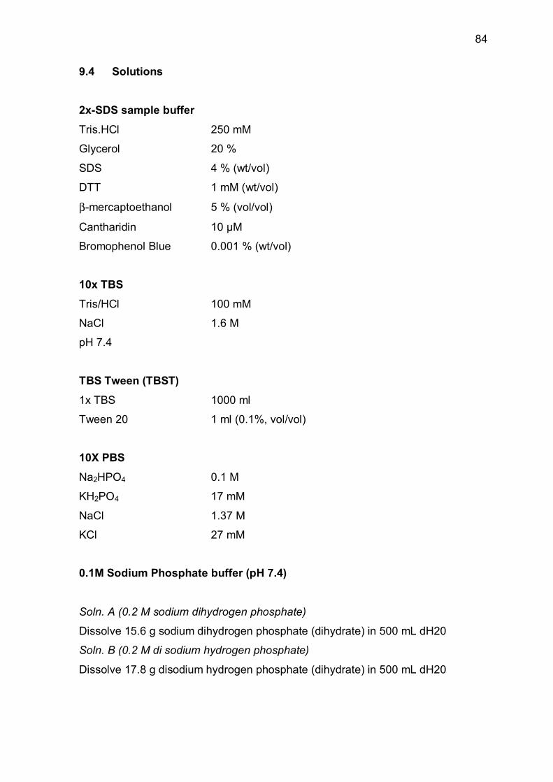
84
9.4 Solutions
2x-SDS sample buffer
Tris.HCl 250 mM
Glycerol 20 %
SDS 4 % (wt/vol)
DTT 1 mM (wt/vol)
β-mercaptoethanol 5 % (vol/vol)
Cantharidin 10 µM
Bromophenol Blue 0.001 % (wt/vol)
10x TBS
Tris/HCl 100 mM
NaCl 1.6 M
pH 7.4
TBS Tween (TBST)
1x TBS 1000 ml
Tween 20 1 ml (0.1%, vol/vol)
10X PBS
Na2HPO4 0.1 M
KH2PO4 17 mM
NaCl 1.37 M
KCl 27 mM
0.1M Sodium Phosphate buffer (pH 7.4)
Soln. A (0.2 M sodium dihydrogen phosphate)
Dissolve 15.6 g sodium dihydrogen phosphate (dihydrate) in 500 mL dH20
Soln. B (0.2 M di sodium hydrogen phosphate)
Dissolve 17.8 g disodium hydrogen phosphate (dihydrate) in 500 mL dH20

85
Mix 57 ml of soln. A, 243 ml of soln. B with 600 ml of water. The pH would itself be
adjusted to 7.4
Urea-glycerol gel buffer
Tris 240 mM
Glycine 276 mM
pH to 8.8 – 8.9
Urea-glycerol gel lysis buffer (10 ml)
Urea 554 mg (8.8 M)
Urea-gel buffer 975 µl
0.5 M DTT 225 µl (10 mM)
1 M Sodium thioglycolate 56.2 µl (5 mM)
100 mM Cantharidin 11.24 µl (10 µM)
6 M Imidazole (pH 8.8) 112.4 µl (60 mM)
Millipore water (qs) 10 ml
Bromophenol blue 0.001% (wt/vol)
Lysis buffer (Immunoprecipitation)
Tris/HCl pH 7.4) 50 mM
NaCl 150 mM
Triton X-100 1 %
Nonidet P - 40 0.5 %
EDTA 1 mM
EGTA 1 mM
NaF 20 mM
Na-orthovanadate 1.5 mM
DTT 10 mM
PMSF 0.5 mM
Complete® protease inhibitor cocktail

86
RhoA pulldown assay
Lysis buffer
HEPES 25mM
NaCl 150 mM
MgCl2 5 mM
EDTA 1 mM
NaF 10 mM
Na-orthovanadate 2 mM
DTT 5 mM
Tritone-X100 0.5%
Glycerol 2 %
PMSF 0.5 mM
Complete® protease inhibitor cocktail
Wash buffer
Tris/HCl (pH 7.4) 25mM
Triton X-100 1%
NaCl 150 mM
MgCl2 10 mm
PMSF 0.5 mM
Complete® protease inhibitor cocktail
Lysis buffer (Membrane fractionation)
Tris/HCl (pH 7.4) 5 mM
Sucrose 250 mM
EDTA 5 mM
MgCl2 1 mM
NaCl 5 mM
DTT 10 mM
PMSF 0.5 mM
Complete® protease inhibitor cocktail

87
10 Acknowledgements
All praises and thanks for Almighty ALLAH who is the ultimate source of all knowledge to mankind, and WHO made me reach at the present pedastal of knowledge with quality of doing something adventurous, novel, thrilling and path bearing.
All respects are for the Holy Prophet Muhammad (Peace be upon him) who is the symbol of guidance, fountain of knowledge, and made mankind to get out of depths of darkness.
Heartiest thanks for my worth supervisor Dr. Thomas Noll for his personal supervision, kind cooperation, constructive criticism, valuable suggestions, sympathetic attituted, freedom of work, and ever inspiring guidance throughout the progress of this work, and Prof. Dr. H.M. Piper for providing me place in his institute and critcally reviewing my work.
I am also greatly thankful to Prof. W. Clauß for supervising me to submit this thesis in faculty of natural sciences.
I am greatly indebted to Dr. Frauke Haertel for providing valuable suggestions and guidance during different technical problems and critically reviewing this manuscript.
My special thanks are for Hermann Holzträger, Anna Reis, Anika Seipp and Annika krautwurst for providing technical assistance.
Cordial thanks for all my lab fellows particularly Muhammad Arshad, Marion Holl, Krishnaveni and Tatyana for their geneial company, time devotion, synergistic help and cooperarion during my stay in the lab. Muhammad Aslam

88
11 Curriculum vitae
Muhammad Aslam
Date of Birth: 25th November 1976
Place of Birth: Sheikhupura (Pakistan)
Marital Status: Single
Nationality: Pakistani
Institution: Physiologisches Institut,
Justus-Liebig-Universität, Giessen, GERMANY
Mailing Address: Aulweg 129,
35392, Giessen, Germany
Phone: 0641 99 47 243
Fax: 0641 99 47 219
E-mail: [email protected]
giessen.de
Doctorate
2003 – 2007 cand. Dr. rer. nat
Graduate College GK534, ´´Biological Basis of Vascular
Medicine’’ Physiologisches Institut, Justus-Liebig-Universität,
Giessen.
Masters
(2001-2003) M.Phil. in Molecular Biology
Center of Excellence in Molecular Biology, University of the
Punjab, Lahore, Pakistan.
Bachelors
(1995-2000) Bachelor of Pharmacy
College of pharmacy, University of the Punjab, Lahore,
Pakistan.

89
12 Publications
1. Härtel FV, Rodewald CW, Aslam M, Gündüz D, Hafer L, Neumann J, Piper
HM, Noll T (2007) Extracellular ATP induces assembly and activation of
myosin light chain phosphatase complex in endothelial cells. Cardiovasc Res; 74:487-96
2. Gündüz D, Kasseckert SA, Härtel FV, Aslam M, Abdallah Y, Schafer M,
Piper HM, Noll T, Schäfer C (2006) Accumulation of extracellular ATP
protects against acute reperfusion injury in rat heart endothelial cells.
Cardiovasc Res. 71: 764-73
3. Pfeil U, Paddenberg R, Aslam M, Noll T, Yu S, Hsu T, Chang CL, Park J,
Weissmann N, Quanz K, Faulhammer P, Goldenberg A, Kummer W (2007)
Intermedin – A novel pulmonary peptide upregulated by hypoxia.
(submitted)
4. Aslam M, Härtel FV, Gündüz D, Piper HM, Noll T cAMP stabilizes
endothelial barrier by activation and assembly of the myosin light chain
phosphatase holoenzyme and inhibition of CPI-17. (Submitted)

90
13 Published abstracts
1. Aslam M, Härtel FV, Gündüz D, Pfeil U, Hsu SY, Kummer W, Piper, HM,
Noll T (2007) Intermedin induces failure of microvascular endothelial barrier
via derangement of actin cytoskeleton and dephosphorylation of focal
adhesion proteins. Circulation; 116(16):II-204 (American Heart
Association-Scientific Session, Nov. 2007, Orlando, FL USA)
2. Aslam M, Härtel FV, Pfeil U, Noll T (2007) Intermedin, a novel peptide,
stabilizes endothelial barrier by rearrangement of actin cytoskeleton via
Rac1 activation. Eur Heart J; 28:714-5 (Annual meeting of european
society of cardiaology, Vienna September 2007, Prize winning abstract)
3. Aslam M, Härtel FV, Piper HM, Noll T (2007) Intermedin disrupts
microvascular endothelial barrier by destroying actin cytoskeleton. Acta
Physiologica; 189:Supp.653:77 (Annual meeting of DPG Hannover, March
2007)
4. Härtel FV, Aslam M, Piper HM, Noll T (2007) CPI-17 is an important
mediator of thrombin-induced endothelial barrier failure. Acta Physiologica; 189:Supp.653:77 (Annual meeting of DPG Hannover, March
2007)
5. Arshad M, Holl M, Härtel FV, Aslam M, Piper HM, Noll T (2007) Transient
hypoxia induces proliferation and tube formation via p38 MAPKα in
endothelial cells. Acta Physiologica; 189:Supp.653:141 (Annual meeting
of DPG Hannover, March 2007)
6. Aslam M, Härtel FV, Holl M, Gündüz D, Arshad M, Piper HM, Noll T (2006)
NO Enhances Endothelial Barrier Function by inactivating the contractile
machinery and Stabilizing Cell – Adhesions. Circulation; 114(18):II-110
(American Heart Association-Scientific Session, Nov. 2006, Chicago, USA)

91
7. Aslam M, Härtel FV, Holl M, Arshad M, Noll T (2006) NO Protects
Endothelial Barrier Failure by Inactivating Contractile Machinery and
Stabilizing Cell-Cell Contacts. J Vasc Res; 43:553 (Annual Meeting of the
German Society for Microcirculation and Vascular Biology)
8. Holl M, Härtel FV, Arshad M, Aslam M, Noll T (2006) Transient Hypoxia
Protects Endothelial Cells from Apoptosis by MEK/ERK-Mediated Bad
Phosphorylation. J Vasc Res; 43:547 (Annual Meeting of the German
Society for Microcirculation and Vascular Biology)
9. Aslam M, Härtel FV, Gündüz D, Arshad M, Piper HM, Noll T (2006) cAMP
stabilisiert die endotheliale Schranke über Aktivierung und Assemblierung
der Myosinleichtkettenphospatase und Hemmung von CPI-17. (72nd Annual
Meeting of German Cardiac society, April 2006, Mannheim, Germany)
10. Härtel FV, Aslam M, Rodewald CW, Gündüz D, Noll T (2006) NO
antagonisiert das Thrombin-induziert endotheliale Schrankenversagen
durch Hemmung des kontraktilen Apparates. (72nd Annual Meeting of
German Cardiac society, April 2006, Mannheim, Germany)
11. Aslam M, Härtel FV, Gündüz D, Holl M, Piper HM, Noll T (2006) CPI-17 is
an important target of cAMP/PKA- mediated activation of MLC phosphatase
and stabilization of endothelial barrier function. Acta Physiologica;
186:S152 (Joint meeting of DPG and FEPS, Munich March 2006)
12. Aslam M, Härtel FV, Arshad M, Noll T, (2006) NO stabilizes endothelial
barrier by inhibition of MLC kinase and activation of MLC Phosphatase.
Acta Physiologica; 186:S71 (Joint meeting of DPG and FEPS, Munich,
March 2006)
13. Holl M, Arshad M, Aslam M, Härtel FV, Piper HM, Noll T (2006) Transient
Hypoxia protects Endothelial Cells from apoptosis by MEK/ERK-mediated
Bad phosphorylation. Acta Physiologica; 186:S63 (Joint meeting of DPG
and FEPS, Munich, March 2006)

92
14. Arshad M, Holl M, Aslam M, Härtel FV, Piper HM, Noll T (2006) Transient
Hypoxia Induces Proliferation via p38 MAPK α-mediated phosphorylation of
Rb protein. Acta Physiologica; 186:S140 (Joint meeting of DPG and
FEPS, Munich, March 2006)
15. Aslam M, Härtel FV, Gündüz D, Arshad M, Noll T (2005) cAMP Stabilizes
Endothelial Barrier by Activation and Assembly of the Myosin Light Chain
Phosphatase holoenzyme and Inhibition of CPI-17. Circulation; 112:II-142
(American Heart Association-Scientific Session Nov. 2005, Dallas USA)
16. Aslam M, Härtel FV, Rodewald CW Gündüz D, Noll T (2005) Nitric Oxide
antagonizes Thrombin Induced endothelial Barrier failure by activation of
Myosin Light Chain Phosphatase and inhibition of MEK/ERK pathway.
Circulation; 112:II-30 (American Heart Association-Scientific Session Nov.
2005, Dallas, USA)
17. Härtel FV, Rodewald CW Aslam M, Gündüz D, Neumann J, Piper HM, Noll
T (2005) Extracellular ATP induces Assembly and Activation of Myosin
Light Chain Phosphatase complex in Endothelial Cells. Circulation; 112:II-
142 (American Heart Association-Scientific Session Nov. 2005, Dallas,
USA)
18. Arshad M, Holl M, Aslam M, Härtel FV, Noll T (2005) Transient Hypoxia
Induces Proliferation of Endothelial Cells via Activation of p38 MAPK α and
γ. Circulation; 112:II-269 (American Heart Association-Scientific Session
Nov. 2005, Dallas, USA)
19. Gündüz D, Härtel FV, Aslam M, Piper HM, Noll T (2005) Accumulation of
extracellular ATP protects against acute endothelial reperfusion injury. Eur
Heart J; 26:264 (Annual meeting of European society of cardiology, Sep.
2005)

93
20. Aslam M, Härtel FV, Gündüz D, Noll T (2005) Stimulation of adenylyl
cyclase enhances endothelial barrier property by activation of the myosin
light chain phosphatase and inhibition of CPI-17. J Vas Res; 42:98-99, (3rd
European meeting on vascular Biology and Medicine, Sep. 2005, Hamburg,
Germany)
21. Aslam M, Härtel FV, Gündüz D, Noll T (2006) Stimulation of adenylyl
cyclase enhances endothelial barrier property via inactivation of contractile
apparatus of the endothelial cells. J Vasc Res; 43:27-60, (Annual Meeting
of the Society for Microcirculation and Vascular Biology, Sep. 2005,
Rostock, Germany)
22. Härtel FV, Aslam M, Rodewald CW, Gündüz D, Haffer L, Neuman J, Piper
HM, Noll T (2005) Extrazellutäres ATP stabilisiert die endotheliale
Schankenfunktion über Aktivierung des Myosinleichtketten-Phospatase-
Komplexes. (71st Annual Meeting of German Cardiac society, April 2005,
Mannheim, Germany)
23. Aslam M, Härtel FV, Peters SC, Noll T (2005) cAMP Stabilizes Endothelial
Barrier and inactivates the contractile machinery by activating myosin
phosphatase. Eur J Phys; 449:S58 (84th Annual Meeting of Deutsche
Physiologische Gesellschaft)
24. Härtel FV, Aslam M, Rodewald CW, Gündüz D, Haffer L, Neumann J, Piper
HM, Noll T (2005) Extracellular ATP Stabilizes endothelial barrier via
Activation of Myosin Light Chain Phosphatase complex. Eur J Phys; 449:
S59 (84th Annual Meeting of Deutsche Physiologische Gesellschaft)
25. Aslam M, Härtel FV, Rodewald CW, Arshad M, Noll T (2005) Exogenous
NO stabilizes macrovascular barrier function via stimulation of myosin light
chain phosphatase complex formation and activation. Eur J Phys; 449:
S106 (84th Annual Meeting of Deutsche Physiologische Gesellschaft)

MU
HA
MM
AD
A
SLA
M R
OLE O
F M
YO
SIN
LIG
HT C
HA
IN
P
HO
SP
HA
TA
SE
VVBédition scientifique
VVB LAUFERSWEILER VERLAG
MUHAMMAD ASLAM
VVB LAUFERSWEILER VERLAGédition scientifique
9 7 8 3 8 3 5 9 5 2 2 0 1
ISBN 3-8359-5220-XVVB LAUFERSWEILER VERLAGST AU FEN BER G R I N G 1 5D - 3 5 3 9 6 G I E S S E N
Tel: 0641-5599888 Fax: -5599890redak t ion@dok to rve r lag .dew w w . d o k t o r v e r l a g . d e
EFFECT OF CAMP/PKA SIGNALING MECHANISM
ON BARRIER FUNCTION OF CULTURED
ENDOTHELIAL CELLS:
INAUGURAL-DISSERTATIONzur Erlangung des Doktorgrades der Naturwissenschaften (Dr. rer. nat.)
des Fachbereichs Biologie und Chemieder Justus-Liebig-Universität Giessen
ROLE OF MYOSIN LIGHT CHAIN PHOSPHATASE
Human umbilical vein endothelial cells stained for actin (red) and ß-catenin (green)