education & research at nyu langone health | nyu ...show many different views on microscope...
TRANSCRIPT
Structure of the Ca21 Pump of Sarcoplasmic Reticulum: A View along theLipid Bilayer at 9-Å Resolution
Haruo Ogawa,*# David L. Stokes,§ Hiroyuki Sasabe,¶ and Chikashi Toyoshima**Institute of Molecular and Cellular Biosciences, University of Tokyo, Bunkyo-ku, Tokyo 113, Japan; #Department of Biological Sciences,Tokyo Institute of Technology, Midori-ku, Yokohama 226, Japan; §Skirball Institute of Biomolecular Medicine, New York UniversityMedical Center, New York, New York 10016, USA; and ¶Frontier Research Program, RIKEN, Wako, Saitama 351-01, Japan
ABSTRACT We have used multilamellar crystals of the ATP-driven calcium pump from sarcoplasmic reticulum to addressthe structural effects of calcium binding to the enzyme. They are stacks of disk-shaped two-dimensional crystals. A densitymap projected along the lipid bilayer was obtained at 9-Å resolution by frozen-hydrated electron microscopy. Although onlyin projection, much more details of the structure were revealed than previously available, especially in the transmembraneregion. Quantitative comparison was made with the model obtained from the tubular crystals of this enzyme formed in theabsence of calcium. Unexpectedly large differences in conformation were found, particularly in the cytoplasmic domain.
INTRODUCTION
Calcium ATPase (Ca21-ATPase) of the sarcoplasmic retic-ulum (SR) is an integral membrane protein ofMr 110,000.It pumps calcium ions from the cytoplasm into the SRagainst a large concentration gradient, thereby causing therelaxation of muscle cells. It is a member of P-type ionpumps that include Na1K1-ATPase and H1K1-ATPase(for a recent review, see Møller et al., 1996). The aminoacid sequence has been determined for many species (Mac-Lennan et al., 1985; Brandl et al., 1986), and extensivepredictions of the secondary structure have been made(Brandl et al., 1986; Taylor and Green, 1989). Site-directedmutagenesis studies have identified four critical amino acidresidues implicated in calcium transport, all on putativetransmembrane helices (Clarke et al., 1990; Chen et al.,1996; Rice and MacLennan, 1996). Hence it is generallybelieved that calcium ions bind directly to the high-affinitysites located within the membrane. The binding of calciumions induces conformational changes in the ATP-bindingsite, resulting in ATP hydrolysis and phosphorylation of theenzyme. The phosphorylation, in turn, causes conforma-tional changes in the calcium-binding site, and the enzymeeventually transfers two calcium ions into the lumen of SRper ATP molecule hydrolyzed (for a review see, e.g., Inesi,1994).
Many studies have characterized these conformationalchanges by various techniques (reviewed by Bigelow andInesi, 1992; Martonosi, 1995). For example, fluoresceinisothiocyanate attached near the ATP binding site (Pick and
Karlish, 1980) and affinity labeling of the ATP binding site(Yamamoto et al., 1989) have been used to determine thatconformational changes do occur around the ATP bindingsite when Ca21 binds to the high affinity site. Nevertheless,hardly any changes in the secondary structure have beendetected, suggesting that the conformational changes aresegmental movements (Csermely et al., 1987; Nakamotoand Inesi, 1986; Girardet and Dupont, 1992). Profile x-raydiffraction studies have demonstrated that long-range struc-tural changes occur with flash photolysis of caged calcium(DeLong and Blasie, 1993), but this method can only detectmovements normal to the membrane. Therefore, direct vi-sualizations are clearly needed to determine the magnitudeof these conformational changes and to understand how thebinding signal at one site is transmitted to the other.
Calcium ATPase from rabbit skeletal muscle SR can becrystallized in several forms suitable for cryoelectron mi-croscopy (for a review see, e.g., Martonosi, 1995). Two-dimensional ordering within the native membrane can beinduced by either lanthanides (Dux et al., 1985) or vanadate(Dux and Martonosi, 1983). The three-dimensional struc-ture has been determined at 14-Å resolution with the tubularcrystals induced by vanadate in the absence of calcium(Toyoshima et al., 1993a). With enzymes solubilized andreconstituted into lipid bilayers, multilamellar three-dimen-sional microcrystals have been obtained (Dux et al., 1987;Stokes and Green, 1990a). These crystals are stacks oftwo-dimensional sheets formed in a high concentration ofcalcium ions; therefore, the enzyme in this type of crystalmust be in a physiological state different from that in thetubular crystals (Dux et al., 1987; Stokes and Lacape`re,1994).
With these crystals, the direction of crystal growth can beadjusted by changing the lipid:protein:detergent ratio(Stokes and Green, 1990b; Cheong et al., 1996). It is pos-sible to make the long axis of the crystal normal to the lipidbilayers. Because such crystals appear like worms, theyshow many different views on microscope grids roughly inthe direction parallel to the lipid bilayer. Nevertheless, the
Received for publication 6 August 1997 and in final form 17 April 1998.
Address reprint requests to Dr. C. Toyoshima, Institute of Molecular andCellular Biosciences, University of Tokyo, Bunkyo-ku, Tokyo 113, Japan.Tel.: 81-3-3814-6347; Fax: 81-3-5689-7227; E-mail: [email protected].
Dr. Ogawa’s present address is Institute for Brain Research, School ofMedicine, University of Tokyo, 7-3-1 Hongo, Bunkyo-ku, Tokyo 113,Japan.
© 1998 by the Biophysical Society
0006-3495/98/07/41/12 $2.00
41Biophysical Journal Volume 75 July 1998 41–52

view along theb axis (shown in Fig. 1) was the one mostfrequently identified, and projection maps at low resolutionhave been published (Stokes and Green, 1990b; Cheong etal., 1996). We managed to collect six such images thatdiffracted to better than 10-Å resolution and describe here aside view of Ca21-ATPase in the presence of calcium. Wealso quantitatively compared the projection map with thosecalculated from the tubular crystals to visualize the confor-mational changes caused by the binding of calcium ions tothe enzyme.
MATERIALS AND METHODS
Preparation and crystallization of Ca21-ATPase
SR was prepared from white skeletal muscles of rabbit legs as described byChampeil et al. (1978). Calcium ATPase was affinity purified with redagarose (Reactive Red 120 from Sigma Chemical Co.) as described by Colland Murphy (1984). A small amount of phospholipid was supplementedduring the purification procedure. Crystallization was carried out essen-tially as described previously (Stokes and Green, 1990a).
ATPase activity
The ATPase activity of the enzyme preparation was measured at 25°C bya coupled enzyme method (Anderson and Murphy, 1983). Final concen-trations in 800ml assay solution were 5mg of Ca21-ATPase, 2 mM ATP,65 mM 3-(N-morpholino)propanesulfonic acid (pH 7), 130 mM KCl, 6mM MgCl2, 0.13 mM CaCl2, 0.5 mM phosphoenolpyruvate, 1 mg/mlC12E8, 0.2 mM NADH, 24 units pyruvate kinase, and 18 units lacticdehydrogenase.
Electron microscopy
Before rapid freezing, the solution containing microcrystals was dialyzedfor 2 h against the crystallization buffer, with no glycerol to reduce theconcentration of glycerol. This dialysis was necessary to prevent too muchsolution from being left on the specimen grids after blotting with filter
paper. The specimen solution was deposited on carbon-coated holey grids.Blotting from the side of the grid opposite that to which the specimensolution was applied (Toyoshima, 1989) was necessary to retain a suffi-cient amount of crystals on the grid.
The specimen was rapidly frozen in ethane slush and kept in liquidnitrogen until use. The specimen grids were mounted on a Gatan 626cryoholder and examined in a JEOL JEM2000EX microscope operated at200 kV accelerating voltage. A double-blade anticontaminator (Gatan651-N) was always in place. All of the images were taken at a nominalmagnification of 40,0003 with the minimal dose system and recorded onKodak SO163 film. The objective lens current was monitored. The mag-nification was calibrated using negatively stained tropomyosin tactoids(Caspar et al., 1969) at various objective lens currents.
Image analysis
Images were first selected by optical diffraction, and those that gave clearspots beyond 10-Å resolution were digitized. The densitometer used was anOrbital Science 1010M with spot size and scanning intervals of 10mm 310 mm. Digitized images were analyzed by the established way for two-dimensional crystals (Amos et al., 1982), including distortion correctionsof the crystal lattice (Henderson et al., 1986).
Because electron diffraction was not feasible with the multilamellarcrystals, compensation of Fourier amplitudes for the uneven contrast trans-fer function (CTF) was necessary. This requires accurate determination ofdefocus parameters from micrographs. At first, defocus parameters wereestimated from the locations of the Thon rings arising presumably fromdisordered phospholipids in the multilamellar crystals, by using a set ofprograms described elsewhere (Tani et al., 1996). Here the amplitudecontrast of 4.8% was assumed (Toyoshima et al., 1993b). Then the Fourierterms from various images were averaged to make a preliminary referencedata set, taking the CTF information thus obtained into account. In the nextstep, defocus parameters were refined by fitting the Fourier terms fromeach image to the preliminary reference data set compensated for theaveraged CTF. In the fitting process, both amplitude variations and phasereversals due to the CTF were examined. These procedures were iteratedseveral times to obtain the final averaged Fourier terms. No temperaturefactor compensation (Schertler et al., 1993) was introduced. Only theeffects of the modulation transfer function of the emulsion and the densi-tometer were compensated for as described by Downing and Grano (1982).In fitting and averaging various images, the envelope function of the CTFwas calculated for each image and was used as a weighting function.
Correlation calculation
To calculate correlation functions, a monomer was cut from the three-dimensional model of the tubular crystals (Toyoshima et al., 1993a). Thedensity cutoff level for the model was chosen so that;100% of theexpected volume was recovered (assuming anMr of 110K and a partialspecific volume of 0.74 cm3/g, based on the amino acid composition). Thecut out monomer was then rotated and projected in a desired direction. Theaverage density along the perimeter of the projected molecule was sub-tracted for floating. Care was taken to match the average densities of thetwo maps to be compared. Correlation functions were then calculated inreciprocal space, using the data to 14-Å resolution, so that the two struc-tures were compared at the same resolution. The top view (Fig. 5,a andc)of the multilamellar crystals was calculated from the published data(Stokes and Green, 1990a), with a resolution limited to 14 Å. Whencorrelation was to be maximized using the top views, two possibilities forthe viewing direction of a projection were also examined. Correlationfunctions for top views were also calculated in real space with an MRCprogram, IMROTRAN.
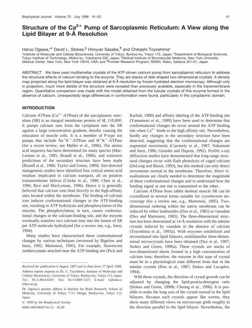
FIGURE 1 A cartoon depicting the configuration of the multilamellarcrystals of Ca21-ATPase. The protein molecules are protruding from bothsides of the bilayers (M) that contain lipids and, presumably, detergents.This type of three-dimensional crystal is essentially a stack of two-dimen-sional crystals. The enzyme molecules are arranged in a C2 lattice withtwofold rotation axes parallel to theb axis (along the membrane plane).The unit cell dimensions area 5 168.4 Å,b 5 56.4 Å,c 5 186.8 Å;a 590°, b 5 83.9°,g 5 90°. The arrow corresponds to the viewing directionof the electron micrograph in Fig. 2a.
42 Biophysical Journal Volume 75 July 1998

RESULTS
A view of multilamellar crystals of Ca21-ATPasealong the plane of the lipid bilayer
Multilamellar crystals of calcium ATPase reconstituted intolipid bilayers were formed in the presence of 10 mM cal-cium at pH 6 (Stokes and Green, 1990a,b). When thelipid-to-protein ratio was decreased, they tended to growprincipally normal to the lipid bilayer and sometimesformed large stacks of two-dimensional crystals (Fig. 1;Stokes and Green, 1990a; Cheong et al., 1996). The packingmodel for the multilamellar crystals is shown in Fig. 1(Taylor et al., 1988; Stokes and Green, 1990b; Cheong etal., 1996). Because these multilamellar crystals consist ofdisk-shaped membranes, they showed many different viewson the microscope grids. Occasionally double bands of lipidbilayers and stripes of protein densities were clearly seen tobe running slightly inclined from normal to these bands(Fig. 2 a). From the lattice parameters, these images werethought to correspond to the views along theb axis of thecrystals (Fig. 1; Cheong et al., 1996). They diffracted tobetter than 10-Å resolution, as demonstrated by the com-puter transform (Fig. 2b). We collected six such images andaveraged them in Fourier space. Merging statistics weregood up to 7-Å resolution, but the twofold phase residualwas not so good at high resolution (Table 1). This may bea result of the orientation of the crystals, such that thedirection of view is slightly offset from theb axis.
The projection map calculated at 9-Å resolution (Fig. 3)showed several distinct features and more details than pre-viously available (Stokes and Green, 1990b; Cheong et al.,
1996). Because of possible overlap of neighboring mole-cules (e.g., two molecules within the unit cell in Fig. 3)pointing in opposite directions, the boundary betweenneighboring enzyme molecules is ambiguous in some parts,particularly around the lipid bilayer. Nevertheless, it isobvious that the “head” piece of the cytoplasmic domainconsists of two well-separated domains that are different insize, and that the transmembrane region consists of at leastthree columns (numbered in Fig. 3), presumably composedof bundles ofa-helices. All transmembrane columns wereinclined by ;30° from vertical (normal to the membraneplane). At this stage it is not clear whether column 3 belongsto the same molecule or to the adjacent one. The largercytoplasmic domain (HL) consists of two barely resolvedsubdomains of nearly equal density, each of which appearsto be connected to the transmembrane column 2 by a thinrod (short barsin Fig. 3). The smaller domain (HS) appearsto have only a tenuous connection with the transmembraneregion and an even weaker one with the larger cytoplasmicdomain (HL).
The split appearance of the cytoplasmic domains of theenzyme in the multilamellar crystals is the most conspicuousfeature and is in marked contrast with those in the tubularcrystals, which constitute a single entity that looks like the headof a bird (Fig. 4,a andb; Toyoshima et al., 1993a). The splitappearance cannot be a result of overlap of enzyme moleculesin the crystal lattice (Fig. 1) or an effect of improved resolution(9 Å in Fig. 3, instead of 14 Å with tubular crystals). Thecorresponding image from the multilamellar crystals calculatedat 14-Å resolution (Fig. 5b) also showed a clear groovebetween the larger and smaller domains on the cytoplasmic
FIGURE 2 (a) Electron micrograph of a multilamellar crystal of Ca21-ATPase exhibiting the view along theb axis. The arrangement of the enzymemolecules in the crystal and the viewing direction are illustrated in Fig. 1. Note that the two leaflets of the lipid bilayers are clearly seen. The specimenis suspended in amorphous ice stretched over a hole in the carbon support film. The scale bar corresponds to 500 Å. (b) Diffraction amplitudes of a lessdefocused image of the crystal shown ina. The orientations of thea* andc* axes are indicated. The strong reflection at 1/9.1 Å21 (with a signal-to-noiseratio of 11.8) is circled. This particular transform appears anisotropic because the image was recorded at a small defocus (3400 Å) with a large amountof astigmatism (4160 Å).
Ogawa et al. A View of the Ca21 Pump at 9-Å Resolution 43

side; at this resolution two transmembrane columns (1 and 3)could not be resolved (Fig. 5b).
Comparison with the structure of Ca21-ATPase inthe tubular crystals
To make a quantitative comparison with the structure of theenzyme in the absence of calcium, monomers of Ca21-ATPase were cut out from the three-dimensional model(Toyoshima et al., 1993a) of the tubular crystals (hereafterreferred to as the 3D model) and arranged in various con-figurations to match the densities in the projection map fromthe multilamellar crystals. To do so, we used two projectionmaps of the multilamellar crystals in (nearly) orthogonaldirections, one projected perpendicular to the membrane(top view, Fig. 5a; Stokes and Green, 1990a), and the otherprojected along the plane of the lipid bilayer (side view, Fig.5 b). Both were calculated at the same resolution (14 Å) asthat of the structure from the tubular crystals, to avoid theeffects of difference in resolution.
Because the direction of the lipid bilayer in the multila-mellar crystals was already known, only one rotationalparameter (in the plane of the lipid bilayer) and threetranslational parameters had to be determined. It was morestraightforward to start with the top view, because therotational and the two translational parameters could bedetermined directly. Therefore, the 3D model was firstprojected normal to the membrane (Fig. 4c) and wascorrelated with the corresponding projection map of themultilamellar crystals. In Fig. 5c is a composite of the 3Dmodel (in red) superimposed on the projection map (topview) of the multilamellar crystal (ingreen) at the bestfitting position. The correlation function showed a broadpeak when plotted against the azimuthal angle. If the centerof the peak was chosen for the azimuth, that is, if the 3Dmodel was rotated by 4° counterclockwise from that shownin Fig. 4 c, the matching of the densities was reasonablygood (Fig. 5c).
The next step was to find the third translational parameterin the plane perpendicular to the lipid bilayer. To do so, the
TABLE 1 Statistics of electron crystallographic data*
Resolution range (Å)
No. ofused
reflectionsCompletedness
(%) Figure of merits#Modified figure
of merits§Merging phaseresiduals¶ (°)
Twofold phaseresiduals\ (°)
;15 109 100.0 0.915 0.816 15.9 8.215–12 42 70.0 0.848 0.664 26.7 26.012–9 82 62.6 0.844 0.597 28.8 36.89–7 92 48.4 0.855 0.573 24.4 40.7
*Only the spots whose signal-to-noise ratio was higher than 2 after merging and to which at least two images contributed were used for this table.#The figure of merit for a reflection is defined as
uOCTF z Fobsu
OuCTF z Fobsu
where CTF is the value of the contrast transfer function, andFobs is the observed Fourier term. This is a conventional figure of merit and is not useful fordetermining the resolution limit when a relatively small and variable number of images contribute to the average. The value is 0 if the phases are random,1 if the phases are the same or only one image contributes. The summations are taken over the images contributing to the same reflection.§The modified figure of merit is defined as
uOS/N z CTF z Fobsu
OS/N z uCTF z Fobsuz ÎNs 2 1
Nmax
whereS/N refers to the signal-to-noise ratio of the observed reflection,Nmax is the maximum number of images constituting the data set,Ns is the numberof images actually contributing to the reflection (Ns # Nmax). This kind of figure of merit is useful when a relatively small and variable number of imagescontribute to the average. The value becomes very close to 1 whenNs is large and the phases are the same. The value becomes smaller if the number ofcontributing images becomes smaller. The value is 0 if only one image contributes (i.e.,Ns 5 1) or if the phases are random. The summations are takenas described in the table footnote above on the definition of the figure of merit for a reflection.¶The merging phase residual for a reflection is defined as
OuCTF z Fobsu z ufavg 2 fu
OuCTF z Fobsu
wherefavg represents the averaged phase, andf the phase of individual transform. The summations are taken as described in the footnotes above.\Twofold phase residuals can be used as a measure of data quality, because twofold rotation axes exist parallel to theb axis of the crystal (normal to theimage). A twofold phase residual for a reflection is defined as
OuCTF z Fobsu z uf2 fd 2 fu
OuCTF z Fobsu
wheref2fd refers to the ideal phase expected from a twofold symmetry (either 0° or 180°, whichever is closer), andf is the phase of individual transform.The summations are taken as described in the footnotes above.
44 Biophysical Journal Volume 75 July 1998

3D model was projected in the direction corresponding tothe b axis of the multilamellar crystals, and the projectionmap was calculated (inred in Fig. 5 d); then it was corre-lated with the side view of the multilamellar crystals (ingreenin Fig. 5d). The projection maps that gave the highestcorrelation are shown superimposed (Fig. 5d). Note that theoverlap of the 3D models is correctly displayed only be-tween the short vertical bars at the top of the figure (Fig. 5d).
It is clear in Fig. 5d that both the cytoplasmic and themembrane domains of the multilamellar crystals (green) aremarkedly different from those of the 3D model (red). Con-firming the visual impression, the connection between thesmaller cytoplasmic domain (HS) and the larger one (HL) isstrong in the 3D model (red), but nearly lost in the multi-lamellar crystals (green). Although the positions of the stalk(short white barsin Fig. 3) roughly coincide, the largercytoplasmic domain (HL) appears to be offset by;14 Åhorizontally. In the transmembrane region, the difference isconspicuous around the position marked by the open circle.Here, the densities of the composite of the 3D models (red)are high, because segments A of two 3D models are seen tooverlap in this view; in fact, density is at a minimum at thisposition in the projection map of the multilamellar crystals(green). The strongest transmembrane column (2, Fig. 3) iscovered by weak densities corresponding to segments B andC in the 3D model (Fig. 4a). The differences appear so
large that we must assume very large conformationalchanges in the membrane region to reconcile these twomaps. For example, if we try to fit segment A (Fig. 4a) tothe transmembrane column 2, we must assume a shift of 17Å in the horizontal direction.
Structural differences in the cytoplasmic region
The correlation function between two structures is domi-nated by the overlap of regions of high density or highcontrast. The contrast is high in the cytoplasmic region,whereas densities are high in the transmembrane region.Therefore, if the cytoplasmic domains had moved withrespect to the membrane region, the correlation functionwould provide a compromise and the matching of densitiescould be poor everywhere. This seems to be the case,because even the densities in the cytoplasmic region do notmatch well. Hence, to see reorganizations in different re-gions and domain movements, we must calculate the cor-relation function based on one region at a time.
To do so, the calculations must begin with the side views,instead of the top views. The 3D model was first rotated byvarious degrees around an axis perpendicular to the mem-brane; then it was projected in the direction parallel to themembrane and correlated with the side view of the multi-
FIGURE 3 A projection map alongtheb axis of a multilamellar crystal ofCa21-ATPase, with thea axis hori-zontal. The map was calculated at 9-Åresolution from the data set averagedover six images; all of the reflectionswere used if the signal-to-noise ratiowas higher than 2 after averaging.Note that the cytoplasmic region con-sists of two well-separated densities(HL and HS), and the transmembraneregion (M) of at least three columns(1, 2, and 3) inclined by;30° fromvertical. HL domain is connected tothe membrane region through a stalk(S), which consists of two thin rods(short bars). One unit cell is enclosed(a-axis dimension is one half of that ofthe three-dimensional unit cell speci-fied in Fig. 1). The asterisk denotes asubsidiary peak in HL domain (seeFig. 7). The scale bar corresponds to20 Å.
Ogawa et al. A View of the Ca21 Pump at 9-Å Resolution 45

lamellar crystals, using only the transmembrane or the cy-toplasmic regions. For either of the two regions, the corre-lation function showed a well-defined peak and reached amaximum when the 3D model (Fig. 4) was rotated clock-wise by 11°. The images in Fig. 6,a andb, are compositesof the 3D models superimposed on the side view of themultilamellar crystals at the best matching positions forthe cytoplasmic (a) and the transmembrane (b) regions,respectively.
When the correlation was maximized for only the cyto-plasmic region, the larger cytoplasmic domain (HL) wascompletely covered by the main body of the headpiece ofthe 3D model (Fig. 6a). The density peaks in the two mapscoincided very well. The smaller cytoplasmic domain (HS)was located slightly below the “beak” of the 3D model (Fig.6 a), but the density and the dimensions were similar. Thelargest change occurred around the groove in the headpiece(Fig. 4a), identified as the ATP binding pocket (Toyoshimaet al., 1993a; Yonekura et al., 1997).
Presumably the most interesting feature to note here is thelocation of the stalk. The positions in the two maps areoffset by;20 Å. In the 3D model (red), its position is justbeneath the density peak of the headpiece, thus giving animpression that the stalk is offset to the left of the head. Incontrast, in the multilamellar crystals, it is offset to the rightof the HL domain.
The overall matching of densities was poor, because thetransmembrane densities of the 3D model were concen-trated on the density minimum in the map of multilamellarcrystals (marked by theopen circlein Fig. 6a). As a result,a large space was left completely uncovered between adja-cent enzyme molecules.
Structural differences in the membrane region
When the correlation function was calculated using thetransmembrane regions alone, the best matching position
FIGURE 4 (a) A cartoon illustrating the structure of Ca21-ATPase in the absence of calcium. The large cytoplasmic domain looks like the head of a bird;the transmembrane domain has three segments (A, B, and C); the small lumenal domain bridges segments A and B. The proposed ATP binding site andion pathway are indicated. The asterisk denotes the connecting density in the cytoplasmic domain (see Fig. 7). (b) A monomer of Ca21-ATPase cut fromthe three-dimensional model of tubular crystals (Toyoshima et al., 1993a) and projected parallel to the plane of the lipid bilayer. (c) A projection normalto the lipid bilayer of a monomer cut from the model of tubular crystals. The scale bar corresponds to 20 Å.
46 Biophysical Journal Volume 75 July 1998

(Fig. 6 b) was shifted slightly (4 Å horizontally) from theposition derived from using the whole molecule (Fig. 5d).The correlation function at this position was 77.6%,whereas the correlation function at the best matching posi-tion for the whole molecule was 76.8%. The difference wassmall between these two positions (,1%). However, thefitting in the transmembrane region was significantly better.In particular, the density minimum (open circlein Fig. 6 b)in the multilamellar crystals (green) was bracketed by seg-ments A of neighboring molecules (red). Yet the transmem-brane column 2 did not coincide exactly with segment A, butwas located between segments A and B. Therefore, if we try tomatch the largest transmembrane segment A in the 3D modelto the strongest transmembrane column 2, we must assume ashift of at least 10 Å in the horizontal direction or an inclinationof 30° of segment A. In that case, the major part of thecytoplasmic domain will be offset by 20 Å.
DISCUSSION
In this paper we have described a view parallel to the lipidbilayer of the multilamellar crystals of Ca21-ATPase. Theenzyme in this crystal, formed in 10 mM Ca21 at pH 6, mustbe in a physiological state that is different from that in thetubular crystals formed in the absence of Ca21 (Dux et al.,1987; Stokes and Lacape`re, 1994). A quantitative compar-
ison between the projection maps of the enzyme has re-vealed large conformational differences between the twotypes of crystals. The most conspicuous difference is ob-served around the ATP-binding site in the cytoplasmicdomain. Because calcium ions are thought to bind directlyto the high-affinity sites located in the transmembrane seg-ments (Clarke et al., 1990; Chen et al., 1996), it is surprisingto see such large conformational differences in the cytoplas-mic region. Of course, we expected to see some conforma-tional changes in the cytoplasmic region, because the bind-ing of calcium must alter the structure around the ATP-binding site, causing hydrolysis of ATP and phosphorylationof the enzyme. Indeed, long-range structural changes havebeen shown to occur with flash photolysis of caged-calcium(DeLong and Blasie, 1993). Yet the differences are so largeand are not localized to a particular part of the enzyme, asdemonstrated by the correlation calculations. This suggeststhat calcium binding elicits dramatic global effects on Ca21-ATPase structure, resulting in domain rearrangements andsegmental movements.
How can the changes in the cytoplasmic domainbe interpreted?
The most conspicuous feature in the projection map of themultilamellar crystals is that the cytoplasmic domain con-
FIGURE 5 (a, b) Projection maps ofmultilamellar crystal calculated at 14-Åresolution. (c, d) Composites of the pro-jection maps at the same resolution ofthe 3D models from tubular crystals(red) and that of multilamellar crystal(green), superimposed to maximize theoverlap of protein densities. The direc-tion of view is perpendicular to themembrane (a, c) or parallel to the planeof the membrane (b axis of the multila-mellar crystals) (b, d). Overlap of den-sities for the 3D models is correctlyreproduced only between the short ver-tical bars (d). The 3D models are locatedat the best fitting positions, based on thecorrelation calculation using the topview. Here the 3D models are rotated by4° counterclockwise from that in Fig. 4around an axis normal to the membrane.Note that mismatching of density is pro-nounced around the position marked bythe open circle. The transmembrane re-gion (M), the stalk region (S), and theorientation of the unit cell are indicated.The scale bar corresponds to 20 Å.
Ogawa et al. A View of the Ca21 Pump at 9-Å Resolution 47

sists of two well-separated densities (HL and HS, Fig. 3).This feature can be explained if we assume a movement ofthe segment connecting the beak and the main body of thecytoplasmic headpiece in the 3D model (Fig. 4b). In this
interpretation, as illustrated in Fig. 7, the beak in the 3Dmodel corresponds to the HS domain, and the connectingsegment (asteriskin Fig. 4a) has moved to form the seconddensity peak closer to the HS domain (asteriskin Fig. 3) in
FIGURE 6 Composites of the projection maps of the 3D models from tubular crystals (red) and that of multilamellar crystal (green), superimposed tomaximize the overlap of protein densities in the cytoplasmic (a) and transmembrane (b) regions. The direction of view is along the plane of the membrane(b axis of the multilamellar crystals). The correlation calculations for locating the 3D models used only the cytoplasmic (a) or the transmembrane (b) regionafter the 3D model was rotated (Fig. 4) by various degrees around an axis normal to the membrane. For either region, the best fit was obtained when the3D model was rotated by 11° clockwise. All of the projection maps were calculated at the same resolution (14 Å). The transmembrane region (M), the stalkregion (S), and the orientation of the unit cell are indicated. The scale bar corresponds to 20 Å.
FIGURE 7 A cartoon illustrating the proposedmovements in the Ca21-ATPase caused by the bind-ing of calcium. (a) A monomer of Ca21-ATPase cutfrom the three-dimensional model of tubular crystalsformed in the absence of calcium (identical to Fig. 4b). (b) A monomer of Ca21-ATPase in the multila-mellar crystals formed in the presence of calcium, cutfrom the projection map shown in Fig. 3. (c, d) Foursegments rearranged by the binding of calcium. Ar-rows indicate the directions of the movements andnumbers the order of the segmental movements. Be-cause the best fit of the 3D model was obtained at thesame azimuthal position for either the cytoplasmic orthe membrane region (Fig. 6), the movements de-picted are solely translational ones. The asterisk inthe HL domain (d) refers to a subsidiary peak thatmight correspond to the connecting density (asteriskin c) in the 3D model (a, c).
48 Biophysical Journal Volume 75 July 1998

the HL domain (also see Fig. 6b). Supporting this interpre-tation, the main body of the cytoplasmic headpiece in the3D model has only one density peak offset to the left (Fig.4 b). In an 8-Å resolution map of the tubular crystals withbound chromium ATP (Yonekura and Toyoshima, unpub-lished observations), the beak forms a domain that is distinctfrom the main body of the headpiece.
It is interesting that such a large change occurs around theputative ATP binding site (Fig. 4a; Toyoshima et al.,1993a; Yonekura et al., 1997), because Ca21-ATPase in themultilamellar crystals cannot hydrolyze ATP (data notshown). The crystallization medium contains 10 mM Ca21,which is high enough to inhibit the ATPase activity (Has-selbach, 1964; MacLennan et al., 1972). The crystals put inthe assay medium containing 0.15 mM Ca21 exhibited noATPase activity, even after 10 min of incubation. However,the addition of detergent, which breaks up the crystals,restored full ATPase activity (data not shown). Hence con-straints posed by forming a crystal may explain the loss ofATPase activity. Nonetheless, the split appearance of thecytoplasmic domains must not be caused by the crystalcontacts between the enzyme molecules in adjacent layersof two-dimensional sheets, because the same density distri-bution was found for the molecules in the uppermost layer(image not shown).
How the binding signal of calcium is transmittedto the cytoplasmic side
The stalk region must have an important role in transmittingthe binding signal of calcium to the ATP-binding site,because the stalk is the only connection between the trans-membrane segments and the headpiece of the cytoplasmicdomain. In this regard, it may be important to note that theposition of the stalk with respect to the headpiece is differ-ent between the two structures: in the tubular crystals, thestalk is offset to the left from the center of the headpiece(Fig. 4 b); it is offset to the right of the HL domain in themultilamellar crystals (Fig. 3). Furthermore, transmembranecolumn 2, which appears to be connected to the stalkdirectly in the projection map of multilamellar crystals, isinclined by 30° from vertical, whereas segment A1, which isa direct extension of the stalk in the tubular crystals, runsnormal to the membrane plane. Thus the binding of calciummight cause the rearrangement of the transmembranesegments, which in turn move the stalk to transmit thebinding signal to the cytoplasmic domains (depicted in Fig.7, c andd).
Rearrangements of thetransmembrane segments
The differences in the transmembrane region are perplex-ingly large between the two maps, as demonstrated by thepoor matching in Fig. 6b, where the correlation of densitiesin the transmembrane region was maximized. The differ-
ences are also difficult to interpret. As already described,the transmembrane region of the enzyme in the tubularcrystals consists of three segments; the largest one is seg-ment A. The largest transmembrane segment is column 2 inthe multilamellar crystals (Fig. 3). However, column 2 andsegment A were not at the same position when the correla-tion in the transmembrane region was maximized (Fig. 6b).Moreover, in the tubular crystals, there is a lumenal densityconnecting segments B and A2; such densities exist, if at all,only in the left side of column 2 in the multilamellar crystals(Fig. 3). In terms of the size and the connectivity to thestalk, it seems appropriate to assign column 2 to segment A.Column 1 is unlikely to be either segment B or C, becauseit is located in the left side of column 2; hence column 1 ismost likely to be a part of segment A. These assignmentsleave column 3, which will then correspond to segments Band C.
To explain the mismatch of the positions of column 2 andsegment A solely by translational movements, segment Amust move 13 Å horizontally (Fig. 6b). Nonetheless, a 30°change in the inclination of transmembrane columns (de-picted in Fig. 7c) would account for a 10-Å displacement.Therefore, it seems possible to reconcile the two maps, buthere again, we are forced to assume large conformationalchanges to explain the differences.
Conformational states of Ca21-ATPase in the twoforms of crystals
How are the structural differences of the enzyme visualizedhere between the two crystal forms related to the reactioncycle of Ca21-ATPase? It is not obvious, because eithertype of crystal is formed in rather special ionic conditions:tubular crystals are formed in the presence of EGTA anddecavanadate, whereas the multilamellar crystals describedhere are formed in the presence of 10 mM calcium. Severalattempts have already been made to address this issue (Duxet al., 1987; Stokes and Lacape`re, 1994).
Because of the absence of calcium ions, Ca21-ATPase inthe tubular crystals has been thought to take a conformationafter transfer of calcium ions to the lumenal side of the SR(Dux and Martonosi, 1983). Limited proteolysis supportsthis idea (Imamura et al., 1984). However, further specifi-cation is difficult, because vanadate is used for inducingcrystals. Decavanadate has been thought to be responsiblefor crystal formation: in fact, strong density peaks that arelikely to represent large vanadate oligomers are observed inthe map of tubular crystals (Toyoshima et al., 1993a). How-ever, stock decavanadate solution inevitably contains otherspecies of oligomers (Csermely et al., 1985) that may mod-ulate the structure of the enzyme in different ways (Coan etal., 1986; Aureliano and Madeira, 1994). Monovanadate hasbeen thought to behave as a phosphate analog (Pick, 1982;Pick and Karlish, 1982), and decavanadate as an ATPanalog (Varga et al., 1985; Coan et al., 1986; Ross andMcIntosh, 1987). Given these results, further characteriza-
Ogawa et al. A View of the Ca21 Pump at 9-Å Resolution 49
tion is clearly needed to identify the physiological state ofthe enzyme in the tubular crystals.
The conformational state of the enzyme in the multila-mellar crystals is not unambiguous either (Dux et al., 1987).Because this type of crystal is formed in high concentrationsof calcium (.0.25 mM at pH 6; Stokes and Lacape`re,1994), the enzyme may be regarded to be in a kind of restingstate after a large amount of calcium ion is transferred to thelumen of the SR. The question is whether this state is thesame as the one in which only low concentrations of cal-cium are present (the so-called E1 state). The apparentdissociation constant of calcium at pH 6 has been measuredto be ;7.6 mM, and the Hill coefficient to be;1.46(Watanabe et al., 1981). These values mean that 99.4% ofthe high-affinity binding sites are occupied at 0.25 mMcalcium; 0.9 mM is required to fill 99.9% of the bindingsites. This number for the dissociation constant is alsoconsistent with the one (pCa1/2 5.15) obtained by the intrin-sic tryptophan fluorescence measurements (Guillain et al.,1980). It is well documented that the intrinsic fluorescenceof tryptophan residues, nearly entirely on putative trans-membrane helices, increases when the calcium concentra-tion is increased. The fluorescence saturates at the millimo-lar range (Guillan et al., 1980, 1981).
A recent study (Juul et al., 1995) showed that high-affinity calcium binding is impaired after digestion withproteinase K in the presence of 0.3 mM Ca21, but not in 10mM Ca21 (at pH 6.5). This result is consistent with the factthat a Ca21 concentration greater than 0.3 mM is required tokeep the high-affinity binding sites continuously full. It mayexplain the result that the long-term stability of Ca21-ATPase is improved by the presence of millimolar Ca21
(Pikula et al., 1988).It is well known that ATPase activity of Ca21-ATPase is
completely inhibited at millimolar calcium. From kineticstudies, it is established that the inhibition of ATPase ac-tivity by a high concentration of calcium is due to thestabilization of the E-P state (Yamada and Ikemoto, 1980),caused by the activation of back-reaction (Trotta and deMeis, 1978). Thus the enzyme in high Ca21 concentrationsretains the ability to proceed to the next steps (i.e., bindingof ATP and phosphorylation) in the reaction cycle.
The conformational changes accompanying the Ca21
binding to the high-affinity sites have been studied usingprofile x-ray diffraction of stacked lamellar vesicles (De-Long and Blasie, 1993). They detected conformationalchanges in three different parts of the enzyme on the releaseof calcium by flash photolysis of caged calcium. The changesin the projected electron densities were small; the increase inthe membrane domain could be attributed to the binding ofCa21 alone, but was accompanied by decreases in the adjacentarea. The small size of the changes might appear to be incom-patible with the current results. However, the differences inconformation visualized here (Fig. 6) are mostly the move-ments parallel to the membrane plane, causing little net massmovement normal to the membrane. The mass movements inthe headpiece they describe (i.e., the decrease at the top part
and the increase at;15 Å below it) are certainly consistentwith the differences depicted in Fig. 7.
Given these results, it seems most appropriate to assumethat the enzyme in the multilamellar crystals is in a statesimilar to, if not identical with, the E1zCa2 state obtainedtransiently at physiological Ca21 concentrations, which issimilar to the conclusion of previous studies (Stokes andLacapere, 1994).
CONCLUSIONS AND PROSPECTS
Conformational differences between the Ca21-ATPase inthe multilamellar crystals and that in the tubular crystalswere unexpectedly large, as visualized in the projectionmaps viewed parallel to the lipid bilayer. These conforma-tional differences most likely represent the changes trig-gered by the binding of calcium ions to the high-affinitybinding sites within the membrane. The binding signalseems to be realized as the rearrangements of the transmem-brane segments and transmitted to the cytoplasmic domainthrough the change in inclination of the stalk segment, asillustrated in Fig. 7c. Large changes around the putativeATP binding site may be largely accounted for by segmen-tal movements of the cytoplasmic domains.
Of course, these structural differences would be muchbetter characterized and understood if the three-dimensionalstructure of the multilamellar crystals were available. It isnow possible to obtain large, flat crystals that diffract to3.6-Å resolution by electron diffraction (Shi et al., 1995). Atthis resolution, it may be feasible to visualize individualamino acids. Thus, in the near future, we may be able tounderstand the structural changes that are involved in theactive transport of calcium ions.
We thank Dr. Nigel Unwin for reading through an initial draft. We alsothank Kazutoshi Tani for some of the programs used in this study.
This work was supported by Grants-in-Aid for Scientific Research and forInternational Scientific Research from the Ministry of Education, Science,Sports and Culture of Japan (to CT) and from the National Institutes ofHealth (AR40997 to DLS).
REFERENCES
Amos, L. A., R. Henderson, and P. N. T. Unwin. 1982. Three-dimensionalstructure determination by electron microscopy of two-dimensionalcrystals.Prog. Biophys. Mol. Biol.39:183–231.
Anderson, K. W., and A. J. Murphy. 1983. Alterations in the structure ofthe ribose moiety of ATP reduce its effectiveness as a substrate for thesarcoplasmic reticulum ATPase.J. Biol. Chem.258:14276–14278.
Aureliano, M., and V. M. C. Madeira. 1994. Interactions of vanadateoligomers with sarcoplasmic reticulum Ca21-ATPase.Biochim. Bio-phys. Acta.1221:259–271.
Bigelow, D. J., and G. Inesi. 1992. Contributions of chemical derivatizationand spectroscopic studies to the characterization of the Ca21 transportATPase of sarcoplasmic reticulum.Biochim. Biophys. Acta.1113:323–338.
Brandl, C. J., N. M. Green, B. Korczak, and D. H. MacLennan. 1986. TwoCa21-ATPase genes: homologies and mechanistic implications of de-duced amino acid sequences.Cell. 44:597–607.
50 Biophysical Journal Volume 75 July 1998

Caspar, D. L. D., C. Cohen, and W. Longley. 1969. Tropomyosin: crystalstructure, polymorphism and molecular interactions.J. Mol. Biol. 41:87–107.
Champeil, P., S. Bu¨schlen-Boucly, F. Bastide, and C. Gary-Bobo. 1978.Sarcoplasmic reticulum ATPase. Spin labeling detection of ligand-induced changes in the relative reactivities of certain sulfhydryl groups.J. Biol. Chem.253:1179–1186.
Chen, L., C. Sumbilla, D. Lewis, L. Zhong, C. Strock, M. E. Kirtley, andG. Inesi. 1996. Short and long range functions of amino acids in thetransmembrane region of the sarcoplasmic reticulum ATPase.J. Biol.Chem.271:10745–10752.
Cheong, G. W., H. S. Young, H. Ogawa, C. Toyoshima, and D. L. Stokes.1996. Lamellar stacking in three-dimensional crystals of Ca21-ATPasefrom sarcoplasmic reticulum.Biophys. J.70:1689–1699.
Clarke, D. M., T. W. Loo, and D. H. MacLennan. 1990. Functionalconsequences of alterations to polar amino acids located in the trans-membrane domain of the Ca21-ATPase of sarcoplasmic reticulum.J. Biol. Chem.265:22223–22227.
Coan, C., D. J. Scales, and A. J. Murphy. 1986. Oligovanadate binding tosarcoplasmic reticulum ATPase.J. Biol. Chem.261:10394–10403.
Coll, R. J., and A. J. Murphy. 1984. Purification of the CaATPase ofsarcoplasmic reticulum by affinity chromatography.J. Biol. Chem.259:14249–14254.
Csermely, P., C. Katopis, B. A. Wallace, and A. Martonosi. 1987. The E1
3 E2 transition of Ca21-transporting ATPase in sarcoplasmic reticulumoccurs without major changes in secondary structure.Biochem. J.241:663–669.
Csermely, P., S. Varga, and A. Martonosi. 1985. Competition betweendecavanadate and fluorescein isothiocyanate on the Ca21-ATPase ofsarcoplasmic reticulum.Eur. J. Biochem.150:455–460.
DeLong, L. J., and J. K. Blasie. 1993. Effect of Ca21 binding on the profilestructure of the sarcoplasmic reticulum membrane using time-resolvedx-ray diffraction.Biophys. J64:1750–1759.
Downing, K. H., and D. A. Grano. 1982. Analysis of photographic emul-sions for electron microscopy of two-dimensional crystalline specimens.Ultramicroscopy.7:381–404.
Dux, L., and A. Martonosi. 1983. Two-dimensional arrays of proteins insarcoplasmic reticulum and purified Ca21-ATPase vesicles treated withvanadate.J. Biol. Chem.258:2599–2603.
Dux, L., S. Pikula, N. Mullner, and A. Martonosi. 1987. Crystallization ofCa21-ATPase in detergent-solubilized sarcoplasmic reticulum.J. Biol.Chem.262:6439–6442.
Dux, L., K. A. Taylor, H. P. Ting-Beall, and A. Martonosi. 1985. Crys-tallization of the Ca21-ATPase of sarcoplasmic reticulum by calciumand lanthanide ions.J. Biol. Chem.260:11730–11743.
Girardet, J-L., and Y. Dupont. 1992. Ellipticity changes of the sarcoplas-mic reticulum Ca21-ATPase induced by cation binding and phosphor-ylation. FEBS Lett.296:103–106.
Guillain, F., P. Champeil, J-J. Lacape`re, and M. P. Gingold. 1981. Stoppedflow and rapid quenching measurement of the transient steps induced bycalcium binding to sarcoplasmic reticulum adenosine triphosphatase.Competition with Ca21-independent phosphorylation.J. Biol. Chem.256:6140–6147.
Guillain, F., M. P. Gingold, S. Bu¨schlen, and P. Champeil. 1980. A directfluorescence study of the transient steps induced by calcium binding tosarcoplasmic reticulum ATPase.J. Biol. Chem.255:2072–2076.
Hasselbach, W. 1964. Relaxing factor and the relaxation of muscle.Prog.Biophys. Biophys. Chem.14:169–222.
Henderson, R., J. M. Baldwin, K. H. Downing, J. Lepault, and F. Zemlin.1986. Structure of purple membrane fromHalobacterium halobium:recording, measurement and evaluation of electron micrographs at 3.5 Åresolution.Ultramicroscopy.19:147–178.
Imamura, Y., K. Saito, and M. Kawakita. 1984. Conformational change ofCa21,Mg21-adenosine triphosphatase of sarcoplsmic reticulum uponbinding of Ca21 and adenyl-59-yl-imidodiphosphate as detected bytrypsin sensitivity analysis.J. Biochem.95:1305–1313.
Inesi, G. 1994. Teaching active transport at the turn of the twenty-firstcentury: recent discoveries and conceptual changes.Biophys. J.66:554–560.
Juul, B., H. Turc, M. L. Durand, A. G. de Gracia, L. Denoroy, J. V. Møller,P. Champeil, and M. le Maire. 1995. Do transmembrane segments in
proteolyzed sarcoplasmic reticulum Ca21-ATPase retain their functionalCa21 binding properties after removal of cytoplasmic fragments byproteinase K?J. Biol. Chem.270:20123–20134.
MacLennan, D. H., C. J. Brandl, B. Korczak, and N. M. Green. 1985.Amino-acid sequence of a Ca21 1 Mg21-dependent ATPase from rabbitmuscle sarcoplasmic reticulum, deduced from its complementary DNAsequence.Nature. 316:696–700.
MacLennan, D. H., C. C. Yip, G. H. Iles, and P. Seeman. 1972. Isolationof sarcoplasmic reticulum proteins.Cold Spring Harb. Symp. Quant.Biol. 37:469–477.
Martonosi, A. N. 1995. The structure and interactions of Ca21-ATPase.Biosci. Rep.15:263–281.
Møller, J. V., B. Juul, and M. le Maire. 1996. Structural organization, iontransport, and energy transduction of P-type ATPases.Biochim. Biophys.Acta.1286:1–51.
Nakamoto, R. K., and G. Inesi. 1986. Retention of ellipticity betweenenzymatic states of the Ca21-ATPase of sarcoplasmic reticulum.FEBS.Lett. 194:258–262.
Pick, U. 1982. The interaction of vanadate ions with the Ca-ATPase fromsarcoplasmic reticulum.J. Biol. Chem.257:6111–6119.
Pick, U., and S. J. D. Karlish. 1980. Indications for an oligomeric structureand for conformational changes in sarcoplasmic reticulum Ca21-ATPaselabelled selectively with fluorescein.Biochem. Biophys. Acta.626:255–261.
Pick, U., and S. J. D. Karlish. 1982. Regulation of the conformationtransition in the Ca-ATPase from sarcoplasmic reticulum by pH, tem-perature, and calcium ions.J. Biol. Chem.257:6120–6126.
Pikula, S., N. Mullner, L. Dux, and A. Martonosi. 1988. Stabilization andcrystallization of Ca21-ATPase in detergent-solubilized sarcoplasmicreticulum.J. Biol. Chem.263:5277–5286.
Rice, W. J., and D. H. MacLennan. 1996. Scanning mutagenesis reveals asimilar pattern of mutation sensitivity in transmembrane sequences M4,M5, and M6, but not in M8, of the Ca21-ATPase of sarcoplasmicreticulum (SERCA1a).J. Biol. Chem.271:31412–31419.
Ross, D. C., and D. B. McIntosh. 1987. Intramolecular cross-linking at theactive site of the Ca21-ATPase of sarcoplasmic reticulum.J. Biol. Chem.262:12977–12983.
Schertler, G. F. X., C. Villa, and R. Henderson. 1993. Projection structureof rhodopsin.Nature.362:770–772.
Shi, D., H-H. Hsiung, R. C. Pace, and D. L. Stokes. 1995. Preparation andanalysis of large, flat crystals of Ca21-ATPase for electron crystallog-raphy.Biophys. J.68:1152–1162.
Stokes, D. L., and N. M. Green. 1990a. Structure of Ca-ATPase: electronmicroscopy of frozen-hydrated crystals at 6 Å resolution in projection.J. Mol. Biol. 213:529–538.
Stokes, D. L., and N. M. Green. 1990b. Three-dimensional crystals ofCa-ATPase from sarcoplasmic reticulum.Biophys. J.57:1–14.
Stokes, D. L., and J. J. Lacape`re. 1994. Conformation of Ca21-ATPase intwo crystal forms.J. Biol. Chem.269:11606–11613.
Tani, K., H. Sasabe, and C. Toyoshima. 1996. A set of programs fordetermining defocus and astigmatism in electron images.Ultramicros-copy.65:31–44.
Taylor, K. A., N. Mullner, S. Pikula, L. Dux, C. Peracchia, S. Varga, andA. Martonosi. 1988. Electron microscope observations on Ca21-ATPasemicrocrystals in detergent-solubilized sarcoplasmic reticulum.J. Biol.Chem.263:5287–5294.
Taylor, W. R., and N. M. Green. 1989. The predicted secondary structuresof the nucleotide-binding sites of six cation-transporting ATPases lead toa probable tertiary fold.Eur. J. Biochem.179:241–248.
Toyoshima, C. 1989. On the use of holey grids in electron crystallography.Ultramicroscopy.30:439–444.
Toyoshima, C., H. Sasabe, and D. L. Stokes. 1993a. Three-dimensionalcryo-electron microscopy of the calcium ion pump in the sarcoplasmicreticulum membrane.Nature.362:469–471.
Toyoshima, C., K. Yonekura, and H. Sasabe. 1993b. Contrast transfer forfrozen-hydrated specimens. II. Amplitude contrast at very low frequen-cies.Ultramicroscopy.48:165–176.
Trotta, E. E., and L. de Meis. 1978. Adenosine 59-triphosphate-orthophosphate exchange catalyzed by the Ca21-transport ATPase ofbrain.J. Biol. Chem.253:7821–7825.
Ogawa et al. A View of the Ca21 Pump at 9-Å Resolution 51

Varga, S., P. Csermely, and A. Martonosi. 1985. The binding of vanadium(V) oligoanions to sarcoplasmic reticulum.Eur. J. Biochem.148:119–126.
Watanabe, T., D. Lewis, R. Nakamoto, M. Kurzmack, C. Fronticelli, andG. Inesi. 1981. Modulation of calcium binding in sarcoplasmic reticulumadenosinetriphosphatase.Biochemistry.20:6617–6625.
Yamada, S., and N. Ikemoto. 1980. Reaction mechanism of calcium-ATPase of sarcoplasmic reticulum.J. Biol. Chem.255:3108–3119.
Yamamoto, H., Y. Imamura, M. Tagaya, T. Fukui, and M. Kawakita. 1989.Ca21-dependent conformational change of the ATP-binding site ofCa21-transporting ATPase of sarcoplasmic reticulum as revealed by analteration of the target-site specificity of adenosine triphosphopyridoxal.J. Biochem.106:1121–1125.
Yonekura, K., D. L. Stokes, H. Sasabe, and C. Toyoshima. 1997. TheATP-binding site of Ca21-ATPase revealed by electron image analysis.Biophys. J.72:997–1005.
52 Biophysical Journal Volume 75 July 1998