ecology - pnas.org · cgi doi 10.1073 pnas.0904305106 pnas september 22, 2009 vol. 106 no. 38...
TRANSCRIPT
BIOCHEMISTRYCorrection for ‘‘The Corynebacterium diphtheriae shaft pilinSpaA is built of tandem Ig-like modules with stabilizing isopep-tide and disulfide bonds,’’ by Hae Joo Kang, Neil G. Paterson,Andrew H. Gaspar, Hung Ton-That, and Edward N. Baker,which appeared in issue 40, October 6, 2009, of Proc Natl AcadSci USA (106:16967–16971; first published September 21, 2009;10.1073/pnas.0906826106).
The authors note that, due to a printer’s error, on page 16967,the keyword ‘‘mas spectromrtry’’ should instead have appearedas ‘‘mass spectrometry.’’ The online version has been corrected.
www.pnas.org/cgi/doi/10.1073/pnas.0911293106
CELL BIOLOGYCorrection for ‘‘CBP and p300 are cytoplasmic E4 polyubiquitinligases for p53,’’ by Dingding Shi, Marius S. Pop, RomanKulikov, Ian M. Love, Andrew Kung, and Steven R. Grossman,which appeared in issue 38, September 22, 2009, of Proc NatlAcad Sci USA (106:16275–16280; first published September 4,2009; 10.1073/pnas.0904305106).
The authors note that the author name Andrew Kung shouldinstead have appeared as Andrew L. Kung. The online versionhas been corrected. The corrected author line and authorcontributions footnote appear below.
Dingding Shi, Marius S. Pop, Roman Kulikov, Ian M. Love,Andrew L. Kung, and Steven R. Grossman
Author contributions: D.S., M.S.P., R.K., and S.R.G. designed research; D.S., M.S.P., R.K., andI.M.L. performed research; A.L.K. contributed new reagents/analytic tools; D.S., M.S.P., R.K.,and S.R.G. analyzed data; and S.R.G. wrote the paper.
www.pnas.org/cgi/doi/10.1073/pnas.0910983106
ECOLOGYCorrection for ‘‘Adaptive shell color plasticity during the earlyontogeny of an intertidal keystone snail,’’ by Patricio H. Man-rı́quez, Nelson A. Lagos, Marı́a Elisa Jara, and Juan CarlosCastilla, which appeared in issue 38, September 22, 2009, of ProcNatl Acad Sci USA (106:16298–16303; first published September2, 2009; 10.1073/pnas.0908655106).
The authors note that on page 16298, right column, first fullparagraph, the first sentence appeared incorrectly in part. ‘‘Inthe rocky intertidal habitats dominated by mussel beds (darkcolored) and barnacle stands (light colored), we found that morethan 95% of early postmetamorphic stages of C. concholepas (�2to 20 mm periostomal length, PL) showed a striking color-matching with the most abundant prey (Fig. 2 A and B)’’ shouldinstead have appeared as ‘‘In the rocky intertidal habitatsdominated by mussel beds (dark colored) and barnacle stands(light colored), we found that more than 95% of early postmeta-morphic stages of C. concholepas (�2 to 20 mm peristomallength, PL) showed a striking color-matching with the mostabundant prey (Fig. 2 A and B).’’ Additionally, the authors notethat on page 16301, right column, first full paragraph, the secondsentence did not appear in full due to a printer’s error. Thesentence should instead have appeared as ‘‘This modulation ofthe shell coloration is restricted to snails of small size (ca. lessthan 3 cm), coincidentally the size at which C. concholepasappear to escape predation by crabs (author’s personal obser-vations).’’ These errors do not affect the conclusions of thearticle.
www.pnas.org/cgi/doi/10.1073/pnas.0910777106
PNAS � October 27, 2009 � vol. 106 � no. 43 � 18427
CORR
ECTI
ON
S
CBP and p300 are cytoplasmic E4 polyubiquitinligases for p53Dingding Shia,1, Marius S. Popa,1, Roman Kulikova, Ian M. Lovea, Andrew L. Kungb, and Steven R. Grossmana,c,d,2
Departments of aCancer Biology and cMedicine, dGastrointestinal Cancer Program, University of Massachusetts Medical School and University ofMassachusetts Memorial Cancer Center, 364 Plantation Street, Worcester, MA 01605; and bDepartment of Pediatric Oncology, Dana-Farber Cancer Institute,44 Binney Street, Boston, MA 02115
Edited by Carol L. Prives, Columbia University, New York, NY, and approved August 3, 2009 (received for review April 22, 2009)
p300 and CREB-binding protein (CBP) act as multifunctional regu-lators of p53 via acetylase and polyubiquitin ligase (E4) activities.Prior work in vitro has shown that the N-terminal 595 aa of p300encode both generic ubiquitin ligase (E3) and p53-directed E4functions. Analysis of p300 or CBP-deficient cells revealed that bothcoactivators were required for endogenous p53 polyubiquitinationand the normally rapid turnover of p53 in unstressed cells. Unex-pectedly, p300/CBP ubiquitin ligase activities were absent in nu-clear extracts and exclusively cytoplasmic. Consistent with thecytoplasmic localization of its E3/E4 activity, CBP deficiency spe-cifically stabilized cytoplasmic, but not nuclear p53. The N-terminal616 aa of CBP, which includes the conserved Zn2�-binding C/H1-TAZ1 domain, was the minimal domain sufficient to destabilize p53in vivo, and it included within an intrinsic E3 autoubiquitinationactivity and, in a two-step E4 assay, exhibited robust E4 activity forp53. Cytoplasmic compartmentalization of p300/CBP’s ubiquitina-tion function reconciles seemingly opposed functions and explainshow a futile cycle is avoided—cytoplasmic p300/CBP E4 activitiesubiquitinate and destabilize p53, while physically separate nuclearp300/CBP activities, such as p53 acetylation, activate p53.
p53, or a component of its tumor suppressor pathway, ismutated or dysregulated in nearly all human cancers (1).
Depending on context, it can signal cells to arrest, senesce, orapoptose through both transcription-dependent as well as inde-pendent mechanisms (2, 3). Its activity is controlled posttrans-lationally by a multitude of covalent modifications, includingphosphorylation, ubiquitination, sumoylation, neddylation,methylation, and acetylation (4, 5).
The ubiquitination and proteasome targeting of p53 by theRING E3 enzyme MDM2 has been considered to generally inhibitp53 function (6). However, the distinction between multiple mo-noubiquitination (MUM) and polyubiquitination of p53 adds ad-ditional complexity to its regulation. MUM has been reported toenhance p53 nuclear export, and in the absence of stress, p53 is verylikely polyubiquitinated and then degraded by the proteasome inthe cytoplasm (7, 8). In addition, recent evidence points to apotential positive role for p53 ubiquitination in its capacity as anuclear transcription factor (4, 9, 10). Adding to the difficulty ofunderstanding p53 ubiquitination, a multitude of ubiquitin ligases(E3s) have been identified for p53 in unstressed cells besidesMDM2, most notably E4F1 (10), COP1 (11), and ARF-BP1 (12),making it unclear how each E3 contributes to overall p53 regulationin any given cell-type or environmental condition (13).
Whether p53 MUM vs. polyubiquitination is actively and specif-ically controlled in cells also remains unclear. In stressed cells, theinduction of MDM2 expression by activated p53, can specificallyincrease the abundance of polyubiquitinated p53 adducts (8). Inunstressed cells, specific polyubiquitin ligases (E4s), or ubiquitin(Ub) chain-extending factors, exist for p53—namely the coactivatorp300 (14) and transcription factor YY1 (15). p300-dependentpolyubiquitination of p53 in vitro required priming of p53 byMDM2-driven p53 MUM (16), although p300, and its paralogCREB-binding protein (CBP), both exhibited robust E3 autoubiq-uitination activity (14). YY1, by contrast, stabilized MDM2/p53
complexes, allowing MDM2 to act more processively toward chainelongation (15). A recognizable E3 or E4 domain could not beidentified in p300/CBP, but the N-terminal 595 aa of p300 appearedto harbor both its E3 (autoubiquitination) and E4 activities (14).Recently, Zn-finger like domains have also been characterized asactive E3 enzymes (10, 17), and p300 does harbor three conservedZn2�-binding Cys-His-rich regions, one of which (C/H1-TAZ1) lieswithin its first 595 aa (18–20).
To better define the role of p300 or CBP in p53 stabilityregulation, p53 turnover was studied in p300- or CBP-deficientcells. p300 or CBP depletion in unstressed cells led to p53stabilization, and p300 and CBP E3 activities were exclusivelycytoplasmic, with detectable amounts of both proteins localizedto the cytoplasm. CBP, like p300, encoded an E3 activity withinits N terminus and exhibited E4 activity toward p53 in vitro. p300and CBP therefore engage in compartmentalized regulation ofp53, by cytoplasmic ubiquitination and presumed nuclear acet-ylation, that is required for the proper homeostasis of p53 inbasal and stressed conditions.
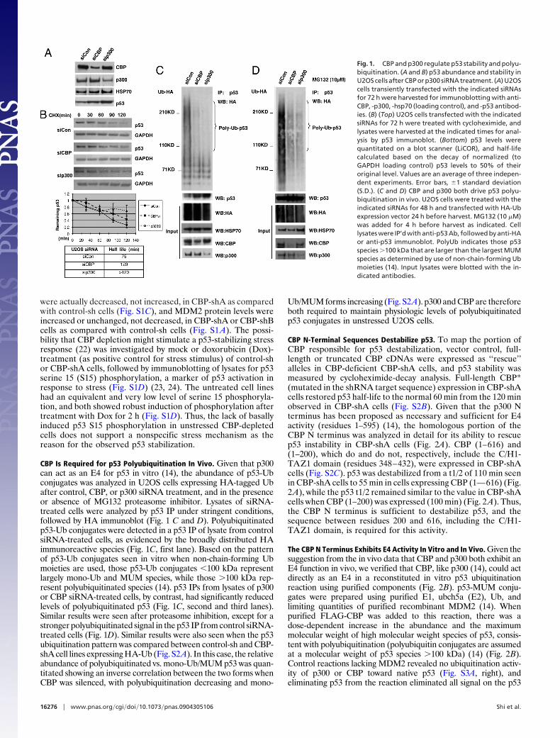
ResultsRegulation of p53 Abundance and Stability by p300 and CBP. To assessthe physiologic contributions of p300 and CBP to p53 regulationin unstressed cells, p300 and CBP were transiently silenced inU2OS cells with specific siRNA duplexes, followed by analysis ofcell lysates for p300, CBP, and p53 levels, as well as determina-tion of p53 half-life (t1/2) by cycloheximide decay (Fig. 1 A andB). p300 siRNA has been reported to increase p53 abundancebut the mechanism was not determined further (21). p300 andCBP siRNAs both caused increases in steady-state p53 abun-dance (Fig. 1 A) and p53 half-life (from 75 min to �2 h and 2 h,respectively; Fig. 1B) compared to that seen in control siRNA-treated cells. Similar gains in p53 abundance were also seen afterstable CBP knockdown by two distinct shRNA sequences (CBP-shA, CBP-shB), and CBP-shA also induced p53 stabilization (Fig.S1A). Demonstrating that these results were not cell type-specificor due to the use of a cancer cell line, siRNA depletion of CBP orp300 in nontransformed MCF10A human breast epithelial cellssimilarly stabilized p53 from t1/2 � 75 min with control siRNA to�2 h for both p300 and CBP siRNAs (Fig. S1B).
Indirect explanations for the increased p53 abundance andstability induced by p300/CBP depletion, such as increased p53mRNA expression, decreased MDM2 expression, or induction ofgenotoxic stress (the latter two of which might stabilize p53) wereinvestigated. RT-PCR analysis showed that p53 mRNA levels
Author contributions: D.S., M.S.P., R.K., and S.R.G. designed research; D.S., M.S.P., R.K., andI.M.L. performed research; A.L.K. contributed new reagents/analytic tools; D.S., M.S.P., R.K.,and S.R.G. analyzed data; and S.R.G. wrote the paper.
The authors declare no conflict of interest.
This article is a PNAS Direct Submission.
1D.S. and M.S.P. contributed equally to this work.
2To whom correspondence should be addressed. E-mail: [email protected].
This article contains supporting information online at www.pnas.org/cgi/content/full/0904305106/DCSupplemental.
www.pnas.org�cgi�doi�10.1073�pnas.0904305106 PNAS � September 22, 2009 � vol. 106 � no. 38 � 16275–16280
CELL
BIO
LOG
Y
were actually decreased, not increased, in CBP-shA as comparedwith control-sh cells (Fig. S1C), and MDM2 protein levels wereincreased or unchanged, not decreased, in CBP-shA or CBP-shBcells as compared with control-sh cells (Fig. S1 A). The possi-bility that CBP depletion might stimulate a p53-stabilizing stressresponse (22) was investigated by mock or doxorubicin (Dox)-treatment (as positive control for stress stimulus) of control-shor CBP-shA cells, followed by immunoblotting of lysates for p53serine 15 (S15) phosphorylation, a marker of p53 activation inresponse to stress (Fig. S1D) (23, 24). The untreated cell lineshad an equivalent and very low level of serine 15 phosphoryla-tion, and both showed robust induction of phosphorylation aftertreatment with Dox for 2 h (Fig. S1D). Thus, the lack of basallyinduced p53 S15 phosphorylation in unstressed CBP-depletedcells does not support a nonspecific stress mechanism as thereason for the observed p53 stabilization.
CBP Is Required for p53 Polyubiquitination In Vivo. Given that p300can act as an E4 for p53 in vitro (14), the abundance of p53-Ubconjugates was analyzed in U2OS cells expressing HA-tagged Ubafter control, CBP, or p300 siRNA treatment, and in the presenceor absence of MG132 proteasome inhibitor. Lysates of siRNA-treated cells were analyzed by p53 IP under stringent conditions,followed by HA immunoblot (Fig. 1 C and D). Polyubiquitinatedp53-Ub conjugates were detected in a p53 IP of lysate from controlsiRNA-treated cells, as evidenced by the broadly distributed HAimmunoreactive species (Fig. 1C, first lane). Based on the patternof p53-Ub conjugates seen in vitro when non-chain-forming Ubmoieties are used, those p53-Ub conjugates �100 kDa representlargely mono-Ub and MUM species, while those �100 kDa rep-resent polyubiquitinated species (14). p53 IPs from lysates of p300or CBP siRNA-treated cells, by contrast, had significantly reducedlevels of polyubiquitinated p53 (Fig. 1C, second and third lanes).Similar results were seen after proteasome inhibition, except for astronger polyubiquitinated signal in the p53 IP from control siRNA-treated cells (Fig. 1D). Similar results were also seen when the p53ubiquitination pattern was compared between control-sh and CBP-shA cell lines expressing HA-Ub (Fig. S2A). In this case, the relativeabundance of polyubiquitinated vs. mono-Ub/MUM p53 was quan-titated showing an inverse correlation between the two forms whenCBP was silenced, with polyubiquitination decreasing and mono-
Ub/MUM forms increasing (Fig. S2A). p300 and CBP are thereforeboth required to maintain physiologic levels of polyubiquitinatedp53 conjugates in unstressed U2OS cells.
CBP N-Terminal Sequences Destabilize p53. To map the portion ofCBP responsible for p53 destabilization, vector control, full-length or truncated CBP cDNAs were expressed as ‘‘rescue’’alleles in CBP-deficient CBP-shA cells, and p53 stability wasmeasured by cycloheximide-decay analysis. Full-length CBP*(mutated in the shRNA target sequence) expression in CBP-shAcells restored p53 half-life to the normal 60 min from the 120 minobserved in CBP-shA cells (Fig. S2B). Given that the p300 Nterminus has been proposed as necessary and sufficient for E4activity (residues 1–595) (14), the homologous portion of theCBP N terminus was analyzed in detail for its ability to rescuep53 instability in CBP-shA cells (Fig. 2A). CBP (1–616) and(1–200), which do and do not, respectively, include the C/H1-TAZ1 domain (residues 348–432), were expressed in CBP-shAcells (Fig. S2C). p53 was destabilized from a t1/2 of 110 min seenin CBP-shA cells to 55 min in cells expressing CBP (1—616) (Fig.2A), while the p53 t1/2 remained similar to the value in CBP-shAcells when CBP (1–200) was expressed (100 min) (Fig. 2 A). Thus,the CBP N terminus is sufficient to destabilize p53, and thesequence between residues 200 and 616, including the C/H1-TAZ1 domain, is required for this activity.
The CBP N Terminus Exhibits E4 Activity In Vitro and In Vivo. Given thesuggestion from the in vivo data that CBP and p300 both exhibit anE4 function in vivo, we verified that CBP, like p300 (14), could actdirectly as an E4 in a reconstituted in vitro p53 ubiquitinationreaction using purified components (Fig. 2B). p53-MUM conju-gates were prepared using purified E1, ubch5a (E2), Ub, andlimiting quantities of purified recombinant MDM2 (14). Whenpurified FLAG-CBP was added to this reaction, there was adose-dependent increase in the abundance and the maximummolecular weight of high molecular weight species of p53, consis-tent with polyubiquitination (polyubiquitin conjugates are assumedat a molecular weight of p53 species �100 kDa) (14) (Fig. 2B).Control reactions lacking MDM2 revealed no ubiquitination activ-ity of p300 or CBP toward native p53 (Fig. S3A, right), andeliminating p53 from the reaction eliminated all signal on the p53
Fig. 1. CBP and p300 regulate p53 stability and polyu-biquitination. (A and B) p53 abundance and stability inU2OS cells after CBP or p300 siRNA treatment. (A) U2OScells transiently transfected with the indicated siRNAsfor 72 h were harvested for immunoblotting with anti-CBP, -p300, -hsp70 (loading control), and -p53 antibod-ies. (B) (Top) U2OS cells transfected with the indicatedsiRNAs for 72 h were treated with cycloheximide, andlysates were harvested at the indicated times for anal-ysis by p53 immunoblot. (Bottom) p53 levels werequantitated on a blot scanner (LiCOR), and half-lifecalculated based on the decay of normalized (toGAPDH loading control) p53 levels to 50% of theiroriginal level. Values are an average of three indepen-dent experiments. Error bars, �1 standard deviation(S.D.). (C and D) CBP and p300 both drive p53 polyu-biquitination in vivo. U2OS cells were treated with theindicated siRNAs for 48 h and transfected with HA-Ubexpression vector 24 h before harvest. MG132 (10 �M)was added for 4 h before harvest as indicated. Celllysates were IP’d with anti-p53 Ab, followed by anti-HAor anti-p53 immunoblot. PolyUb indicates those p53species �100 kDa that are larger than the largest MUMspecies as determined by use of non-chain-forming Ubmoieties (14). Input lysates were blotted with the in-dicated antibodies.
16276 � www.pnas.org�cgi�doi�10.1073�pnas.0904305106 Shi et al.
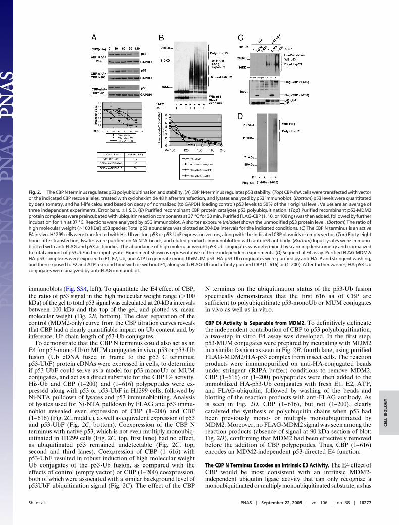
immunoblots (Fig. S3A, left). To quantitate the E4 effect of CBP,the ratio of p53 signal in the high molecular weight range (�100kDa) of the gel to total p53 signal was calculated at 20-kDa intervalsbetween 100 kDa and the top of the gel, and plotted vs. meanmolecular weight (Fig. 2B, bottom). The clear separation of thecontrol (MDM2-only) curve from the CBP titration curves revealsthat CBP had a clearly quantifiable impact on Ub content and, byinference, Ub chain length of p53-Ub conjugates.
To demonstrate that the CBP N terminus could also act as anE4 for p53-mono-Ub or MUM conjugates in vivo, p53 or p53-Ubfusion (Ub cDNA fused in frame to the p53 C terminus;p53-UbF) protein cDNAs were expressed in cells, to determineif p53-UbF could serve as a model for p53-monoUb or MUMconjugates, and act as a direct substrate for the CBP E4 activity.His-Ub and CBP (1–200) and (1–616) polypeptides were ex-pressed along with p53 or p53-UbF in H1299 cells, followed byNi-NTA pulldown of lysates and p53 immunoblotting. Analysisof lysates used for Ni-NTA pulldown by FLAG and p53 immu-noblot revealed even expression of CBP (1–200) and CBP(1–616) (Fig. 2C, middle), as well as equivalent expression of p53and p53-UbF (Fig. 2C, bottom). Coexpression of the CBP Nterminus with native p53, which is not even multiply monoubiq-uitinated in H1299 cells (Fig. 2C, top, first lane) had no effect,as ubiquitinated p53 remained undetectable (Fig. 2C, top,second and third lanes). Coexpression of CBP (1–616) withp53-UbF resulted in robust induction of high molecular weightUb conjugates of the p53-Ub fusion, as compared with theeffects of control (empty vector) or CBP (1–200) coexpression,both of which were associated with a similar background level ofp53UbF ubiquitination signal (Fig. 2C). The effect of the CBP
N terminus on the ubiquitination status of the p53-Ub fusionspecifically demonstrates that the first 616 aa of CBP aresufficient to polyubiquitinate p53-monoUb or MUM conjugatesin vivo as well as in vitro.
CBP E4 Activity Is Separable from MDM2. To definitively delineatethe independent contribution of CBP to p53 polyubiquitination,a two-step in vitro E4 assay was developed. In the first step,p53-MUM conjugates were prepared by incubating with MDM2in a similar fashion as seen in Fig. 2B, fourth lane, using purifiedFLAG-MDM2/HA-p53 complex from insect cells. The reactionproducts were immunopurified on anti-HA-conjugated beadsunder stringent (RIPA buffer) conditions to remove MDM2.CBP (1–616) or (1–200) polypeptides were then added to theimmobilized HA-p53-Ub conjugates with fresh E1, E2, ATP,and FLAG-ubiquitin, followed by washing of the beads andblotting of the reaction products with anti-FLAG antibody. Asis seen in Fig. 2D, CBP (1–616), but not (1–200), clearlycatalyzed the synthesis of polyubiquitin chains when p53 hadbeen previously mono- or multiply monoubiquitinated byMDM2. Moreover, no FLAG-MDM2 signal was seen among thereaction products (absence of signal at 90-kDa section of blot;Fig. 2D), confirming that MDM2 had been effectively removedbefore the addition of CBP polypeptides. Thus, CBP (1–616)encodes an MDM2-independent p53-directed E4 function.
The CBP N Terminus Encodes an Intrinsic E3 Activity. The E4 effect ofCBP would be most consistent with an intrinsic MDM2-independent ubiquitin ligase activity that can only recognize amonoubiquitinated or multiply monoubiquitinated substrate, as has
Fig. 2. The CBP N terminus regulates p53 polyubiquitination and stability. (A) CBP N-terminus regulates p53 stability. (Top) CBP-shA cells were transfected with vectoror the indicated CBP rescue alleles, treated with cycloheximide 48 h after transfection, and lysates analyzed by p53 immunoblot. (Bottom) p53 levels were quantitatedby densitometry, and half-life calculated based on decay of normalized (to GAPDH loading control) p53 levels to 50% of their original level. Values are an average ofthree independent experiments. Error bars, �1 S.D. (B) Purified recombinant CBP protein catalyzes p53 polyubiquitination. (Top) Purified recombinant p53-MDM2protein complexes were preincubated with ubiquitin reaction components at 37 °C for 30 min. Purified FLAG-CBP (1, 10, or 100 ng) was then added, followed by furtherincubation for 1 h at 37 °C. Reactions were analyzed by p53 immunoblot. A shorter exposure (middle) shows the unmodified p53 protein level. (Bottom) The ratio ofhigh molecular weight (�100 kDa) p53 species: Total p53 abundance was plotted at 20-kDa intervals for the indicated conditions. (C) The CBP N terminus is an activeE4 in vivo. H1299 cells were transfected with His-Ub vector, p53 or p53-UbF expression vectors, along with the indicated CBP plasmids or empty vector. (Top) Forty-eighthours after transfection, lysates were purified on Ni-NTA beads, and eluted products immunoblotted with anti-p53 antibody. (Bottom) Input lysates were immuno-blotted with anti-FLAG and p53 antibodies. The abundance of high molecular weight p53-Ub conjugates was determined by scanning densitometry and normalizedto total amount of p53UbF in the input lysate. Experiment shown is representative of three independent experiments. (D) Sequential E4 assay. Purified FLAG-MDM2/HA-p53 complexes were exposed to E1, E2, Ub, and ATP to generate mono-Ub/MUM p53. HA-p53-Ub conjugates were purified by anti-HA IP and stringent washing,and then exposed to E2 and ATP a second time with or without E1, along with FLAG-Ub and affinity purified CBP (1–616) or (1–200). After further washes, HA-p53-Ubconjugates were analyzed by anti-FLAG immunoblot.
Shi et al. PNAS � September 22, 2009 � vol. 106 � no. 38 � 16277
CELL
BIO
LOG
Y

been observed for p300 (14). We investigated whether sequencesarising from the CBP N terminus, which demonstrate E4 activity(Fig. 2 C and D) and promote p53 instability in vivo (Fig. 2A),encode an intrinsic E3 activity that could be clearly separated fromMDM2. Prokaryotically synthesized, purified, GST-CBP (1–616),(1–451), and (452–721) were incubated with Ub, E1, E2 (ubch5a),and ATP in an autoubiquitination reaction, and the reactionproducts detected by anti-Ub immunoblot. Titration of GST-CBP(1–616) or (1–451) with standard ubiquitin reaction components,revealed robust E3 activity (Fig. S3C). GST-CBP (452–721), how-ever, was inactive as an E3 (Fig. S3C). Thus, the CBP polypeptidethat promotes p53 degradation in vivo and p53 polyubiquitinationin vivo and in vitro, also encodes an intrinsic, MDM2-independent,E3 ubiquitin ligase domain. The core sequences responsible for thisactivity appear to lie within at least the N-terminal 451 aa of CBP,and include the C/H1-TAZ1 domain.
CBP Promotes p53 Degradation in the Cytoplasm. Since p300/CBP areconsidered to be nuclear coactivators (25), but p53 and MDM2 bothshuttle between nucleus and cytoplasm (26), the cellular localiza-tion of CBP’s regulation of p53 ubiquitination and degradation wasexplored further. Levels of unmodified p53 were proportionatelyincreased in both nuclear and cytoplasmic fractions in CBP-shA vs.control cells, and controls for nuclear (Rb) and cytoplasmic (actin)proteins revealed that there was little cross-contamination of thefractions (Fig. 3A). Mono-Ub/MUM p53 was found predominantly
in the cytoplasm, and levels of this species in the cytoplasm weresignificantly increased in CBP-shA vs. control cells, either due to theoverall increase in total p53, or a loss of an (E4) activity requiredto convert p53-mono-Ub or p53-MUM conjugates to polyubiquiti-nated forms (Fig. 3A).
To determine if the differential ubiquitination of nuclear andcytoplasmic p53 correlated with different rates of turnover,nuclear and cytoplasmic p53 half-life was measured by cyclo-heximide decay in control and CBP-shA cells (Fig. 3B and Fig.S3D). PARP (nuclear marker) and �-tubulin (cytoplasmicmarker) immunoblots of the fractions from each time pointrevealed negligible cross-contamination (Fig. 3B). Surprisingly,CBP silencing led to stabilization of the normally rapidly de-graded cytoplasmic p53 (t1/2 increase from 50 to 85 min, P �0.01), but had no significant effect on the slower turnover ofnuclear p53 (t1/2 increase from 95 to 100 min, P � 0.63; Fig. 3Band Fig. S3D). Given that p300/CBP are considered to be nuclearproteins (27, 28), but the major effect of CBP depletion was oncytoplasmic p53 half-life, the possibility that cytoplasmic poolsof CBP or p300 might influence p53 metabolism was explored.
CBP/p300 Are Partly Cytoplasmic. CBP and p300 are localized inPML oncogenic domains (PODs) and senescence-associated nu-clear bodies, respectively (27, 29). To gain a more precise under-standing of p300 and CBP subcellular localization, their abundancein cytoplasmic and nuclear fractions of U2OS cells was determined(Fig. 3C). The nuclear:cytoplasmic abundance ratios of Rb andactin, as determined by densitometry, revealed that the upper limitof cross-contamination of one fraction into the other was no greaterthan 10% in either direction (Fig. 3C). CBP and p300 werepredominantly nuclear, as expected, but a substantial fraction ofCBP was cytoplasmic, whereas a small but reproducible fraction ofp300 was also observed in the cytoplasmic fraction (Fig. 3C). Thesefractionation results were further confirmed by immunofluorescentstaining of U2OS cells for CBP and p300, where a clear cytoplasmicsignal was detected for CBP, but p300 appeared predominantlynuclear (Fig. S3E). The specificity of the CBP immunofluorescencesignal was confirmed by the near complete loss of CBP staining seenin CBP-shA cells (Fig. S3E).
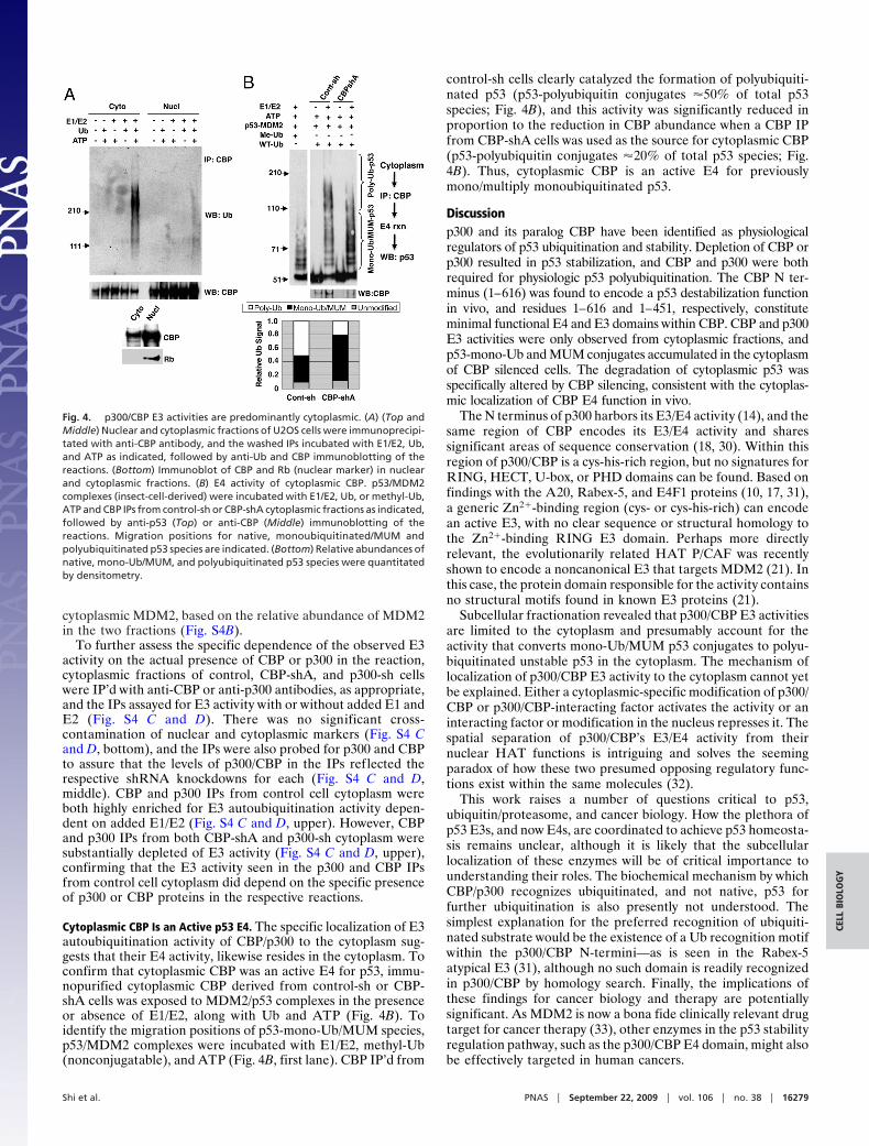
CBP/p300 E3 Activities Are Exclusively Cytoplasmic. To determine ifnuclear and cytoplasmic CBP or p300 molecules have anydifferential capacities to influence p53 ubiquitination, nuclearand cytoplasmic CBP/p300 were assayed for E3 autoubiquitina-tion activity. Nuclear and cytoplasmic fractions of U2OS cellswere IP’d with anti-CBP or p300 antibody, and the IPs wereassayed for E3 activity by the addition of ubiquitin, E1, E2(ubch5a), and ATP, followed by Ub, CBP, or p300 immunoblot-ting (Fig. 4A and Fig. S4A). Dropout control reactions lackingE1/E2, Ub, or ATP were also performed to demonstrate that anyUb chains observed were the result of bona fide in vitro E3activity and not contamination by cellular Ub chains bindingnonspecifically to the IPs (Fig. 4A and Fig. S4A). CBP and p300were observed in the cytoplasm and nuclear fractions as ex-pected, and controls indicated little cross-contamination (Fig.4A, bottom, and Fig. S4A, bottom). Surprisingly, CBP and p300derived from cytoplasm demonstrated robust E3 autoubiquiti-nation activity dependent on the simultaneous addition ofE1/E2, Ub, and ATP, whereas nuclear-derived CBP/p300 exhib-ited much lower or undetectable activity (Fig. 4A and Fig. S4A).CBP/p300 immunoblotting of the reactions revealed that the lackof E3 activity associated with nuclear CBP/p300 could not beexplained by lesser abundance (Fig. 4A and Fig. S4A). The lackof nuclear CBP autoubiquitination activity could also not beexplained by nonspecific inhibition by extraction conditions orinhibitory factors within the nuclear fraction, as immunopurifiednuclear MDM2 exhibited easily detected autoubiquitinationactivity, which if anything, had a higher specific activity than
Fig. 3. Subcellular localization of CBP/p300 E3 activity. (A) p53 localization inCBP-deficient cells. Nuclear or cytoplasmic fractions of control or CBP-shA cellswere analyzed by immunoblotting with Rb (nuclear marker), actin (cytoplasmicmarker), and p53 antibodies. (B) CBP regulation of p53 turnover in the cytoplasmand nucleus. Control and CBP-shA U2OS cells treated with cycloheximide werefractionated into nuclear and cytoplasmic fractions at the indicated time points.(Top three panels) The fractions were immunoblotted for p53, PARP (nuclearmarker),andactin (cytoplasmicmarker). (Lower left)Subcellular fractions (time�0) from control and CBP-shA cells were immunoblotted with anti-CBP, anti-PARP,anti-�-tubulin, and anti-p53 antibodies. (Lower right) Determination of nuclearandcytoplasmicp53half-lifefromCont-shandCBP-shAcells.Result is theaveragehalf-life from four separate experiments. * indicates significant P � 0.01 fordifference in cytoplasmic p53 t1/2 between Cont-sh and CBP-shA. (C) Subcellularlocalization of p300 and CBP. U2OS cells were fractionated into nuclear andcytoplasmic fractions, and the fractions were immunoblotted for CBP, p300, Rb(nuclear marker), and actin (cytoplasmic marker).
16278 � www.pnas.org�cgi�doi�10.1073�pnas.0904305106 Shi et al.
cytoplasmic MDM2, based on the relative abundance of MDM2in the two fractions (Fig. S4B).
To further assess the specific dependence of the observed E3activity on the actual presence of CBP or p300 in the reaction,cytoplasmic fractions of control, CBP-shA, and p300-sh cellswere IP’d with anti-CBP or anti-p300 antibodies, as appropriate,and the IPs assayed for E3 activity with or without added E1 andE2 (Fig. S4 C and D). There was no significant cross-contamination of nuclear and cytoplasmic markers (Fig. S4 Cand D, bottom), and the IPs were also probed for p300 and CBPto assure that the levels of p300/CBP in the IPs reflected therespective shRNA knockdowns for each (Fig. S4 C and D,middle). CBP and p300 IPs from control cell cytoplasm wereboth highly enriched for E3 autoubiquitination activity depen-dent on added E1/E2 (Fig. S4 C and D, upper). However, CBPand p300 IPs from both CBP-shA and p300-sh cytoplasm weresubstantially depleted of E3 activity (Fig. S4 C and D, upper),confirming that the E3 activity seen in the p300 and CBP IPsfrom control cell cytoplasm did depend on the specific presenceof p300 or CBP proteins in the respective reactions.
Cytoplasmic CBP Is an Active p53 E4. The specific localization of E3autoubiquitination activity of CBP/p300 to the cytoplasm sug-gests that their E4 activity, likewise resides in the cytoplasm. Toconfirm that cytoplasmic CBP was an active E4 for p53, immu-nopurified cytoplasmic CBP derived from control-sh or CBP-shA cells was exposed to MDM2/p53 complexes in the presenceor absence of E1/E2, along with Ub and ATP (Fig. 4B). Toidentify the migration positions of p53-mono-Ub/MUM species,p53/MDM2 complexes were incubated with E1/E2, methyl-Ub(nonconjugatable), and ATP (Fig. 4B, first lane). CBP IP’d from
control-sh cells clearly catalyzed the formation of polyubiquiti-nated p53 (p53-polyubiquitin conjugates �50% of total p53species; Fig. 4B), and this activity was significantly reduced inproportion to the reduction in CBP abundance when a CBP IPfrom CBP-shA cells was used as the source for cytoplasmic CBP(p53-polyubiquitin conjugates �20% of total p53 species; Fig.4B). Thus, cytoplasmic CBP is an active E4 for previouslymono/multiply monoubiquitinated p53.
Discussionp300 and its paralog CBP have been identified as physiologicalregulators of p53 ubiquitination and stability. Depletion of CBP orp300 resulted in p53 stabilization, and CBP and p300 were bothrequired for physiologic p53 polyubiquitination. The CBP N ter-minus (1–616) was found to encode a p53 destabilization functionin vivo, and residues 1–616 and 1–451, respectively, constituteminimal functional E4 and E3 domains within CBP. CBP and p300E3 activities were only observed from cytoplasmic fractions, andp53-mono-Ub and MUM conjugates accumulated in the cytoplasmof CBP silenced cells. The degradation of cytoplasmic p53 wasspecifically altered by CBP silencing, consistent with the cytoplas-mic localization of CBP E4 function in vivo.
The N terminus of p300 harbors its E3/E4 activity (14), and thesame region of CBP encodes its E3/E4 activity and sharessignificant areas of sequence conservation (18, 30). Within thisregion of p300/CBP is a cys-his-rich region, but no signatures forRING, HECT, U-box, or PHD domains can be found. Based onfindings with the A20, Rabex-5, and E4F1 proteins (10, 17, 31),a generic Zn2�-binding region (cys- or cys-his-rich) can encodean active E3, with no clear sequence or structural homology tothe Zn2�-binding RING E3 domain. Perhaps more directlyrelevant, the evolutionarily related HAT P/CAF was recentlyshown to encode a noncanonical E3 that targets MDM2 (21). Inthis case, the protein domain responsible for the activity containsno structural motifs found in known E3 proteins (21).
Subcellular fractionation revealed that p300/CBP E3 activitiesare limited to the cytoplasm and presumably account for theactivity that converts mono-Ub/MUM p53 conjugates to polyu-biquitinated unstable p53 in the cytoplasm. The mechanism oflocalization of p300/CBP E3 activity to the cytoplasm cannot yetbe explained. Either a cytoplasmic-specific modification of p300/CBP or p300/CBP-interacting factor activates the activity or aninteracting factor or modification in the nucleus represses it. Thespatial separation of p300/CBP’s E3/E4 activity from theirnuclear HAT functions is intriguing and solves the seemingparadox of how these two presumed opposing regulatory func-tions exist within the same molecules (32).
This work raises a number of questions critical to p53,ubiquitin/proteasome, and cancer biology. How the plethora ofp53 E3s, and now E4s, are coordinated to achieve p53 homeosta-sis remains unclear, although it is likely that the subcellularlocalization of these enzymes will be of critical importance tounderstanding their roles. The biochemical mechanism by whichCBP/p300 recognizes ubiquitinated, and not native, p53 forfurther ubiquitination is also presently not understood. Thesimplest explanation for the preferred recognition of ubiquiti-nated substrate would be the existence of a Ub recognition motifwithin the p300/CBP N-termini—as is seen in the Rabex-5atypical E3 (31), although no such domain is readily recognizedin p300/CBP by homology search. Finally, the implications ofthese findings for cancer biology and therapy are potentiallysignificant. As MDM2 is now a bona fide clinically relevant drugtarget for cancer therapy (33), other enzymes in the p53 stabilityregulation pathway, such as the p300/CBP E4 domain, might alsobe effectively targeted in human cancers.
Fig. 4. p300/CBP E3 activities are predominantly cytoplasmic. (A) (Top andMiddle) Nuclear and cytoplasmic fractions of U2OS cells were immunoprecipi-tated with anti-CBP antibody, and the washed IPs incubated with E1/E2, Ub,and ATP as indicated, followed by anti-Ub and CBP immunoblotting of thereactions. (Bottom) Immunoblot of CBP and Rb (nuclear marker) in nuclearand cytoplasmic fractions. (B) E4 activity of cytoplasmic CBP. p53/MDM2complexes (insect-cell-derived) were incubated with E1/E2, Ub, or methyl-Ub,ATP and CBP IPs from control-sh or CBP-shA cytoplasmic fractions as indicated,followed by anti-p53 (Top) or anti-CBP (Middle) immunoblotting of thereactions. Migration positions for native, monoubiquitinated/MUM andpolyubiquitinated p53 species are indicated. (Bottom) Relative abundances ofnative, mono-Ub/MUM, and polyubiquitinated p53 species were quantitatedby densitometry.
Shi et al. PNAS � September 22, 2009 � vol. 106 � no. 38 � 16279
CELL
BIO
LOG
Y

Materials and MethodsCell Culture and Plasmids. U2OS cells were grown in DMEM supplemented with10% FBS and antibiotics. Cells were treated, where noted, with 100 �g/mLcycloheximide, 2 �M Dox, or 10 �M MG-132 (Sigma). Plasmid transfection wasdone with Fugene 6 (Roche), and siRNA transfections used Oligofectamine(Invitrogen). pRSV-CBPmyc (34) and pcDNA-UbHA (9) have been described. Togenerate pN8.Flag-CBP (1–616) and -CBP (1–200), CBP fragments were PCR-amplified from pRSV-CBPmyc and cloned into pN8.Flag vector using BamHIand EcoR1. An shRNA-resistant CBP allele (pRSV-CBP*myc) was generated bysilent mutation of the shRNA target sequence (AACTCCAATAGC mutated toAATAGTAACTCT; CBP residues 190–193) within pRSV-CBPmyc. pGEX-CBP (1–616) and (1–200) were generated by cloning the indicated PCR-amplifiedfragments into pGEX-2tk. pCDNA3-p53-UbF, which has the Ub cDNA clonedin-frame to the 3� end of the p53 cDNA, was the generous gift of ChristineBlattner, and MT107 His-Ub expression vector has been described (35).
Western Blotting, Immunofluorescence, and Immunoprecipitation. For westernblotanalyses, cellswere lysed incoldNETN240buffer (20mMHEPES,pH7.4,2mMMgCl2, 10 �M ZnCl2, 240 mM NaCl, 0.2% Triton-X 100), supplemented withcomplete EDTA-free tablets (Roche). For immunoprecipitation of whole celllysates, cells were lysed in cold RIPA buffer [50 mM Tris-HCl, pH 7.4, 250 mM NaCl,10 mM MgCl2, 10 �M ZnCl2, 1% Triton X-100, 0.5% DOC supplemented with fresh5mMNEMandcompleteEDTA-freetablets (Roche)]. Immunoprecipitationsfromwhole cell lysates were performed in the lysis buffer overnight, followed bycapture with Protein A agarose (Upstate) and five washes in lysis buffer. Westernblot signals were quantified after visualization of primary antibody by HRP-conjugated secondary antibody and enhanced chemiluminescence or by fluores-cent-labeled secondary antibody and detection by Odyssey blot scanner (LiCor),using ImageJ National Institutes of Health (NIH) software.
In Vitro E3 Assays. To determine CBP/p300 or MDM2 E3 ligase activity, CBP,p300, or MDM2 were immunoprecipitated from 0.7 mg cytoplasm or 0.2 mgnuclear fractions diluted with high salt buffer [10 mM HEPES (pH 7.5), 150 mMNaCl, 150 mM KCl, 1 mM MgCl2, 0.5% Triton X-100, supplemented with fresh5 mM NEM and protease inhibitors) using A-22, N-15, or D-7 antibodiesfollowed by protein A Sepharose. The IPs were washed in high salt buffer threetimes followed by two washes in Ub buffer (25 mM HEPES, pH 7.4, 10 mM NaCl,3 mM MgCl2, 0.05% Triton X-100, 0.5 mM DTT, 3 mM Mg-ATP). The washedand equilibrated IPs were then incubated with 100 ng E1 (rabbit; BostonBiochem), 25 ng E2 (UbcH5a, human recombinant; Boston Biochem), and 5 �gUb (human recombinant; Boston Biochem) for 60 min at 37 °C. Reactions were
then stopped by the addition of sample buffer, followed by SDS-PAGE andimmunoblotting. E3 assays using purified GST-CBP polypeptides were per-formed as described (14). GST-CBP (1–616), (1–451), and (452–721) (36) wereexpressed in BL21 cells and purified using glutathione-Sepharose (GE Health-care). Purified GST-CBP (0.1, 0.2 or 0.5 �g) (1–616), (1–451), or (452–721)proteins were added to ubiquitin reaction components as indicated andincubated at 37 °C for 60 min, followed by analysis of the reaction productswith anti-Ub antibody.
One-Step E4 Assay. To determine CBP E4 ligase activity, insect-cell-derivedhuman p53 and FLAG-MDM2 were purified as a complex using FLAG M2(Sigma) agarose as described (14), then incubated with ubiquitin reactioncomponents (100 ng E1, rabbit; Boston Biochem), 25 ng E2 (UbcH5a, humanrecombinant; Boston Biochem), and 5 ng Ub (human recombinant; BostonBiochem) at 37 °C for 30 min. FLAG-CBP (1, 10, or 100 �g) immunopurifiedfrom baculovirus-infected Sf9 insect cells (with anti-FLAG resin/FLAG peptideelution) (35) or CBP IP’d from U2OS cytoplasmic fraction (see In Vitro E3 Assaysection) was then added, followed by further incubation of the reaction at37 °C for 60 min and p53 immunoblot of the reaction products.
Two-Step E4 Assay. To determine E4 ligase activity of CBP, a two-step ubiquiti-nation reaction was performed. First, insect-cell-derived p53 was ubiquitinatedby MDM2 as described above. To remove MDM2 and nontagged ubiquitin, p53was then immunoprecipitated and washed three times with RIPA buffer, twotimes with PBS, and two times with Ub buffer. Immobilized p53 was then mixedwith second ubiquitination reaction components [100 ng E1, 25 ng UbcH5a, 5 �gFlag-tagged Ub (Boston Biochem)] along with affinity-purified Flag-CBP (1–200)or (1–616) polypeptides obtained from transfected U2OS cells and incubated at37 °C for 60 min. To remove CBP autoubiquitination products, beads werewashedthreetimeswithRIPAbuffer.p53waselutedbyboilingwithNuPAGELDSsample buffer, separated by SDS-PAGE, and visualized by immunoblotting withanti-Flag antibodies.
Additional Materials and Methods are available in SI Materials andMethods.
ACKNOWLEDGMENTS. We thank C.M. Chiang (UT Southwestern, Dallas, TX)for generously providing FLAG-CBP baculovirus, C. Blattner (Forschungszen-trum Karlsruhe, Karlsruhe, Germany) for providing pCDNA3.p53-UbF, andmembers of the Altieri and Grossman labs for helpful discussions. S.G. wassupported by a Kimmel Scholar Award, and R01CA107532 from the NationalCancer Institute (NCI).
1. Michael D, Oren M (2002) The p53 and Mdm2 families in cancer. Curr Opin Genet Dev12:53–59.
2. Moll UM, Wolff S, Speidel D, Deppert W (2005) Transcription-independent pro-apo-ptotic functions of p53. Curr Opin Cell Biol 17:631–636.
3. Slee EA, O’Connor DJ, Lu X (2004) To die or not to die: How does p53 decide? Oncogene23:2809–2818.
4. Bode AM, Dong Z (2004) Post-translational modification of p53 in tumorigenesis. NatRev Cancer 4:793–805.
5. Morgunkova A, Barlev NA (2006) Lysine methylation goes global. Cell Cycle 5:1308–1312.6. Brooks CL, Gu W (2003) Ubiquitination, phosphorylation, and acetylation: The molec-
ular basis for p53 regulation. Curr Opin Cell Biol 15:164–171.7. Brooks CL, Li M, Gu W (2004) Monoubiquitination: The signal for p53 nuclear export?
Cell Cycle 3:436–438.8. Li M, et al. (2003) Mono- versus polyubiquitination: Differential control of p53 fate by
Mdm2. Science 302:1972–1975.9. Kaur M, Pop M, Shi D, Brignone C, Grossman SR (2006) hHR23B is required for
genotoxic-specific activation of p53 and apoptosis. Oncogene 26:1231–1237.10. Le Cam L, et al. (2006) E4F1 is an atypical ubiquitin ligase that modulates p53 effector
functions independently of degradation. Cell 127:775–788.11. Dornan D, et al. (2004) The ubiquitin ligase COP1 is a critical negative regulator of p53.
Nature 429:86–92.12. Chen D, et al. (2005) ARF-BP1/Mule is a critical mediator of the ARF tumor suppressor.
Cell 121:1071–1083.13. Brooks CL, Gu W (2006) p53 ubiquitination: Mdm2 and beyond. Mol Cell 21:307–315.14. Grossman SR, et al. (2003) Polyubiquitination of p53 by a ubiquitin ligase activity of
p300. Science 300:342–344.15. Sui G, et al. (2004) Yin Yang 1 is a negative regulator of p53. Cell 117:859–872.16. Lai Z, et al. (2001) Human mdm2 mediates multiple mono-ubiquitination of p53 by a
mechanism requiring enzyme isomerization. J Biol Chem 276:31357–31367.17. Wertz IE, et al. (2004) De-ubiquitination and ubiquitin ligase domains of A20 down-
regulate NF-kappaB signalling. Nature 430:694–699.18. Arany Z, Sellers WR, Livingston DM, Eckner R (1994) E1A-associated p300 and CREB-
associated CBP belong to a conserved family of coactivators. Cell 77:799–800.19. Freedman SJ, et al. (2002) Structural basis for recruitment of CBP/p300 by hypoxia-
inducible factor-1 alpha. Proc Natl Acad Sci USA 99:5367–5372.20. De Guzman RN, Wojciak JM, Martinez-Yamout MA, Dyson HJ, Wright PE (2005)
CBP/p300 TAZ1 domain forms a structured scaffold for ligand binding. Biochemistry44:490–497.
21. Linares LK, et al. (2007) Intrinsic ubiquitination activity of PCAF controls the stability ofthe oncoprotein Hdm2. Nat Cell Biol 9:331–338.
22. Oren M, et al. (2002) Regulation of p53: Intricate loops and delicate balances. Ann NY Acad Sci 973:374–383.
23. Shieh SY, Ikeda M, Taya Y, Prives C (1997) DNA damage-induced phosphorylation ofp53 alleviates inhibition by MDM2. Cell 91:325–334.
24. Siliciano JD, et al. (1997) DNA damage induces phosphorylation of the amino terminusof p53. Genes Dev 11:3471–3481.
25. Goodman RH, Smolik S (2000) CBP/p300 in cell growth, transformation, and develop-ment. Genes Dev 14:1553–1577.
26. Tao W, Levine AJ (1999) Nucleocytoplasmic shuttling of oncoprotein Hdm2 is requiredfor Hdm2-mediated degradation of p53. Proc Natl Acad Sci USA 96:3077–3080.
27. LaMorte VJ, Dyck JA, Ochs RL, Evans RM (1998) Localization of nascent RNA and CREBbinding protein with the PML-containing nuclear body. Proc Natl Acad Sci USA95:4991–4996.
28. Eckner R, et al. (1994) Molecular cloning and functional analysis of the adenovirusE1A-associated 300-kD protein (p300) reveals a protein with properties of a transcrip-tional adaptor. Genes Dev 8:869–884.
29. Pedeux R, et al. (2005) ING2 regulates the onset of replicative senescence by inductionof p300-dependent p53 acetylation. Mol Cell Biol 25:6639–6648.
30. Lundblad JR, Kwok RP, Laurance ME, Harter ML, Goodman RH (1995) AdenoviralE1A-associated protein p300 as a functional homologue of the transcriptional co-activator CBP. Nature 374:85–88.
31. Mattera R, Tsai YC, Weissman AM, Bonifacino JS (2006) The Rab5 guanine nucle-otide exchange factor Rabex-5 binds ubiquitin (Ub) and functions as a Ub ligasethrough an atypical Ub-interacting motif and a zinc finger domain. J Biol Chem281:6874 – 6883.
32. Grossman SR (2001) p300/CBP/p53 interaction and regulation of the p53 response. EurJ Biochem 268:2773–2778.
33. Vassilev LT (2007) MDM2 inhibitors for cancer therapy. Trends Mol Med 13:23–31.34. Kazantsev A, Preisinger E, Dranovsky A, Goldgaber D, Housman D (1999) Insoluble
detergent-resistant aggregates form between pathological and nonpathologicallengths of polyglutamine in mammalian cells. Proc Natl Acad Sci USA 96:11404 –11409.
35. Treier M, Staszewski LM, Bohmann D (1994) Ubiquitin-dependent c-Jun degradation invivo is mediated by the delta domain. Cell 78:787–798.
16280 � www.pnas.org�cgi�doi�10.1073�pnas.0904305106 Shi et al.