ecological and evolutionary science crossm · decades (3–6).anthracis b. has two circular,...
TRANSCRIPT
Genomic Characterization and Copy Number Variation ofBacillus anthracis Plasmids pXO1 and pXO2 in a HistoricalCollection of 412 Strains
Angela Pena-Gonzalez,a Luis M. Rodriguez-R,d Chung K. Marston,b Jay E. Gee,b Christopher A. Gulvik,b Cari B. Kolton,b
Elke Saile,b Michael Frace,c Alex R. Hoffmaster,b Konstantinos T. Konstantinidisa,d
aSchool of Biological Sciences, Georgia Institute of Technology, Atlanta, Georgia, USAbBacterial Special Pathogens Branch, Division of High-Consequence Pathogens and Pathology, NationalCenter for Emerging and Zoonotic Infectious Diseases, Centers for Disease Control and Prevention, Atlanta,Georgia, USA
cBiotechnology Core Facility Branch, Division of Scientific Resources, National Center for Emerging andZoonotic Infectious Diseases, Centers for Disease Control and Prevention, Atlanta, Georgia, USA
dSchool of Civil & Environmental Engineering, Georgia Institute of Technology, Atlanta, Georgia, USA
ABSTRACT Bacillus anthracis plasmids pXO1 and pXO2 carry the main virulence fac-tors responsible for anthrax. However, the extent of copy number variation withinthe species and how the plasmids are related to pXO1/pXO2-like plasmids in otherspecies of the Bacillus cereus sensu lato group remain unclear. To gain new insightsinto these issues, we sequenced 412 B. anthracis strains representing the total phylo-genetic and ecological diversity of the species. Our results revealed that B. anthracisgenomes carried, on average, 3.86 and 2.29 copies of pXO1 and pXO2, respectively,and also revealed a positive linear correlation between the copy numbers of pXO1and pXO2. No correlation between the plasmid copy number and the phylogeneticrelatedness of the strains was observed. However, genomes of strains isolated fromanimal tissues generally maintained a higher plasmid copy number than genomes ofstrains from environmental sources (P � 0.05 [Welch two-sample t test]). Compari-sons against B. cereus genomes carrying complete or partial pXO1-like and pXO2-likeplasmids showed that the plasmid-based phylogeny recapitulated that of the mainchromosome, indicating limited plasmid horizontal transfer between or within thesespecies. Comparisons of gene content revealed a closed pXO1 and pXO2 pange-nome; e.g., plasmids encode �8 unique genes, on average, and a single large frag-ment deletion of pXO1 in one B. anthracis strain (2000031682) was detected. Collec-tively, our results provide a more complete view of the genomic diversity ofB. anthracis plasmids, their copy number variation, and the virulence potential ofother Bacillus species carrying pXO1/pXO2-like plasmids.
IMPORTANCE Bacillus anthracis microorganisms are of historical and epidemiologicalimportance and are among the most homogenous bacterial groups known, eventhough the B. anthracis genome is rich in mobile elements. Mobile elements cantrigger the diversification of lineages; therefore, characterizing the extent of genomicvariation in a large collection of strains is critical for a complete understanding ofthe diversity and evolution of the species. Here, we sequenced a large collection ofB. anthracis strains (�400) that were recovered from human, animal, and environ-mental sources around the world. Our results confirmed the remarkable stability ofgene content and synteny of the anthrax plasmids and revealed no signal of plas-mid exchange between B. anthracis and pathogenic B. cereus isolates but rather pre-dominantly vertical descent. These findings advance our understanding of the biol-ogy and pathogenomic evolution of B. anthracis and its plasmids.
Received 7 May 2018 Accepted 28 July2018 Published 14 August 2018
Citation Pena-Gonzalez A, Rodriguez-R LM,Marston CK, Gee JE, Gulvik CA, Kolton CB, SaileE, Frace M, Hoffmaster AR, Konstantinidis KT.2018. Genomic characterization and copynumber variation of Bacillus anthracis plasmidspXO1 and pXO2 in a historical collection of 412strains. mSystems 3:e00065-18. https://doi.org/10.1128/mSystems.00065-18.
Editor Rup Lal, University of Delhi
Copyright © 2018 Pena-Gonzalez et al. This isan open-access article distributed under theterms of the Creative Commons Attribution 4.0International license.
Address correspondence to Alex R. Hoffmaster,[email protected], or Konstantinos T.Konstantinidis, [email protected].
RESEARCH ARTICLEEcological and Evolutionary Science
crossm
July/August 2018 Volume 3 Issue 4 e00065-18 msystems.asm.org 1
on Septem
ber 16, 2018 by guesthttp://m
systems.asm
.org/D
ownloaded from
KEYWORDS Bacillus anthracis, anthrax-like B. cereus, pXO1, pXO2, pathogenomics,phylogenomics
Bacillus anthracis, the etiological agent of anthrax, is a Gram-positive endospore-forming bacterium belonging to the Bacillus cereus sensu lato group (1, 2). Dormant
spores represent the infecting form of the bacterium and can remain viable in soils fordecades (3–6). B. anthracis has two circular, extrachromosomal DNA plasmids, pXO1and pXO2, which carry the major virulence factors required for pathogenesis (6, 7).pXO1 carries the genes that encode the following anthrax toxin proteins: protectiveantigen (PA), lethal factor (LF), and edema factor (EF). These proteins act in binarycombinations to produce the two anthrax toxins edema toxin (PA and EF) and lethaltoxin (PA and LF) (6–9). Plasmid pXO2 harbors the genes that encode the cap operonresponsible for the production of a polyglutamate capsule, which allows the pathogento evade the host immune response by protecting itself from phagocytosis (6–9).
Given the severity of the disease and the fact that this microorganism can be usedas a biological weapon, it is important to characterize the diversity of the two virulenceplasmids in a large collection of strains. Currently, plasmid detection is mainly accom-plished by amplification of specific markers through PCR (7, 10). Although this approachis relatively rapid, it can miss plasmids that have diverged in sequence and cannotreveal the full gene content of plasmids. In addition, the plasmid copy number and theextent of copy number variation among members of B. anthracis are still unclear. Forexample, by using quantitative PCR (qPCR), Coker et al. reported ratios of up to 40.5copies of plasmid pXO1 and 5.4 copies of plasmid pXO2 per genome (8), while Pilo etal. reported 10.89 as the average number of copies for pXO1 and 1.59 for pXO2 (11).Using digital PCR (dPCR) in analyses of three isolates, Straub et al. reported that thereare likely 3 to 4 copies of pXO1 per cell and 1 to 2 copies of pXO2 (12). Sequence-basedprojects have also revealed that there are likely 2 to 3 copies of pXO1 per chromosomecopy (13). An important limitation in those previous estimates was that they wereperformed with a relatively small number of isolates, which can bias the characteriza-tion of the population copy number variation. In addition, previous studies havesuggested that the virulence levels of B. anthracis strains carrying both plasmids candiffer depending on the copy number of the plasmids (8). These results underscore thenecessity to accurately quantify plasmid copy variation in a large collection of diverseB. anthracis isolates and evaluate whether plasmid copy number is a phylogeneticallyconserved trait. High-throughput, sequence-based methods not only can detect andquantify plasmid copy number but also can elucidate gene content and sequencediversity, which ultimately will allow better understanding of the pathogenomic evo-lution within the group and with other close relatives. Several studies have alreadycharacterized the phylogenetic relationships and population structure of hundreds ofB. anthracis isolates in France, the Netherlands, and the United States using high-resolution, sequence-based methods such as those analyzing single nucleotide poly-morphisms (SNPs) (1–3). The major results from these studies have shown that B. an-thracis isolates are highly clonal with remarkably stable genomes and low intraspeciesdiversity and can be placed into 1 of 12 conserved lineages defined by canonical SNPs(CanSNPs). However, no studies to date have focused on characterizing plasmid diver-sity and copy number variation in large B. anthracis data sets.
Further, the B. anthracis genome, despite its observed stability, is rich in mobileelements (transposases, resolvases, and integrases), which could be an important factorin plasmid gene content diversification and horizontal transfer (14). Whether or not thepXO1 and pXO2 plasmids are mobile and can be transferred between B. anthracisgenomes as well as to and from other members of the Bacillus cereus sensu lato groupremains speculative, but that issue might be directly related to the virulence of thegenomes and the evolutionary history of the plasmids. Gene transfer and deletion arealso important for classification since these species are typically classified based ontheir plasmid and virulent factor content (as opposed to phylogeny) in this group.
Pena-Gonzalez et al.
July/August 2018 Volume 3 Issue 4 e00065-18 msystems.asm.org 2
on Septem
ber 16, 2018 by guesthttp://m
systems.asm
.org/D
ownloaded from

Finally, the phylogenetic relationships within the Bacillus cereus sensu lato group arestill problematic. B. anthracis belongs to the B. cereus sensu lato group, which alsoincludes two other main species: B. cereus sensu stricto and B. thuringiensis (15, 16).These species were initially recognized and established because they exhibited thefollowing distinct phenotypic traits: B. anthracis was identified as the causative agent ofanthrax (15); B. thuringiensis was recognized as an entomopathogenic bacterium char-acterized by the production of parasporal crystal proteins (Cry and Cyt), which havebeen widely used as a natural pesticide (16); and finally, B. cereus sensu stricto, initiallyrecognized as a common soil-dwelling microorganism, colonizes the gut of inverte-brates as a symbiotic microorganism and is also an opportunistic human pathogen (17,18). DNA hybridization techniques, 16S rRNA-based typing, and multilocus sequencetyping (MLST) schemes have progressively revealed limited genomic dissimilaritiesexisting among these species, demonstrating that they are more closely related thanhad initially been considered (19). This, and the fact that the main phenotypic traits forclassification are carried in plasmids, has led to discussion on whether or not themembers of the B. cereus sensu lato group should be considered a single species withcharacterized ecotypes and pathotypes (16, 19).
Therefore, full-genome analysis of newly sequenced B. anthracis strains and repre-sentative strains in the B. cereus sensu lato group is critical to further elucidate the truephylogenetic relationships within the group. In addition, B. cereus strains encodinggenetic determinants that confer pathogenic capabilities similar to those of B. anthracishave been described previously (20–24). Marston et al. (20) and Hoffmaster et al. (21)reported the isolation of B. cereus strains producing anthrax-like diseases in humanswith clinical presentations of pneumonia and cutaneous lesions in North America,respectively. More recently, Antonation et al. reported the collection of four atypicalB. cereus isolates (designated B. cereus bv. anthracis) from dead mammals (chimpan-zees, gorillas, elephants, and goats) in west and central Africa (23). These isolatesharbored virulence plasmids similar to those of the B. anthracis Ames strain. We havealso recently described the genome of B. cereus strain LA2007, a human-pathogenicisolate carrying a pXO1-like plasmid that showed 99.70% average nucleotide identity(ANI) to B. anthracis Ames pXO1 (25). Interestingly, the pXO1-like plasmids of thepathogenic B. cereus strains reported to date are similar but not identical to those foundin B. anthracis. Therefore, determining the genetic backbone and phylogenetic diversityof the pXO1/pXO2-like plasmids is critical, not only to develop more accurate detectiontools, but also to understand the pathogenomic evolution of virulence determinantswithin the B. cereus sensu lato group.
In this study, we used next-generation sequencing data to detect, quantify, andcharacterize the full genomic content of B. anthracis plasmids pXO1 and pXO2 in acollection of 412 newly sequenced strains that represent the global diversity of thespecies recovered to date. We also compared the phylogenetic diversity of B. anthracisrepresentatives with that of a set of 106 available/reference B. cereus sensu lato strainsthat included nonpathogenic strains as well as pathogenic strains carrying pXO1-likeplasmids.
RESULTSEstimated plasmid copy number and covariance. In this study, a total of 412
B. anthracis strains were newly sequenced. The results of the whole-genome compar-ison of these genomes will be reported elsewhere; here, we focused our analyses on theplasmid sequences. The libraries had an average sequencing depth of 135.4�, with amedian value of 128.3� and a minimum value of 9.8�. To estimate pXO1 and pXO2copy numbers, we calculated the ratio of plasmid sequence depth (using B. anthracisAmes ancestor plasmid sequences as references to recruit reads) to the averagesequencing depth for the chromosome. We identified a total of 58 and 42 strains thatcompletely lacked pXO1 and pXO2, respectively, or that had too few reads (i.e., �2�
sequencing depth after subsampling; see Materials and Methods for details) mappingon the plasmid (i.e., 42 and 62 strains for pXO1 and pXO2, respectively) and that were
B. anthracis Plasmid Diversity and Copy Number Estimation
July/August 2018 Volume 3 Issue 4 e00065-18 msystems.asm.org 3
on Septem
ber 16, 2018 by guesthttp://m
systems.asm
.org/D
ownloaded from
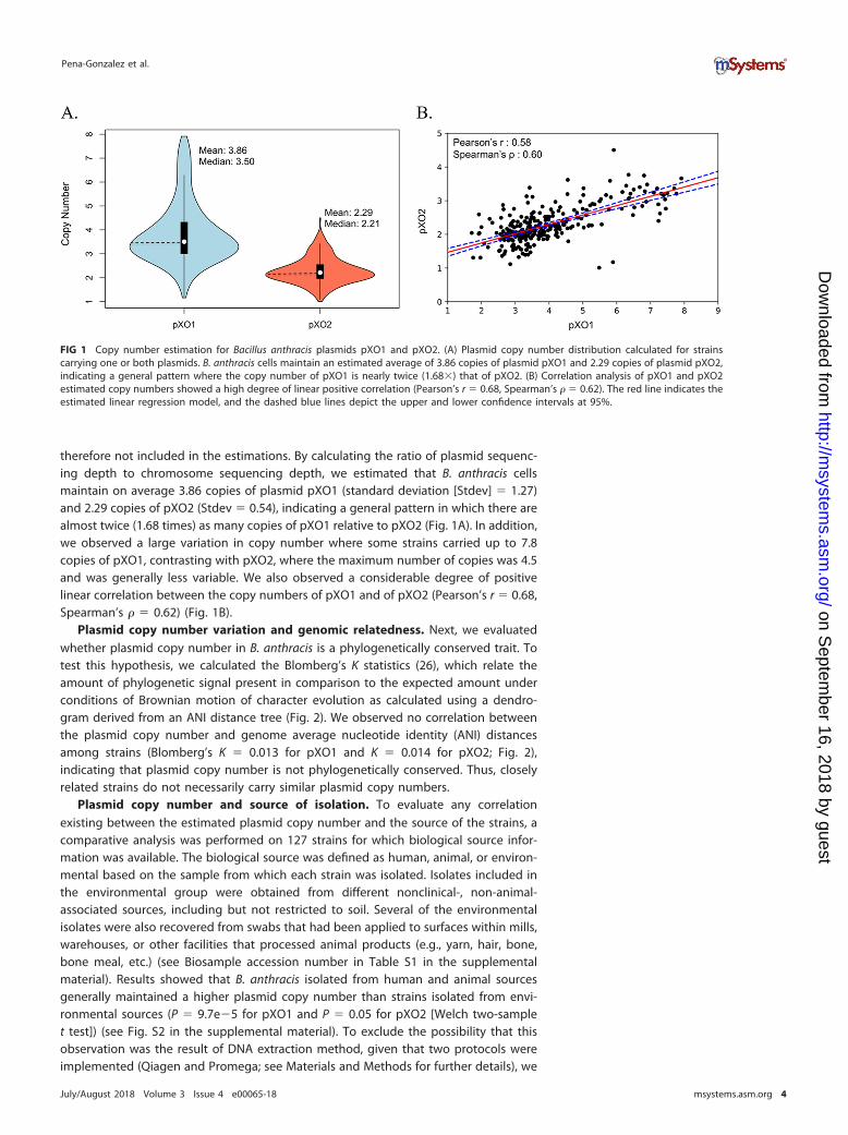
therefore not included in the estimations. By calculating the ratio of plasmid sequenc-ing depth to chromosome sequencing depth, we estimated that B. anthracis cellsmaintain on average 3.86 copies of plasmid pXO1 (standard deviation [Stdev] � 1.27)and 2.29 copies of pXO2 (Stdev � 0.54), indicating a general pattern in which there arealmost twice (1.68 times) as many copies of pXO1 relative to pXO2 (Fig. 1A). In addition,we observed a large variation in copy number where some strains carried up to 7.8copies of pXO1, contrasting with pXO2, where the maximum number of copies was 4.5and was generally less variable. We also observed a considerable degree of positivelinear correlation between the copy numbers of pXO1 and of pXO2 (Pearson’s r � 0.68,Spearman’s � � 0.62) (Fig. 1B).
Plasmid copy number variation and genomic relatedness. Next, we evaluatedwhether plasmid copy number in B. anthracis is a phylogenetically conserved trait. Totest this hypothesis, we calculated the Blomberg’s K statistics (26), which relate theamount of phylogenetic signal present in comparison to the expected amount underconditions of Brownian motion of character evolution as calculated using a dendro-gram derived from an ANI distance tree (Fig. 2). We observed no correlation betweenthe plasmid copy number and genome average nucleotide identity (ANI) distancesamong strains (Blomberg’s K � 0.013 for pXO1 and K � 0.014 for pXO2; Fig. 2),indicating that plasmid copy number is not phylogenetically conserved. Thus, closelyrelated strains do not necessarily carry similar plasmid copy numbers.
Plasmid copy number and source of isolation. To evaluate any correlationexisting between the estimated plasmid copy number and the source of the strains, acomparative analysis was performed on 127 strains for which biological source infor-mation was available. The biological source was defined as human, animal, or environ-mental based on the sample from which each strain was isolated. Isolates included inthe environmental group were obtained from different nonclinical-, non-animal-associated sources, including but not restricted to soil. Several of the environmentalisolates were also recovered from swabs that had been applied to surfaces within mills,warehouses, or other facilities that processed animal products (e.g., yarn, hair, bone,bone meal, etc.) (see Biosample accession number in Table S1 in the supplementalmaterial). Results showed that B. anthracis isolated from human and animal sourcesgenerally maintained a higher plasmid copy number than strains isolated from envi-ronmental sources (P � 9.7e�5 for pXO1 and P � 0.05 for pXO2 [Welch two-samplet test]) (see Fig. S2 in the supplemental material). To exclude the possibility that thisobservation was the result of DNA extraction method, given that two protocols wereimplemented (Qiagen and Promega; see Materials and Methods for further details), we
FIG 1 Copy number estimation for Bacillus anthracis plasmids pXO1 and pXO2. (A) Plasmid copy number distribution calculated for strainscarrying one or both plasmids. B. anthracis cells maintain an estimated average of 3.86 copies of plasmid pXO1 and 2.29 copies of plasmid pXO2,indicating a general pattern where the copy number of pXO1 is nearly twice (1.68�) that of pXO2. (B) Correlation analysis of pXO1 and pXO2estimated copy numbers showed a high degree of linear positive correlation (Pearson’s r � 0.68, Spearman’s � � 0.62). The red line indicates theestimated linear regression model, and the dashed blue lines depict the upper and lower confidence intervals at 95%.
Pena-Gonzalez et al.
July/August 2018 Volume 3 Issue 4 e00065-18 msystems.asm.org 4
on Septem
ber 16, 2018 by guesthttp://m
systems.asm
.org/D
ownloaded from

performed a two-sample t test analysis comparing plasmid copy numbers between thetwo extraction methods. The results revealed no significant difference (P � 0.11 forpXO1 and P � 0.81 for pXO2). In addition, we performed an analysis of variance(ANOVA) to determine the influence of DNA extraction method and biological source(two independent variables) in explaining the values of plasmid copy number (thecontinuous dependent variable). The results showed that the variation explained by thebiological source was significant (F � 6.23, P � 0.01) whereas the variation explainedby the extraction method was not (F � 0.072, P � 0.7).
FIG 2 Lack of phylogenetic conservatism of Bacillus anthracis plasmid copy number. A dendrogram was constructed based on the average nucleotide identity(ANI) distances calculated for 412 B. anthracis strains. Presence or absence of pXO1 (inner circle in blue) and pXO2 (inner circle in red) and estimated plasmidcopy number data (bar plots) are shown. Strains with high and low plasmid copy numbers were found to be dispersed across the three main clades, i.e., cladeA (n � 397), clade B (n � 12), and clade C (n � 3), and no apparent clusters were evident. The tree scale corresponds to 1 � ANI distance.
B. anthracis Plasmid Diversity and Copy Number Estimation
July/August 2018 Volume 3 Issue 4 e00065-18 msystems.asm.org 5
on Septem
ber 16, 2018 by guesthttp://m
systems.asm
.org/D
ownloaded from

Plasmid-based versus chromosome-based phylogenetic relationships. To de-termine whether plasmid-based ANI clustering resembled that shown by the chromo-some, we analyzed strains for which plasmid pXO1 and/or pXO2 were detected, inaddition to incorporating 36 B. anthracis reference strains that were sequenced previ-ously (see Table S2 and Table S3). Initial characterization of genomic relatedness basedon the ANI distance values determined for the chromosome showed that the strains inthe total set were grouped in three main clades: clade A (397 strains), clade B (12strains), and clade C (3 strains), with clade A containing the majority of strains, similarlyto what has been previously described with other typing methods such as multilocusvariable-number tandem-repeat analysis (MLVA) (Fig. 2). When we compared theclustering profiles of both plasmids versus that of the chromosome, we observed a highlevel of topological correlation. To quantify the strength of the correlation we used twometrics: (i) the cophenetic distance, defined as the intergroup distance at which twoobservations are first combined into a single cluster, and (ii) the Baker’s � index,defined as the rank correlation between the stages at which the pairs of observationscombine in each one of the two dendrograms being compared. For pXO1, the calcu-lated cophenetic correlation was 0.70, and the Baker’s � index correlation was 0.62. ForpXO2, the calculated cophenetic correlation was 0.89, and the Baker’s � correlation was0.93, indicating that, in general, the pXO1 and pXO2 phylogenies recapitulate that ofthe chromosome.
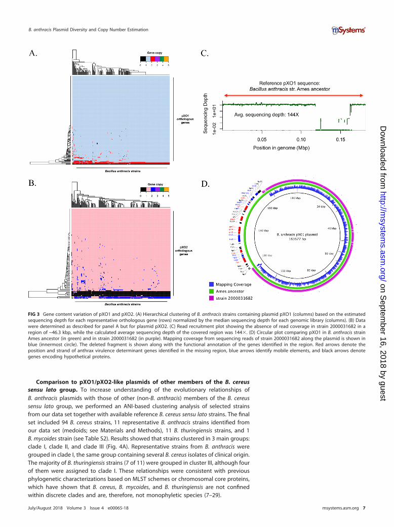
Gene content variation of pXO1 and pXO2. To avoid limitations of the assemblyprocess, such as gaps or truncated genes and misassemblies, we assessed gene contentvariations of the plasmids by recruiting high-quality (trimmed) Illumina reads againstthe predicted genes on the plasmid and determining gene presence/absence by thenumber of reads recruited (or not) on the gene. In general, genomes containing one orboth plasmids showed highly conserved gene content (Fig. 3A and B). The calculatedpXO1 pangenome was composed of 197 orthologous genes; 179 (91%) of them werepresent in all strains (strict core), and 195 (99%) were present in at least 95% of all thestrains (relaxed core). Only two genes were found to be variable in pXO1. These geneswere annotated (using the nonredundant UniProtKB/SwissProt databases) as a trans-posase for insertion sequence (IS) element IS231E (reported previously in Bacillusthuringiensis serovar finitimus) and G-protein-coupled receptor 98. In pXO2, 108 genescomposed the pangenome; 96 genes were part of the strict core (89%), while 102 geneswere part of the relaxed core. Only six genes composed the variable genome. Three ofthe six variable genes were annotated as encoding uncharacterized/hypothetical pXO2proteins, two genes were annotated as encoding putative pXO2 trans-acting regulators,and one gene had partial homology (query coverage � 35%, identity � 45%) to thegene encoding subunit ssr1 of the chromatin structure remodeling complex (RSC). Allvariable genes had hypothetical or poorly characterized functions or were mobileelements (e.g., transposases). Although the plasmid gene content diversity generallyobserved between any two genomes analyzed was not large, we identified a largefragment deletion in the pXO1 plasmid of one strain, i.e., strain 2000031682 (Fig. 3C).The deleted fragment was about ~46.3 kbp in length and contained 39 genes in total,including the following genes encoding the main virulence factors responsible foranthrax toxin: cya, pagA, lef, and the atxA transcriptional activator gene. We alsoidentified a number of genes encoding integrases, resolvases, and transposases in andaround the deleted fragment (Fig. 3D). Resequencing and reassembling of the strainconfirmed the large gene deletion. In addition, we identified orthologous genespresent in both plasmids that showed sequencing depth levels greater than those seenwith the majority of plasmid genes. These genes were most likely multicopy genes. InpXO1, we identified three multicopy genes that were observed to have two copies, onaverage, consistently across the complete set of strains. In pXO2, we identified threegenes with a consistent multicopy pattern in most of the strains characterized in thisanalysis. In both plasmids, these genes corresponded to transposases for insertionsequence elements (IS231F, IS231C, IS231B, IS231E, IS231A, and IS1151).
Pena-Gonzalez et al.
July/August 2018 Volume 3 Issue 4 e00065-18 msystems.asm.org 6
on Septem
ber 16, 2018 by guesthttp://m
systems.asm
.org/D
ownloaded from
Comparison to pXO1/pXO2-like plasmids of other members of the B. cereussensu lato group. To increase understanding of the evolutionary relationships ofB. anthracis plasmids with those of other (non-B. anthracis) members of the B. cereussensu lato group, we performed an ANI-based clustering analysis of selected strainsfrom our data set together with available reference B. cereus sensu lato strains. The finalset included 94 B. cereus strains, 11 representative B. anthracis strains identified fromour data set (medoids; see Materials and Methods), 11 B. thuringiensis strains, and 1B. mycoides strain (see Table S2). Results showed that strains clustered in 3 main groups:clade I, clade II, and clade III (Fig. 4A). Representative strains from B. anthracis weregrouped in clade I, the same group containing several B. cereus isolates of clinical origin.The majority of B. thuringiensis strains (7 of 11) were grouped in cluster III, although fourof them were assigned to clade I. These relationships were consistent with previousphylogenetic characterizations based on MLST schemes or chromosomal core proteins,which have shown that B. cereus, B. mycoides, and B. thuringiensis are not confinedwithin discrete clades and are, therefore, not monophyletic species (7–29).
FIG 3 Gene content variation of pXO1 and pXO2. (A) Hierarchical clustering of B. anthracis strains containing plasmid pXO1 (columns) based on the estimatedsequencing depth for each representative orthologous gene (rows) normalized by the median sequencing depth for each genomic library (columns). (B) Datawere determined as described for panel A but for plasmid pXO2. (C) Read recruitment plot showing the absence of read coverage in strain 2000031682 in aregion of ~46.3 kbp, while the calculated average sequencing depth of the covered region was 144�. (D) Circular plot comparing pXO1 in B. anthracis strainAmes ancestor (in green) and in strain 2000031682 (in purple). Mapping coverage from sequencing reads of strain 2000031682 along the plasmid is shown inblue (innermost circle). The deleted fragment is shown along with the functional annotation of the genes identified in the region. Red arrows denote theposition and strand of anthrax virulence determinant genes identified in the missing region, blue arrows identify mobile elements, and black arrows denotegenes encoding hypothetical proteins.
B. anthracis Plasmid Diversity and Copy Number Estimation
July/August 2018 Volume 3 Issue 4 e00065-18 msystems.asm.org 7
on Septem
ber 16, 2018 by guesthttp://m
systems.asm
.org/D
ownloaded from
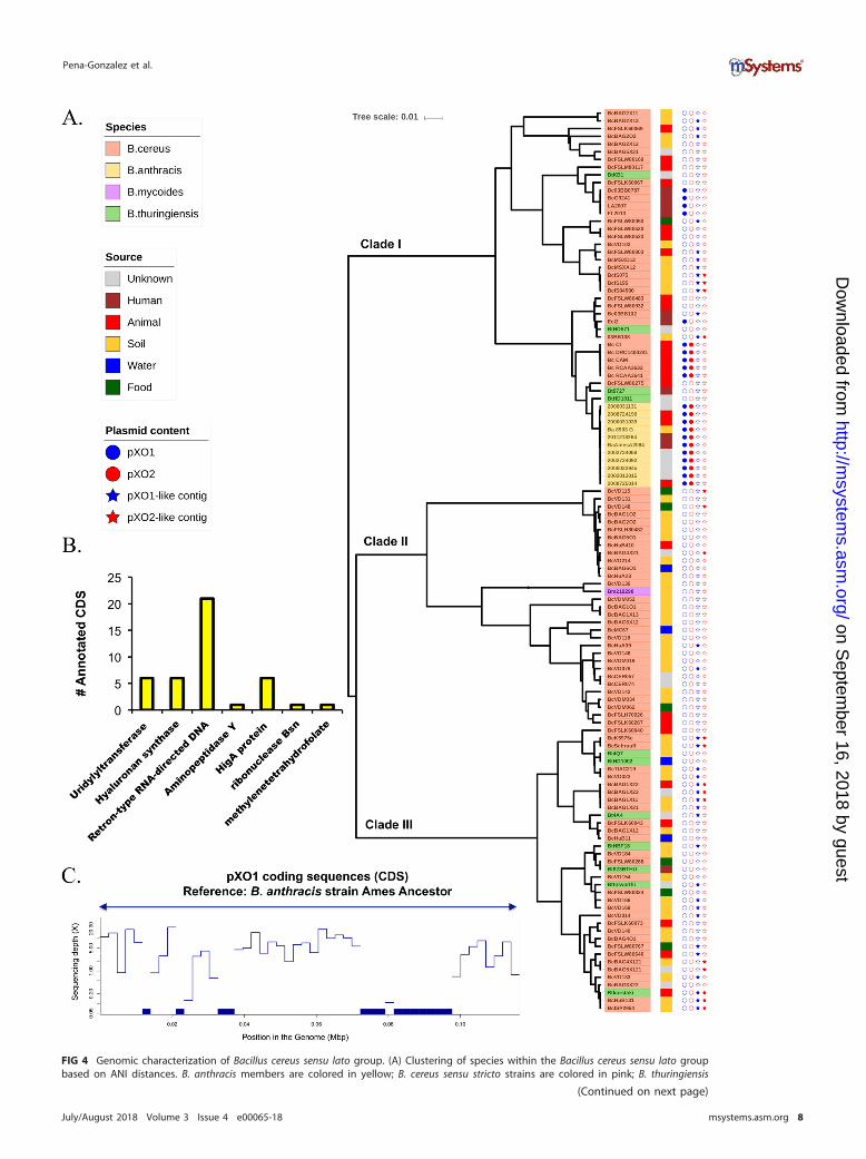
FIG 4 Genomic characterization of Bacillus cereus sensu lato group. (A) Clustering of species within the Bacillus cereus sensu lato groupbased on ANI distances. B. anthracis members are colored in yellow; B. cereus sensu stricto strains are colored in pink; B. thuringiensis
(Continued on next page)
Pena-Gonzalez et al.
July/August 2018 Volume 3 Issue 4 e00065-18 msystems.asm.org 8
on Septem
ber 16, 2018 by guesthttp://m
systems.asm
.org/D
ownloaded from
We then attempted to identify non-B. anthracis genomes that carried a complete orpartial genomic backbone with pXO1 and/or pXO2. To achieve this goal, since ourgenome sequences were incomplete (draft), we followed two approaches. First, weidentified large (�500-bp) contigs with �80% identity and �80% sequence coveragewith respect to reference pXO1 and pXO2 plasmids from the B. anthracis Ames ancestor(we called these contigs “pXO1/2-like contigs”); second, we generated read recruitmentplots to visualize and quantify the sequencing depth coverage provided by reads of thegenomic library of the corresponding strain along with the reference plasmid sequence(see Materials and Methods). We identified 33 genomes containing pXO1-like contigs,12 of which were assignable to clade I, 2 to clade II, and 20 to clade III. We alsoidentified 17 strains containing pXO2-like contigs; 4 were assignable to clade I, 3 toclade II, and 10 to clade III (see, e.g., Fig. 4A). Functional characterization of the genespredicted in pXO1-like contigs (365 in total) showed that the majority of those genescorresponded to hypothetical proteins (97%) and that only 42 (3%) could be function-ally annotated. Among these, five genes encoded hyaluronan synthase (Fig. 4B). Inaddition, we screened our B. cereus sensu lato data set for the hasABC operon and foundthat, among 116 genomes, only 11 strains harbored the functional gene set. These 11genomes corresponded to pathogenic B. cereus strains (03BB87, 03BB102, CAM, CI,RCA-A-364-1, RCA-A-363-2, DRC14-0024-1, FL2013, LA2007, G9241, and Elc2). In allcases, the genes were colocated, and the hasA gene for strain Elc2 was the mostdivergent one (see Fig. S6). However, no anthrax toxin genes were identified amongthese sequences.
We also included in this analysis 11 previously characterized pathogenic B. cereusgenomes carrying complete pXO1 and/or pXO2 plasmids. In particular, five B. cereus bv.anthracis strains (RCA_A_364-1, RCA_A_363-2, DRC_14-0024-1, CAM, and CI) wereisolated from dead mammals (chimpanzees, gorillas, elephants, and goats) with anillness consistent with anthrax in west and central Africa (23), and six pathogenicB. cereus strains (G9241, BcFL2013, LA2007, 03BB87, 03BB102, and Elc2) were isolatedfrom human cases of pneumonia or cutaneous lesions. These strains were compared to11 representative B. anthracis genomes (from our data set) and to an additionalB. cereus strain (03BB108, isolated from dust at a worksite where a Texas weldercontracted fatal pneumonia in 2003) that carried partial homology to the backbone ofpXO1. Clustering analysis based on ANI dissimilarity revealed four groups (Fig. S3A). Thefirst group was highly clonal and was composed of B. anthracis strains with an averageintragroup ANI distance of 0.04 (i.e., 99.96% identity). The second group containedB. cereus bv. anthracis isolates which were also highly similar, with an average ANIdistance of 0.03. The third group, labeled B. cereus group I, was composed of threeB. cereus isolates (03BB108, 03BB102, and Ecl2) that had among them an average ANIdistance of 1.01. Finally, the fourth group, labeled B. cereus group II, was composed offour human-pathogenic B. cereus strains (LA2007, BcFL2013, G9241, and 03BB87) andshowed an average of a 0.01 intragroup ANI distance, representing the smallestobserved intragroup diversity value. B. cereus group II was the most divergent from allother groups, with an average intergroup ANI distance of 5.27 (Fig. S3B).
Plasmid detection and quantification based on read coverage confirmed that allB. cereus bv. anthracis strains carried complete pXO1-like and pXO2-like plasmids, whilestrains BcFL2013, G9241, 03BB87, LA2007, and Elc2 harbored a complete pXO1-likeplasmid but not a pXO2-like plasmid (Fig. S3C). 03BB102, which was isolated from apatient with a fatal case of pneumonia in Texas, differed from the other strains in thatit did not harbor a full-length pXO1-like or pXO2-like plasmid, although partial se-
FIG 4 Legend (Continued)members are colored in green; and B. mycoides is colored in purple. The right colored strip indicates the source of isolation. Filled or emptycircles indicate the presence (filled) or absence (empty) of pXO1 (in blue) and pXO2 (in red). Filled stars denote the strains for whichpXO1-like (blue) or pXO2-like (red) contigs were identified. (B) The number of functionally annotated coding sequences (excludinghypothetical sequences) predicted in pXO1-like contigs (42 in total, 3% of the total number). (C) Example of B. cereus strain VD014 showingpartial pXO1-like backbone homologous to pXO1 from B. anthracis Ames ancestor.
B. anthracis Plasmid Diversity and Copy Number Estimation
July/August 2018 Volume 3 Issue 4 e00065-18 msystems.asm.org 9
on Septem
ber 16, 2018 by guesthttp://m
systems.asm
.org/D
ownloaded from

quence homology to the pathogenicity island was detected (51.72% of the total genesof the island were present) (22). Sequence-based analysis revealed that 03BB102harbors the typical anthrax virulence genes but lacks about half of the canonical pXO1gene content (Fig. S3C). Further gene-based characterization showed that this plasmid(p03BB102) carried a complete, 5.1-kbp pXO2 cap locus with 93% nucleotide identity tothe Ames strain cap locus (GenBank accession number AE017335) and was flanked by5 IS elements. In addition, duplicate homologs of protective antigen genes (pagA andpagR) were identified; one homolog showed ~99% nucleotide identity to its orthologin pXO1 of the Ames ancestor strain, while the second showed 92% and 94% (respec-tively), indicating that these homologs have already begun to diverge (Fig. 5). AlthoughpXO1/pXO2-like plasmids seem to be remarkably conserved in terms of gene contentand synteny, strain 03BB102 is an exception to this rule, which suggests that the levelof plasmid diversity in nature may be higher than previously thought.
We also calculated plasmid copy numbers in the set of B. cereus strains carryingB. anthracis-like plasmids (Table S1). The estimated average pXO1 copy number inB. cereus bv. anthracis strains was 1.8, while that for the set of human-pathogenicB. cereus strains was 2.32, which was similar to the estimated average for B. anthracis(3.86). For pXO2, the estimated average copy number in B. cereus bv. anthracis was 2.12,which is similar to the estimated copy number in B. anthracis (2.29).
Assessing origins and vertical versus horizontal transmission of plasmids. Todetermine if the pXO1/pXO2-like plasmids have been transferred between members ofB. cereus and B. anthracis, we contrasted the phylogenetic relationships among thegenomes on the basis of comparisons of the chromosomal genes to those of theplasmids. Phylogenetic reconstruction based on plasmid core orthologous genes ofthe strains harboring a complete pXO1 plasmid (139 genes) and/or pXO2 plasmid (88genes) showed a topology similar to that observed with whole-genome ANI distanceevaluations based on tanglegram analysis (Fig. 6), indicating limited lateral mobilizationof the plasmids between the strains (Fig. S4). For pXO1, B. cereus bv. anthracis strainswere closer to those in the B. anthracis group than they were to the set of human-pathogenic B. cereus group II strains, and Elc2 was the most divergent strain. For pXO2,three main clades were observed: (i) one containing all B. anthracis strains, (ii) anothercontaining strain B. cereus CI, and (iii) a final clade containing the remaining B. cereusbv. anthracis strains (RCA_A_364-1, RCA_A_363-2, DRC_14-0024-1, and CAM) (Fig. S4D).
FIG 5 Gene content comparison between B. anthracis pXO1/pXO2 and B. cereus strain 03BB102 plasmids. The connecting lines show the presence and locationof shared genes, while the gray scale represents the level of nucleotide identity. p03BB102 carries a cap locus (~5.1 kbp) that shows 93% nucleotide identityto the cap locus in reference plasmid pXO2 (B. anthracis “Ames ancestor”; GenBank accession number AE017335) and is flanked by IS elements. A duplicatehomolog of pagA and pagR genes is also present in p03BB102, with one homolog showing 99% identity to its pXO1 B. anthracis homolog (B. anthracis “Amesancestor” GenBank accession number AE017336) for both genes, while the second shows 92% and 94% identity, respectively.
Pena-Gonzalez et al.
July/August 2018 Volume 3 Issue 4 e00065-18 msystems.asm.org 10
on Septem
ber 16, 2018 by guesthttp://m
systems.asm
.org/D
ownloaded from

Strain CI was more closely related to the B. anthracis group than it was to the otherB. cereus bv. anthracis isolates.
Further, clustering based on the presence/absence of the variable genes of bothplasmids showed a grouping pattern similar to that of the chromosome (Fig. S4B andE), indicating that (higher) gene content variation largely correlates to (higher) genome
FIG 6 Assessment of plasmid lateral transfer between representative B. anthracis and pathogenic B. cereus strainscarrying complete pXO1/pXO2-like plasmids. (A and B) Comparison of phylogenetic relationships based on the coregenome for the chromosome and pXO1 (A) and for the chromosome and pXO2 (B) in strains carrying one or bothplasmids. Phylogenetic reconstructions shown in panel A were based on the alignment of 210,123 variablepositions found in the concatenated alignment of 4,233 core orthologous genes for the chromosome and 458variable positions identified in the concatenated alignment of 149 pXO1 core orthologous genes. Phylogeneticrelationships in panel B were constructed from the alignment of 74,389 variable positions found in the concate-nated alignment of 4,616 core orthologous genes for the chromosome and 120 variable positions identified in theconcatenated alignment of 88 pXO2 core orthologous genes. No signal of plasmid lateral transfer between the twophylogroups was apparent.
B. anthracis Plasmid Diversity and Copy Number Estimation
July/August 2018 Volume 3 Issue 4 e00065-18 msystems.asm.org 11
on Septem
ber 16, 2018 by guesthttp://m
systems.asm
.org/D
ownloaded from
divergence. Collectively, these results indicated limited horizontal transfer of the plas-mid between B. cereus and B. anthracis. Accordingly, B. cereus genomes harboring theB. anthracis plasmids appear to have maintained these plasmids since their last com-mon ancestor with B. anthracis. However, we did observe topological incongruencesbetween the chromosome and pXO1 core gene trees within B. anthracis (entangle-ment � 0.20, Fig. S5A), indicating that the plasmid might have undergone hori-zontal transfer within the group. For instance, the chromosome-based and plasmid-based topologies were significantly incongruent by all three tests applied, i.e., theone-sided maximum likelihood (ML) Kishino-Hasegawa test (KH) (30), the Shimodaira-Hasegawa test (SH) (31), and the expected likelihood weight test (ELW) (32) (pval-1sKH � 0.005, pval-SH � 0.002, c-ELW � 0.002 [where pval is P value, 1sKH is one-sidedKishino-Hasegawa test, and c-ELW is cumulative expected likelihood weight test]; alltests were applied with a 5% significance level and 1,000 resamplings using theresampling of estimated log-likelihoods [RELL] method). However, further analysisshowed that the topological differences mentioned above were predominantly due torecombination and/or varied selection pressures only in genes encoding five prod-ucts (OG20, a hypothetical protein containing a DUF87 domain; OG149, the Edemafactor [cya] component in the anthrax toxin; OG39, a type IV secretion system protein;OG133, the protective antigen [pagA] in the anthrax toxin; OG135, a germinationprotein [gerKC]) and were not plasmid-wide. When these genes were removed from thecore gene alignment, the plasmid tree and the chromosome were topologically morecongruent (entanglement � 0.05) (Fig. S5B). We also observed that the complete pXO1and chromosome trees grouped in the same cluster (less distance between them) in aminimally dimensional representation of the topological variability of the trees evalu-ated using the Kendall and Colijn metric (Fig. S5C and D) (see Materials and Methods).Collectively, our analyses revealed no strong evidence of plasmid lateral transferbetween or within B. anthracis and B. cereus.
DISCUSSION
In this study, we estimated plasmid copy numbers in a large collection of newlysequenced B. anthracis strains, characterized their full plasmid gene content, andcompared the levels of phylogenetic diversity of representative genomes with those ofother Bacillus species carrying complete or partial pXO1/pXO2-like plasmids. Our majorfindings showed that B. anthracis cells maintain, on average, 3.86 copies of pXO1 and2.29 copies of pXO2 and revealed that there is a positive linear correlation in thenumbers of copies of both plasmids which was consistent with two previously reportedsequence-based studies (12, 13). The gene content of these B. anthracis plasmids wasremarkably stable, although a few genomes (e.g., that of strain 2000031682) lackedlarge parts of the plasmids. Furthermore, the number of plasmid copies that B. anthracisgenomes harbored seemed to be influenced by the source from where the strains wereisolated (animal or environmental) but not by phylogeny. We also identified severalenvironmental B. cereus sensu lato strains containing pXO1-like and pXO2-like contigs,some of which had been previously reported (33). We found no strong evidence ofplasmid exchange between B. anthracis and B. cereus sensu lato genomes, suggestingplasmid maintenance since the last common ancestor of the two species.
Our estimates revealed a lower number of pXO1 copies per chromosome, onaverage (n � 3.86), than had been reported from earlier studies based on molecularmethods such as qPCR. For example, Coker et al. estimated ratios of up to 40 copies ofpXO1 (8), while Pilo et al. reported 10 to 11 copies of the same plasmid (11). In bothcases, the estimation based on quantities of a portion of a single gene per replicon,representing ~0.1% of the total replicon length, seemed to be inflated compared withour more comprehensive shotgun sequence-based estimations. However, for pXO2,qPCR and sequencing provided similar estimates of approximately 1 to 2 copies percell. This indicated that PCR may have overestimated pXO1 abundance since thecompeting hypothesis, that sequencing was biased against pXO1 abundance but notagainst that of pXO2, appears to be less parsimonious. However, we have also identified
Pena-Gonzalez et al.
July/August 2018 Volume 3 Issue 4 e00065-18 msystems.asm.org 12
on Septem
ber 16, 2018 by guesthttp://m
systems.asm
.org/D
ownloaded from

strains carrying up to 4.5 copies of pXO2. The fact that the plasmid copy number wasnot a phylogenetically conserved trait but was influenced by the source of isolationsuggested that extrinsic forces (e.g., environmental factors such as temperature, pH, soilmoisture, and cation levels, among others) might play a more important role indetermining the number of replicons that B. anthracis cells maintain. In other words,the plasmid copy number may become adjusted in response to environmental cues.Studies of the ecology of B. anthracis have shown that the global distribution of anthraxwas largely determined by climatic factors and land features, where, for example, soilswith high calcium levels and a pH above 6.1 foster better spore survival (6, 9, 34). Itshould also be mentioned that although the prevailing assumption was that B. anthra-cis remains primarily dormant in soil as spores, several recent studies have suggestedgrowth in soil/rhizosphere. For example, Saile and Koehler showed that B. anthracisstrains can germinate on and around roots, suggesting that even environmental strainscan grow and be metabolically active under specific conditions (35). Thus, the trendreported here of higher plasmid copy numbers in strains from animal sources, includinghuman tissues, likely reflected a real ecological adaptation between different sourcesrather than just the effect of prolonged time in the spore stage for environmentalstrains.
We also characterized the gene content diversity of the plasmids across the set ofstrains carrying one or both plasmids. Our results confirmed that the highly conservedgene content and synteny for both plasmids (�97% of total plasmid genes shared)were similar to what has been previously described for this species. In addition, weidentified a single strain (2000031682) with a large fragment deletion in the pXO1plasmid. The deleted fragment size was approximately 46.3 kbp, and the fragmentcarried the main virulence genes responsible for anthrax toxin production: cya, pagA,lef, and atxA. While the history of this strain is not clear, it was originally archived at CDCin 1964 on an agar slant and stored at room temperature. The strain was recoveredfrom the slant and frozen at �70°C in 2001. We previously reported that numerousstrains in this collection were cured of plasmids during decades in room temperaturestorage (36). We cannot ascertain whether this strain was received under this conditionor if the deletion might have occurred during storage.
In this study, we also characterized environmental B. cereus sensu lato strainspossessing partial or complete pXO1/pXO2-like plasmids and B. cereus bv. anthracisstrains possessing complete B. anthracis plasmids. Through bioinformatic approaches,we identified 50 strains with contigs homologous to those of pXO1 and/or pXO2. Weconfirmed that pXO1-like and pXO2-like contigs were widely prevalent in environmen-tal isolates of the B. cereus sensu lato group, similarly to what was previously revealedby Van der Auwera et al. using PCR-based approaches (33). The annotation of the genespresent in pXO1/pXO2-like contigs showed that most of them were identified asencoding hypothetical proteins, with few of them predicted to be involved in DNAinsertion and transposition (for example, retron-type RNA-directed DNAases and ribo-nucleases; see Fig. 4B), but no genes encoding anthrax toxins were identified. However,we found genes encoding hyaluronic acid (HA) capsule formation in these contigs. AnHA capsule provides pathogenic bacilli with capsular material used to escape the innatehost immune response and is involved in the pathogenesis of anthrax-like diseases (37,38). Given that all the B. cereus sensu lato strains analyzed here were of environmentalorigin, these findings might indicate that at least some of the virulence factors encodedon the B. anthracis plasmids (e.g., HA capsule formation) may be important for survivalin the environment outside the human host.
A potential limitation in our study was the possibility that some strains could havelost part(s) or all of their plasmids during successive subculturings or that their plasmidcopy number was adjusted upon cultivation under laboratory conditions compared tothe natural environment. The rate of plasmid loss during recurrent subculturing couldhave been accelerated under laboratory conditions of stress (36) which might poten-tially have also biased our estimation of the “true” copy number variation. To minimizethis issue, the original culture stock, rather than derived subcultures, was used for
B. anthracis Plasmid Diversity and Copy Number Estimation
July/August 2018 Volume 3 Issue 4 e00065-18 msystems.asm.org 13
on Septem
ber 16, 2018 by guesthttp://m
systems.asm
.org/D
ownloaded from
preparing DNA for sequencing. To investigate this limitation further, a larger effort withrespect to soil/field sampling would be necessary to evaluate how frequently B. cereusgroup strains carry complete or partial pXO1/pXO2-like plasmids and the natural copynumber of B. cereus/B. anthracis strains. The fact that we did observe differencesbetween strains of environmental origin and of clinical origin, even though all strainshad been maintained under laboratory conditions since their isolation, further indi-cated that significant biological/ecological differences likely underlay the plasmid copynumber variation observed here.
Phylogenetic reconstruction of B. anthracis medoids and the B. cereus bv. anthracisand human-pathogenic B. cereus genomes based on analysis of pXO2 core genesshowed that strain CI was more closely related to the members of the B. anthracis groupthan it was to other B. cereus bv. anthracis strains (see Fig. S4D in the supplementalmaterial). Antonation and colleagues reported that strain CI is closer to other B. cereusbv. anthracis strains than B. anthracis, even though this strain showed the largestintragroup distance to other B. cereus bv. anthracis members (23). This inconsistency ismost likely due to differences between the two studies in the bioinformatic approachesused to build trees. Antonation et al. estimated phylogenetic relationships based onSNP data of core plasmid genes, while our tree was based on the concatenatedalignment of core orthologous genes (88 in total).
In summary, by using next-generation sequencing data, we have estimated B. an-thracis plasmid copy numbers, characterized their genomic diversity, and comparedrepresentative strains to clinical and environmental B. cereus sensu lato strains carryingpXO1/pXO2-like plasmids. The results derived from this study have advanced ourunderstanding of the biology of the B. cereus group, improved the ecological andevolutionary framework used to classify species, and appropriately defined phyloge-netic relationships and taxonomic assignments within the B. cereus sensu lato group.Our results also highlighted the advantages of using genomic relatedness (as measuredby ANI, for example), instead of plasmid-encoded traits, to assign taxonomy androbustly resolve the relationships among closely related members of the B. cereus sensulato group. These results and interpretations were also consistent with previous studiesof plasmid-encoded traits in other bacterial species such as Clostridium botulinum (39).Therefore, the results derived from our study will help to improve the ecological andevolutionary framework used to classify species and appropriately define phylogeneticrelationships, particularly in bacterial groups that exhibit high phenotypic diversity suchas the B. cereus sensu lato group.
Although the collection size of the B. anthracis strains sequenced in this study andthe number of strains deposited in public databases are likely still small compared tothe total natural diversity of the species, to the best of our knowledge, this is the largeststudy characterizing B. anthracis plasmid copy number variation and gene contentdiversity using sequencing data to date. Hence, the data presented here shouldfacilitate future studies of B. anthracis and its virulence plasmids. Finally, the bioinfor-matic approaches used in this study can also be applied as a reference framework forepidemiological studies involving this and other microorganisms of medical relevance.
MATERIALS AND METHODSCollection description. The collection of genomes analyzed in this study is part of the Zoonoses and
Select Agent Laboratory’s historical strain collection at the Centers for Disease Control and Prevention.The strains included in the study were acquired from human, animal, and environmental sourcesworldwide from the 1950s to 2013. The complete set has been deposited in the NCBI sequence readarchive (SRA) under BioProject identifier (ID) 264742 (see Table S1 in the supplemental material).
Growth conditions, DNA extraction, and sequencing. DNA was extracted from isolates using aQIAamp DNA blood Minikit (Qiagen, Valencia, CA) or a Maxwell 16 instrument (Promega, Madison, WI).For the QIAamp extraction, cells were grown overnight in heart infusion broth (Remel, Lenexa, KS). Cellswere pelleted by centrifugation for 10 min at 5,000 � g. Broth was removed, and DNA was extractedusing a Qiagen QIAamp DNA blood Minikit and following the manufacturer’s protocol for isolatinggenomic DNA from Gram-positive bacteria. For DNA extractions performed using the Maxwell instru-ment, the manufacturer’s protocol was followed. Briefly, cells were grown overnight on Trypticase soyagar with 5% sheep blood and then mechanically disrupted by vortex mixing for 2 min in a suspensionof silica beads and Tris-EDTA (TE) buffer (Promega; Maxwell RSC). The suspension was centrifuged for 30 s
Pena-Gonzalez et al.
July/August 2018 Volume 3 Issue 4 e00065-18 msystems.asm.org 14
on Septem
ber 16, 2018 by guesthttp://m
systems.asm
.org/D
ownloaded from
at 10,000 � g. A 300-�l volume of the resulting supernatant was used for DNA extraction following themanufacturer’s protocol for blood and cells. Sequencing was performed on an Illumina GAIIx systemusing TruSeq chemistry.
Read quality control, assembly, and gene prediction. Raw reads were initially screened foradaptor sequences using Scythe (40) and trimmed at both the 5= and 3= ends based on a PHRED scorecutoff of 20 using SolexaQA�� (41). Reads that were �50 bp in length after trimming were discarded.Quality-filtered reads were de novo assembled using IDBA-UD with precorrections (42), and the percent-ages of contamination and genome completeness were assessed based on either recovery of lineage-specific marker genes using CheckM (43) or recovery of essential genes (single copy) in bacterial andarchaeal genomes using the script HMM.essential.rb available at the Enveomics collection (44). Protein-coding sequences were predicted using MetaGeneMark (45), and 16S rRNA gene sequences wereidentified using barrnap 0.6 (https://github.com/tseemann/barrnap). All predicted genes from the as-semblies were taxonomically annotated using MyTaxa (46), and the taxonomic distributions of adjacentgenes (in windows of 10 genes) in the concatenated assembly were inspected for possible contaminationthrough the use of bar plots. The methods and scripts for read quality control, assembly, and geneprediction described above were used as part of MiGA (Microbial Genomes Atlas), a system developedin our laboratory for data management and processing of microbial genomes and metagenomes(http://microbial-genomes.org/).
B. anthracis and B. cereus sensu lato reference genomes. Assembled sequencing data for 36additional B. anthracis strains and raw sequencing reads for 130 B. cereus sensu lato reference strains weredownloaded from the nucleotide database or the sequencing read archive (SRA) at NCBI (https://www.ncbi.nlm.nih.gov/sra) with the accession numbers listed in Table S2. Reference strains were processed inparallel with the CDC B. anthracis collection as described above. After quality control inspection, 26B. cereus reference strains showing �20% contamination as calculated with CheckM (see above) wereexcluded from the analysis.
Plasmid copy number estimation. Whole-genome sequencing enabled estimation of the copynumber of each plasmid relative to the number of chromosome copies in each sequence library. Copynumber was estimated as the ratio of the average sequencing depth across the whole plasmid sequenceto the average sequencing depth across the chromosome. The effects of short regions with very high orvery low sequencing depth on average sequencing depth were negligible. To speed up computationalprocessing, read sets were randomly subsampled to a level where conclusions would not change. Aftercreating various library sizes, we calculated the pXO1 copy number for three libraries of different sizes(large, medium, and small), and a level of as little as 10% of the library size did not have an effect in copynumber estimation (see Fig. S1 in the supplemental material). Quality-filtered sequence libraries weretherefore subsampled to 10% of their size and blastn mapped to three targets: the reference B. anthracisAmes ancestor (GCF_000008445.1), plasmids pXO1 (NC_007322.2) and pXO2 (NC_007323.2), and eachassembled genome. Read depths were calculated for each library using the function “enve.recplot”incorporated in the R package “enveomics.R” (44). Using the same R function, read recruitment plots weregenerated for each library to quantify and visualize the coverage across the full length of the referenceplasmids to determine the presence/absence of the entire plasmid. Presence data were considered trueif the calculated average sequencing depth across the full reference was �2� in the subsampled library.
Average nucleotide identity (ANI) distances and medoids. The average nucleotide identity (ANI)(47, 48) between the sets of genomes was calculated using the command line interface of MiGA(Microbial Genome Atlas; https://github.com/bio-miga/miga). Briefly, MiGA calculated a matrix of dis-tances with 1 � ANI for all pairs of genomes considered in the database. Subsequently, clusters in thematrix were identified using the PAM algorithm (partitioning around the medoids) (49) with k medoids,where k was determined by the local gain in the average Silhouette width (50) for each level of clusteringuntil a group of five or fewer genomes was reached. Here, the medoids were representative strains in thediversity space. Afterward, a dendrogram was built based on ANI distances (1 � ANI) using hierarchicalagglomerative clustering with the Ward criterion (26).
Phylogenetic signal in plasmid copy number. Phylogenetic conservatism of plasmid copy numberswas determined through the calculation of the Blomberg’s K statistic (51) included in the function“phylosignal” of the R package “Picante” (52). K values of 1 correspond to a Brownian motion process,which implies some degree of phylogenetic signal or conservatism. K values closer to zero correspondto a random or convergent pattern of evolution, while K values greater than 1 indicate strongphylogenetic signal and conservatism of traits.
Phylogenomic relationship of plasmids and chromosome based on ANI. Large (�500-bp) contigswith �80% identity and �80% query coverage with respect to either pXO1 or pXO2 B. anthracis Amesancestor reference sequences were considered to be pXO1 or pXO2 homologous and were extractedfrom the assemblies. Dendrograms based on ANI distances were built for the plasmids and chromosomesas described previously and subsequently compared through tanglegrams using the R package Dendex-tend, version 1.2.0 (53). Statistical correlation between pairs of dendrograms was evaluated with twoparameters: Baker’s � index correlation (54) and the cophenetic distant correlation (55). Both areincluded in the R package Dendextend.
Read-based genomic gene content analysis. pXO1 and pXO2 ortholog genes among B. anthracisgenomes were identified using a reciprocal-best match (RBM) blastn approach as described by Weigandet al. (39). In brief, the sequences of the predicted genes in the plasmid sequence of one strain weresearched against the predicted genes of all of the remaining strains in the set in a pairwise fashion usingblastn (56). Reciprocal best matches were identified when the best match was bidirectional for the pairof strains being compared and when there was at least 70% nucleotide identify and 70% query gene
B. anthracis Plasmid Diversity and Copy Number Estimation
July/August 2018 Volume 3 Issue 4 e00065-18 msystems.asm.org 15
on Septem
ber 16, 2018 by guesthttp://m
systems.asm
.org/D
ownloaded from
coverage using rbm.rb (44). Next, orthologous groups (OGs) in reciprocal best matches were identifiedusing the unsupervised Markov cluster algorithm (MCL) implemented in ogs.mcl.rb in the Enveomicscollection (44) with the following default settings: 1.5 inflation parameter and bit score as parameter toweight edges. Descriptive statistics on the set of orthology groups were estimated using the scriptogs.stats.rb (Enveomics collection). Genes conserved in all genomes were identified as core orthologousgenes. Genes conserved in some but not all of the strains were identified as variable orthologous genes.Representative orthologous genes from the previous analysis (including both core and variable genes)were randomly selected and extracted to generate a pangenome or “bag of genes.” To better determinethe presence/absence of the genes included in the pangenome, we recruited raw sequencing readsagainst the predicted genes on the plasmid. For this, FastA libraries were subsampled to 500,000 readsper sample and mapped against the set of representative orthologous genes using blastn. The maximumnumber of target sequences in the database was set to 1 (best match). The observed and estimatedsequencing depths as well as the number of reads mapping to each gene in the database werecalculated using the script “BlastTab.seqdepth_ZIP.pl” from the Enveomics collection (44), assuming azero-inflated Poisson distribution to correct for noncovered positions with parameters estimated asdescribed by Beckett et al. (57). Orthologous genes with zero inflation values of �0.3, which representthe fraction of the gene that is not covered, were excluded. Thus, only genes with �70% coverage wereconsidered to be present. The calculated average sequencing depths for the genes in pXO1 were 32.2�and 19.9� for the genes in pXO2. To determine the copy number of the genes in each plasmid, thesequencing depth calculated for each gene in each strain was normalized by the median sequencingdepth of each strain and reported through a dendrogram of hierarchical clustering.
Genomic characterization of B. anthracis and B. cereus strains carrying pXO1-like and/orpXO2-like plasmids. Orthologous genes among B. anthracis medoids and B. cereus genomes carryingB. anthracis-like plasmids were identified through the reciprocal-best match (RBM) blastn approach asdescribed above. Core genes were extracted and aligned to estimate phylogenetic relationships betweenB. cereus and B. anthracis. The sets of core genes were filtered to remove in-paralogous genes and alignedusing MUSCLE v3.8.31 (58) with default parameters. The aligned outputs were saved in FastA format, andthe script Aln.cat.rb from the Enveomics collection was used to concatenate the multiple alignments intoa single file and to remove invariable sites, defined as columns with only one state and undefinedcharacters. Phylogenetic reconstructions were performed with either RAxML version 8.1.21 (59) orFastTree version 2.1.7 (60) with the GTR model for nucleotides used in both cases. The collection ofvariable genes, defined as genes absent in 1 or more genomes, was identified as described above. Thepresence or absence of these variable genes was used to cluster genomes hierarchically using a completelinkage across a centered Pearson correlation similarity range using the function “heatmap2” containedin the R package gplots v3.0.1 (https://CRAN.R-project.org/package�gplots). Functional annotation ofvariable genes was bioinformatically inferred through a BLASTP search against the RefSeq protein andthe UniProtKB/Swiss-Prot databases with identity greater than 45% and minimal query coverage of 70%.
Tests for incongruence between phylogenetic trees were performed using TREE-PUZZLE 5.2 andmaximum likelihood (ML) (61). ML analysis was carried out using empirically derived base frequencies,ratios of transitions to transversions estimated from data sets, the HKY model of substitution, a gammadistribution model for site rate variation with �-parameter estimated from data set, and 4 gamma ratecategories. Topological variability or distances among trees derived from individual OGs were calculatedusing the Kendall and Colijn metric (62) implemented in R package “treeSpace” (63). Tanglegramentanglements were calculated as described previously (53).
Data availability. The complete set of strains sequenced in this study has been deposited in theNCBI Sequence Read Archive (SRA) under BioProject ID 264742 (see Table S1 for strain-specific accessionnumbers).
SUPPLEMENTAL MATERIALSupplemental material for this article may be found at https://doi.org/10.1128/
mSystems.00065-18.FIG S1, EPS file, 0.4 MB.FIG S2, EPS file, 1.6 MB.FIG S3, EPS file, 2.9 MB.FIG S4, EPS file, 2.6 MB.FIG S5, EPS file, 2.3 MB.FIG S6, EPS file, 0.8 MB.TABLE S1, DOCX file, 0.1 MB.TABLE S2, XLSX file, 0.1 MB.TABLE S3, XLSX file, 0.04 MB.
ACKNOWLEDGMENTSThe findings and conclusions in this report are ours and do not necessarily represent
the official position of the Centers for Disease Control and Prevention (CDC). Mentionof company names or products does not constitute endorsement by the CDC.
This work was supported by United States National Science Foundation award
Pena-Gonzalez et al.
July/August 2018 Volume 3 Issue 4 e00065-18 msystems.asm.org 16
on Septem
ber 16, 2018 by guesthttp://m
systems.asm
.org/D
ownloaded from
number 1356288 and DHHS/PHS/CDC award no. RF023 to K.T.K. A.P.-G. was partiallysupported by Colciencias—Colombian Administrative Department for Science, Tech-nology and Innovation—through a doctoral fellowship.
REFERENCES1. Derzelle S, Girault G, Kokotovic B, Angen Ø. 2015. Whole genome-
sequencing and phylogenetic analysis of a historical collection of Bacil-lus anthracis strains from Danish cattle. PLoS One 10:e0134699. https://doi.org/10.1371/journal.pone.0134699.
2. Van Ert MN, Easterday WR, Huynh LY, Okinaka RT, Hugh-Jones ME, RavelJ, Zanecki SR, Pearson T, Simonson TS, U’Ren JM, Kachur SM, Leadem-Dougherty RR, Rhoton SD, Zinser G, Farlow J, Coker PR, Smith KL, WangB, Kenefic LJ, Fraser-Liggett CM, Wagner DM, Keim P. 2007. Globalgenetic population structure of Bacillus anthracis. PLoS One 2:e461.https://doi.org/10.1371/journal.pone.0000461.
3. Girault G, Blouin Y, Vergnaud G, Derzelle S. 2014. High-throughputsequencing of Bacillus anthracis in France: investigating genome diver-sity and population structure using whole-genome SNP discovery. BMCGenomics 15:288. https://doi.org/10.1186/1471-2164-15-288.
4. Hendricks KA, Wright ME, Shadomy SV, Bradley JS, Morrow MG, Pavia AT,Rubinstein E, Holty JC, Messonnier NE, Smith TL, Pesik N, Treadwell TA,Bower WA. 2014. Centers for Disease Control and Prevention expertpanel meetings on prevention and treatment of anthrax in adults. EmergInfect Dis 20:e130687. https://doi.org/10.3201/eid2002.130687.
5. Pilo P, Frey J. 2011. Bacillus anthracis: molecular taxonomy, populationgenetics, phylogeny and patho-evolution. Infect Genet Evol 11:1218 –1224. https://doi.org/10.1016/j.meegid.2011.05.013.
6. Hugh-Jones M, Blackburn J. 2009. The ecology of Bacillus anthracis. MolAspects Med 30:356 –367. https://doi.org/10.1016/j.mam.2009.08.003.
7. Riojas MA, Kiss K, McKee ML, Hazbón MH. 2015. Multiplex PCR forspecies-level identification of Bacillus anthracis and detection of pXO1,pXO2, and related plasmids. Health Sec 13:122–129. https://doi.org/10.1089/hs.2014.0056.
8. Coker PR, Smith KL, Fellows PF, Rybachuck G, Kousoulas KG, Hugh-JonesME. 2003. Bacillus anthracis virulence in guinea pigs vaccinated withanthrax vaccine adsorbed is linked to plasmid quantities and clonality. JClin Microbiol 41:1212–1218. https://doi.org/10.1128/JCM.41.3.1212-1218.2003.
9. Bergman NH (ed). 2011. Bacillus anthracis and anthrax. John Wiley &Sons, Hoboken, NJ.
10. Irenge LM, Gala JL. 2012. Rapid detection methods for Bacillus anthracisin environmental samples: a review. Appl Microbiol Biotechnol 93:1411–1422. https://doi.org/10.1007/s00253-011-3845-7.
11. Pilo P, Rossano A, Bamamga H, Abdoulkadiri S, Perreten V, Frey J. 2011.Bovine Bacillus anthracis in Cameroon. Appl Environ Microbiol 77:5818 –5821. https://doi.org/10.1128/AEM.00074-11.
12. Straub T, Baird C, Bartholomew RA, Colburn H, Seiner D, Victry K, ZhangL, Bruckner-Lea CJ. 2013. Estimated copy number of Bacillus anthracisplasmids pXO1 and pXO2 using digital PCR. J Microbiol Methods 92:9 –10. https://doi.org/10.1016/j.mimet.2012.10.013.
13. Ravel J, Jiang L, Stanley ST, Wilson MR, Decker RS, Read TD, Worsham P,Keim PS, Salzberg SL, Fraser-Liggett CM, Rasko DA. 2009. The completegenome sequence of Bacillus anthracis Ames “Ancestor”. J Bacteriol191:445– 446. https://doi.org/10.1128/JB.01347-08.
14. Papazisi L, Rasko DA, Ratnayake S, Bock GR, Remortel BG, Appalla L, LiuJ, Dracheva T, Braisted JC, Shallom S, Jarrahi B, Snesrud E, Ahn S, Sun Q,Rilstone J, Okstad OA, Kolstø AB, Fleischmann RD, Peterson SN. 2011.Investigating the genome diversity of B. cereus and evolutionary aspectsof B. anthracis emergence. Genomics 98:26 –39. https://doi.org/10.1016/j.ygeno.2011.03.008.
15. Okinaka RT, Keim P. 2016. The phylogeny of Bacillus cereus sensu lato.Microbiology Spectrum 4. https://doi.org/10.1128/microbiolspec.TBS-0012-2012.
16. Økstad OA, Kolstø A-B. 2011. Genomics of bacillus species, p 29 –53. InGenomics of foodborne bacterial pathogens. Springer, Berlin, Germany.
17. Granum PE, Lund T. 1997. Bacillus cereus and its food poisoning toxins.FEMS Microbiol Lett 157:223–228. https://doi.org/10.1111/j.1574-6968.1997.tb12776.x.
18. Helgason E, Caugant DA, Olsen I, Kolstø AB. 2000. Genetic structure ofpopulation of Bacillus cereus and B. thuringiensis isolates associated
with periodontitis and other human infections. J Clin Microbiol 38:1615–1622.
19. Maughan H, Van der Auwera G. 2011. Bacillus taxonomy in the genomicera finds phenotypes to be essential though often misleading. InfectGenet Evol 11:789 –797. https://doi.org/10.1016/j.meegid.2011.02.001.
20. Marston CK, Ibrahim H, Lee P, Churchwell G, Gumke M, Stanek D, Gee JE,Boyer AE, Gallegos-Candela M, Barr JR, Li H, Boulay D, Cronin L, QuinnCP, Hoffmaster AR. 2016. Anthrax toxin-expressing Bacillus cereus iso-lated from an anthrax-like eschar. PLoS One 11:e0156987. https://doi.org/10.1371/journal.pone.0156987.
21. Hoffmaster AR, Ravel J, Rasko DA, Chapman GD, Chute MD, Marston CK,De BK, Sacchi CT, Fitzgerald C, Mayer LW, Maiden MC, Priest FG, BarkerM, Jiang L, Cer RZ, Rilstone J, Peterson SN, Weyant RS, Galloway DR, ReadTD, Popovic T, Fraser CM. 2004. Identification of anthrax toxin genes ina Bacillus cereus associated with an illness resembling inhalation anthrax.Proc Natl Acad Sci U S A 101:8449 – 8454. https://doi.org/10.1073/pnas.0402414101.
22. Hoffmaster AR, Hill KK, Gee JE, Marston CK, De BK, Popovic T, Sue D,Wilkins PP, Avashia SB, Drumgoole R, Helma CH, Ticknor LO, Okinaka RT,Jackson PJ. 2006. Characterization of Bacillus cereus isolates associatedwith fatal pneumonias: strains are closely related to Bacillus anthracisand harbor B. anthracis virulence genes. J Clin Microbiol 44:3352–3360.https://doi.org/10.1128/JCM.00561-06.
23. Antonation KS, Grützmacher K, Dupke S, Mabon P, Zimmermann F,Lankester F, Peller T, Feistner A, Todd A, Herbinger I, de Nys HM,Muyembe-Tamfun JJ, Karhemere S, Wittig RM, Couacy-Hymann E,Grunow R, Calvignac-Spencer S, Corbett CR, Klee SR, Leendertz FH. 2016.Bacillus cereus biovar anthracis causing anthrax in sub-Saharan Africa—chromosomal monophyly and broad geographic distribution. PLoS NeglTrop Dis 10:e0004923. https://doi.org/10.1371/journal.pntd.0004923.
24. Klee SR, Özel M, Appel B, Boesch C, Ellerbrok H, Jacob D, Holland G,Leendertz FH, Pauli G, Grunow R, Nattermann H. 2006. Characterizationof Bacillus anthracis-like bacteria isolated from wild great apes from Coted’Ivoire and Cameroon. J Bacteriol 188:5333–5344. https://doi.org/10.1128/JB.00303-06.
25. Pena-Gonzalez A, Marston CK, Rodriguez-R LM, Kolton CB, Garcia-Diaz J,Theppote A, Frace M, Konstantinidis KT, Hoffmaster AR. 2017. Draftgenome sequence of Bacillus cereus LA2007, a human-pathogenic iso-late harboring anthrax-like plasmids. Genome Announc 5:e00181-17.https://doi.org/10.1128/genomeA.00181-17.
26. Murtagh F, Legendre P. 2014. Ward’s hierarchical agglomerative cluster-ing method: which algorithms implement ward’s criterion? J Classif31:274 –295. https://doi.org/10.1007/s00357-014-9161-z.
27. Zwick ME, Joseph SJ, Didelot X, Chen PE, Bishop-Lilly KA, Stewart AC,Willner K, Nolan N, Lentz S, Thomason MK, Sozhamannan S, MateczunAJ, Du L, Read TD. 2012. Genomic characterization of the Bacillus cereussensu lato species: backdrop to the evolution of Bacillus anthracis.Genome Res 22:1512–1524. https://doi.org/10.1101/gr.134437.111.
28. Tourasse NJ, Kolstø AB. 2008. SuperCAT: a supertree database for com-bined and integrative multilocus sequence typing analysis of the Bacilluscereus group of bacteria (including B. cereus, B. anthracis and B. thu-ringiensis). Nucleic Acids Res 36:D461–D468. https://doi.org/10.1093/nar/gkm877.
29. Tourasse NJ, Helgason E, Økstad OA, Hegna IK, Kolstø AB. 2006. TheBacillus cereus group: novel aspects of population structure and ge-nome dynamics. J Appl Microbiol 101:579 –593. https://doi.org/10.1111/j.1365-2672.2006.03087.x.
30. Kishino H, Hasegawa M. 1989. Evaluation of the maximum likelihoodestimate of the evolutionary tree topologies from DNA sequence data,and the branching order in Hominoidea. J Mol Evol 29:170 –179. https://doi.org/10.1007/BF02100115.
31. Shimodaira H, Hasegawa M. 1999. Multiple comparisons of log-likelihoods with applications to phylogenetic inference. Mol Biol Evol16:1114 –1116. https://doi.org/10.1093/oxfordjournals.molbev.a026201.
32. Strimmer K, Rambaut A. 2002. Inferring confidence sets of possibly
B. anthracis Plasmid Diversity and Copy Number Estimation
July/August 2018 Volume 3 Issue 4 e00065-18 msystems.asm.org 17
on Septem
ber 16, 2018 by guesthttp://m
systems.asm
.org/D
ownloaded from

misspecified gene trees. Proc Biol Sci 269:137–142. https://doi.org/10.1098/rspb.2001.1862.
33. Van der Auwera GA, Feldgarden M, Kolter R, Mahillon J. 2013. Whole-genome sequences of 94 environmental isolates of Bacillus cereus sensulato. Genome Announc 1:e00380-13. https://doi.org/10.1128/genomeA.00380-13.
34. World Health Organization, International Office of Epizootics. 2008. An-thrax in humans and animals. World Health Organization, Geneva,Switzerland.
35. Saile E, Koehler TM. 2006. Bacillus anthracis multiplication, persistence,and genetic exchange in the rhizosphere of grass plants. Appl EnvironMicrobiol 72:3168 –3174. https://doi.org/10.1128/AEM.72.5.3168-3174.2006.
36. Marston CK, Hoffmaster AR, Wilson KE, Bragg SL, Plikaytis B, Brachman P,Johnson S, Kaufmann AF, Popovic T. 2005. Effects of long-term storageon plasmid stability in Bacillus anthracis. Appl Environ Microbiol 71:7778 –7780. https://doi.org/10.1128/AEM.71.12.7778-7780.2005.
37. Brézillon C, Haustant M, Dupke S, Corre JP, Lander A, Franz T, Monot M,Couture-Tosi E, Jouvion G, Leendertz FH, Grunow R, Mock ME, Klee SR,Goossens PL. 2015. Capsules, toxins and AtxA as virulence factors ofemerging Bacillus cereus biovar anthracis. PLoS Neglect Trop Dis9:e0003455. https://doi.org/10.1371/journal.pntd.0003455.
38. Oh SY, Budzik JM, Garufi G, Schneewind O. 2011. Two capsular polysac-charides enable Bacillus cereus G9241 to cause anthrax-like disease.Mol Microbiol 80:455– 470. https://doi.org/10.1111/j.1365-2958.2011.07582.x.
39. Weigand MR, Pena-Gonzalez A, Shirey TB, Broeker RG, Ishaq MK, Kon-stantinidis KT, Raphael BH. 2015. Implications of genome-based discrim-ination between Clostridium botulinum group I and Clostridium sporo-genes strains for bacterial taxonomy. Appl Environ Microbiol 81:5420 –5429. https://doi.org/10.1128/AEM.01159-15.
40. Buffalo V. 2014. Scythe—a Bayesian adapter trimmer (version 0.994beta). https://github.com/vsbuffalo/scythe.
41. Cox MP, Peterson DA, Biggs PJ. 2010. SolexaQA: at-a-glance qualityassessment of Illumina second-generation sequencing data. BMC Bioin-formatics 11:485. https://doi.org/10.1186/1471-2105-11-485.
42. Peng Y, Leung HCM, Yiu SM, Chin FYL. 2012. IDBA-UD: a de novoassembler for single-cell and metagenomic sequencing data with highlyuneven depth. Bioinformatics 28:1420 –1428. https://doi.org/10.1093/bioinformatics/bts174.
43. Parks DH, Imelfort M, Skennerton CT, Hugenholtz P, Tyson GW. 2015.CheckM: assessing the quality of microbial genomes recovered fromisolates, single cells, and metagenomes. Genome Res 25:1043–1055.https://doi.org/10.1101/gr.186072.114.
44. RodriguezR LM, Konstantinidis KT. 2016. The enveomics collection: atoolbox for specialized analyses of microbial genomes and metag-enomes. PeerJ Preprints 4:e1900v1. https://peerj.com/preprints/1900/.
45. Lukashin AV, Borodovsky M. 1998. GeneMark.hmm: new solutions forgene finding. Nucleic Acids Res 26:1107–1115. https://doi.org/10.1093/nar/26.4.1107.
46. Luo C, Rodriguez-R LM, Konstantinidis KT. 2014. MyTaxa: an advancedtaxonomic classifier for genomic and metagenomic sequences. NucleicAcids Res 42:e73.
47. Goris J, Konstantinidis KT, Klappenbach JA, Coenye T, Vandamme P,
Tiedje JM. 2007. DNA-DNA hybridization values and their relationship towhole-genome sequence similarities. Int J Syst Evol Microbiol 57:81–91.https://doi.org/10.1099/ijs.0.64483-0.
48. Konstantinidis KT, Tiedje JM. 2005. Towards a genome-based taxonomyfor prokaryotes. J Bacteriol 187:6258 – 6264. https://doi.org/10.1128/JB.187.18.6258-6264.2005.
49. Kaufman L, Rousseeuw PJ. 1990. Partitioning around medoids (programpam), p 68 –125. Finding groups in data: an introduction to clusteranalysis statistics. John Wiley & Sons, Hoboken, NJ. https://doi.org/10.1002/9780470316801.ch2.
50. Rousseeuw PJ. 1987. Silhouettes: a graphical aid to the interpretationand validation of cluster analysis. J Comp Appl Math 20:53– 65. https://doi.org/10.1016/0377-0427(87)90125-7.
51. Blomberg SP, Garland T, Jr, Ives AR. 2003. Testing for phylogenetic signalin comparative data: behavioral traits are more labile. Evolution 57:717–745. https://doi.org/10.1111/j.0014-3820.2003.tb00285.x.
52. Kembel SW, Cowan PD, Helmus MR, Cornwell WK, Morlon H, Ackerly DD,Blomberg SP, Webb CO. 2010. Picante: R tools for integrating phylog-enies and ecology. Bioinformatics 26:1463–1464. https://doi.org/10.1093/bioinformatics/btq166.
53. Galili T. 2015. Dendextend: an R package for visualizing, adjusting andcomparing trees of hierarchical clustering. Bioinformatics 31:3718 –3720.https://doi.org/10.1093/bioinformatics/btv428.
54. Baker FB. 1974. Stability of two hierarchical grouping techniques case I:sensitivity to data errors. J Am Stat Assoc 69:440 – 445. https://doi.org/10.1080/01621459.1974.10482971.
55. Sokal RR, Rohlf FJ. 1962. The comparison of dendrograms by objectivemethods. Taxon 11:33– 40. https://doi.org/10.2307/1217208.
56. Altschul SF, Gish W, Miller W, Myers EW, Lipman DJ. 1990. Basic localalignment search tool. J Mol Biol 215:403– 410. https://doi.org/10.1016/S0022-2836(05)80360-2.
57. Beckett S, Jee J, Ncube T, Pompilus S, Washington Q, Singh A, Pal N.2014. Zero-inflated Poisson (ZIP) distribution: parameter estimation andapplications to model data from natural calamities. Involve. Involve7:751–767. https://projecteuclid.org/euclid.involve/1513733747.
58. Edgar RC. 2004. MUSCLE: a multiple sequence alignment method withreduced time and space complexity. BMC Bioinformatics 5:113. https://doi.org/10.1186/1471-2105-5-113.
59. Stamatakis A. 2014. RAxML version 8: a tool for phylogenetic analysisand post-analysis of large phylogenies. Bioinformatics 30:1312–1313.https://doi.org/10.1093/bioinformatics/btu033.
60. Price MN, Dehal PS, Arkin AP. 2010. FastTree 2-approximately maximum-likelihood trees for large alignments. PLoS One 5:e9490. https://doi.org/10.1371/journal.pone.0009490.
61. Schmidt HA, Strimmer K, Vingron M, Von Haeseler A. 2002. TREE-PUZZLE:maximum likelihood phylogenetic analysis using quartets and parallel com-puting. Bioinformatics 18:502–504. https://doi.org/10.1093/bioinformatics/18.3.502.
62. Kendall M, Colijn C. 2016. Mapping phylogenetic trees to reveal distinctpatterns of evolution. Mol Biol Evol 33:2735–2743. https://doi.org/10.1093/molbev/msw124.
63. Jombart T, Kendall M, Almagro-Garcia J, Colijn C. 2017. TREESPACE:statistical exploration of landscapes of phylogenetic trees. Mol EcolResour 17:1385–1392. https://doi.org/10.1111/1755-0998.12676.
Pena-Gonzalez et al.
July/August 2018 Volume 3 Issue 4 e00065-18 msystems.asm.org 18
on Septem
ber 16, 2018 by guesthttp://m
systems.asm
.org/D
ownloaded from