eau guidelines on renal cell carcinoma - uroweb.org · 80 renal cell carcinoma table 1: the 2017...
TRANSCRIPT
79Renal Cell Carcinoma
EAU GUIDELINES ON RENAL CELL CARCINOMA
(Limited text update March 2018)
B. Ljungberg (Chair), L. Albiges, K. Bensalah, A. Bex (Vice-chair), R.H. Giles (Patient Advocate), M. Hora, M.A. Kuczyk, T. Lam, L. Marconi, A.S. Merseburger, T. Powles, M. Staehler, A. VolpeGuidelines Associates: Y. Abu-Ghanem, S. Dabestani, S. Fernández-Pello Montes, F. Hofmann, R. Tahbaz
EpidemiologyThe use of imaging techniques such as ultrasound (US) and computerised tomography (CT) has increased the detection of asymptomatic renal cell cancer (RCC). The peak incidence of RCC occurs between 60 and 70 years of age, with a 3 : 2 ratio of men to women. Aetiological factors include lifestyle factors, such as smoking, obesity and hypertension. Having a first-degree relative with RCC is associated with a significantly increased risk of RCC.
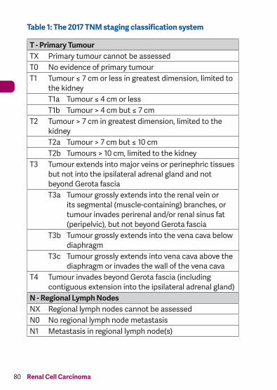
Staging systemThe current UICC 2017 TNM (Tumour Node Metastasis) classification is recommended for the staging of RCC (Table 1).
08 Renal Cell Carcinoma_2018.indd 79 22-02-18 10:25
80 Renal Cell Carcinoma
Table 1: The 2017 TNM staging classification system
T - Primary TumourTX Primary tumour cannot be assessedT0 No evidence of primary tumourT1 Tumour ≤ 7 cm or less in greatest dimension, limited to
the kidneyT1a Tumour ≤ 4 cm or lessT1b Tumour > 4 cm but ≤ 7 cm
T2 Tumour > 7 cm in greatest dimension, limited to the kidneyT2a Tumour > 7 cm but ≤ 10 cmT2b Tumours > 10 cm, limited to the kidney
T3 Tumour extends into major veins or perinephric tissues but not into the ipsilateral adrenal gland and not beyond Gerota fasciaT3a Tumour grossly extends into the renal vein or
its segmental (muscle-containing) branches, or tumour invades perirenal and/or renal sinus fat (peripelvic), but not beyond Gerota fascia
T3b Tumour grossly extends into the vena cava below diaphragm
T3c Tumour grossly extends into vena cava above the diaphragm or invades the wall of the vena cava
T4 Tumour invades beyond Gerota fascia (including contiguous extension into the ipsilateral adrenal gland)
N - Regional Lymph NodesNX Regional lymph nodes cannot be assessedN0 No regional lymph node metastasisN1 Metastasis in regional lymph node(s)
08 Renal Cell Carcinoma_2018.indd 80 22-02-18 10:25
81Renal Cell Carcinoma
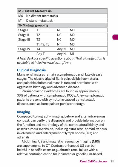
M - Distant MetastasisM0 No distant metastasisM1 Distant metastasisTNM stage groupingStage I T1 N0 M0Stage II T2 N0 M0Stage III T3 N0 M0
T1, T2, T3 N1 M0Stage IV T4 Any N M0
Any T Any N M1A help desk for specific questions about TNM classification is available at http://www.uicc.org/tnm.
Clinical DiagnosisMany renal masses remain asymptomatic until late disease stages. The classic triad of flank pain, visible haematuria, and palpable abdominal mass is rare and correlates with aggressive histology and advanced disease. Paraneoplastic syndromes are found in approximately 30% of patients with symptomatic RCCs. A few symptomatic patients present with symptoms caused by metastatic disease, such as bone pain or persistent cough.
ImagingComputed tomography imaging, before and after intravenous contrast, can verify the diagnosis and provide information on the function and morphology of the contralateral kidney and assess tumour extension, including extra-renal spread, venous involvement, and enlargement of lymph nodes (LNs) and adrenals. Abdominal US and magnetic resonance imaging (MRI) are supplements to CT. Contrast-enhanced US can be helpful in specific cases (e.g., chronic renal failure with a relative contraindication for iodinated or gadolinium-based
08 Renal Cell Carcinoma_2018.indd 81 22-02-18 10:25
82 Renal Cell Carcinoma
contrast media, complex cystic masses, and differential diagnosis of peripheral vascular disorders such as infarction and cortical necrosis). Magnetic resonance imaging can be used in patients with possible venous involvement, or allergy to intravenous contrast. Chest CT is the most accurate for chest staging and is recommended in the primary work–up of patients with suspected RCC. In patients with hereditary RCC who are worried about the radiation exposure of frequent CT scans, MRI may be offered as alternative for follow-up imaging.
BiopsyPercutaneous renal tumour biopsies are used:• to obtain histology of radiologically indeterminate renal
masses;• to select patients with small renal masses for active
surveillance;• to obtain histology before, or simultaneously with, ablative
treatments;• to select the most suitable form of medical and surgical
strategy in the setting of metastatic disease.
In patients with any sign of impaired renal function, a renal scan and total renal function evaluation using estimated glomerular filtration rate should always be undertaken to optimise the treatment decision. Renal biopsy is not indicated for comorbid and frail patients who can be considered only for conservative management (watchful waiting) regardless of biopsy results.
08 Renal Cell Carcinoma_2018.indd 82 22-02-18 10:25
83Renal Cell Carcinoma
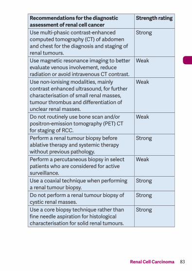
Recommendations for the diagnostic assessment of renal cell cancer
Strength rating
Use multi-phasic contrast-enhanced computed tomography (CT) of abdomen and chest for the diagnosis and staging of renal tumours.
Strong
Use magnetic resonance imaging to better evaluate venous involvement, reduce radiation or avoid intravenous CT contrast.
Weak
Use non-ionising modalities, mainly contrast enhanced ultrasound, for further characterisation of small renal masses, tumour thrombus and differentiation of unclear renal masses.
Weak
Do not routinely use bone scan and/or positron-emission tomography (PET) CT for staging of RCC.
Weak
Perform a renal tumour biopsy before ablative therapy and systemic therapy without previous pathology.
Strong
Perform a percutaneous biopsy in select patients who are considered for active surveillance.
Weak
Use a coaxial technique when performing a renal tumour biopsy.
Strong
Do not perform a renal tumour biopsy of cystic renal masses.
Strong
Use a core biopsy technique rather than fine needle aspiration for histological characterisation for solid renal tumours.
Strong
08 Renal Cell Carcinoma_2018.indd 83 22-02-18 10:25
84 Renal Cell Carcinoma
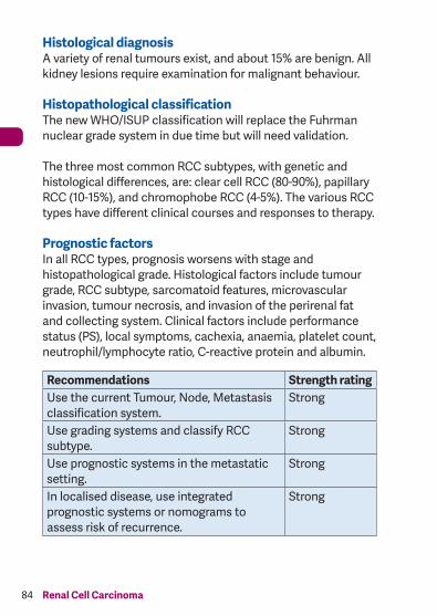
Histological diagnosisA variety of renal tumours exist, and about 15% are benign. All kidney lesions require examination for malignant behaviour.
Histopathological classificationThe new WHO/ISUP classification will replace the Fuhrman nuclear grade system in due time but will need validation.
The three most common RCC subtypes, with genetic and histological differences, are: clear cell RCC (80-90%), papillary RCC (10-15%), and chromophobe RCC (4-5%). The various RCC types have different clinical courses and responses to therapy.
Prognostic factorsIn all RCC types, prognosis worsens with stage and histopathological grade. Histological factors include tumour grade, RCC subtype, sarcomatoid features, microvascular invasion, tumour necrosis, and invasion of the perirenal fat and collecting system. Clinical factors include performance status (PS), local symptoms, cachexia, anaemia, platelet count, neutrophil/lymphocyte ratio, C-reactive protein and albumin.
Recommendations Strength ratingUse the current Tumour, Node, Metastasis classification system.
Strong
Use grading systems and classify RCC subtype.
Strong
Use prognostic systems in the metastatic setting.
Strong
In localised disease, use integrated prognostic systems or nomograms to assess risk of recurrence.
Strong
08 Renal Cell Carcinoma_2018.indd 84 22-02-18 10:25
85Renal Cell Carcinoma
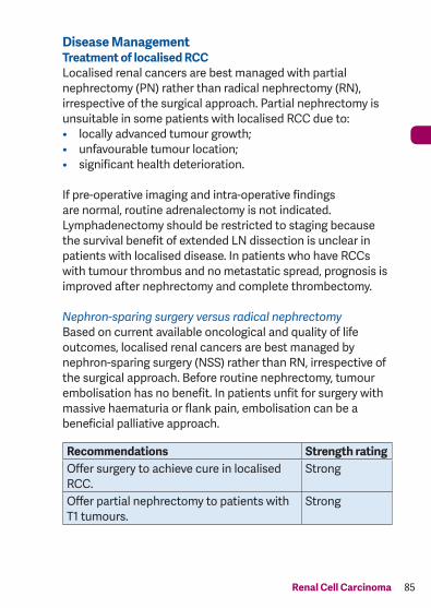
Disease ManagementTreatment of localised RCCLocalised renal cancers are best managed with partial nephrectomy (PN) rather than radical nephrectomy (RN), irrespective of the surgical approach. Partial nephrectomy is unsuitable in some patients with localised RCC due to:• locally advanced tumour growth;• unfavourable tumour location;• significant health deterioration.
If pre-operative imaging and intra-operative findings are normal, routine adrenalectomy is not indicated.Lymphadenectomy should be restricted to staging because the survival benefit of extended LN dissection is unclear in patients with localised disease. In patients who have RCCs with tumour thrombus and no metastatic spread, prognosis is improved after nephrectomy and complete thrombectomy.
Nephron-sparing surgery versus radical nephrectomyBased on current available oncological and quality of life outcomes, localised renal cancers are best managed by nephron-sparing surgery (NSS) rather than RN, irrespective of the surgical approach. Before routine nephrectomy, tumour embolisation has no benefit. In patients unfit for surgery with massive haematuria or flank pain, embolisation can be a beneficial palliative approach.
Recommendations Strength ratingOffer surgery to achieve cure in localised RCC.
Strong
Offer partial nephrectomy to patients with T1 tumours.
Strong
08 Renal Cell Carcinoma_2018.indd 85 22-02-18 10:25
86 Renal Cell Carcinoma
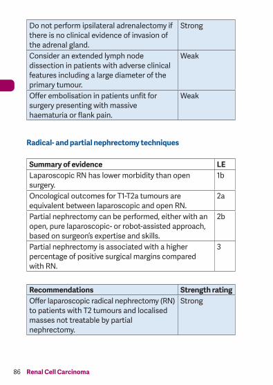
Do not perform ipsilateral adrenalectomy if there is no clinical evidence of invasion of the adrenal gland.
Strong
Consider an extended lymph node dissection in patients with adverse clinical features including a large diameter of the primary tumour.
Weak
Offer embolisation in patients unfit for surgery presenting with massive haematuria or flank pain.
Weak
Radical- and partial nephrectomy techniques
Summary of evidence LELaparoscopic RN has lower morbidity than open surgery.
1b
Oncological outcomes for T1-T2a tumours are equivalent between laparoscopic and open RN.
2a
Partial nephrectomy can be performed, either with an open, pure laparoscopic- or robot-assisted approach, based on surgeon’s expertise and skills.
2b
Partial nephrectomy is associated with a higher percentage of positive surgical margins compared with RN.
3
Recommendations Strength ratingOffer laparoscopic radical nephrectomy (RN) to patients with T2 tumours and localised masses not treatable by partial nephrectomy.
Strong
08 Renal Cell Carcinoma_2018.indd 86 22-02-18 10:25
87Renal Cell Carcinoma
Do not perform minimally invasive RN in patients with T1 tumours for whom a partial nephrectomy is feasible by any approach, including open.
Strong
Do not perform minimally invasive surgery if this approach may compromise oncological, functional and perioperative outcomes.
Strong
Alternatives to surgerySurveillanceElderly and comorbid patients with incidental small renal masses have a low RCC-specific mortality and significant competing-cause mortality. In selected patients with advanced age and/or comorbidities, active surveillance (AS) is appropriate to initially monitor small renal masses, followed, if required, by treatment for progression. The concept of AS differs from the concept of watchful waiting. Watchful waiting is reserved for patients whose comorbidities contraindicate any subsequent active treatment and who do not require follow-up imaging, unless clinically indicated.
Cryoablation and radiofrequency ablationCurrently there are no data showing oncological benefit of cryoablation or radiofrequency ablation (RFA) techniques over PN.
Recommendations Strength ratingOffer active surveillance, radiofrequency ablation and cryoablation to elderly and/or comorbid patients with small renal masses.
Weak
08 Renal Cell Carcinoma_2018.indd 87 22-02-18 10:25
88 Renal Cell Carcinoma
Treatment of locally advanced RCCManagement of clinically positive lymph nodes (cN+)In the presence of clinically positive LNs (cN+), LND is always justified but the extent of LND is still controversial. Low level data suggest that tumour thrombus in the setting of non-metastatic disease should be excised. Adjunctive procedures such as tumour embolisation or inferior vena cava filter do not appear to offer any benefits in the treatment of tumour thrombus. In patients unfit for surgery, or with non-resectable disease, embolisation can control symptoms, including visible haematuria or flank pain. At present there is no evidence for the use of adjuvant therapy following surgery.
Treatment of advanced/metastatic RCCManagement of RCC with venous tumour thrombus
Recommendations Strength ratingIn patients with clinically enlarged lymph nodes, perform lymph node dissection for staging purposes or local control.
Weak
In case of venous involvement, remove the renal tumour and thrombus.
Strong
Cytoreductive nephrectomyTumour nephrectomy is curative only if all tumour deposits are excised. This includes patients with the primary tumour in place and single- or oligo-metastatic resectable disease. For most patients with metastatic disease, cytoreductive nephrectomy is palliative and systemic treatments are necessary.
08 Renal Cell Carcinoma_2018.indd 88 22-02-18 10:25
89Renal Cell Carcinoma
Summary of evidence LECytoreductive nephrectomy combined with interferon-alpha (INF-α) improves survival in patients with metastatic RCC and good performance status.
1a
Deferred cytoreductive nephrectomy with presurgical sunitinib in intermediate-risk patients with clear-cell metastatic RCC leads to a survival benefit in secondary endpoint analysis and selects out patients with inherent resistance to systemic therapy.
2b
Cytoreductive nephrectomy for patients with simultaneous complete resection of a single metastasis or oligometastases may improve survival and delay systemic therapy.
3
Patients with IMDC poor risk (≥ four risk factors) do not benefit.
2b
Recommendations Strength ratingOffer cytoreductive nephrectomy to favourable- and intermediate-risk patients with metastatic RCC (mRCC).
Weak
Do not offer cytoreductive nephrectomy in IMDC poor-risk patients with ≥ four risk factors.
Weak
Perform immediate cytoreductive nephrectomy in patients with oligometastases when complete resection can be achieved.
Weak
Offer deferred cytoreductive nephrectomy to intermediate-risk patients with clear-cell mRCC who require systemic therapy with sunitinib.
Weak
IMDC = The Metastatic Renal Cancer Database Consortium.
08 Renal Cell Carcinoma_2018.indd 89 22-02-18 10:25

90 Renal Cell Carcinoma
Local therapy of metastases in metastatic RCC (mRCC)A systematic review of the local treatment of metastases from RCC in any organ was undertaken. The heterogeneity of the data will only allow for cautious recommendations.
Summary of evidence LEAll included studies were retrospective non-randomised comparative studies, resulting in a high risk of bias associated with non-randomisation, attrition, and selective reporting.
3
With the exception of brain and possibly bone metastases, metastasectomy remains by default the only local treatment for most sites.
3
Retrospective comparative studies consistently point towards a benefit of complete metastasectomy in mRCC patients in terms of overall survival, cancer-specific survival and delay of systemic therapy.
3
Radiotherapy to bone and brain metastases from RCC can induce significant relief from local symptoms (e.g. pain).
3
Recommendations Strength ratingOffer local therapy for metastatic disease (including metastasectomy) to patients with a favourable disease profile in whom complete resection is achievable or when local symptoms need to be controlled.
Weak
Offer stereotactic radiotherapy for clinically relevant bone or brain metastases for local control and symptom relief.
Weak
08 Renal Cell Carcinoma_2018.indd 90 22-02-18 10:25
91Renal Cell Carcinoma
Systemic therapy for advanced/metastatic RCCChemotherapy
Summary of evidence LEIn metastatic RCC, 5-fluorouracil combined with immunotherapy has equivalent efficacy to INF-α.
1b
In metastatic RCC, chemotherapy is otherwise not effective with the exception of gemcitabine and doxorubicine in sarcomatoid and rapidly progressive disease.
3
Recommendation Strength ratingDo not offer chemotherapy as first-line therapy in patients with clear-cell metastatic RCC.
Strong
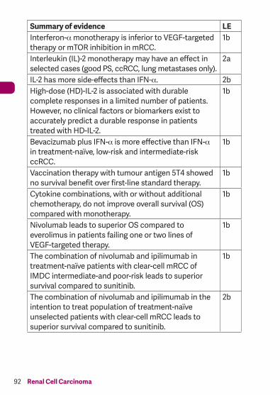
ImmunotherapyInterferon-α may only be effective in some patient subgroups, including patients with clear-cell RCC (ccRCC), favourable-risk criteria, and lung metastases only. Interleukin-2 (IL-2), vaccines and targeted immunotherapy have no place in the standard treatment of advanced/mRCC.
Immune checkpoint inhibition of programmed death receptor (PD-1) and ligand (PD-L1) inhibition have been investigated in mRCC. Randomised data support the use of nivolumab (a PD-1 inhibitor) in vascular endothelial growth factor (VEGF)- refractory disease. A combination of two immune checkpoint inhibitors ipilimumab and nivolumab versus sunitinib in a phase III study on metastatic RCC showed superior survival for a combination of ipilimumab and nivolumab in intermediate- and poor-risk patients.
08 Renal Cell Carcinoma_2018.indd 91 22-02-18 10:25
92 Renal Cell Carcinoma
Summary of evidence LEInterferon-α monotherapy is inferior to VEGF-targeted therapy or mTOR inhibition in mRCC.
1b
Interleukin (IL)-2 monotherapy may have an effect in selected cases (good PS, ccRCC, lung metastases only).
2a
IL-2 has more side-effects than IFN-α. 2bHigh-dose (HD)-IL-2 is associated with durable complete responses in a limited number of patients.However, no clinical factors or biomarkers exist to accurately predict a durable response in patients treated with HD-IL-2.
1b
Bevacizumab plus IFN-α is more effective than IFN-α in treatment-naïve, low-risk and intermediate-risk ccRCC.
1b
Vaccination therapy with tumour antigen 5T4 showed no survival benefit over first-line standard therapy.
1b
Cytokine combinations, with or without additional chemotherapy, do not improve overall survival (OS) compared with monotherapy.
1b
Nivolumab leads to superior OS compared to everolimus in patients failing one or two lines of VEGF-targeted therapy.
1b
The combination of nivolumab and ipilimumab in treatment-naïve patients with clear-cell mRCC of IMDC intermediate-and poor-risk leads to superior survival compared to sunitinib.
1b
The combination of nivolumab and ipilimumab in the intention to treat population of treatment-naïve unselected patients with clear-cell mRCC leads to superior survival compared to sunitinib.
2b
08 Renal Cell Carcinoma_2018.indd 92 22-02-18 10:25
93Renal Cell Carcinoma
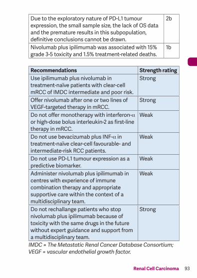
Due to the exploratory nature of PD-L1 tumour expression, the small sample size, the lack of OS data and the premature results in this subpopulation, definitive conclusions cannot be drawn.
2b
Nivolumab plus ipilimumab was associated with 15% grade 3-5 toxicity and 1.5% treatment-related deaths.
1b
Recommendations Strength ratingUse ipilimumab plus nivolumab in treatment-naïve patients with clear-cell mRCC of IMDC intermediate and poor risk.
Strong
Offer nivolumab after one or two lines of VEGF-targeted therapy in mRCC.
Strong
Do not offer monotherapy with interferon-α or high-dose bolus interleukin-2 as first-line therapy in mRCC.
Weak
Do not use bevacizumab plus INF-α in treatment-naïve clear-cell favourable- and intermediate-risk RCC patients.
Weak
Do not use PD-L1 tumour expression as a predictive biomarker.
Weak
Administer nivolumab plus ipilimumab in centres with experience of immune combination therapy and appropriate supportive care within the context of a multidisciplinary team.
Weak
Do not rechallange patients who stop nivolumab plus ipilimumab because of toxicity with the same drugs in the future without expert guidance and support from a multidisciplinary team.
Strong
IMDC = The Metastatic Renal Cancer Database Consortium; VEGF = vascular endothelial growth factor.
08 Renal Cell Carcinoma_2018.indd 93 22-02-18 10:25
94 Renal Cell Carcinoma
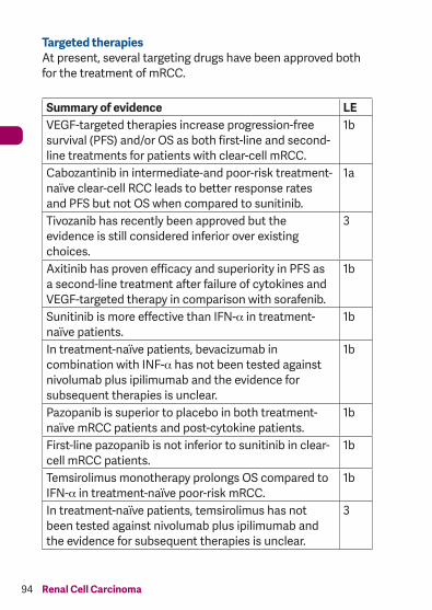
Targeted therapiesAt present, several targeting drugs have been approved both for the treatment of mRCC.
Summary of evidence LEVEGF-targeted therapies increase progression-free survival (PFS) and/or OS as both first-line and second-line treatments for patients with clear-cell mRCC.
1b
Cabozantinib in intermediate-and poor-risk treatment-naïve clear-cell RCC leads to better response rates and PFS but not OS when compared to sunitinib.
1a
Tivozanib has recently been approved but the evidence is still considered inferior over existing choices.
3
Axitinib has proven efficacy and superiority in PFS as a second-line treatment after failure of cytokines and VEGF-targeted therapy in comparison with sorafenib.
1b
Sunitinib is more effective than IFN-α in treatment-naïve patients.
1b
In treatment-naïve patients, bevacizumab in combination with INF-α has not been tested against nivolumab plus ipilimumab and the evidence for subsequent therapies is unclear.
1b
Pazopanib is superior to placebo in both treatment-naïve mRCC patients and post-cytokine patients.
1b
First-line pazopanib is not inferior to sunitinib in clear-cell mRCC patients.
1b
Temsirolimus monotherapy prolongs OS compared to IFN-α in treatment-naïve poor-risk mRCC.
1b
In treatment-naïve patients, temsirolimus has not been tested against nivolumab plus ipilimumab and the evidence for subsequent therapies is unclear.
3
08 Renal Cell Carcinoma_2018.indd 94 22-02-18 10:25
95Renal Cell Carcinoma
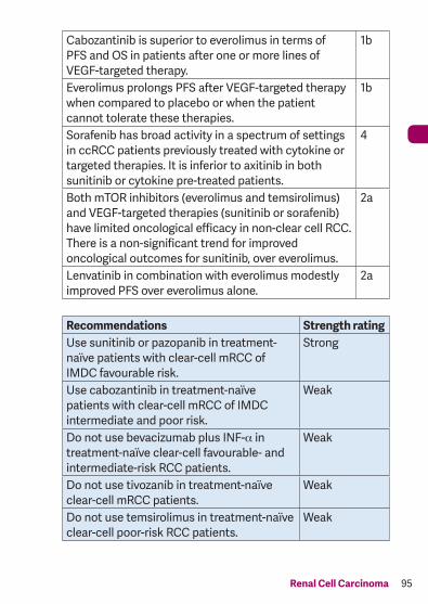
Cabozantinib is superior to everolimus in terms of PFS and OS in patients after one or more lines of VEGF-targeted therapy.
1b
Everolimus prolongs PFS after VEGF-targeted therapy when compared to placebo or when the patient cannot tolerate these therapies.
1b
Sorafenib has broad activity in a spectrum of settings in ccRCC patients previously treated with cytokine or targeted therapies. It is inferior to axitinib in both sunitinib or cytokine pre-treated patients.
4
Both mTOR inhibitors (everolimus and temsirolimus) and VEGF-targeted therapies (sunitinib or sorafenib) have limited oncological efficacy in non-clear cell RCC. There is a non-significant trend for improved oncological outcomes for sunitinib, over everolimus.
2a
Lenvatinib in combination with everolimus modestly improved PFS over everolimus alone.
2a
Recommendations Strength ratingUse sunitinib or pazopanib in treatment-naïve patients with clear-cell mRCC of IMDC favourable risk.
Strong
Use cabozantinib in treatment-naïve patients with clear-cell mRCC of IMDC intermediate and poor risk.
Weak
Do not use bevacizumab plus INF-α in treatment-naïve clear-cell favourable- and intermediate-risk RCC patients.
Weak
Do not use tivozanib in treatment-naïve clear-cell mRCC patients.
Weak
Do not use temsirolimus in treatment-naïve clear-cell poor-risk RCC patients.
Weak
08 Renal Cell Carcinoma_2018.indd 95 22-02-18 10:25
96 Renal Cell Carcinoma
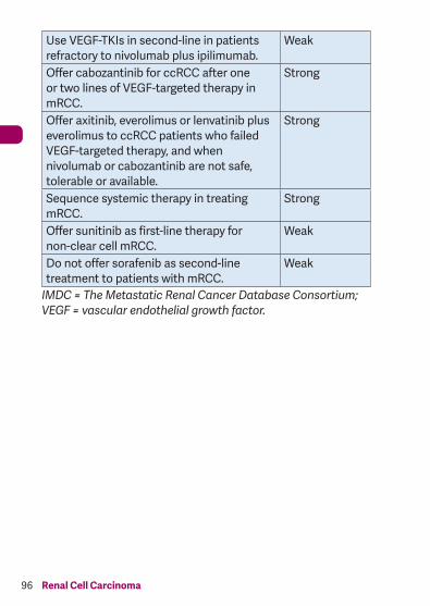
Use VEGF-TKIs in second-line in patients refractory to nivolumab plus ipilimumab.
Weak
Offer cabozantinib for ccRCC after one or two lines of VEGF-targeted therapy in mRCC.
Strong
Offer axitinib, everolimus or lenvatinib plus everolimus to ccRCC patients who failed VEGF-targeted therapy, and when nivolumab or cabozantinib are not safe, tolerable or available.
Strong
Sequence systemic therapy in treating mRCC.
Strong
Offer sunitinib as first-line therapy for non-clear cell mRCC.
Weak
Do not offer sorafenib as second-line treatment to patients with mRCC.
Weak
IMDC = The Metastatic Renal Cancer Database Consortium; VEGF = vascular endothelial growth factor.
08 Renal Cell Carcinoma_2018.indd 96 22-02-18 10:25
97Renal Cell Carcinoma
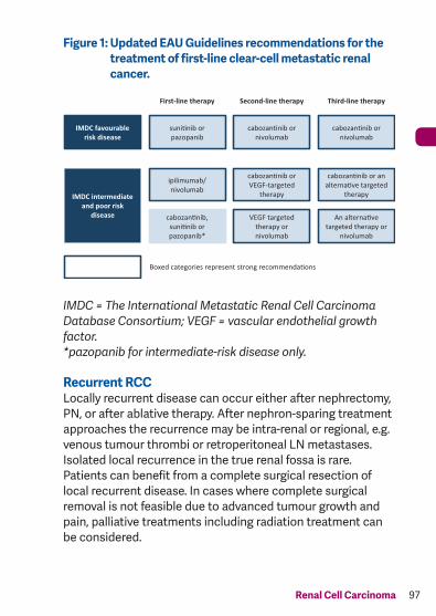
Figure 1: Updated EAU Guidelines recommendations for the treatment of first-line clear-cell metastatic renal cancer.
IMDC = The International Metastatic Renal Cell Carcinoma Database Consortium; VEGF = vascular endothelial growth factor. *pazopanib for intermediate-risk disease only.
Recurrent RCCLocally recurrent disease can occur either after nephrectomy, PN, or after ablative therapy. After nephron-sparing treatment approaches the recurrence may be intra-renal or regional, e.g. venous tumour thrombi or retroperitoneal LN metastases. Isolated local recurrence in the true renal fossa is rare. Patients can benefit from a complete surgical resection of local recurrent disease. In cases where complete surgical removal is not feasible due to advanced tumour growth and pain, palliative treatments including radiation treatment can be considered.
First-line therapy Second-line therapy Third-line therapy
IMDC favourable risk disease
suni�nib or pazopanib
cabozan�nib or nivolumab
cabozan�nib or nivolumab
IMDC intermediate and poor risk
disease
ipilimumab/ nivolumab
cabozan�nib, suni�nib or pazopanib*
cabozan�nib or VEGF-targeted
therapy
VEGF targeted therapy or nivolumab
cabozan�nib or an alterna�ve targeted
therapy
An alterna�ve targeted therapy or
nivolumab
Boxed categories represent strong recommenda�ons
08 Renal Cell Carcinoma_2018.indd 97 22-02-18 10:25
98 Renal Cell Carcinoma
Surveillance following surgery for RCCThe aim of surveillance is to detect either local recurrence or metastatic disease while the patient is still surgically curable. Surveillance after treatment for RCC allows the urologist to identify:• postoperative complications;• renal function;• local recurrence;• recurrence in the contralateral kidney;• development of metastases.
Depending on the availability of new effective treatments, more intensive follow-up schedules may be required, particularly as there is a higher local recurrence rate after cryotherapy and RFA. At present there is no evidence-based standard for the follow-up of patients with RCC, or for the optimal duration of follow-up. An example of a surveillance algorithm monitoring patients after treatment for RCC that recognises not only the patient’s risk profile but also treatment efficacy is provided in Table 2. For patients with metastatic disease, individualised follow-up is indicated.
08 Renal Cell Carcinoma_2018.indd 98 22-02-18 10:25
99Renal Cell Carcinoma
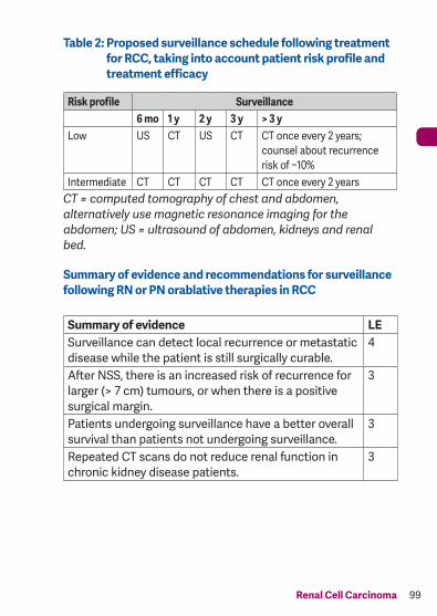
Table 2: Proposed surveillance schedule following treatment for RCC, taking into account patient risk profile and treatment efficacy
Risk profile Surveillance6 mo 1 y 2 y 3 y > 3 y
Low US CT US CT CT once every 2 years; counsel about recurrence risk of ~10%
Intermediate CT CT CT CT CT once every 2 yearsCT = computed tomography of chest and abdomen, alternatively use magnetic resonance imaging for the abdomen; US = ultrasound of abdomen, kidneys and renal bed.
Summary of evidence and recommendations for surveillance following RN or PN orablative therapies in RCC
Summary of evidence LESurveillance can detect local recurrence or metastatic disease while the patient is still surgically curable.
4
After NSS, there is an increased risk of recurrence for larger (> 7 cm) tumours, or when there is a positive surgical margin.
3
Patients undergoing surveillance have a better overall survival than patients not undergoing surveillance.
3
Repeated CT scans do not reduce renal function in chronic kidney disease patients.
3
08 Renal Cell Carcinoma_2018.indd 99 22-02-18 10:25
100 Renal Cell Carcinoma
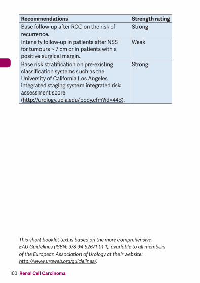
Recommendations Strength ratingBase follow-up after RCC on the risk of recurrence.
Strong
Intensify follow-up in patients after NSS for tumours > 7 cm or in patients with a positive surgical margin.
Weak
Base risk stratification on pre-existing classification systems such as the University of California Los Angeles integrated staging system integrated risk assessment score (http://urology.ucla.edu/body.cfm?id=443).
Strong
This short booklet text is based on the more comprehensive EAU Guidelines (ISBN: 978-94-92671-01-1), available to all members of the European Association of Urology at their website: http://www.uroweb.org/guidelines/.
08 Renal Cell Carcinoma_2018.indd 100 22-02-18 10:25