early perception-action coupling: eye movements and the ... · after habituation, two new test...
TRANSCRIPT
Early perception-action coupling: Eye movements and thedevelopment of object perception
Scott P. Johnson*, Kerri L. Johnson
Department of Psychology, Uris Hall, Cornell University, Ithaca, NY 14853, USA
Received 10 July 2000; received in revised form 8 September 2000; accepted 4 October 2000
Abstract
We investigated the scanning strategies used by 2- to 3.5-month-old infants when viewing partlyoccluded object displays. Eye movements were recorded with a corneal reflection system as the infantsobserved stimuli depicting two rod parts above and below an occluding box. Stimulus parameters werechosen on the basis of past research demonstrating the importance of motion, occluder width, and edgealignment to perception of object unity. Results indicated that the infants tailored scanning to displaycharacteristics, engaging in more extensive scanning when unity perception was challenged by a wideoccluder or misaligned edges. In addition, older infants tended to scan the lower parts of the displaysmore frequently than did younger infants. Exploration of individual differences, however, revealedmarked contrasts in specific scanning styles across infants. The findings are consistent with views ofperceptual development stressing the importance of information processing skills and self-directedaction to the acquisition of object knowledge. © 2000 Elsevier Science Inc. All rights reserved.
Keywords: Eye movements; Perception-action coupling; Object perception; Perceptual development; Cognitivedevelopment; Information processing vs. core principles perspectives
Veridical perception of object layout is a necessary requirement for the selection ofappropriate action schemes (Gibson, 1966). Before 4 to 6 months, infants are limited toinaccurate reaching and grasping, and extensive manual exploration of objects is largelyprecluded (Bushnell, 1985; von Hofsten, 1984). The oculomotor system, however, is rela-tively mature in young infants when compared to other action systems, and infants engagein active visual exploration of the environment from birth (Slater, 1995; von Hofsten &
* Corresponding author. Tel.: �(607) 255-6392; fax: �(607) 255-8433.E-mail address: [email protected] (S.P. Johnson).
Infant Behavior & Development 23 (2000) 461–483
0163-6383/00/$ – see front matter © 2000 Elsevier Science Inc. All rights reserved.PII: S0163-6383(01)00057-1
Rosander, 1998). These burgeoning perceptual skills are soon used effectively to perceiveobject layout: There is evidence of rapid development of veridical object perception in thefirst 4 months after birth, as revealed by experiments investigating perception of object unity(Johnson & Aslin, 1995, 1996; Johnson & Nanez, 1995; Kellman & Spelke, 1983).
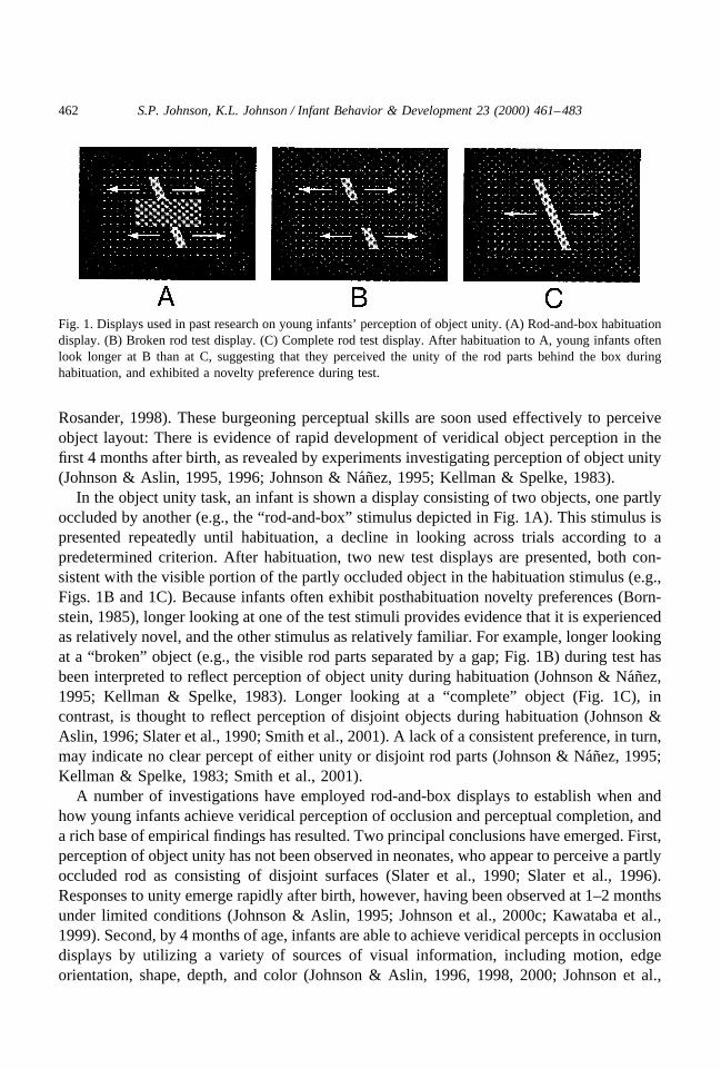
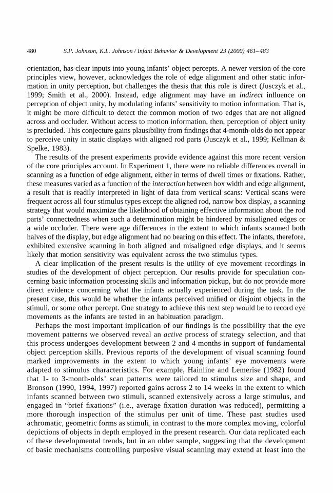
In the object unity task, an infant is shown a display consisting of two objects, one partlyoccluded by another (e.g., the “rod-and-box” stimulus depicted in Fig. 1A). This stimulus ispresented repeatedly until habituation, a decline in looking across trials according to apredetermined criterion. After habituation, two new test displays are presented, both con-sistent with the visible portion of the partly occluded object in the habituation stimulus (e.g.,Figs. 1B and 1C). Because infants often exhibit posthabituation novelty preferences (Born-stein, 1985), longer looking at one of the test stimuli provides evidence that it is experiencedas relatively novel, and the other stimulus as relatively familiar. For example, longer lookingat a “broken” object (e.g., the visible rod parts separated by a gap; Fig. 1B) during test hasbeen interpreted to reflect perception of object unity during habituation (Johnson & Nanez,1995; Kellman & Spelke, 1983). Longer looking at a “complete” object (Fig. 1C), incontrast, is thought to reflect perception of disjoint objects during habituation (Johnson &Aslin, 1996; Slater et al., 1990; Smith et al., 2001). A lack of a consistent preference, in turn,may indicate no clear percept of either unity or disjoint rod parts (Johnson & Nanez, 1995;Kellman & Spelke, 1983; Smith et al., 2001).
A number of investigations have employed rod-and-box displays to establish when andhow young infants achieve veridical perception of occlusion and perceptual completion, anda rich base of empirical findings has resulted. Two principal conclusions have emerged. First,perception of object unity has not been observed in neonates, who appear to perceive a partlyoccluded rod as consisting of disjoint surfaces (Slater et al., 1990; Slater et al., 1996).Responses to unity emerge rapidly after birth, however, having been observed at 1–2 monthsunder limited conditions (Johnson & Aslin, 1995; Johnson et al., 2000c; Kawataba et al.,1999). Second, by 4 months of age, infants are able to achieve veridical percepts in occlusiondisplays by utilizing a variety of sources of visual information, including motion, edgeorientation, shape, depth, and color (Johnson & Aslin, 1996, 1998, 2000; Johnson et al.,
Fig. 1. Displays used in past research on young infants’ perception of object unity. (A) Rod-and-box habituationdisplay. (B) Broken rod test display. (C) Complete rod test display. After habituation to A, young infants oftenlook longer at B than at C, suggesting that they perceived the unity of the rod parts behind the box duringhabituation, and exhibited a novelty preference during test.
462 S.P. Johnson, K.L. Johnson / Infant Behavior & Development 23 (2000) 461–483

2000a; Johnson et al., 2000b; Kellman & Spelke, 1983; Needham, 1998; Smith et al., 2001;see Johnson, 1997, 2000 for reviews).
Experiments employing the object unity paradigm, then, have revealed a fundamental shiftin how infants perceive the world, from birth through the next few months. With the onsetof visual experience, neonates may perceive the environment as consisting of a “sensorytableaux,” or disconnected fragments that do not cohere into tangible, bounded objects(Piaget, 1954). Veridical perception of occlusion, however, emerges rapidly. The periodbetween 2 to 4 months seems especially important for the development of those attentionaland cognitive skills necessary to detect and utilize visual information that supports percep-tion of object unity (Johnson, 1997).
Although much is known currently about the emergence of unity perception at adescriptive level, decisive explanations of underlying mechanisms of development havebeen more difficult to achieve. How exactly do veridical percepts arise in the infant?Several candidate accounts have been proposed. Spelke, for example (1990, Spelke &Van de Walle, 1993), has suggested that infants experience objects in accord with a setof core principles. Development, on this account, consists of refinements and enrichmentof an incipient conceptual system that is predisposed to perceive objects as bounded,solid entities. This system is modular, or self-encapsulated, and independent of percep-tion (Spelke & Hermer, 1996). In contrast to this core knowledge hypothesis, Johnson(2000, in press) proposed that veridical object perception and object knowledge aredependent on subsidiary information processing skills, along with experience viewingobjects that become occluded and are again fully visible. The lower-level perceptualabilities are required to detect the visual information necessary for segmentation of theoptic array into its constituent surfaces, alongside the conjoining of these visible surfacefragments across spatial and temporal gaps into percepts of coherent objects. The visualexperience is critical for the building of associations of partial views of surfaces to viewsof fully visible objects. Object knowledge, then, arises from lower-level perceptualproficiency that develops over the first few months after birth, and exposure to partlyoccluded and unoccluded objects in the visual environment (see Johnson, 2000, in press;Jusczyk et al., 1999; Mareschal & Johnson, in press for further discussion.)
A key prediction of the information processing perspective is that the development ofveridical object perception is accompanied by improvements in the effectiveness with whichinfants sample the optic array. Clearly, without adequate scrutiny of the visual informationthat specifies object layout (such as relative depth, orientation, and surface appearance),accurate percepts of the environment are precluded. The goal of the present experiments,therefore, was to explore the relation between perception of partly occluded objects and eyemovements. We reasoned that one potential limitation in infants’ perception of object unitymay be rooted in inefficient scanning strategies, such that very young infants, relative toolder infants, would be less likely to scan the entire stimulus, and to limit fixations touninformative regions of the display. We also explored whether scanning strategies mightvary as a function of display characteristics, by including stimuli in which unity either wouldor would not likely be perceived.
463S.P. Johnson, K.L. Johnson / Infant Behavior & Development 23 (2000) 461–483
1. Experiment 1
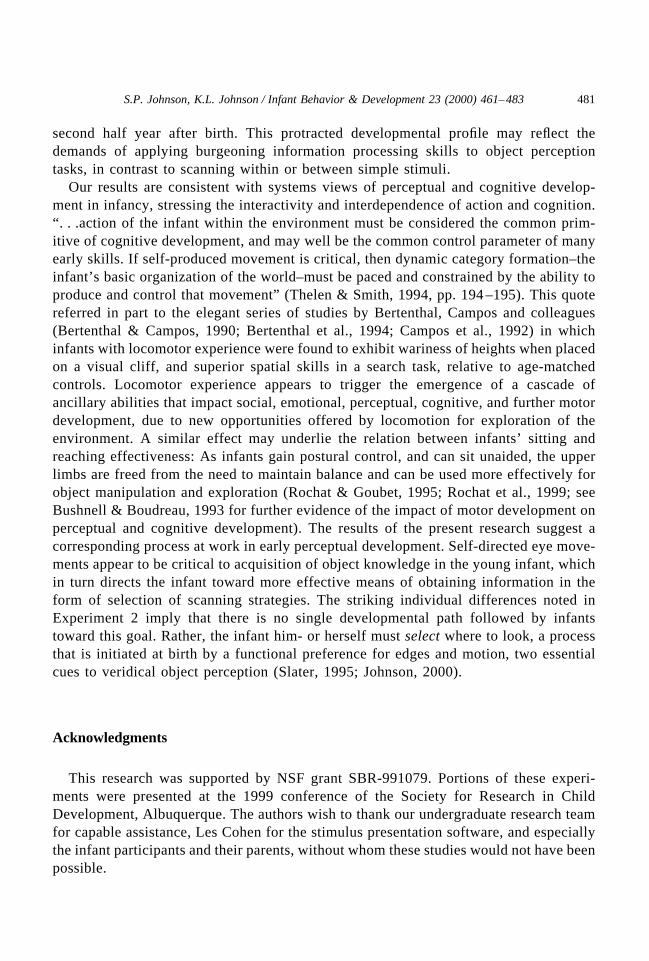
In the first experiment, infants were presented with four displays depicting two rod partsabove and below an occluding box. Stimuli were chosen on the basis of past researchshowing that responses to object unity in rod-and-box displays vary both as a function ofoccluder width, and of the alignment of the rod edges across the occluder: Two-month-oldshave been found to perceive unity when the occluder was narrow, but not wide (Johnson &Aslin, 1995; Johnson & Nanez, 1995), and 4-month-olds have been found to perceive unitywhen the rod edges were aligned, but unity perception is attenuated when rod parts aremisaligned (Johnson & Aslin, 1996; Johnson et al., 2000a, b; Smith et al., 2001). The fourstimuli employed in Experiment 1, therefore, were varied along these two dimensions,yielding two levels of occluder width (wide and narrow) and two levels of edge alignment(aligned and misaligned) (see Fig. 2). Infants were presented each of the four displays twiceand their eye movements recorded.
1.1. Method
1.1.1 ParticipantsFourteen full-term infants (5 females) comprised the final sample, ranging in age from 59
to 127 days. An additional 17 infants were observed but not included in the analyses, due tofussiness (2 infants), equipment failure (3), experimenter error (3), an inability to obtain areliable point of gaze (POG) for unknown reasons (2), excessive movement on the part of theinfant, such that we were unable to record eye movements (3), or poor calibration of the POG(4; see subsequent discussion of calibration).
1.1.2. Apparatus and stimuliA Macintosh 7600 computer and 76 cm Barco color monitor were used to present the
stimuli. The infants were shown one of four rod-and-box displays as eye movements wererecorded. Each stimulus was viewed twice (one infant viewed only one of each stimulus dueto excessive fussiness), and was presented for an average 21.6 s (SD � 8.4). Betweenrod-and-box displays, an “attention-getter” stimulus was shown, to keep the infant engagedin the task. Stimulus duration was controlled by the experimenter (see Procedures section).
Each rod-and-box display was presented against a black background with a 12 � 20 gridof white dots serving as texture elements (see Fig. 2). (Background texture has been foundto lead to longer looking, and therefore perhaps greater attentional engagement, in habitu-ation experiments; background texture also provides depth information to aid in perceptualsegregation of stimulus elements; see Johnson & Aslin, 1996.) The background measured32.5 � 23 cm (15.4° � 10.9° visual angle, at the infant’s 120 cm viewing distance). The fourrod-and-box displays each contained a blue occluding box and two green rod parts, butdiffered with respect to occluder width and alignment of the rod parts. In the wide occluderdisplays, the box measured 25.2 � 6.0 cm (11.9° � 2.8°). In the narrow occluder displays,the occluder was half this height. In the aligned rod displays, the rod measured 17.3 � 1.5cm (8.2° � 0.7°) and was oriented 22° counterclockwise. The rod parts’ edges were alignedacross the occluder. In the misaligned rod displays, the bottom rod part was displaced 2.7 cm
464 S.P. Johnson, K.L. Johnson / Infant Behavior & Development 23 (2000) 461–483
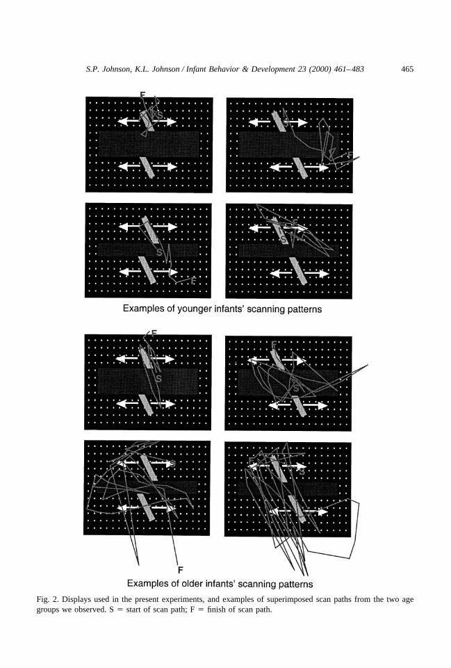
Fig. 2. Displays used in the present experiments, and examples of superimposed scan paths from the two agegroups we observed. S � start of scan path; F � finish of scan path.
465S.P. Johnson, K.L. Johnson / Infant Behavior & Development 23 (2000) 461–483

(1.3°) to the left. The attention-getter display consisted of a target-patterned ball, presentedagainst the same dot background, that expanded and contracted rhythmically (each cyclelasting 2 s) in time with a gentle beep. At its maximum size, the ball measured 10.1 cm (4.8°)in diameter; at its smallest, it measured 2.2 cm (1.0°).
An Applied Science Laboratories Model 504 corneal reflection eye tracking system wasused to collect looking time data. A remote pupil camera with a pan/tilt base was placed onthe table below the stimulus monitor. The stimulus viewed by the infant was importeddirectly into the eye tracker from the Macintosh for purposes of off-line data coding (seeResults section). Data were saved on the hard drive of the PC as X-Y coordinates of the POG,recorded at 60 Hz on a 260 � 240 grid of points on the stimulus. The eye tracker also feda signal into a videotape recorder in the form of crosshairs superimposed on the stimulus.
1.1.3. ProcedureInfants were seated in a parent’s lap 120 cm from the stimulus monitor. Two experiment-
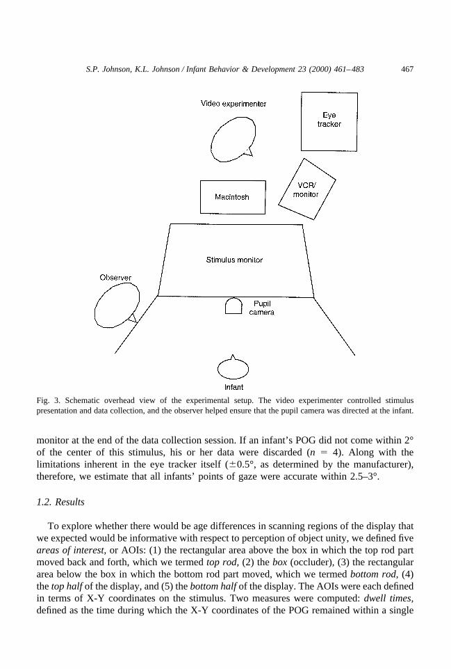
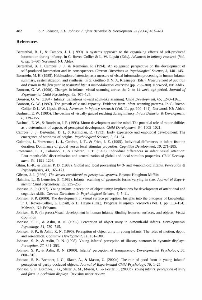
ers worked in concert to collect data. The “observer” watched the infant, and held a remotecontrol that directed the pupil camera, through a peephole in one of two partitions extendingout from either side of the stimulus monitor. The “video experimenter” sat behind thestimulus monitor and watched an image of the infant’s pupil on a 25 cm achromatic monitor,and the POG and stimulus on the VTR monitor. Both the observer and the video experi-menter were out of sight of the infant (see Fig. 3).
The room lights were first turned off and the infant shown a Mickey Mouse cartoon toengage his or her interest, as the observer directed the pupil camera toward the infant’s eyewith the remote control. After the eye was in view, the video experimenter changed from this“manual” mode of camera control to an automatic mode, during which the camera remaineddirected at the pupil despite small displacements of the infant’s head (via an algorithm builtinto the eye tracker). (Occasionally during the experiment, the infant moved his or her headmore quickly than the camera could follow, such that the pupil was lost from view. At thispoint the video experimenter changed from the automatic mode back to manual, the observeragain located the pupil in the camera, and automatic control was resumed.) Followingacquisition of the pupil image, and as the infant watched the cartoon, adjustments were madeon the eye tracker to maximize robustness of the POG. This varied somewhat from infant toinfant with respect to reflectance of infrared and visible illumination (corneal and pupilreflection, respectively). The infant was then shown the four rod-and-box displays, orderedaccording to a balanced Latin-square design. Between displays, the attention-getter was presented.
Trial length varied according to the video experimenter’s judgment of the infant’s interestlevel. The video experimenter attempted to obtain consistent tracking of as many displays aspossible (up to a maximum of 8), for as long as possible, which was accomplished bykeeping trials short and switching stimuli frequently. The data collection session usuallylasted about 4 min.
1.1.4. CalibrationThe eye tracker was calibrated on the second author’s left eye with a 9-point calibration
routine (i.e., the POG for 9 known points was entered). Individual infants were not calibrated,but accuracy of POG was checked by presenting the attention-getter at 5 points across the
466 S.P. Johnson, K.L. Johnson / Infant Behavior & Development 23 (2000) 461–483
monitor at the end of the data collection session. If an infant’s POG did not come within 2°of the center of this stimulus, his or her data were discarded (n � 4). Along with thelimitations inherent in the eye tracker itself (�0.5°, as determined by the manufacturer),therefore, we estimate that all infants’ points of gaze were accurate within 2.5–3°.
1.2. Results
To explore whether there would be age differences in scanning regions of the display thatwe expected would be informative with respect to perception of object unity, we defined fiveareas of interest, or AOIs: (1) the rectangular area above the box in which the top rod partmoved back and forth, which we termed top rod, (2) the box (occluder), (3) the rectangulararea below the box in which the bottom rod part moved, which we termed bottom rod, (4)the top half of the display, and (5) the bottom half of the display. The AOIs were each definedin terms of X-Y coordinates on the stimulus. Two measures were computed: dwell times,defined as the time during which the X-Y coordinates of the POG remained within a single
Fig. 3. Schematic overhead view of the experimental setup. The video experimenter controlled stimuluspresentation and data collection, and the observer helped ensure that the pupil camera was directed at the infant.
467S.P. Johnson, K.L. Johnson / Infant Behavior & Development 23 (2000) 461–483
AOI, and fixations, defined as individual segments in the data stream during which the X-Ycoordinates of the POG remained within 0.5° for at least 100 ms. Dwell time and fixationdata were computed by the “Eyenal” software included in the eye tracker system.
Infants were divided into two age groups for purposes of analyses, a younger group (n �6; M age � 69.8 days, SD � 8.1), and an older group (n � 8; M age � 98.5 days, SD �14.5). Initial analyses revealed no significant age differences in mean time of displaypresentation, t(12) � 1.70, ns (M � 26.2 s, SD � 9.0 for younger infants; M � 18.8 s, SD �7.2, for older infants), total number of fixations, t(12) � �1.09, ns (M � 98.3, SD � 40.1for younger infants; M � 125.9, SD � 50.8, for older infants), or total dwell time, t(12) �0.72, ns (M � 64.7 s, SD � 30.5 for younger infants; M � 52.5 s, SD � 32.1, for olderinfants). As seen in the examples shown in Fig. 2, however, there were distinct differencesin the manner in which younger and older infants scanned the displays, a conclusionconfirmed by analyses of dwell times and fixations.
1.2.1. Dwell time dataPrior to analysis, dwell times were equated for differences in display duration by con-
verting them to proportions, relative to time of each display presentation. A 2 (age) � 3(AOI: box, top rod, or bottom rod) � 2 (box width: wide vs. narrow) � 2 (edge alignment:misaligned vs. aligned) mixed ANOVA, with repeated measures on the second, third, andfourth factors, revealed a significant main effect of AOI, F(2, 24) � 8.10, p � .01, which wasqualified by a significant box width � edge alignment interaction, F(1, 12) � 8.48, p � .05,an AOI � box width interaction, F(2, 24) � 16.07, p � .001, and an AOI � box width �edge alignment interaction, F(2, 26) � 13.32, p � .001. As seen in Fig. 4A (left panel), theseeffects were due to greater dwell times in the box region when the edges were aligned vs.when edges were misaligned, but only when the occluder was wide, as revealed by simpleeffects tests, F(1, 12) � 15.43, p � .01; no other comparisons of dwell times in each AOIas a function of box width and edge alignment reached significance, all Fs � 2.9, ns (thereasons for this pattern of looking are explored in more detail subsequently, in the section onVertical scans).
There was also a significant age � AOI interaction, F(2, 24) � 3.81, p � .05. As seen inFig. 4A (right panel), younger infants’ dwell times in the top rod region were greater thanin box region, F(1, 12) � 6.29, p � .05, and dwell times in the box region were greater thanin the bottom rod region, F(1, 12) � 4.88, p � .05. In contrast, all comparisons betweendwell times in each AOI fell short of significance for the older infants, all Fs � 1.9, ns.
1.2.2. Fixation dataLike dwell time data, fixation data were converted to proportions to equate for differences
across trials in display time. Fixations/s were analyzed to investigate age differences inscanning across the three AOIs, as a function of occluder width and edge alignment, with anage � AOI � box width � edge alignment mixed ANOVA. This analysis yielded asignificant age difference, F(1, 12) � 5.70, p � .05: Older infants tended to exhibit morefixations/s (M � 0.40, SD � 0.15) than did younger infants (M � 0.24, SD � 0.08). Therewas also a significant effect of AOI, F(2, 24) � 4.41, p � .05, which was qualified by asignificant box width � AOI interaction, F(2, 24) � 10.79, p � .001, and a significant edge
468 S.P. Johnson, K.L. Johnson / Infant Behavior & Development 23 (2000) 461–483

alignment � box width � AOI interaction, F(2, 24) � 5.14, p � .05. These effects paralleledthose obtained in the analysis of dwell times (see Fig. 4B, left panel): more fixations/s in thewide vs. narrow box AOI when the edges were aligned, F(1, 12) � 5.46, p � .05; no other
Fig. 4. Data from Experiment 1. Both dwell times (A, left panel) and fixations per s (B, left panel) revealed ascanning strategy geared toward determining the unity of the two rod parts. The larger proportion of dwell times andfixations in the box AOI in the aligned rod, wide box display reflects greater vertical scanning between the top andbottom halves of the display. There was also a high rate of vertical scanning in the misaligned rod, wide box andmisaligned rod, narrow box displays, but not the aligned rod, narrow box display. The right panels show age differencesin dwell times (A) and fixations (B) in scanning AOIs, and reveal that older infants engaged in more fixations per s,and that dwell times and fixations per s were more evenly distributed across AOIs, relative to younger infants.
469S.P. Johnson, K.L. Johnson / Infant Behavior & Development 23 (2000) 461–483
comparisons of fixations/s in each AOI as a function of box width and edge alignmentreached significance, all Fs � 3.5, ns (again, the reasons for this pattern are examined inmore detail in the Vertical scans section).
There was also a marginally significant age � AOI interaction, F(2, 24) � 2.90, p � .07.(Because age differences in scanning are a central focus of this article, we followed up onthis finding with simple effects tests even though the interaction failed to reach statisticalsignificance.) Both younger and older infants fixated the box more when it was wide thanwhen it was narrow, F(1, 12) � 18.18, p � .01, and this difference did not vary reliably asa function of age, F(1, 12) � 2.17, ns. Age differences were revealed, however, in the extentto which the infants fixated the top and bottom rods (Fig. 4B, right panel). For older infants,there was no reliable difference in fixations toward the top and bottom rods, F(1, 12) � 0.19,ns. Fixations in these two AOIs, however, varied as a function of box width, F(1, 12) � 6.34,p � .05. In the wide box displays, there were more fixations toward the bottom rod, anonsignificant difference, F(1, 12) � 0.75, ns, and in the narrow box displays, there weremore fixations toward the top rod, but this difference was only marginally significant, F(1,12) � 4.04, p � .07. Younger infants, in contrast, fixated the top rod more than the bottomrod, F(1, 12) � 6.58, p � .05, and this difference did not vary reliably as a function of boxwidth, F(1, 12) � 0.07, ns.
1.2.3. Vertical scansThe next analyses explored the extent to which infants scanned between the top and
bottom portions of the displays. We reasoned that one way in which age-related improve-ments in perception of object unity might be revealed is in increased vertical scanning, tofacilitate detection of edge alignment and common motion of the two visible rod parts.Vertical scans were defined as changes in fixation/s from one half of the display to the other(top to bottom or vice versa, between AOIs incorporating the entire top or bottom half of thedisplay). These measures were also examined as a function of display type. Vertical scanswere investigated with an age � box width � edge alignment mixed ANOVA that yieldeda significant effect of age, F(1, 12) � 8.78, p � .05, due to more vertical scans by olderinfants (M � 0.25, SD � 0.12) relative to younger infants (M � 0.09, SD � 0.07). There wasalso a significant box width � edge alignment interaction, F(1, 12) � 5.06, p � .05, and noother significant effects. Simple effects tests revealed no significant difference in verticalscans in misaligned rod displays as a function of occluder height, F(1, 12) � 0.04, ns (wideoccluder M � 0.20, SD � 0.18; narrow occluder M � 0.20, SD � 0.19). When the rod wasaligned, in contrast, there were more vertical scans when the box was wide (M � 0.22, SD �0.15) than when it was narrow (M � 0.13, SD � 0.12), although the difference was onlymarginally significant, F(1, 12) � 4.28, p � .06.
1.3. Discussion
The results from dwell time and fixation data suggest important age differences inscanning strategies when infants view rod-and-box displays. Scans within the three AOIswere relatively proportional for older infants, whereas younger infants tended to concentratescans in the top portions of the stimulus (the top rod and the box). Older infants also scanned
470 S.P. Johnson, K.L. Johnson / Infant Behavior & Development 23 (2000) 461–483

between the top and bottom halves of the stimulus more frequently than did younger infants,a strategy that would be expected to facilitate extraction of important visual information thatsupports perception of object unity.
The finding that younger infants did not often fixate the bottom rod part appears to beinconsistent with reports that 2-month-olds perceive object unity under some circumstances(Johnson & Aslin, 1995; Johnson et al., 2000c). It seems unlikely that this effect wouldobtain unless infants inspected the entire stimulus. These discrepant results might be due tothe limited amount of time during which the display was available for inspection in thepresent study, necessitated by the need to keep the infants’ interest level high throughout theprocedure. Does this mean that in the present study, younger infants will look more at thebottom rod part if provided additional exposure to the stimulus? This question was exploredby computing correlations between display time and number of fixations (not fixations/s) ineach of the AOIs (box, top rod, bottom rod). For the younger infants, the only correlation thatapproached significance was that between display time and fixations in the vicinity of thebottom rod, r � 0.79, p � .06. For the older infants, none of the correlations reachedsignificance. Given extra time, then, the younger infants engaged in somewhat more exten-sive scanning, a pattern characteristic of the older infants. Although this finding must beconsidered tentative, as the correlation was only marginally significant statistically, it isconsistent with recent reports that young infants may follow a local-to-global processingstrategy, and older infants are more adept at processing global information (e.g., Colombo etal., 1995; Freeseman et al., 1993; Ghim & Eimas, 1988; Johnson et al., 2000a).
Another important consideration is the interaction of box width and edge alignment onmeasures of dwell times, fixations, and vertical scans: All these measures were greater whenthe box was wide and the edges were aligned. These results are consistent with theinterpretation that edge orientation is central to perception of object unity. Eye movementpatterns, then, would be expected to reveal a scanning strategy dedicated to obtaininginformation concerning whether or not two edges across an occluder are aligned. When therod edges were aligned, the infants engaged in more vertical scanning in the wide-occluderdisplay, relative to the narrow-occluder display. This suggests that the wider spatial gapchallenged perception of the edge alignment, and more scans were needed to detect the edgerelations. Many of these scans crossed the midline (between the top and bottom halves of thedisplay) and fell in the box AOI, because it was centrally located and intersected both the topand bottom rod parts. Vertical scans were comparable in frequency to the wide box, alignededge display when infants were presented with the two misaligned rod displays, suggestingactive comparison of the edge relations in an attempt to determine alignment. In contrast,when the infants viewed the display with aligned edges across the narrow occluder, deter-mination of alignment was more readily accomplished, necessitating fewer scans. Theseconclusions must be considered speculative as we have no independent evidence (e.g., fromhabituation data) concerning percepts of unity or disjoint objects among our sample ofinfants (see General Discussion). Nevertheless, the infants’ scanning patterns conformed tothe hypothesis that the task of perceiving object unity is based on a prior determination ofedge relations, a central tenet of the information-processing view.
471S.P. Johnson, K.L. Johnson / Infant Behavior & Development 23 (2000) 461–483

2. Experiment 2
The second experiment explored scanning patterns in a small sample of infants observedlongitudinally, across an age range between 2 and 5 months. The purpose of Experiment 2was to explore individual differences in scanning patterns, with the goal of revealing bothcommonalities across infants (to confirm the results of Experiment 1), and continuity overtime in individual infants’ responses. That is, we asked whether individual infants wouldshow an increase with age in the type of extensive scanning observed in the older infants inExperiment 1, as well as distinct “styles” of scanning patterns that remained stable over thefirst few months.
2.1. Method
2.1.1. ParticipantsFive full-term infants (1 female) were observed for five to eight data collection sessions,
spaced at least one week apart. An additional five infants were observed but not included inthe sample because they completed fewer than four sets of data (either for reasons describedin Experiment 1, or because they were not brought to the lab for scheduled appointments).Across the sample observed for the present experiment, five sessions failed to result in usabledata, due to an inability to obtain a reliable POG (1) or excessive movement on the part ofthe infant (4).
2.1.2. Apparatus, stimuli, procedure, and calibrationAll other methodological aspects of Experiment 2 were identical to those of Experiment 1.
2.2. Results
Data from each of the five infants in Experiment 2 are considered separately with a seriesof analyses targeted at specific questions arising from Experiment 1. First, do data fromindividual infants provide evidence of age-related changes in dwell times and fixations in thebottom rod AOI? Second, do these data reflect age-related improvements in vertical scan-ning? Third, are these vertical scans more frequent in the aligned rod, wide box condition?Fourth, is there evidence of other, more idiosyncratic scanning strategies on the part ofindividual infants? Each infants’ data were first subjected to box width � edge alignment �AOI mixed ANOVAs (collapsed across age) on dwell times, fixations/s, and vertical scans/s,along with a series of correlations among age and the dependent measures.
Figs. 5–9 plot dwell times, fixations/s, and vertical scans/s as a function of AOI (panelsa-c, respectively), and dwell time (panel d) and fixation data (panel e) as a function of AOIand age for each of the five participants. Inspection of Figs. 5–9 reveals marked individualdifferences in scanning strategies across the five infants. However, as discussed subse-quently, these infants’ data also provide confirmation of the conclusions reached fromExperiment 1.
Infant SH was observed six times from 64 to 99 days (Fig. 5). His data exhibit several ofthe patterns observed in Experiment 1. First, dwell times varied with box width and edge
472 S.P. Johnson, K.L. Johnson / Infant Behavior & Development 23 (2000) 461–483
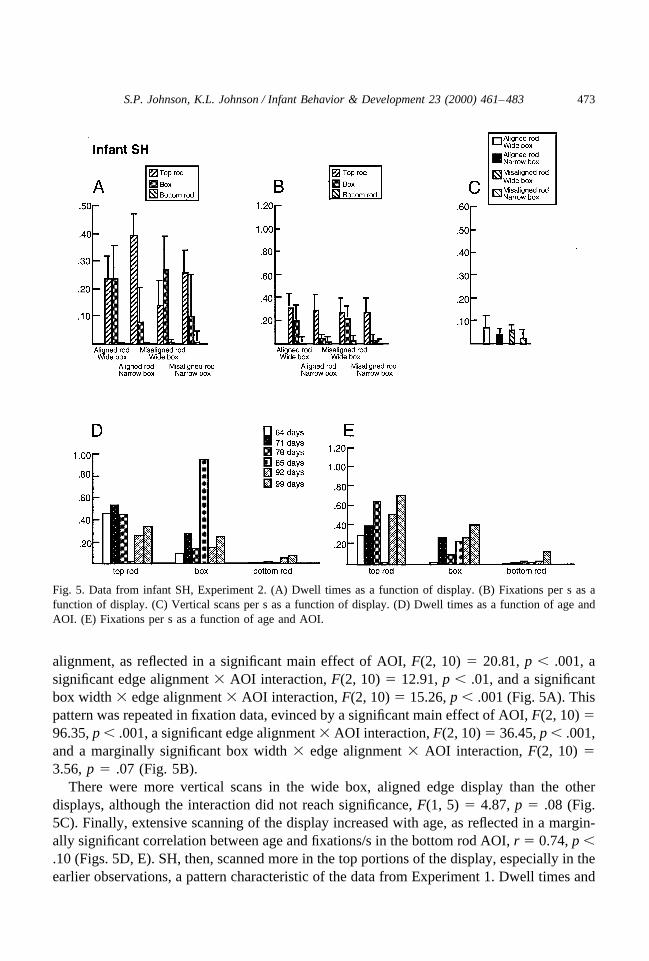
alignment, as reflected in a significant main effect of AOI, F(2, 10) � 20.81, p � .001, asignificant edge alignment � AOI interaction, F(2, 10) � 12.91, p � .01, and a significantbox width � edge alignment � AOI interaction, F(2, 10) � 15.26, p � .001 (Fig. 5A). Thispattern was repeated in fixation data, evinced by a significant main effect of AOI, F(2, 10) �96.35, p � .001, a significant edge alignment � AOI interaction, F(2, 10) � 36.45, p � .001,and a marginally significant box width � edge alignment � AOI interaction, F(2, 10) �3.56, p � .07 (Fig. 5B).
There were more vertical scans in the wide box, aligned edge display than the otherdisplays, although the interaction did not reach significance, F(1, 5) � 4.87, p � .08 (Fig.5C). Finally, extensive scanning of the display increased with age, as reflected in a margin-ally significant correlation between age and fixations/s in the bottom rod AOI, r � 0.74, p �.10 (Figs. 5D, E). SH, then, scanned more in the top portions of the display, especially in theearlier observations, a pattern characteristic of the data from Experiment 1. Dwell times and
Fig. 5. Data from infant SH, Experiment 2. (A) Dwell times as a function of display. (B) Fixations per s as afunction of display. (C) Vertical scans per s as a function of display. (D) Dwell times as a function of age andAOI. (E) Fixations per s as a function of age and AOI.
473S.P. Johnson, K.L. Johnson / Infant Behavior & Development 23 (2000) 461–483

fixations were concentrated primarily on the top rod AOI when the box was narrow, and splitmore evenly between the top rod and box when the box was wide. (An exception to thispattern was a high proportion of dwell times in the box AOI at 85 days. There were fewfixations at the same time, suggesting that SH was not as actively engaged in the task thatday as in the other sessions.) There was also a nonsignificant trend toward more verticalscans in the wide box, aligned edges display.
Infant ED was observed five times from 76 to 125 days (Fig. 6). Like SH, her data revealtendencies to scan in the top portions of the display, and varied with both box width and edgealignment. Interestingly, however, effects of box width were evident only in the dwell timeanalysis, and effects of edge alignment were evident only in the fixation analysis. Theanalysis of dwell time yielded a significant main effect of AOI, F(2, 8) � 5.66, p � .05, anda significant box width � AOI interaction, F(2, 8) � 5.88, p � .05 (Fig. 6A). The analysisof fixation data yielded significant main effects of edge alignment, F(1, 4) � 11.10, p � .05,
Fig. 6. Data from infant ED, Experiment 2. (A) Dwell times as a function of display. (B) Fixations per s as afunction of display. (C) Vertical scans per s as a function of display. (D) Dwell times as a function of age andAOI. (E) Fixations per s as a function of age and AOI.
474 S.P. Johnson, K.L. Johnson / Infant Behavior & Development 23 (2000) 461–483
and AOI, F(2, 8) � 12.38, p � .01, and a significant box width � AOI interaction, F(2, 8) �13.61, p � .01 (Fig. 6B). Vertical scans were more frequent in aligned-edge displays, F(1,4) � 13.36, p � .05, but there were no significant differences as a function of box width, nora box width � edge alignment interaction (Fig. 6C). Finally, there were no significantincreases with age in any scanning measures (Fig. 6D, E). ED, then, demonstrated a scanningpattern similar to that of SH: Dwell times and fixations were centered largely on the top rodAOI when the box was narrow, and distributed more consistently between the top rod andbox when the box was wide. Unlike SH and the sample in Experiment 1, however, there wasno evidence of age differences in scanning patterns in ED’s data.
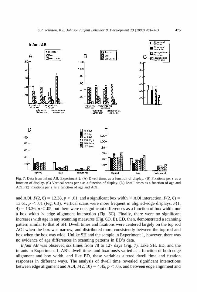
Infant AB was observed six times from 78 to 127 days (Fig. 7). Like SH, ED, and theinfants in Experiment 1, AB’s dwell times and fixations/s varied as a function of both edgealignment and box width, and like ED, these variables altered dwell time and fixationresponses in different ways. The analysis of dwell time revealed significant interactionsbetween edge alignment and AOI, F(2, 10) � 4.45, p � .05, and between edge alignment and
Fig. 7. Data from infant AB, Experiment 2. (A) Dwell times as a function of display. (B) Fixations per s as afunction of display. (C) Vertical scans per s as a function of display. (D) Dwell times as a function of age andAOI. (E) Fixations per s as a function of age and AOI.
475S.P. Johnson, K.L. Johnson / Infant Behavior & Development 23 (2000) 461–483
box width, F(2, 10) � 16.67, p � .001 (Fig. 7A). The analysis of fixation data revealedsignificant main effects of box width, F(1, 5) � 7.29, p � .05 and AOI, F(2, 10) � 10.68,p � .01, along with a significant box width � AOI interaction, F(2, 10) � 5.00, p � .05 (Fig.7B). There was also a marginally significant difference in vertical scans as a function of boxwidth, with fewer vertical scans in narrow box displays, F(1, 5) � 6.18, p � .06 (Fig. 7C).Finally, there were increases with age in looking to the bottom rod region, as reflected incorrelations between age and dwell time in the bottom rod AOI, r � 0.75, p � .10, fixations/sto the bottom rod AOI, r � 0.88, p � .05, and vertical scans, r � 0.73, p � .10 (Figs. 7D,E). These patterns are comparable, by and large, to those of SH and ED: more looking in thetop portion of the displays, and a greater proportion of fixations in the top rod AOI relativeto the box AOI in the narrow-box displays. Like SH, there was more extensive scanning withage across the display. In contrast to the other infants, however, vertical scans showed a trendtoward less active scanning in the narrow box, misaligned rod display.
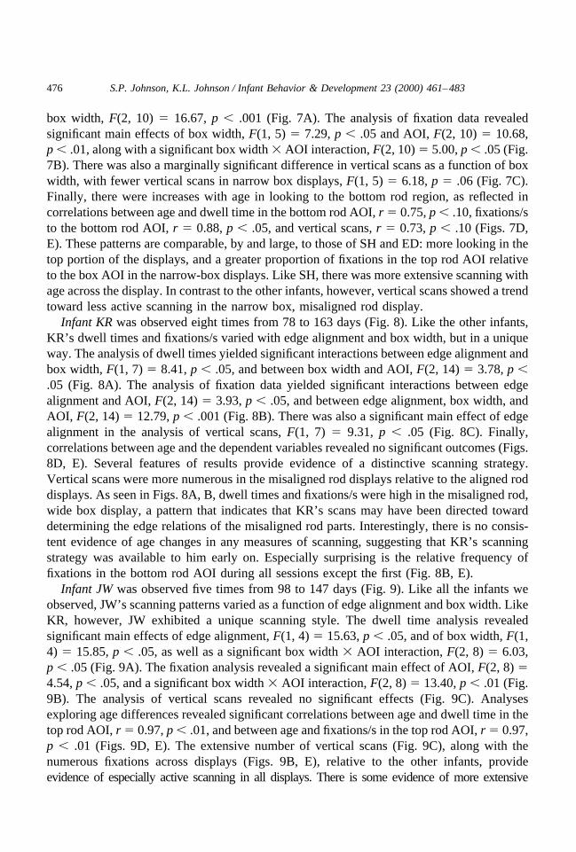
Infant KR was observed eight times from 78 to 163 days (Fig. 8). Like the other infants,KR’s dwell times and fixations/s varied with edge alignment and box width, but in a uniqueway. The analysis of dwell times yielded significant interactions between edge alignment andbox width, F(1, 7) � 8.41, p � .05, and between box width and AOI, F(2, 14) � 3.78, p �.05 (Fig. 8A). The analysis of fixation data yielded significant interactions between edgealignment and AOI, F(2, 14) � 3.93, p � .05, and between edge alignment, box width, andAOI, F(2, 14) � 12.79, p � .001 (Fig. 8B). There was also a significant main effect of edgealignment in the analysis of vertical scans, F(1, 7) � 9.31, p � .05 (Fig. 8C). Finally,correlations between age and the dependent variables revealed no significant outcomes (Figs.8D, E). Several features of results provide evidence of a distinctive scanning strategy.Vertical scans were more numerous in the misaligned rod displays relative to the aligned roddisplays. As seen in Figs. 8A, B, dwell times and fixations/s were high in the misaligned rod,wide box display, a pattern that indicates that KR’s scans may have been directed towarddetermining the edge relations of the misaligned rod parts. Interestingly, there is no consis-tent evidence of age changes in any measures of scanning, suggesting that KR’s scanningstrategy was available to him early on. Especially surprising is the relative frequency offixations in the bottom rod AOI during all sessions except the first (Fig. 8B, E).
Infant JW was observed five times from 98 to 147 days (Fig. 9). Like all the infants weobserved, JW’s scanning patterns varied as a function of edge alignment and box width. LikeKR, however, JW exhibited a unique scanning style. The dwell time analysis revealedsignificant main effects of edge alignment, F(1, 4) � 15.63, p � .05, and of box width, F(1,4) � 15.85, p � .05, as well as a significant box width � AOI interaction, F(2, 8) � 6.03,p � .05 (Fig. 9A). The fixation analysis revealed a significant main effect of AOI, F(2, 8) �4.54, p � .05, and a significant box width � AOI interaction, F(2, 8) � 13.40, p � .01 (Fig.9B). The analysis of vertical scans revealed no significant effects (Fig. 9C). Analysesexploring age differences revealed significant correlations between age and dwell time in thetop rod AOI, r � 0.97, p � .01, and between age and fixations/s in the top rod AOI, r � 0.97,p � .01 (Figs. 9D, E). The extensive number of vertical scans (Fig. 9C), along with thenumerous fixations across displays (Figs. 9B, E), relative to the other infants, provideevidence of especially active scanning in all displays. There is some evidence of more extensive
476 S.P. Johnson, K.L. Johnson / Infant Behavior & Development 23 (2000) 461–483
scanning with age, but overall, JW’s characteristic frequent scans were seen during all sessions. LikeKR, but unlike the other infants in Experiment 2, JW made frequent fixations in the bottom rod AOI.
2.3. Discussion
The findings of Experiment 2 provide evidence of both similarities and differences inscanning strategies across individual infants. Notably, scanning patterns for the five infantsappear to have been adapted for purposes of effective acquisition of relevant visual infor-mation concerning edge connectedness in each display, similar to the results obtained inExperiment 1. All the infants, for example, modified the proportion of dwell times andfixations across the AOIs as a function of display type. Support for hypothesized age-relatedimprovements in scanning in the bottom rod AOI was obtained in only two infants, and inonly one infant for improvements in vertical scanning. This is likely a result of the limited
Fig. 8. Data from infant KR, Experiment 2. (A) Dwell times as a function of display. (B) Fixations per s as afunction of display. (C) Vertical scans per s as a function of display. (D) Dwell times as a function of age andAOI. (E) Fixations per s as a function of age and AOI.
477S.P. Johnson, K.L. Johnson / Infant Behavior & Development 23 (2000) 461–483

age range across which the infants were observed. Nevertheless, the two infants whoexhibited greater scanning in the bottom rod AOI (SH and AB), and the one infant whoengaged in more vertical scans with age (AB), were in the same age range as the youngerinfants in Experiment 1, who also showed these effects. It is also possible that the repeatedexposure to the same stimuli across sessions contributed to differences in performance acrossExperiments 1 and 2.
The most important outcome of Experiment 2, however, was that distinctive processingstyles were revealed. Consistent with studies of motor development (e.g., Thelen et al.,1993), the results of the present studies suggest that there are multiple routes to achieving agoal during the development of a new skill: When the objective is to obtain visual infor-mation via scanning patterns, our data suggest that individual infants have strikingly differentmeans to this end.
Fig. 9. Data from infant JW, Experiment 2. (A) Dwell times as a function of display. (B) Fixations per s as afunction of display. (C) Vertical scans per s as a function of display. (D) Dwell times as a function of age andAOI. (E) Fixations per s as a function of age and AOI.
478 S.P. Johnson, K.L. Johnson / Infant Behavior & Development 23 (2000) 461–483
3. General discussion
Eye movements were recorded as young infants viewed partly occluded object displays,to address questions of underlying mechanisms of the development of object perception. InExperiment 1, evidence was obtained in a cross-sectional sample for differences in infants’scanning as a function of age and stimulus characteristics. Older infants tended to engage inmore extensive scanning, as revealed by both more frequent scanning in the lower part of thedisplay, and more frequent vertical scans. Both these scanning patterns would be expectedto have the effect of imparting more information concerning the alignment and commonmotion of the partly occluded rod edges. The infants also exhibited more extensive scanswhen determination of edge alignment might be challenged, either by misalignment or by arelatively wide occluder. Evidence was also obtained, in Experiment 2, for individualdifferences in scanning strategies with a longitudinal sample. Three infants’ scanningpatterns conformed with some of the principal findings of Experiment 1, but two did not, andeach infant’s scanning was unique in its own way.
These data bear important implications for theories of perceptual development. As notedin the Introduction, two current views provide contrasting predictions with respect toperceptual organization in infants and the role of visual information in development ofperception of object unity. According to an account stressing “core knowledge,” younginfants’ object percepts are guided by a limited set of reasoning principles (Spelke & Van deWalle, 1993). One of these is the contact principle: two surfaces undergoing a commonmotion belong to the same object. Static information, in contrast, has no inputs to initialpercepts of unity. On this view, then, motion information alone dictates perception of objectunity, and information such as edge and surface orientation is excluded from the process untillater in the first year after birth (cf. Kellman & Banks, 1998). According to an opposingaccount stressing information processing skills, the development of object perception derivesfrom improvements in the detection and utilization of available visual information, accom-panied by cortical maturation and visual experience, rather than core principles (Johnson,2000, in press). This view is consistent with evidence that static information such as edgealignment and global form has a strong influence on 4-month-olds’ perception of objectunity. Edge alignment, in particular, appears to be an important cue for unity, for both infants(Johnson & Aslin, 1996; Johnson et al., 2000a, b) and adults (Jusczyk et al., 1999). When tworod edges are misaligned, for example, unity perception is attenuated, even when the twosurfaces undergo common motion. Edge alignment without motion, nevertheless, is insuf-ficient to specify unity to young infants under many circumstances (Jusczyk et al., 1999;Kellman & Spelke, 1983; but see Needham, 1998). Johnson (1997, 2000; Johnson & Aslin,1996) proposed a threshold model to account for these and other results, stipulating thatperception of object unity depends on both the visual information available to the observer(to specify the depth relations among display elements and edge interpolation behind theoccluder), as well as the readiness of the observer to attend to that information. That is, acertain threshold of information must be exceeded for veridical percepts to obtain, and thisthreshold is higher among infants than adults.
The bulk of extant research would appear to be more consistent with the informationprocessing view, rather than the core principles view: Static information, such as edge
479S.P. Johnson, K.L. Johnson / Infant Behavior & Development 23 (2000) 461–483
orientation, has clear inputs into young infants’ object percepts. A newer version of the coreprinciples view, however, acknowledges the role of edge alignment and other static infor-mation in unity perception, but challenges the thesis that this role is direct (Jusczyk et al.,1999; Smith et al., 2000). Instead, edge alignment may have an indirect influence onperception of object unity, by modulating infants’ sensitivity to motion information. That is,it might be more difficult to detect the common motion of two edges that are not alignedacross and occluder. Without access to motion information, then, perception of object unityis precluded. This conjecture gains plausibility from findings that 4-month-olds do not appearto perceive unity in static displays with aligned rod parts (Jusczyk et al., 1999; Kellman &Spelke, 1983).
The results of the present experiments provide evidence against this more recent versionof the core principles account. In Experiment 1, there were no reliable differences overall inscanning as a function of edge alignment, either in terms of dwell times or fixations. Rather,these measures varied as a function of the interaction between box width and edge alignment,a result that is readily interpreted in light of data from vertical scans: Vertical scans werefrequent across all four stimulus types except the aligned rod, narrow box display, a scanningstrategy that would maximize the likelihood of obtaining effective information about the rodparts’ connectedness when such a determination might be hindered by misaligned edges ora wide occluder. There were age differences in the extent to which infants scanned bothhalves of the display, but edge alignment had no bearing on this effect. The infants, therefore,exhibited extensive scanning in both aligned and misaligned edge displays, and it seemslikely that motion sensitivity was equivalent across the two stimulus types.
A clear implication of the present results is the utility of eye movement recordings instudies of the development of object perception. Our results provide for speculation con-cerning basic information processing skills and information pickup, but do not provide moredirect evidence concerning what the infants actually experienced during the task. In thepresent case, this would be whether the infants perceived unified or disjoint objects in thestimuli, or some other percept. One strategy to achieve this next step would be to record eyemovements as the infants are tested in an habituation paradigm.
Perhaps the most important implication of our findings is the possibility that the eyemovement patterns we observed reveal an active process of strategy selection, and thatthis process undergoes development between 2 and 4 months in support of fundamentalobject perception skills. Previous reports of the development of visual scanning foundmarked improvements in the extent to which young infants’ eye movements wereadapted to stimulus characteristics. For example, Hainline and Lemerise (1982) foundthat 1- to 3-month-olds’ scan patterns were tailored to stimulus size and shape, andBronson (1990, 1994, 1997) reported gains across 2 to 14 weeks in the extent to whichinfants scanned between two stimuli, scanned extensively across a large stimulus, andengaged in “brief fixations” (i.e., average fixation duration was reduced), permitting amore thorough inspection of the stimulus per unit of time. These past studies usedachromatic, geometric forms as stimuli, in contrast to the more complex moving, colorfuldepictions of objects in depth employed in the present research. Our data replicated eachof these developmental trends, but in an older sample, suggesting that the developmentof basic mechanisms controlling purposive visual scanning may extend at least into the
480 S.P. Johnson, K.L. Johnson / Infant Behavior & Development 23 (2000) 461–483
second half year after birth. This protracted developmental profile may reflect thedemands of applying burgeoning information processing skills to object perceptiontasks, in contrast to scanning within or between simple stimuli.
Our results are consistent with systems views of perceptual and cognitive develop-ment in infancy, stressing the interactivity and interdependence of action and cognition.“. . .action of the infant within the environment must be considered the common prim-itive of cognitive development, and may well be the common control parameter of manyearly skills. If self-produced movement is critical, then dynamic category formation–theinfant’s basic organization of the world–must be paced and constrained by the ability toproduce and control that movement” (Thelen & Smith, 1994, pp. 194 –195). This quotereferred in part to the elegant series of studies by Bertenthal, Campos and colleagues(Bertenthal & Campos, 1990; Bertenthal et al., 1994; Campos et al., 1992) in whichinfants with locomotor experience were found to exhibit wariness of heights when placedon a visual cliff, and superior spatial skills in a search task, relative to age-matchedcontrols. Locomotor experience appears to trigger the emergence of a cascade ofancillary abilities that impact social, emotional, perceptual, cognitive, and further motordevelopment, due to new opportunities offered by locomotion for exploration of theenvironment. A similar effect may underlie the relation between infants’ sitting andreaching effectiveness: As infants gain postural control, and can sit unaided, the upperlimbs are freed from the need to maintain balance and can be used more effectively forobject manipulation and exploration (Rochat & Goubet, 1995; Rochat et al., 1999; seeBushnell & Boudreau, 1993 for further evidence of the impact of motor development onperceptual and cognitive development). The results of the present research suggest acorresponding process at work in early perceptual development. Self-directed eye move-ments appear to be critical to acquisition of object knowledge in the young infant, whichin turn directs the infant toward more effective means of obtaining information in theform of selection of scanning strategies. The striking individual differences noted inExperiment 2 imply that there is no single developmental path followed by infantstoward this goal. Rather, the infant him- or herself must select where to look, a processthat is initiated at birth by a functional preference for edges and motion, two essentialcues to veridical object perception (Slater, 1995; Johnson, 2000).
Acknowledgments
This research was supported by NSF grant SBR-991079. Portions of these experi-ments were presented at the 1999 conference of the Society for Research in ChildDevelopment, Albuquerque. The authors wish to thank our undergraduate research teamfor capable assistance, Les Cohen for the stimulus presentation software, and especiallythe infant participants and their parents, without whom these studies would not have beenpossible.
481S.P. Johnson, K.L. Johnson / Infant Behavior & Development 23 (2000) 461–483
References
Bertenthal, B. I., & Campos, J. J. (1990). A systems approach to the organizing effects of self-producedlocomotion during infancy. In C. Rovee-Collier & L. W. Lipsitt (Eds.), Advances in infancy research (Vol.6, pp. 1–60) Norwood, NJ: Ablex.
Bertenthal, B. I., Campos, J. J., & Kermoian, R. (1994). An epigenetic perspective on the development ofself-produced locomotion and its consequences. Current Directions in Psychological Science, 3, 140–145.
Bornstein, M. H. (1985). Habituation of attention as a measure of visual information processing in human infants:summary, systematization, and synthesis. In G. Gottlieb & N. A. Krasnegor (Eds.), Measurement of auditionand vision in the first year of postnatal life: A methodological overview (pp. 253–300). Norwood, NJ: Ablex.
Bronson, G. W. (1990). Changes in infants’ visual scanning across the 2- to 14-week age period. Journal ofExperimental Child Psychology, 49, 101–125.
Bronson, G. W. (1994). Infants’ transitions toward adult-like scanning. Child Development, 65, 1243–1261.Bronson, G. W. (1997). The growth of visual capacity: Evidence from infant scanning patterns. In C. Rovee-
Collier & L. W. Lipsitt (Eds.), Advances in infancy research (Vol. 11, pp. 109–141). Norwood, NJ: Ablex.Bushnell, E. W. (1985). The decline of visually guided reaching during infancy. Infant Behavior & Development,
8, 139–155.Bushnell, E. W., & Boudreau, J. P. (1993). Motor development and the mind: The potential role of motor abilities
as a determinant of aspects of perceptual development. Child Development, 64, 1005–1021.Campos, J. J., Bertenthal, B. I., & Kermoian, R. (1992). Early experience and emotional development: The
emergence of wariness of heights. Psychological Science, 3, 61–64.Colombo, J., Freeseman, L. J., Coldren, J. T., & Frick, J. E. (1995). Individual differences in infant fixation
duration: Dominance of global versus local stimulus properties. Cognitive Development, 10, 271–285.Freeseman, L. J., Colombo, J., & Coldren, J. T. (1993). Individual differences in infant visual attention:
Four-month-olds’ discrimination and generalization of global and local stimulus properties. Child Develop-ment, 64, 1191–1203.
Ghim, H.-R., & Eimas, P. D. (1988). Global and local processing by 3- and 4-month-old infants. Perception &Psychophysics, 43, 165–171.
Gibson, J. J. (1966). The senses considered as perceptual systems. Boston: Houghton Mifflin.Hainline, L., & Lemerise, E. (1982). Infants’ scanning of geometric forms varying in size. Journal of Experi-
mental Child Psychology, 33, 235–256.Johnson, S. P. (1997). Young infants’ perception of object unity: Implications for development of attentional and
cognitive skills. Current Directions in Psychological Science, 6, 5–11.Johnson, S. P. (2000). The development of visual surface perception: Insights into the ontogeny of knowledge.
In C. Rovee-Collier, L. Lipsitt, & H. Hayne (Eds.), Progress in infancy research (Vol. 1, pp. 113–154).Mahwah, NJ: Erlbaum.
Johnson, S. P. (in press).Visual development in human infants: Binding features, surfaces, and objects. VisualCognition
Johnson, S. P., & Aslin, R. N. (1995). Perception of object unity in 2-month-old infants. DevelopmentalPsychology, 31, 739–745.
Johnson, S. P., & Aslin, R. N. (1996). Perception of object unity in young infants: The roles of motion, depth,and orientation. Cognitive Development, 11, 161–180.
Johnson, S. P., & Aslin, R. N. (1998). Young infants’ perception of illusory contours in dynamic displays.Perception, 27, 341–353.
Johnson, S. P., & Aslin, R. N. (2000). Infants’ perception of transparency. Developmental Psychology, 36,808–816.
Johnson, S. P., Bremner, J. G., Slater, A., & Mason, U. (2000a). The role of good form in young infants’perception of partly occluded objects. Journal of Experimental Child Psychology, 76, 1–25.
Johnson, S. P., Bremner, J. G., Slater, A. M., Mason, U., & Foster, K. (2000b). Young infants’ perception of unityand form in occlusion displays. Revision under review.
482 S.P. Johnson, K.L. Johnson / Infant Behavior & Development 23 (2000) 461–483

Johnson, S. P., Cohen, L. B., Marks, K., & Lawson, K. D. (2000c). Young infants’ perception of object unity inrotating displays: further evidence. Manuscript in preparation.
Johnson, S. P., & Nanez, J. E. (1995). Young infants’ perception of object unity in two-dimensional displays.Infant Behavior & Development, 18, 133–143.
Jusczyk, P. W., Johnson, S. P., Spelke, E. S., & Kennedy, L. J. (1999). Synchronous change and perception ofobject unity: Evidence from adults and infants. Cognition, 71, 257–288.
Kawataba, H., Gyoba, J., Inoue, H., & Ohtsubo, H. (1999). Visual completion of partly occluded grating in infantsunder 1 month of age. Vision Research, 39, 3586–3591.
Kellman, P. J., & Banks, M. S. (1998). Infant visual perception. In W. Damon (Series Ed.) & R. Siegler and D.Kuhn (Vol. Eds.), Handbook of child psychology: Vol. 2. Cognition, Perception, and Language (5th ed., pp.103–146). New York: Wiley.
Kellman, P. J., & Spelke, E. S. (1983). Perception of partly occluded objects in infancy. Cognitive Psychology,15, 483–524.
Mareschal, D., & Johnson, S. P. (in press). Learning to perceive object unity: A connectionist account.Developmental Science.
Needham, A. (1998). Infants’ use of featural information in the segregation of stationary objects. Infant Behaviorand Development, 21, 47–76.
Piaget, J. (1954). The construction of reality in the child. New York: Basic Books.Rochat, P., & Goubet, N. (1995). Development of sitting and reaching in 5- to 6-month-old infants. Infant
Behavior & Development, 18, 53–68.Rochat, P., Goubet, N., & Senders, S. J. (1999). To reach or not to reach? Perception of body effectivities by
young infants. Infant & Child Development, 8, 129–148.Slater, A. (1995). Visual perception and memory at birth. In C. Rovee-Collier & L. P. Lipsitt (Eds.), Advances
in infancy research (Vol. 9, pp. 107–162). Norwood, NJ: Ablex.Slater, A., Johnson, S. P., Brown, E., & Badenoch, M. (1996). Newborn infants’ perception of partly occluded
objects. Infant Behavior and Development, 19, 145–148.Slater, A., Morison, V., Somers, M., Mattock, A., Brown, E., & Taylor, D. (1990). Newborn and older infants’
perception of partly occluded objects. Infant Behavior and Development, 13, 33–49.Smith, W. C., Johnson, S. P., & Spelke, E. S. (2001). Motion and edge sensitivity in perception of object unity.
Revision under review.Spelke, E. S., & Hermer, L. (1996). Early cognitive development: Objects and space. In E. Carterette & M.
Friedman (Series Eds.) & R. Gelman and T. Kit-Fong Au (Vol. Eds.), Handbook of perception and cognition:Perceptual and cognitive development (2nd ed., pp. 71–114). New York: Academic Press.
Spelke, E. S., & Van de Walle, G. (1993). Perceiving and reasoning about objects: Insights from infants. In N.Eilan, R. A. McCarthy, & B. Brewer (Eds.), Spatial representation: Problems in philosophy and psychology(pp. 132–161). Oxford: Blackwell.
Thelen, E., Corbetta, D., Kamm, K., Spencer, J. P., Schneider, K., & Zernicke, R. F. (1993). The transition toreaching: Mapping intention and intrinsic dynamics. Child Development, 64, 1058–1098.
Thelen, E., & Smith, L. B. (1994). A dynamic systems approach to the development of cognition and action.Cambridge, MA: Bradford Books.
von Hosten, C. (1984). Developmental changes in the organization of prereaching movements. DevelopmentalPsychology, 20, 378–388.
von Hofsten, C., & Rosander, K. (1998). The establishment of gaze control in early infancy. In F. Simion & G.Butterworth (Eds.), The development of sensory, motor, and cognitive capacities in early infancy: fromperception to cognition (pp. 49–66). East Sussex, UK: Psychology Press.
483S.P. Johnson, K.L. Johnson / Infant Behavior & Development 23 (2000) 461–483