early diagnosis saves lives in esophageal...
TRANSCRIPT
939
http://journals.tubitak.gov.tr/medical/
Turkish Journal of Medical Sciences Turk J Med Sci(2013) 43: 939-945© TÜBİTAKdoi:10.3906/sag-1210-45
Early diagnosis saves lives in esophageal perforations
Ayşen TASLAK ŞENGÜL1, Yasemin BİLGİN BÜYÜKKARABACAK1,*, Tülin DURGUN YETİM2,Mehmet Gökhan PİRZİRENLİ1, Burçin ÇELİK1, Ahmet BAŞOĞLU1
1Department of Thoracic Surgery, Faculty of Medicine, Ondokuz Mayıs University, Samsun, Turkey2Department of Thoracic Surgery, Faculty of Medicine, Mustafa Kemal University, Hatay, Turkey
* Correspondence: [email protected]
1. IntroductionEsophageal perforation is not common, but if it is not diagnosed early and treated properly, it quickly leads to mediastinitis, sepsis, and multiorgan failure, which ultimately result in mortality (1). The frequent use of endoscopic procedures in the diagnosis and treatment of gastrointestinal disorders has led to an increase in the incidence of esophageal perforation (2).
Treatment of this rare injury, which has high morbidity and mortality, should be patient-specific depending on the localization and severity of the injury and the time elapsed before the diagnosis. The factor that determines the morbidity and mortality in esophageal perforation is the length of time elapsed between diagnosis and the onset of treatment (3,4).
In this study, the patients treated in our clinic for esophageal perforation were evaluated according to the etiologies and methods of diagnosis and treatment.
2. Materials and methodsTwenty-two patients who were diagnosed with esophageal perforation in Ondokuz Mayıs University Faculty of Medicine’s thoracic surgery clinics between 2000 and 2011 were retrospectively studied. Patients were evaluated for age, sex, etiology, localization of the perforation, diagnostic procedures, treatment method, morbidity, and mortality. Diagnosis was made on the basis of physical examination and radiological and endoscopic findings.
Nine of the patients were female and 13 were male. The mean age was 61.4 years (range: 22–79). The mean hospital stay was 9.5 days (range: 5–31). The patients presenting within the first 24 h were classified as “early patients”, and the ones presenting after 24 h were classified as “late patients”. The cause of perforation was foreign body in 17 patients, dilatation with bougie in 2, balloon dilatation in 2, and spontaneous rupture in 1. General features of the patients are given in Table 1, and primary pathologies and causes of perforation are given in Table 2.
Aim: Esophageal perforations are rare but highly fatal pathologies. This study aims to discuss the treatment methods for esophageal perforations.
Materials and methods: Twenty-two patients who were diagnosed with esophageal perforation in the Ondokuz Mayıs University Faculty of Medicine’s thoracic surgery clinics between 2000 and 2011 were retrospectively evaluated.
Results: The cause of perforation was foreign body in 17 patients, dilatation with bougie in 2, balloon dilatation in 2, and spontaneous rupture in 1. Eight patients had cervical, 12 had thoracal, and 2 had thoracoabdominal esophagus perforations. The period between perforation occurrence and treatment was longer than 24 h in 10 patients and shorter than 24 h in 12 patients. Eight patients were treated with primary repair and debridement, 5 with chest tube drainage and conservative treatment, and 1 with self-opening stent, and 1 patient underwent resection. On the other hand, 7 patients were followed with conservative therapy after the removal of the foreign body with esophagoscopy. There was 1 mortality in the surgically treated group, while there were 4 in the conservatively treated group.
Conclusion: Surgery is the “gold standard” for the treatment of esophageal perforations. Conservative therapy should be applied only in selected patients under careful monitoring. The most important factor for morbidity and mortality is early diagnosis and determination of the treatment method that best suits the patient.
Key words: Esophagus, perforation, treatment
Received: 12.10.2012 Accepted: 01.01.2013 Published Online: 02.10.2013 Printed: 01.11.2013
Research Article
940
TASLAK ŞENGÜL et al. / Turk J Med Sci
Six of the patients diagnosed with perforation due to foreign body underwent primary repair and debridement, 2 were treated with the cervical approach, and 4 were treated with a thoracotomy. In patients who underwent early primary repair, the repaired area was supported by the surrounding tissues. Seven patients received conservative therapy after the removal of the foreign body with esophagoscopy. Four patients who presented with sepsis due to mediastinitis after 24 h were intubated, followed by bilateral chest tube drainage.
One patient was directed to our clinic with a diagnosis of spontaneous esophageal perforation due to Boerhaave syndrome. In the first session, the esophagus was closed with a stapler from the proximal and distal side of the perforation site, and the perforation site was debrided and repaired primarily. Mediastinal debridement, pleural decortications, and drainage were performed. Following
infection treatment, the patient underwent subtotal esophagectomy plus cervical esophagogastrostomy in the second session.
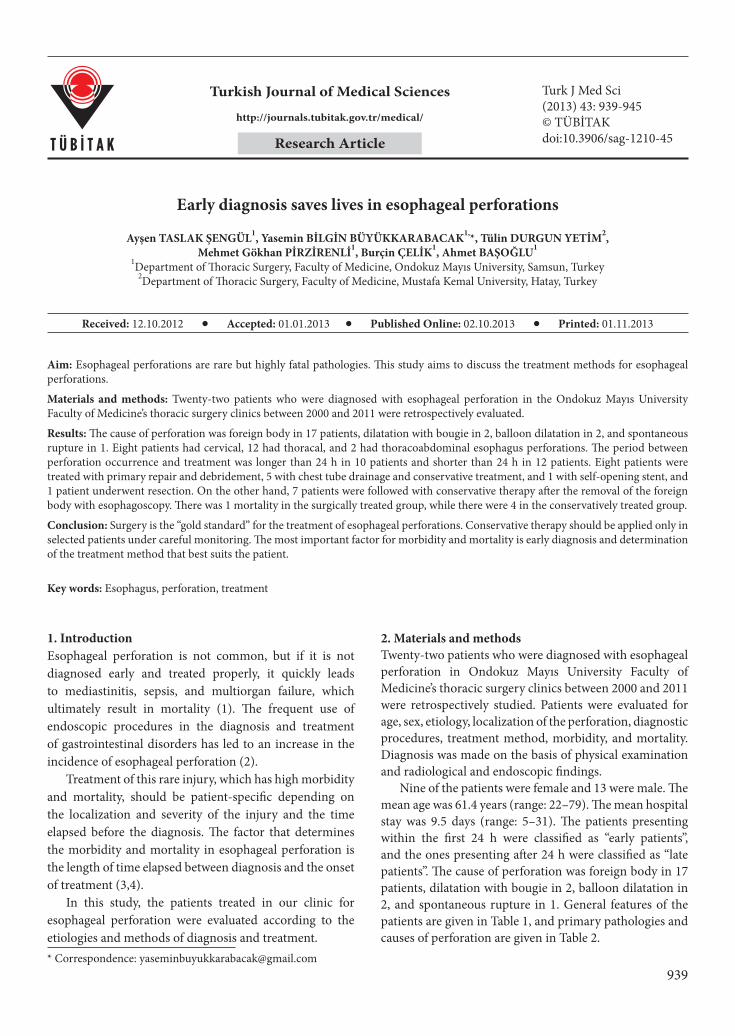
Of the 2 patients diagnosed with perforation due to dilatation, 1 was treated with conservative medical therapy. The other patient developed bilateral empyema. Bilateral chest tube drainage was performed. Sepsis was treated with parenteral nutrition and antibiotic therapy. Since no oral passage could be achieved, a passage was provided via descending dilatation of the esophagus with laparotomy, and a stent was placed into the stenosis site. Figure 1 shows the chest X-ray of the patient after stent placement.
In the 2 patients who underwent dilatation due to achalasia, perforation was diagnosed in 1 patient right after dilatation and in the other on the third day. Primary repair with thoracotomy and debridement was performed on both patients. Oral feeding was stopped in patients receiving conservative therapy. If necessary, the pleural cavity was drained with tube thoracostomy; broad-spectrum antibiotic therapy was started preoperatively. Total parenteral nutrition was provided with products rich in energy and protein, and enteral nutrition products were given with jejunostomy.
Table 1. General features.
Sex N
Male 13
Female 9
Localization of perforation N
Cervical 8
Thoracal 12
Thoracoabdominal 2
Mean age 61.4 years (22–79)
Mean hospital stay 9.5 days (5–31)Admittance time N
Within the first 24 h 12
After 24 h 10
Table 2. Primary disease and cause of perforation.
Primary disease Cause of perforation N
Foreign body Foreign body 17
Achalasia Balloon dilatation 2
Esophageal stenosis due to radiation therapy Bougie dilatation 2
Boerhaave syndrome Spontaneous 1
Figure 1. Chest X-ray of patient with perforation due to bougie dilatation after stent placement.
941
TASLAK ŞENGÜL et al. / Turk J Med Sci
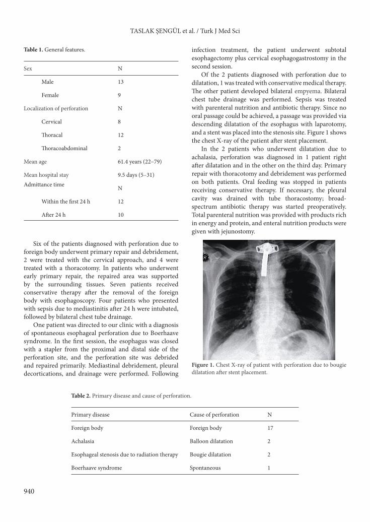
3. ResultsThe perforations detected were at the cervical part of the esophagus in 8 patients, at the thoracal part in 12, and at the thoracoabdominal part in 2. The most common symptoms were dysphagia (n: 14) and back pain (n: 8) (Figure 2). Five patients were admitted with mediastinitis and in a state of septic shock. Four of them had foreign body perforations at the thoracal part of the esophagus, and 1 was admitted due to Boerhaave syndrome; all of them died. Of these 5 patients, 2 were admitted to the hospital on the 7th day and the other 3 on the 3rd day.
Radiological study revealed bilateral empyema in 5 patients, unilateral empyema in 1, subcutaneous emphysema in the neck in 7, mediastinal emphysema in 3, pneumothorax in 2, and hydropneumothorax in 1 patient.
Ten patients were late patients and 12 patients were early patients. Perforations that occurred due to endoscopic procedures were diagnosed early. The spontaneous perforations and perforations that occurred due to foreign body were diagnosed in the late period.
Drainage was performed in 1 patient who developed esophagopleural fistula and empyema after primary repair, and on the postoperative 16th day, the patient underwent re-thoracotomy for decortication. In 1 patient, aspiration pneumonia was observed, and in 3 patients, wound site infection.
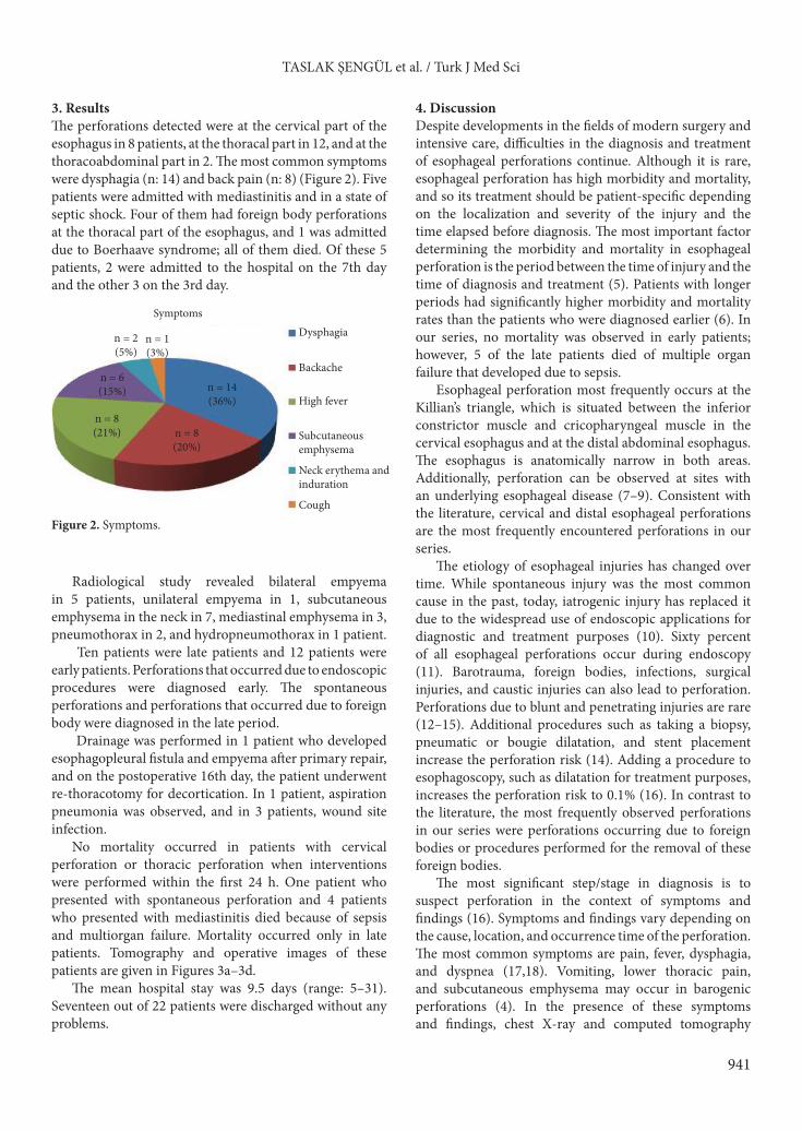
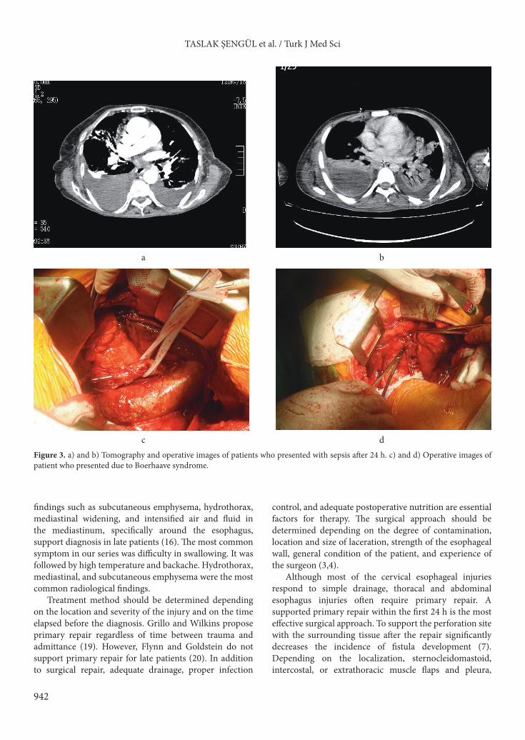
No mortality occurred in patients with cervical perforation or thoracic perforation when interventions were performed within the first 24 h. One patient who presented with spontaneous perforation and 4 patients who presented with mediastinitis died because of sepsis and multiorgan failure. Mortality occurred only in late patients. Tomography and operative images of these patients are given in Figures 3a–3d.
The mean hospital stay was 9.5 days (range: 5–31). Seventeen out of 22 patients were discharged without any problems.
4. DiscussionDespite developments in the fields of modern surgery and intensive care, difficulties in the diagnosis and treatment of esophageal perforations continue. Although it is rare, esophageal perforation has high morbidity and mortality, and so its treatment should be patient-specific depending on the localization and severity of the injury and the time elapsed before diagnosis. The most important factor determining the morbidity and mortality in esophageal perforation is the period between the time of injury and the time of diagnosis and treatment (5). Patients with longer periods had significantly higher morbidity and mortality rates than the patients who were diagnosed earlier (6). In our series, no mortality was observed in early patients; however, 5 of the late patients died of multiple organ failure that developed due to sepsis.
Esophageal perforation most frequently occurs at the Killian’s triangle, which is situated between the inferior constrictor muscle and cricopharyngeal muscle in the cervical esophagus and at the distal abdominal esophagus. The esophagus is anatomically narrow in both areas. Additionally, perforation can be observed at sites with an underlying esophageal disease (7–9). Consistent with the literature, cervical and distal esophageal perforations are the most frequently encountered perforations in our series.
The etiology of esophageal injuries has changed over time. While spontaneous injury was the most common cause in the past, today, iatrogenic injury has replaced it due to the widespread use of endoscopic applications for diagnostic and treatment purposes (10). Sixty percent of all esophageal perforations occur during endoscopy (11). Barotrauma, foreign bodies, infections, surgical injuries, and caustic injuries can also lead to perforation. Perforations due to blunt and penetrating injuries are rare (12–15). Additional procedures such as taking a biopsy, pneumatic or bougie dilatation, and stent placement increase the perforation risk (14). Adding a procedure to esophagoscopy, such as dilatation for treatment purposes, increases the perforation risk to 0.1% (16). In contrast to the literature, the most frequently observed perforations in our series were perforations occurring due to foreign bodies or procedures performed for the removal of these foreign bodies.
The most significant step/stage in diagnosis is to suspect perforation in the context of symptoms and findings (16). Symptoms and findings vary depending on the cause, location, and occurrence time of the perforation. The most common symptoms are pain, fever, dysphagia, and dyspnea (17,18). Vomiting, lower thoracic pain, and subcutaneous emphysema may occur in barogenic perforations (4). In the presence of these symptoms and findings, chest X-ray and computed tomography
Figure 2. Symptoms.
Dysphagia
n = 14(36%)
n = 8(20%)
n = 8(21%)
n = 6(15%)
n = 2(5%)
n = 1(3%)
Backache
High fever
Subcutaneousemphysema
Neck erythema andinduration
Cough
Symptoms
942
TASLAK ŞENGÜL et al. / Turk J Med Sci
findings such as subcutaneous emphysema, hydrothorax, mediastinal widening, and intensified air and fluid in the mediastinum, specifically around the esophagus, support diagnosis in late patients (16). The most common symptom in our series was difficulty in swallowing. It was followed by high temperature and backache. Hydrothorax, mediastinal, and subcutaneous emphysema were the most common radiological findings.
Treatment method should be determined depending on the location and severity of the injury and on the time elapsed before the diagnosis. Grillo and Wilkins propose primary repair regardless of time between trauma and admittance (19). However, Flynn and Goldstein do not support primary repair for late patients (20). In addition to surgical repair, adequate drainage, proper infection
control, and adequate postoperative nutrition are essential factors for therapy. The surgical approach should be determined depending on the degree of contamination, location and size of laceration, strength of the esophageal wall, general condition of the patient, and experience of the surgeon (3,4).
Although most of the cervical esophageal injuries respond to simple drainage, thoracal and abdominal esophagus injuries often require primary repair. A supported primary repair within the first 24 h is the most effective surgical approach. To support the perforation site with the surrounding tissue after the repair significantly decreases the incidence of fistula development (7). Depending on the localization, sternocleidomastoid, intercostal, or extrathoracic muscle flaps and pleura,
Figure 3. a) and b) Tomography and operative images of patients who presented with sepsis after 24 h. c) and d) Operative images of patient who presented due to Boerhaave syndrome.
a
c
b
d
943
TASLAK ŞENGÜL et al. / Turk J Med Sci
pericardium, omentum, and diaphragm are the ideal live support tissues. In intraabdominal perforations, the fundus of the stomach can also be used to support the repair line (11,21). In the literature, the rate of fistula for primary repair alone has been reported to be 40%. However, it has also been reported that the fistula rate reduces to 10% in supported primary repair cases (22). In all our patients who undergo primary repair and resection, we prefer to strengthen the perforation site and anastomosis line. In the cervical area, we provide support with surrounding muscle tissue and in the thorax, with pleura, mediastinal tissues, and intercostal muscles. In late patients with esophagus edema in whom primary repair is not possible, placing a thick biliary T-tube into the perforation to prevent mediastinal contamination can be a treatment option. A wide T-tube is inserted into the perforation and its tip is taken out of the thorax. After the recovery of sepsis, in 6 months to 1 year the T-tube is removed. The patient is reevaluated to decide whether an additional surgical procedure is needed or not. However, this treatment method is rarely performed, and in the literature there are no clear data on its results (7). Linden et al. (23) reported that the mortality rate in a patient series treated with T-tube was 9%, but they also reported that 30% of these patients needed reoperation.
Resection can be considered for a malignant lesion, for patients with numerous benign strictures, for unrepairable esophageal injuries, for a serious and inadequately drained mediastinitis, or in cases of primary repair site dehiscence. Mortality rate after resection has been reported at 15%–40%. However, it is worse in caustic injuries and in patients with poor general health. In many studies performed on late patients, it has been demonstrated that patients who have undergone primary repair have higher mortality risk than patients who have undergone resection (2).
Especially in cases of widespread mediastinitis and pleural contamination, the transhiatal approach yields better results because the transthoracic approach enables a more controlled debridement (2). We also prefer the transthoracic approach, since it provides not only a better pleural and mediastinal debridement but also wider decortication and right drainage. Anastomosis is then performed on the neck to address hygienic concerns. In the current series, a patient who presented with esophageal rupture on the second day and developed fistula after primary repair underwent resection and cervical esophagogastrostomy. The patient died due to sepsis on the postoperative 35th day.
Conservative therapy can be applied to selected patients with suspected or limited perforation, minimal symptoms, perforation in the cervical esophagus, and minimal pleural or mediastinal contamination (2–11). According to the criteria defined by Cameron et al. (24) and modified by
Altorjay et al. (25), conservative therapy can be applied to well-marginated circumferential perforations, to cavities with contrast agents that drain back into the lumen, to cases with minimal symptoms, and to cases where there is no cancer obstruction or abdominal esophageal leak. In late patients we also prefer conservative therapy until the general condition of the patient improves and the infection at the perforation site is controlled. In this approach, oral feeding is stopped in patients fitting the criteria and broad-spectrum antibiotic therapy and total parenteral nutrition are started. If there is pleural effusion (empyema), chest tube drainage should be performed. In the meantime, a nasogastric tube can be placed endoscopically into the esophagus proximal to the perforation, and continuous irrigation can be performed with saline or antibiotic-containing solutions. After following this procedure for 7–10 days, if the patient is stable, the perforation is controlled with contrast radiography. If there is no contrast escape, oral therapy is started, but if the escape continues, surgical treatment choices should be considered (2).
In our clinic, saline or water is given orally to irrigate the fibrin and necrotic material in the fistula area. We believe that this procedure will help the closure of the fistula and contribute to the success of the next surgical procedure or the stent application.
Coated, self-expandable metallic esophageal stents are reported to be a good treatment option for the cases in which surgical treatment is risky, such as in patients with unresectable esophageal cancer, benign esophageal diseases, or iatrogenic perforations that have been diagnosed early. Recently, this method has become more common due to the short hospitalization period, low cost, and early onset of oral nutrition (26).
Hunerbain et al. compared patients treated with conservative therapy and patients treated with stent. They reported that the group treated with stent had shorter hospital stays and earlier oral feeding, and they developed no complications (27). Johnson et al. emphasized that regardless of all criteria, coated, self-expandable metallic stent application is an effective treatment choice for esophageal perforations (28). Although there can be complications such as migrations (5%–23%) or stent re-placements after stent applications (26), the success rates, recovery rates after stent application, and mortality rates have been reported as 92%–100%, 13%–69%, and 0%–33%, respectively (11). While determining the method of treatment, the location and size of the perforation, the time elapsed up to the diagnosis, and the general condition (infection and sepsis) of the patient can affect the results. Endoscopic methods seem to be preferable due to advantages such as short hospitalization periods and low cost. The differences in the success rates in the literature indicate that selection of the correct case is as
944
TASLAK ŞENGÜL et al. / Turk J Med Sci
important as the experience. Since we do not have studies with large series, the effectiveness and reliability of the methods are not exactly known (7). In one patient in our series too, esophageal perforation occurred after bougie dilatation. Bilateral pleural drainage was performed, and broad-spectrum antibiotic therapy and parenteral hyperalimentation were given. A self-expandable metal stent was then placed. Oral feeding was started on the postoperative 5th day, and the patient was discharged with a normal diet on the 7th day.
In a recent study, the overall mortality in esophageal perforations was reported to be 18%. Barogenic perforations had the highest mortality rate with 36%, followed by instrumental injuries with 19% and traumatic perforations with 7%. In this study, it was emphasized that the location of the perforation was the most important factor affecting the prognosis. The mortality rate was 6% in cervical esophagus perforations while it was 27% in thoracic esophageal perforations. In the study of Okten et al. (29), the mortality rate was 36% for thoracic esophagus perforations, 0% for abdominal injuries, and 20% for cervical esophagus perforations; on the other hand, in the study of Eroglu et al. (2), rates were 16.7%, 16.7%, and 0%, respectively. A longer interval between the trauma and the treatment is one of the criteria of poor prognosis. In the literature, the mortality rates have been reported as 0%–18% in early patients and 7%–37.5% in late patients. Treatment method has also been defined as an important prognostic criterion (11).
In the series of Brinster et al. with 726 patients (8), the patients who underwent simple drainage only had the highest mortality rate (36%). This was followed by exclusion (24%), conservative therapy (17%), and esophagectomy (17%). The lowest mortality was 12% in
patients who underwent primary repair. In our study, the highest mortality rate was 25%, and it was observed in the group of late presented patients with thoracic esophageal perforation due to foreign body. No mortality was observed in patients with cervical perforation or in early patients, regardless of the location and etiology.
Nutritional support is significant in the postoperative care of the patients with esophageal perforation. Nutrition can be provided by parenteral nutrition, or, as we frequently do with our patients, with enteral nutrition products through a jejunostomy catheter (3). The normal daily and increased calorie requirements of the patient can be provided at a level as close to the natural feeding as possible with jejunostomy. Furthermore, by protecting the integrity of the gastrointestinal tract mucosa, the immune system is strengthened (30). It is also known that in such patients who are hospitalized for long periods, enteral nutrition is more economical than total parenteral nutrition (31).
In conclusion, surgery is the “gold standard” in the treatment of esophageal perforations. While the emerging interventional procedures increase the incidence of esophageal perforations, they will be treated more easily and effectively with the development of minimally invasive surgeries. The main principles in the surgical approach are to repair the esophageal leak as quickly as possible and to provide mediastinal and pleural drainage, proper broad-spectrum antibiotic therapy, and adequate nutrition. Conservative therapy should only be applied to selected patients with careful monitorization. Early diagnosis and determination of the treatment method that fits the patient are the most important factors in determining morbidity and mortality.
References
1. Balumuka DD, Chalya PL, Mahalu W. Oesophageal perforation: a diagnostic and therapeutic challenge in a resource limited setting. A report of three cases. J Cardiothorac Surg 2011; 6: 116.
2. Eroglu A, Turkyilmaz A, Aydin Y, Yekeler E, Karaoglanoglu N. Current management of esophageal perforation: 20 years experience. Dis Esophagus 2009; 22: 374–80.
3. Özçelik C, İnci İ. Özofagus yaralanmaları. In: Yüksel M, Başoğlu A, editors. Özofagus Hastalıklarının Tıbbi ve Cerrahi Tedavisi. İstanbul: Bilmedya Grup; 2002. pp. 77–108 (in Turkish).
4. Erdoğan A, Öz N, Sarper A, Dertsiz L, Demircan A, Işın E. Özofagus perforasyonları: 11 olgunun analizi. GKDC Dergisi 1999; 7: 57–62 (in Turkish).
5. Eroglu A, Kurkcuoglu IC, Karaoglanoglu N, Tekinbas C, Yimaz O, Basoglu M. Esophageal perforation: the importance of early diagnosis and primary repair. Dis Esophagus 2004; 17: 91–4.
6. Ghai A, Wadher R, Kamal K, Verma V. Mediastinitis after oesophagoscopy: a case report. SAJAA 2009; 15: 33–4.
7. Wright C (translators: Çağırıcı U, Turhan K). Özofagus perforasyonunun tedavisi. In: Sugerbaker D, editor (translation editor: Yüksel M) Erişkin Özofagus Cerrahisi. İstanbul: Nobel Tıp Kitapevi; 2011. pp. 353–60 (in Turkish).
8. Brinster CJ, Singhal S, Lee L, Marshall MB, Kaiser LR, Kucharczuk JC. Evolving options in the management of esophageal perforation. Ann Thorac Surg 2004; 77: 1475–83.
9. Türkyılmaz A, Eroğlu A, Aydın Y, Yılmaz Ö, Karaoğlanoğlu N. Survival in esophageal cancer patients with hematogenous distant organ metastases. Turk J Med Sci 2009; 39: 415–21.
10. Eisen GM, Baron TH, Dominitz JA, Faigel DO, Goldstein JL, Johanson JF, Mallery JS, Raddawi HM, Vargo JJ 2nd, Waring JP et al. Complications of upper GI endoscopy. Gastrointest Endosc 2002; 55: 784–93.
945
TASLAK ŞENGÜL et al. / Turk J Med Sci
11. Chirica M, Champault A, Dray X, Sulpice L, Munoz-Bongrand N, Sarfati E, Cattan P. Esophageal perforations. J Visc Surg 2010; 147: 117–28.
12. Weiman DS, Walker WA, Brosnan KM, Pate JW, Fabian TC. Noniatrogenic esophageal trauma. Ann Thorac Surg 1995; 59: 845–50.
13. Gaissert HA, Roper CL, Patterson GA, Grillo HC. Infectious necrotizing esophagitis: outcome after medical and surgical intervention. Ann Thorac Surg 2003; 75: 342–7.
14. Wolfsen HC, Hemminger LL, Achem SR, Loeb DS, Stark ME, Bouras EP, DeVault KR. Complications of endoscopy of the upper gastrointestinal tract: a single-center experience. Mayo Clinic Proc 2004; 79: 1264–7.
15. Soyer T, Ayva Ş, Somuncu S, Atasoy P, Kanmaz T, Çakmak M. Caustic esophageal injury decreases the number of interstitial cells of Cajal in the rat esophagus. Turk J Med Sci 2010; 40: 599–604.
16. Yenigün B, Çelik A, Cangır AK. Özofagus yaralanmaları. TTD Toraks Cerrahisi Bülteni 1, January 2010 (in Turkish).
17. Younes Z, Johnson D. The spectrum of spontaneous and iatrogenic esophageal injury: perforations, Mallory–Weiss tears, and hematomas. J Clin Gastroenterol 1999; 29: 306–17.
18. Pasricha PJ, Fleischer DE, Kalloo AN. Endoscopic perforations of the upper digestive tract: a review of their pathogenesis, prevention and management. Gastroenterology 1994; 106: 787–802.
19. Grillo HC, Wilkins EW. Esophageal repair following late diagnosis of intrathoracic perforation. Ann Thorac Surg 1975; 20: 387–99.
20. Flynn AE, Verrier ED, Way LW, Thomas AN, Pellegrini CA. Esophageal perforation. Arch Surg 1989; 124: 1211–5.
21. Kotzampassakis N, Christodoulou M, Krueger T, Demartines N, Vuillemier H, Cheng C, Dorta G, Ris HB. Esophageal leaks repaired by a muscle onlay approach in the presence of mediastinal sepsis. Ann Thorac Surg 2009; 88: 966–72.
22. Wright CD, Mathisen DJ, Wain JC, Moncure AC, Hilgenberg AD, Grillo HC. Reinforced primary repair of thoracic esophageal perforation. Ann Thorac Surg 1995; 60: 245–9.
23. Linden PA, Bueno R, Mentzer SJ, Zellos L, Lebenthal A, Colson YL, Sugarbaker DJ, Jaklitsch MT. Modified T-tube repair of delayed esophageal perforation results in a low mortality rate similar to that seen with acute perforations. Ann Thorac Surg 2007; 83: 1129–33.
24. Cameron JL, Kieffer RF, Hendrix TR, Mehigan DG, Baker RR. Selective nonoperative management of contained intrathoracic esophageal disruptions. Ann Thorac Surg 1979; 27: 404–8.
25. Altorjay A, Kiss J, Voros A, Bohak A. Nonoperative management of esophageal perforations. Is it justified? Ann Surg 1997 Apr; 225: 415–21.
26. Moyes LH, MacKay CK, Forshaw MJ. The use of self-expanding plastic stents in the management of oesophageal leaks and spontaneous oesophageal perforations. Diagn Ther Endosc 2011; 2011: 418103.
27. Hunerbein M, Stroszczynski C, Moesta KT, Schlag PM. Treatment of thoracic anastomotic leaks after esophagectomy with self-expanding plastic stents. Ann Surg 2004; 240: 801–7.
28. Johnsson E, Lundell L, Liedman B. Sealing of esophageal perforation or ruptures with expandable metallic stents: a prospective controlled study on treatment efficacy and limitations. Dis Esophagus 2005; 18: 262–6.
29. Okten I, Cangir AK, Ozdemir N, Kavukcu S, Akay H, Yavuzer S. Management of esophageal perforation. Surg Today 2001; 31: 36–9.
30. Yürüker S, Topgül K, Anadol AZ. Cerrahi sonrası planlanmamış enteral beslenme seçenekleri: üç farklı olgu. İnönü Üniversitesi Tıp Fakültesi Dergisi 2006; 13: 121–5 (in Turkish).
31. Bankhead R, Boullata J, Brantley S, Corkins M, Guenter P, Krenitsky J, Lyman B, Metheny NA, Mueller C, Robbins S et al. Enteral nutrition practice recommendations. JPEN J Parenter Enteral Nutr 2009; 33: 122–67.