eanm guidelines for radionuclide imaging of...
TRANSCRIPT
1
EANM/SNMMI 2019 guidelines for radionuclide imaging of pheochromocytoma and 1
paraganglioma 2
3
David Taïeb1, Rodney J Hicks2, Elif Hindié3, Benjamin A. Guillet4, Anca Avram5, Pietro 4
Ghedini6, Henri J. Timmers7, Aaron T Scott8, Saeed Elojeimy9, Domenico Rubello10, Irène J 5
Virgolini11, Stefano Fanti6, Sona Balogova12,13, Neeta Pandit-Taskar14, Karel Pacak15 6
7
1. Department of Nuclear Medicine, La Timone University Hospital, CERIMED, Aix-8
Marseille Univ, France. 9
2. Centre for Cancer Imaging, Peter MacCallum Cancer Centre, Melbourne, VIC, 10
Australia. 11
3. Department of Nuclear Medicine, Bordeaux University Hospitals, Hôpital Haut-12
Lévêque, Pessac, France. 13
4. Department of Radiopharmacy, La Timone University Hospital, CERIMED, Aix-14
Marseille Univ, France. 15
5. Nuclear Medicine/Radiology, University of Michigan, Ann Arbor, USA 16
6. Nuclear Medicine Unit, Medicina Nucleare Metropolitana, University Hospital 17
S.Orsola-Malpighi, Bologna. 18
7. Department of Endocrinology, Radboud University Nijmegen Medical Centre, 19
Nijmegen, The Netherlands. 20
8. John Hopkins hospital, Baltimore, USA. 21
9. Department of Radiology, University of New Mexico, Albuquerque, NM. 22
10. Department of Nuclear Medicine, Radiology, Neuroradiology, Medical Physics, 23
Clinical Laboratory, Microbiology, Pathology, Trasfusional Medicine, Santa Maria 24
della Misericordia Hospital, Rovigo, Italy. 25
11. Department of Nuclear Medicine, Medical University Innsbruck, Anichstrasse 35, 26
6020, Innsbruck, Austria. 27
12. Department of nuclear medicine, Comenius University and St.Elisabeth oncology 28
institute, Heydukova 10, 81250 Bratislava, Slovakia. 29
13. Department of Nuclear Medicine, Hôpital Tenon Assistance Publique-Hôpitaux de 30
Paris and Sorbonne University, Paris, France. 31
14. Department of Radiology, Molecular Imaging and Therapy Service, Memorial Sloan 32
Kettering Cancer Center, New York, USA. 33
2
15. Program in Reproductive and Adult Endocrinology, Eunice Kennedy Shriver National 1
Institutes of Child Health and Human Development, National Institutes of Health, 2
Bethesda MD, USA. 3
4
5
Correspondence: Prof David Taïeb, M.D., Ph.D. 6
Department of Nuclear Medicine, La Timone University Hospital, CERIMED, Aix-7
Marseille Univ, France: +33-4-91-38-55-58, Fax: +33-4-91-38-47-69, david.taieb@ap-8
hm.fr 9
10
Running title: Imaging of pheochromocytoma and paraganglioma 11
12
Keywords: Guidelines, radionuclide imaging, paraganglioma, pheochromocytoma, 13
radionuclide therapy, PET, SPECT, somatostatin analogs, nuclear, gene, hypoxia, hereditary 14
15
3
Abbreviations 1
2
PPGL = pheochromocytoma and paraganglioma, HNPGL = head and neck paraganglioma; 3
RET = rearranged during transfection proto-oncogene ; VHL= von Hippel-Lindau ; SDH = 4
succinate dehydrogenase; SDHA, -B, -C, -D, = succinate dehydrogenase subunits A, B, C, 5
and D; SDHx = succinate dehydrogenase subunits; NF1 = neurofibromin 1; MAX = myc-6
associated factor X; EGLN2/PHD1 = egl-9 family hypoxia-inducible factor 2; 7
EGLN1/PHD2= egl-9 family hypoxia inducible factor 1; TMEM127 = transmembrane 8
protein 127; EPAS1/HIF2A = endothelial PAS domain protein 2/hypoxia-inducible factor 9
2α, FH= fumarate hydratase, SSAs = somatostatin analogues; MIBG = 10
metaiodobenzyguianidine; RCC = renal cell carcinoma; GIST = gastroinstestinal stromal 11
tumor; MTC = medullary thyroid carcinoma ; WHO = World Health Organization ; MEN = 12
multiple endocrine neoplasia; BAT = brown adipose tissue ; NPV = negative predictive 13
value; PPV = positive predictive value; NE = norepinephrine; EPI = epinephrine; DA = 14
dopamine; NET = neuroendocrine tumor; GI = gastrointestinal; SRS = somatostatin receptor 15
scintigraphy ; COMT = catechol-O-methyltransferase; AADC = aromatic L-amino acid 16
decarboxylase. 17
18
19
20
4
Abstract 1
2
There are diverse radionuclide imaging techniques available for the diagnosis, staging 3
and follow-up of pheochromocytoma and paraganglioma (PPGL). Beyond their ability to 4
detect and localize disease, these imaging approaches variably characterize these tumors at 5
cellular and molecular levels and can guide therapy. In particular, the excellent results 6
obtained with gallium-68(68Ga)-labelled somatostatin receptor analogs (SSAs) in recent years 7
have simplified the imaging approach that can also be used for selecting patients for peptide 8
receptor radionuclide therapy (PRRT) as a potential alternative or complement to the 9
traditional theranostic approach with iodine-123(123I)- or iodine-131(131I)-labelled meta-iodo-10
benzyl-guanidine (MIBG). Genomic characterization of subgroups with differing risks of 11
developing lesions and of subsequent metastatic spread are refining the use of molecular 12
imaging for surveillance of patients in combination with catecholamine assays. 13
The purpose of the present guidelines is to assist nuclear medicine practitioners in selecting, 14
performing, interpreting, and reporting the results of the currently available SPECT and PET 15
imaging procedures. Guidelines from related fields and relevant literature have been taken 16
into consideration in consultation with leading experts involved in the management of PPGL 17
patients. The information provided should be taken in the context of local laws and 18
regulations as well as availability of various radiopharmaceuticals. 19
20
Purpose 21
22
The purpose of these guidelines is to assist nuclear medicine practitioners in: 23
24
1. Understanding the role and challenges of radionuclide imaging of PPGL. 25
2. Providing practical information for performing different imaging procedures for these 26
tumors. 27
3. Providing an algorithm for selecting the most appropriate imaging procedure in 28
specific clinical situations. 29
30
5
Background information and definitions 1
2
Tumor origin and location 3
4
Pheochromocytoma (PHEO) and paraganglioma (PGL) belong to the same family of 5
neural crest-derived neoplasms (also called PPGL). PPGLs occur with a frequency of up to 6
1:500 in patients with hypertension and up to 1:1000 in unselected post-mortem studies [1]. 7
They are widely distributed from the skull base to the pelvis but almost uniquely develop in 8
close relationship with the sympathetic and parasympathetic divisions of the autonomic 9
nervous system. The sympathetic axis includes the adrenal medulla and chromaffin cells 10
located in posterior mediastinum (in the periaortic regions), or the retroperitoneum, while the 11
parasympathetic paraganglia are mainly located in the head and neck region and 12
anterior/middle mediastinum. Based on the classification published in 2004 by the World 13
Health Organization (WHO), the term pheochromocytoma should be reserved solely for 14
adrenal PGL [2]. The term PGL is used to describe extradrenal tumors, regardless of their 15
location (sympathetic or parasympathetic). 16
17
Clinical presentation 18
19
PHEO accounts for about 4% of adrenal incidentalomas. However, since their 20
prevalence is higher in autopsy series, there is probably underdiagnosis during life. PHEO 21
usually causes symptoms of catecholamine oversecretion (e.g., sustained or paroxysmal 22
elevations in blood pressure, headache, episodic profuse sweating, palpitations, pallor, and 23
apprehension or anxiety). By contrast, head and neck PGLs are often incidentally discovered 24
on imaging studies or are revealed by symptoms and signs of compression or infiltration of 25
adjacent structures (e.g., hearing loss, tinnitus, dysphagia, cranial nerve palsies). 26
27
Spectrum of hereditary syndromes 28
29
Around 5-10% of solitary PHEOs are hereditary, whereas the presence of multiple 30
PHEOs or the combination of a PHEO with synchronous or metachronous extra-adrenal PGL 31
are related to currently known germline/somatic mutations in more of 70% of cases. These are 32
now recognized to be caused by at least 16 susceptibility genes [3]. 33
Most important correlations between the gene(s) involved and a specific tumor 34
6
anatomical predispostion have been found as described below: 1
1: PHEO: RET, VHL, SDHx, NF1, MAX, TMEM127,and EPAS1/HIF2A . 2
2: Sympathetic-PGLs: SDHx, VHL, RET, PHD1/2 and EPAS1/HIF2A (somatic 3
mutation). 4
3: Head and neck PGL (HNPGL): SDHx. 5
Major predictors for hereditary PPGL include a family history of PPGL, characteristic 6
syndromic presentation, young age at diagnosis, a previous personal history of PPGL, 7
multifocality, unusual location (e.g. heart, urinary bladder), and/or tumor recurrence, 8
particularly in the adrenal gland and a combined elevation of normetanephrine and 3-9
methoxytyramine [4, 5]. Renal cell carcinoma (RCC), gastrointestinal stromal tumor (GIST), 10
pituitary adenoma and, rarely, pulmonary chondroma, neuroblastoma and neuroendocrine 11
neoplasms (carcinoids) can also be related to SDHx mutations [6-8]. Other manifestations can 12
also be suggestive of certain gene mutations. For example, prior medullary thyroid cancer 13
(MTC) for RET, café-au-lait spots/neurofibromas for NF1, renal cell carcinoma 14
(RCC)/hemangioblastomas/pancreatic tumors for VHL, congenital polycythemia and 15
duodenal somatostatinoma for EPAS1/HIF2A, and RCC/leiomyomas for FH (Table 1). 16
17
Furthermore, PPGL with an underlying SDHB mutation are associated with a higher 18
risk of aggressive behavior leading ultimately to death, particularly due to the development of 19
metastatic disease. The risk of malignancy in SDHB mutation-associated tumors has been 20
estimated to range from 31% to 71%. In addition to impacting the distribution of disease, the 21
genomically-distinct subgroups of PPGLs have different patterns of catecholamine secretion 22
and the expression of cell membrane receptors and transporters, which impact their imaging 23
phenotype, particularly the uptake of catecholamines or their precursors [9]. 24
Clinical indications for nuclear imaging 25
26
Confirmation of diagnosis of PPGL 27
28
The diagnosis of PHEO is often based on the presence of high levels of plasma or 29
urinary metanephrines and methoxytyramine in combination with suspicious characteristic 30
clinical features. Some specific radiologic features of anatomic imaging (CT/MRI) may also 31
be suggestive of the diagnosis [10]. Typically, a PHEO demonstrates avid enhancement but 32
can have a heterogeneous appareance due to cystic, necrotic, or fibrotic regions within the 33
7
lesion. In cases of a non-secreting adrenal mass, the high specificity of functional imaging 1
may sometimes contribute to its diagnosis. 2
In the presence of a retroperitonal, extra-adrenal non-renal mass, it is important to 3
differentiate a PGL from other tumors or lymph node involvement including metastases. 4
Although specific functional imaging is very helpful to distinguish PGL from other tumors, it 5
is usually not done before biochemical results are available. 6
In head and neck locations, there are also many differential diagnoses such as lymph 7
node metastasis, neurogenic tumors (e.g. schwannoma, neurofibroma, ganglioneuroma), 8
jugular meningioma, internal jugular vein thrombosis, internal carotid artery aneurysm, 9
hemangioma and vascular malposition. 10
11
Staging at initial presentation of PPGL 12
13
Generally, PPGLs are benign and progress slowly. Metastatic rates vary from less than 14
1% to more than 70%, depending on tumor location, size, biochemical phenotype and, 15
particularly, genetic background. Functional imaging is probably not necessary in the 16
preoperative work-up of patients meeting the following criteria: > 40 yrs, absence of a family 17
history, small (less than 3.0 cm), no multiplicity, adrenergic biochemical phenotype (uniquely 18
pointing towards a tumor in the adrenal gland only) and negative genetic testing. Since the 19
genetic status is often not available before surgery, the presence of multifocal or metastatic 20
disease, substantial locoregional extension, or extremely high methoxytyramine levels calls 21
for nuclear imaging, which is very useful in this regard since it may point towards SDHx 22
mutation. HNPGLs require a specific approach and although metastatic disease is rare, 23
locoregional extension and multifocality are common and often warrant the use of functional 24
imaging modalities as well as detailed anatomical correlation with arterial and venous phase 25
CT or MRI. Early treatment is crucial since most of these tumors become difficult to resect if 26
they are large or involve the skull base. 27
28
Restaging and follow-up 29
30
Nuclear imaging may be used for restaging following completion of treatment of 31
aggressive tumors or as follow-up of non-secretory and asymptomatic lesions. It can also 32
localize occult tumor sites in cases of positive biochemical results, suspicion of disease 33
recurrence or to clarify equivocal findings on anatomic imaging. PPGLs associated with 34
8
adverse pathological features, and/or large tumors, elevated methoxytyramine/dopamine 1
and/or the presence of specific germline/somatic mutations should alert a clinician to carry 2
out extended and prolonged (often lifelong) monitoring. In HNPGL, anatomical identification 3
of tumor remnants or recurrences can be difficult after treatment due to postoperative or 4
radiation-induced morphological changes (e.g., fibrosis, edema, necrosis, or presence of 5
surgical material). Conversely, nuclear imaging using specific radiopharmaceuticals is only 6
minimally influenced by the post-treatment sequelae and therefore, enables accurate diagnosis 7
of tumor recurrences that could be missed by structural imaging [11]. Nuclear imaging can 8
also be helpful in assessing responses to therapies in metastatic or in inoperable PPGL. 9
10
Selection for targeted molecular radiotherapy 11
12
Recently, the success of PRRT with 177Lu Lu-DOTATATE (oxodotreotide) or other 13
90Y or 177Lu radiolabeled somatostatin analogs in patients with inoperable/metastatic GEP 14
NET has given a great impetus toward the use of PRRT for inoperable/metastatic PPGL. 15
Nuclear imaging (PET or SPECT) gives valuable information when planning targeted 16
radionuclide therapy with radiolabeled 131IMIBG or PRRT with 177LuLu-DOTATATE or 17
other related agents [12]. Besides confirming uptake in lesions, it also helps in personalized 18
dosimetric evaluation of at-risk organs and tumor target. 19
20
Radiotherapy planning 21
22
Nowadays, integration of multi-modality imaging into radiotherapy planning has 23
brought greater precision to the delivery of radiation. Molecular imaging can complement 24
MRI in difficult situations (especially in the evaluation of venous extension from large 25
jugular PGL or tumor reccurences in the surgical bed) and, therefore, could lead to a more 26
accurate definition of biological target volumes, thereby potentially decreasing the likelihood 27
of complications within surrounding normal tissues. This is particularly true for stereotactic 28
radiosurgery, which enables highly elevated biological effective dose delivery to the tumor. 29
Useful Clinical information for optimal Imaging and interpretation 30
31
A nuclear medicine physician should obtain the following information whenever possible: 32
33
9
1. Personal history of PPGL and/or other tumors. 1
2. Personal history of surgery, chemotherapy, and radiotherapy (including timing and 2
frequency). 3
3. Known genetic mutation or documented family history of PPGL. 4
4. Results of PPGL-related laboratory tests (metanephrines, methoxytyramine, 5
calcitonin, chromogranin A). 6
5. Results of previous anatomic and functional imaging modalities (including baseline 7
and nadir on-treatment imaging for the assessment of tumor response(s)). 8
6. Drugs (e.g. proton pump inhibitors, histamine type-2 receptor antagonists) or 9
conditions (gastric disorders, impaired kidney function, chronic heart failure, 10
hypertension, rheumatoid arthritis, inflammatory bowel disease, non-neuroendocrine 11
neoplasms) that may interfere with the accuracy of procedures and measurements 12
(particularly for chromogranin A). 13
14
General considerations for image acquisition and interpretation: 15
16
1. PPGL have different preferential sites of origin that must be known. The integration of 17
functional and anatomical imaging on hybrid SPECT/CT or PET/CT devices is 18
strongly preferred. 19
2. Images are usually acquired from the top of the skull (for a large jugular PGL) to the 20
pelvis. In case of suspicion of recurrent or metastatic disease, whole-body images are 21
needed. 22
3. Malignancy is defined only by the presence of metastases at sites where chromaffin 23
cells are normally absent (according to current 2017 WHO Classification of Tumours 24
of Endocrine Organs, this includes only bones and lymph nodes). 25
4. The presence of retroperitoneal PGL or multifocal tumors increases the chance of 26
hereditary syndrome and requires extensive search for additional PPGL and any other 27
syndromic lesions (e.g., most commonly GIST, RCC, pancreatic tumor, 28
hemangioblastoma, medullary thyroid carcinoma, pituitary adenoma, neuroblastoma, 29
somatostatinoma, and/or pancreatic tumor). 30
5. All non-physiologic and suspicious foci of tracer uptake must be described since PGL 31
may arise in various atypical locations (e.g., orbital, intrathyroidal, hypoglossal, 32
cardiac, pericardial, gallbladder, urinary bladder, liver, cauda equina). 33
10
6. Metastases from PPGL are often small and numerous and could be difficult to 1
precisely localize on co-registered CT images of combined SPECT/CT and PET/CT 2
(unenhanced procedure, thick anatomical sections, or positional shift between CT and 3
PET images). 4
5
General points to consider while reporting: 6
7
1. The clinical setting and the clinical question that is raised. 8
2. Details of patient preparation, including concomitant drugs that were withheld. 9
3. The procedure: radiopharmaceutical, activity administered, acquisition protocol, CT 10
parameters in case of hybrid imaging and patient exposure, including the 11
radiopharmaceutical dose and the CT parameters of radiation exposure (volume 12
computed Tomography Dose Index/CTDIVOL, Dose-Lengh Product/DLP). 13
4. The positive findings and interpretation for each anatomical region (i.e., head and 14
neck, chest, abdomen and pelvis, bone/bone marrow). 15
5. Comparative data analysis with other imaging studies or previous nuclear imaging. 16
6. Conclusion: The report should present findings in terms of their consistency with a 17
particular diagnosis followed by a listing of study limitations. When conclusive 18
evidence requires additional diagnostic functional or morphologic examinations or an 19
adequate follow-up, this request should be included in the report as could suitability 20
for radionuclide therapy in the case of metastatic disease. Where a hereditary 21
syndrome is suggested, the implications for genetic testing could be raised. 22
23
24
11
123I-Iobenguane/123I-Metaiodobenzylguanidine scintigraphy 1
2
Radiopharmaceutical 3
4
MIBG is commercially available labeled with 123I or 131I. 123IMIBG scintigraphy is highly 5
preferable to 131IMIBG scintigraphy because (a) it provides higher-quality images (the 159 6
keV emission of 123I is better adapted to detection with conventional gamma cameras); (b) the 7
lower radiation burden of 123I allows a higher permissible administered activity, resulting in a 8
higher count rate; (c) SPECT can more feasibly be performed with 123I; (d) less time elapses 9
between injection and imaging with 123IMIBG scintigraphy (24 hours) than with 10
131IMIBG scintigraphy (48–72 hours). Nevertheless, ]IMIBG might not be available to 11
every nuclear medicine facility. Although 131IMIBG can be used in such circumstances, it is 12
not recommended because of low sensitivity and unfavourable dosimetry. 13
14
Mechanism of cellular uptake 15
16
MIBG, an iodinated analog of guanidine, is structurally similar to NE. Guanidine analogues 17
share the same transport pathway as norepinephrine via the cell membrane NE transporter 18
(NET) system. A non-specific uptake has also been reported for MIBG uptake in PPGL 19
tissues [13, 14]. In the cytoplasmic compartment, MIBG is stored in the neurosecretory 20
granules via vesicular monoamine transporters 1 and 2 (VMAT 1, 2) [15]. MIBG specifically 21
concentrates in tissues expressing catecholamine-secreting tumors, allowing specific detection 22
of other neuroendocrine tumors and, to some degree, in the normal adrenal medulla. 23
24
Pharmacokinetics 25
26
After IV administration, MIBG concentrates in liver (33%), lungs (3%), heart (0.8%), spleen 27
(0.6%) and salivary glands (0.4%) [16]. In the vascular compartment, the small amount of the 28
remaining MIBG concentrates in platelets through the serotonin (5HT) transporter. Tracer 29
uptake in the normal adrenal glands is weak; the normal adrenals can be faintly visible, 30
especially using 123IMIBG. The majority of MIBG [16] is excreted unaltered by the kidneys 31
(60-90% of the injected dose is eliminated via urine within 4 days from the day of its 32
administration; 50% within 24h), fecal elimination is minor (<2% up to day 4). In PPGL 33
12
patients, uptake in heart and liver is lowered by approximately 40%, most likely as a 1
consequence of competition with circulating catecholamines [17]. 2
3
Synthesis and quality control 4
5
MIBG labeled with 123I or 131I is currently commercially available in a “ready to use” 6
formulation that conforms to the European/US Pharmacopeia. The labeled product is 7
available in a sterile solution for intravenous use. The solution is colorless or slightly yellow, 8
contains 0.15-0.5 mg/ml of MIBG, is stable 60 h after synthesis and can be diluted in sterile 9
water or saline. The activity of MIBG should be measured in a calibrated ionization chamber, 10
and radiochemical purity can be evaluated using thin-layer chromatography (TLC). 11
12
Drug interactions 13
14
Many drugs modify the uptake and storage of MIBG and may interfere with MIBG 15
scintigraphy [18]. It should be underlined that most of the therapeutic pharmaceuticals 16
introduced in the last 25 years and in common use today have never been tested for effect on 17
MIBG uptake [19, 20]. Reported interfering agents include opioids, tricyclic or other 18
antidepressants, sympathomimetics, antipsychotics and antihypertensive agents [21-23]. 19
Labetalol, for example, has been reported to cause false negative scans and should be stopped 20
more than 10 days prior to MIBG administration if a patient’s clinical status allows that [24, 21
25]. A single oral dose of amytriptiline, a tricyclic antidepressant, significantly enhanced 22
cardiac MIBG washout and compete on the NET with catecholamines resulting in less 23
accumulation of MIBG in PPGL [26]. Post-therapy MIBG scintigraphy failed to detect the 24
vast majority of metastatic PPGL lesions in a polytoxicomanic patient in whom the diagnostic 25
scan was positive [27]. Nifedipine, on the other hand, can cause prolonged retention of MIBG 26
in PPGL [28]. Calcium antagonists are usually listed as medications that interfere with MIBG 27
uptake, but a definitive proof is lacking and probably it is not necessary to withdraw them 28
[19]. Very high serum catecholamines levels may be associated with a lower MIBG 29
accumulation [29-32]. To date, many of these interactions are suspected according to in 30
vitro/preclinical observations or only expected according to their pharmacological properties 31
and thus should be interpreted with caution. Furthermore, mechanisms involved in MIBG 32
uptake or retention may differ between models. For example, specific uptake of MIBG is 33
mediated by 5HT transporters in platelets and by NET in PPGL. 34
13
1
Side effects 2
• Rare adverse events (tachycardia, pallor, vomiting, abdominal pain) can be minimized 3
by slow injection. 4
• No adverse allergic reactions is expected, although a single case has been reported 5
with 131IMIBG [33]. 6
7
Recommended activity 8
9
131IMIBG: 40-80 MBq (adult), 123IMIBG: 200-400 MBq (adult). Radioactive activity 10
administered to children should be calculated on the basis of a reference dose for an adult, 11
scaled to body weight (for 123IMIBG : 5.2 MBq/kg with a minimum of 37 MBq and a 12
maximum of 370 MBq for the North American consensus guidelines and a maximum of 400 13
MBq for the EANM paediatric dosage card) ; for 123IMIBG : 35-78 MBq [23]. 14
15
Administration 16
17
Intravenous injection. Slow injection is recommended (over at least 1 minute). 18
19
Radiation dosimetry 20
21
The effective dose is 0.013 mSv/MBq for 123IMIBG and 0.14 mSv/MBq for 131IMIBG in 22
adult and is 0.037 mSv/MBq for 123IMIBG and 0.43 mSv/MBq for 131IMIBG in children 23
(5 years) [34]. There is an increased radiation dose from CT in SPECT/CT protocols, the 24
value being dependent on parameter scan. 25
26
Pregnancy 27
28
The use of radiopharmaceuticals is generally contraindicated in pregnancy. However, if 29
clinically necessary, a decision to perform imaging should be based on the benefits against the 30
possible harm to fetus from the procedure in case of pregnancy, whether known or suspected. 31
Proper institutional and local guidelines should be followed in relation to radiation effects. 32
33

14
Breast feeding 1
2
Breast feeding should be discontinued for at least 3 days after scintigraphy using 123IMIBG 3
non-contaminated by 124I or 125I. Breast feeding should be stopped completely when using 4
131IMIBG. 5
6
Renal insufficiency 7
8
Reduced plasma clearance of 123IMIBG occurs in patients with renal insufficiency and is not 9
cleared by dialysis [35]. Therefore, reduced administered activity by 50% should be 10
considered and possible delayed imaging is recommended. 11
12
Patient preparation 13
14
• Thyroid blockade (130 mg/day of potassium iodine; equivalent to 100 mg of iodine) 15
started one day before tracer injection and continued 7-10 days for 131IMIBG [36], 16
but use of this agent for diagnostic imaging is strongly discouraged. Potassium 17
perchlorate may be substituted for Lugol’s iodine in iodine-allergic patients and 18
started 4 hours before tracer injection and continued for 2 days (400-600 mg/day). 19
• Discontinuation of drugs interfering with MIBG uptake and retention (as mentioned in 20
prior section). All have to be withheld for 1–3 days, except for labetalol, which should 21
be discontinued 10 days prior, and antipsychotics, for which the withdrawal period 22
should be about 3-4 weeks. Regarding antipsychotic medications, it can be dangerous 23
to stop these medications, despite their potential effects on MIBG uptake. Therefore, 24
this should only be performed based on a very careful consultation with the patient’s 25
psychiatric care team. 26
• Decision to discontinue other medication should be weighed with the clinical setting 27
and in very good coordination with the managing clinician. 28
29
Image acquisition and reconstruction 30
31
123IMIBG scans should be obtained approximately 24 hours after tracer injection. 32
• Imaging parameters 33

15
Anterior and posterior planar static images of the head and neck (+ right and left lateral 1
oblique views), thorax, abdomen and pelvis are obtained for 10-15 minutes per image (or 2
about 500 kcounts) (using a 256 × 256 matrix, with a large-field-of-view camera, a low-3
energy collimator, and with a 20% window centered at the 159 keV photopeak). Whole-body 4
images can be an alternative to planar views and include anterior and posterior acquisitions 5
into 1024 x 512 word or 1024 x 256 word matrix for a minimum of 30 min (speed 6 cm/min 6
max) [37]. 7
Many centers prefer medium energy collimators because they reduce scatter and septal 8
penetration yields of high energy photons that are part of the 123I decay scheme. However, 9
longer acquisition times required with medium-energy collimators. 10
SPECT (SPECT/CT) over the anatomical regions showing pathological tracer uptake on 11
planar images often complete the image session and can replace lateral oblique planar images 12
of the head and neck region. The SPECT images are performed for a 360° orbit (128 × 128 13
word matrix, 6-degree angle steps, 30-45s per stop). Co-registered CT images (100-130kV, 14
mAs modulation recommended) from SPECT/CT cameras enable attenuation and facilitate 15
precise localization of any focus of increased of tracer. 16
17
• Reconstruction 18
19
Iterative SPECT reconstruction or other validated reconstruction protocols that accurately 20
visualize lesions can be adapted to the clinical setting. CT-based attenuation correction 21
(CTAC) is used for SPECT/CT. 22
23
Imaging interpretation 24
25
• Visual analysis 26
Physiological distribution: Normal uptake of 123IMIBG is observed in myocardium, salivary 27
glands, lacrimal glands, thyroid gland, liver, lungs, adrenal glands (slight uptake can be seen 28
in up to 80% of cases), bowel and uterine uptake during menstrual period. 123IMIBG uptake 29
in the adrenal glands is considered normal if mild (less or equal to liver uptake), symmetric 30
and when the glands are not enlarged on CT. Large intestinal activity may also be visible, 31
especially at later imaging times. Brown adipose tissue (BAT) uptake should be considered as 32
a normal distribution of MIBG. Although this may be more common in children compared to 33
16
adults, BAT can also be enhanced in the presence of PPGL and can potentially mask head and 1
neck and adrenal lesions. Beta adrenoceptor blockade can reduce this finding. However, the 2
use of beta adrenoceptor blockade alone is contraindicated in PPGL patients. 3
4
Pathological uptake: As normal adrenal uptake is faint, distinctly increased uptake or 5
asymmetric adrenal uptake in presence of enlarged gland should be considered to reflect 6
potential pathology until proven otherwise. Extra-adrenal sites of uptake that are focal and 7
cannot be explained by normal physiological distribution are considered abnormal. 8
SPECT/CT is helpful for better localization of abnormal uptake and is generally 9
recommended 10
11
12
• Quantification 13
Quantification of uptake is not routinely utilized as the methodology is not fully clinically 14
established. Quantitative SPECT/CT has been developed to provide dosimetry for therapeutic 15
radionuclides including 177Lu [38, 39] but should not be used in routine clinical practice 16
unless it has been properly validated [40]. 17
18
Common pitfalls 19
20
• False positives 21
CT-based attenuation correction often leads to enhanced physiological visualization of the 22
adrenal medulla and may therefore induce false-positive interpretation unless the 23
interpretation priniciples detailed above are followed and adapted to allow moderate uptake as 24
a normal finding. In MEN-2 patients, 123IMIBG scintigraphy is of limited value for 25
discriminating physiological uptake from diffuse adrenal medullary hyperplasia or PHEO. 26
False-positive cases have also been related to tracer uptake by other neuroendocrine lesions 27
(carcinoid tumor, medullary thyroid carcinoma, Merkel cell carcinoma, ganglioneuroma). 28
Rarely, MIBG uptake has been reported in adrenocortical adenomas or carcinomas, 29
retroperitoneal angiomyolipomas and hemangiomas. SPECT/CT may avoid misleading 30
accumulations in the liver (inhomogeneous uptake, hepatic hemangioma, hepatocellular 31
carcinoma), in the renal parenchyma (diffuse for renal artery stenosis, focal for acute 32
pyelonephritis) or in the urinary tract (hydronephrosis, renal cysts), atelectasis, pneumonia, 33
17
vascular malformations, accessory spleen, adrenal abscess, foregut duplication cysts, 1
hemorrhagic cysts, ovarian torsion, chronic inflammatory foci [23]. 2
3
• False negatives 4
HNPGL, SDHx-related PPGLs, small lesions, large PHEO with cystic degeneration, necrosis, 5
or hemorrhage, poorly-differentiated tumors (low expression of VMAT-1) are more prone to 6
yield false-negative results [41-45]. False negative results may also result from interfering 7
medications (drug-interference). 8
9
Diagnostic accuracy 10
11
123IMIBG scintigraphy has a sensitivity ranging from 83–100% and a high specificity (95–12
100%) for PHEO. Studies, which have included high numbers of extra-adrenal, multiple, or 13
hereditary PGLs, found reduced sensitivity of 123IMIBG scintigraphy (52-75%) [41-44]. 14
123IMIBG scintigraphy may be suboptimal in cases with special genotypic features such as 15
SDHB-related PPGLs [45-47]. In patients with metastatic disease, 123IMIBG scintigraphy 16
may lead to a significant underestimation of the extent of disease with potential to 17
inappropriately guide management. The sensitivity of 123I-MIBG scintigraphy is overall low 18
in HNPGLs (from 18 to 50%). However, importantly, 123IMIBG scintigraphy can be used to 19
select potential candidates for 131IMIBG therapy. 20
21
22
Indium-111(111In)-Pentetreotide scintigraphy 23
24
Radiopharmaceutical 25
26
111InIn-DTPA-pentetreotide (OctreoScan) is a 111InIn(DTPA)]0 conjugate of octreotide, a 27
long-acting somatostatin analog. 28
29
Mechanism of uptake 30
31
111InIn-DTPA-pentetreotide specifically binds to somatostatin (SST) receptors expressed on 32
18
cell membranes, especially subtypes 2 and 5. SSTR are expressed on many cells of 1
neuroendocrine origin, and therefore tumours derived from those cell types. 2
3
Pharmacokinetics 4
5
111InIn-DTPA-pentetreotide is cleared from the blood, primarily through the kidneys (50% 6
of the injected dose is recovered in the urine by 6 h, 85% within 24 h). Some of the tracer is, 7
however, retained in tubular cells. Hepatobiliary excretion and spleen trapping are 8
respectively 2% and 2.5 % of the administered dose. However, the bowel is often visualized 9
on delayed imaging and the gallbladder can also contain activity and cause diagnostic 10
uncertainty unless carefully correlated with anatomical imaging. Pituitary and thyroid can also 11
be visualized [48, 49]. 12
13
Synthesis and quality control 14
15
111InIn-DTPA-pentetreotide is commercially available (Octreoscan®, Covidien) and supplied 16
as a monodose kit for radiolabeling. The kit contains two sterile vials containing lyophilized 17
pentetreotide (10 µg) and 111InIndium Chloride (122 MBq/1.1mL at ART). 18
The radiopharmaceutical is prepared by adding the desired activity of 111In-chloride into the 19
vial containing pentetreotide at room temperature. The preparation should be used within 6 20
hours and can be diluted in sterile saline (2-3 mL). 21
The preparation of the 111InIn-DTPA-pentetreotide is stable for 6 hours, and should not be 22
used if radiochemical purity is less than 98%. The pH of the solution ranges between 3.8 and 23
4.3. 24
Thin layer chromatography can be used to check radiochemical purity as follows: solid phase 25
ITLC, mobile-phase 0.1N sodium citrate adjusted with HCl to pH 5, Rf: 111In-pentetreotide 26
0.0, unbound 111In 1.0. 27
28
Drug interactions and side effects 29
30
It has been proposed to temporarily interrupt somatostatin analogue therapy to avoid possible 31
somatostatin receptor blockade, but this is not universally applicable. 32
33
19
Recommended activity 1
2
185–222 MBq in adults and 5 3 MBq/kg in children [50]. 3
4
Administration 5
6
Slow (1-2 minute) intravenous infusion. 7
8
Radiation dosimetry 9
10
The effective dose is about 0.054 mSv/MBq in adults and 0.16 mSv/MBq in children (5 11
years) [51]. There is an increased radiation dose from CT in SPECT/CT protocols, the value 12
being dependent on a parameter scan. 13
14
Renal failure 15
16
For patients with significant renal failure, high blood pool activity may hamper visualization 17
of uptake foci. After haemodialysis, an interpretable scintigram can be obtained. 18
19
Pregnancy 20
Generally use of radiopharmaceuticals is contraindicated in pregnancy. Clinical decision to 21
use, should consider the benefits against the possible harm of carrying out the procedure in 22
case of pregnancy, whether known or suspected. 23
24
Breast feeding 25
26
It is not necessary to discontinue breast-feeding after diagnostic use of 111InIn-DTPA-27
pentetreotide. However, close contact with infants should be restricted during the first 2 days 28
after administration. 29
30
Patient preparation 31
32
• Somatostatin analogs are rarely used in the treatment of PPGLs. Short-acting 33
somatostatin analogs may be discontinued for 24 h before 111InIn-DTPA-34
20
pentetreotide administration, while long-acting preparations are preferably stopped 4–1
6 weeks before the study, and patients should be switched to short-acting 2
formulations, if necessary [50, 52]. 3
• Laxatives are advised, especially when the abdomen is the area of interest. It is of note 4
that this is a controversial when SPECT-CT is planned due to less difficulties for 5
avoiding potential pitfalls due to physiological bowel activity. 6
• To reduce radiation exposure, patients should be well hydrated before and for at least 7
1 day after injection. 8
9
Image acquisition 10
11
• Timing of imaging 12
Scans are obtained 4 hours and 24 hours after tracer injection. 13
14
• Imaging field 15
- Acquisitions are performed using two energy peaks at 171 and 245 keV and large-16
field-of-view medium-energy collimators. 17
- Anterior and posterior planar views of the head & neck (+ right and left lateral oblique 18
views) and thorax, abdomen and pelvis are acquired at 4 hours and 24 hours (512 x 19
512 or 256 x 256 word matrix and are obtained for a 10-15 minutes per view (or about 20
300 kcounts for the head and neck and 500 kcounts for the rest of the body) [49, 50]. 21
- Whole-body imaging can be used instead of planar views. For whole-body images: 22
anterior and posterior images are acquired into 1024 x 512 word or 1024 x 256 word 23
matrix for a minimum of 30 min (speed 6 cm/min max). 24
25
- SPECT over the anatomical regions showing pathological tracer uptake on planar 26
images are clearly helpful. The SPECT images are performed for a 360° orbit (128 × 27
128 word matrix, 6-degree angle steps, 30-45s per stop). Co-registered CT images 28
(100-130 kV, mAs modulation recommended) from SPECT/CT cameras enable 29
attenuation and facilitate precise localization of any focus of increased tracer. 30
SPECT/CT images are particularly useful over the abdomen. If only one SPECT 31
acquisition is obtained, acquisition at 24 hour is preferred because of higher target-to-32
background ratio. 33
34
21
• Optional images 1
- Acquisitions (SPECT and/or planar views) may be repeated at 48 hours for 2
clarification of equivocal abdominal findings. 3
4
Reconstruction 5
6
Iterative SPECT reconstruction or other validated reconstruction protocols that accurately 7
visualize lesions can be adapted to the clinical setting. CT-based attenuation correction 8
(CTAC) for SPECT/CT. 9
10
Image analysis 11
12
• Visual analysis 13
Physiological distribution: Uptake is seen in spleen, kidney, liver, bowel (visible on the 24-14
hour images), and the gallbladder if fasting. Normal adrenal glands can also be faintly visible. 15
Other sites with faint uptake are the pituitary gland and the thyroid (increased diffuse uptake 16
in case of thyroiditis). 17
18
Pathological uptake: Accumulation of radioactivity at an abnormal site is considered to 19
represent somatostatin receptor binding especially if it is present on the scintigrams on the 20
two standard imaging time points. 21
22
• Quantification 23
The optimal time interval to quantify tumors is at 24 hr post-injection or later. Tumor uptake 24
can be scored according to Krenning as follows: 0 = no uptake; 1 = very low/equivocal 25
uptake; 2 = clear, but faint uptake (less than or equal to liver uptake); 3 = moderate uptake 26
(higher than liver uptake); 4 = intense uptake (equal to or greater than that in the spleen). This 27
scoring is mainly used in selecting candidates for PRRT. A modified Krenning score has also 28
been used for SPECT using the same reference tissues but is not necessarily equivalent, 29
especially for small lesions in the liver that may be Krenning 2 on planar imaging but visible 30
above hepatic parenchyma on SPECT and therefore, modified Krenning score 3. 31
32
Pitfalls 33
34
22
• False positives 1
Renal parapelvic cysts, accessory splenic tissue or abdominal hernia. False positive cases may 2
be related to tracer uptake by other neuroendocrine lesions or other SST2-expressing tumors 3
(thyroid or breast disease are the most frequent causes of false-positives foci) [50]. Since 4
SST2 is overexpressed in activated lymphocytes and macrophages, FP images may also be 5
related to granulomatous and inflammatory diseases. 6
7
• False negatives 8
Small HNPGLs, abdominal PGLs. 9
10
Diagnostic accuracy 11
12
Considering parasympathetic HNPGLs, several studies have demonstrated the superiority of 13
111InIn-DTPA-pentetreotide scintigraphy compared to 131I/123IMIBG scintigraphy, with 14
sensitivities of 89-100% and 18-50%, respectively [53-58]. However, its sensitivity needs to 15
be revised in patients with hereditary syndromes because some additional lesions can be at the 16
millimeter range and not detectable by conventional scintigraphy [59]. 17
The sensitivity of 111InIn-DTPA-pentetreotide is inferior to that of 123IMIBG scintigraphy 18
in abdominal and metastatic PPGL (except those related to SDHx mutations), even though it 19
can provide additional information in some patients with rapidly progressing metastatic PPGL 20
[41, 60-62]. 111InIn-DTPA-pentetreotide may also detect other syndromic lesions (e.g., 21
neuroendocrine pancreatic tumor, medullary thyroid carcinoma, neuroblastoma, or pituitary 22
tumors). Both single photon agents have been shown to have inferior diagnostic performance 23
compared to the PET/CT techniques described below. 24
25
PET imaging with 68Ga-conjugated somatostatin analogs 26
27
Radiopharmaceuticals 28
29
Imaging somatostatin receptors with PET tracers has been obtained with 3 different DOTA-30
coupled somatostatin agonists (SSTa), Tyr3-octreotide (DOTATOC, edotreotide), Tyr3-31
octreotate (DOTATATE, oxodotreotide), and Nal3-octreotide (DOTANOC). All of them are 32
radiolabeled with 68Ga elutate (68GaGa-SSTa) obtained from 68Ge/68Ga generators eluate. 33
23
68Ge/68Ga generators, GalliaPharm® (Eckert Zeigler) and Galli Eo® (IRE ELIT) have 1
received marketing authorization worldwilde. DOTATATE and DOTATOC were recently 2
approved by respectively US Food and Drug Adminsitration (FDA) and European Medicines 3
Agency (EMA). 4
Due to the short half-life of 68Ga (68 min), 68GaGa-SSTa are synthesized exclusively less 5
than 4 h before patient administration on the day of examination. According to each country's 6
regulations, 68GaGa-SSTa manufacturing can either be centralized and the agent be shipped 7
to Nuclear Medicine Departments, or the agent can by synthesized in local radiopharmacies 8
meeting good radiopharmaceutical practices (GMP). 9
10
Mechanism of cellular uptake 11
12
68GaGa-SSTa target the somatostatin receptor subtype 2 (SST2), which is the most 13
commonly over-expressed receptor in PPGLs, and is internalized into the cells. SST1 is also 14
strongly expressed in some PGLs, while other receptor subtypes are only slightly expressed or 15
are entirely missing. The low expression of SST5 observed in PGLs constitutes a major 16
difference with some endocrine tumors of the gastrointestinal tract. DOTATOC (Tyr3-17
octreotide), DOTATATE (Tyr3-octreotate), and DOTANOC (Nal3-octreotide) have excellent 18
affinity for SSTR2 (IC50: 2.5 nM; 0.2 nM; and 1.9 nM respectively). DOTANOC also binds 19
specifically to SSTR3, SSTR4 and SSTR5. DOTATOC also binds to SSTR5 (although with 20
lower affinity than DOTANOC) [63-65]. 21
22
Tracer binding and retention depends on density of somatostatin receptors (SSTR) on the cell 23
surface and, the degree of internalization of the ligand/receptor complex. Recently, SSTR 24
antagonists such as 68GaGa-NODAGA-JR11 [66] have been developed, and are still under 25
evaluation. SST antagonists should decrease DOTA-peptide washout and increase target 26
residence time, despite the lack of internalization, as these remain radiopharmaceuticals 27
remain anchored within the cell membrane. 68Ga radiolabeled antagonists can also be applied 28
as a theranostic agents [66]. 29
30
31
Pharmacokinetics 32
33
24
After IV administration, SSTa concentrate to all SSTR2-expressing organs: pituitary, thyroid, 1
spleen, adrenals, kidney, pancreas, prostate, liver, and salivary glands. There is no uptake in 2
the cerebral cortex or in the heart is reached in 60 to 120 minutes [67]. DOTA peptides 3
showed a bi-exponential blood half-life (2 and 50 min), and are mainly excreted by the kidney 4
(40 to 75% of the injected dose respectively at 3 and 24 h after injection). Less than 2% of the 5
injected dose is excreted in the feces up to 48 h and no radiolabeled metabolites have been 6
reported during the first 4 h. 7
8
Synthesis and quality control 9
10
Currently, two 68Ge/68Ga generators and DOTATOC and DOTATATE have marketing 11
authorization. 68Ga can be harvested daily through 68Ge/68Ga generators elution for several 12
months. 13
DOTATOC and DOTATATE are available as commercial kits for radiolabeling. 14
Radiolabelling is 68Ga is robust (radiochemical purity >95%), requires between 20 and 30 15
minutes, and procedure can be performed manually or by mean of automatic devices. 16
Briefly, the labeling procedure is divided into different steps and performed using suitable 17
shielding to reduce radiation exposure. 18
- 68Ga chloride elution: 68Ge/68Ga generators are eluted with hydrochloric acid solution 19
(0.1-1N). 20
- DOTA-peptide radiolabeling: A defined volume of 68Ga elution chloride and pH 21
adjuster buffer are successively added in aseptic conditions and mixture is heated (95°C, 7-8 22
min) according to manufacturer recommendations. 23
- Purification: if required based on generator characteristics, a purification step can be 24
performed using an accessory cartridge to reduce the amount of 68Ge to lower than 0.001%. 25
Radionuclidic identity is then mandatory and tested by half-life measurements, gamma-ray 26
spectral analysis and determination of any long-lived radionuclidic contaminant 24 hours after 27
EOS . 28
- Quality controls: Appearance, pH and radiochemical purity must be assessed before 29
injection using validated methods (i.e., Thin Layer Chromatography or HPLC). 30

25
Radiochemical purity of 68GaGa-DOTATATE and 68GaGa-DOTATOC must be higher 1
than 92% and 95 %, respectively. Ongoing routine samples are tested periodically for sterility. 2
3
Drug interactions and side effects 4
5
Somatostatin analogues may affect the tracer accumulation in organ, and in tumour sites [68]. 6
7
Recommended activity 8
9
• The activity administered is 2 MBq/kg (100 to 200 MBq). 10
• The amount of administred SSTa should not exceed 40 mcg. 11
12
Administration 13
14
Slow (1-2 minuten0 intravenous infusion. 15
16
Radiation dosimetry 17
18
The effective dose from radiopharmaceutical is approximately 0.021 mSv/MBq in adult [69]. 19
There is an increased radiation dose from CT in SPECT/CT protocols, the value being 20
dependent on a parameter scan. 21
22
Pregnancy 23
24
Clinical decision is necessary to consider the benefits against the possible harm of carrying 25
out any procedure in case of pregnancy, whether known or suspected. See 18FF-FDG PET 26
section. 27
28
Breast feeding 29
30
Breast feeding should be discontinued for 12 h after injection. 31
32

26
Patient preparation 1
2
No need for fasting before injection. It has been recommended to discontinue octreotide 3
therapy (1 day for short lived molecules and 3-4 weeks for long-acting analogues). To date, 4
this issue is still not definitively clarified. 5
6
Image acquisition 7
8
Scans are usually obtained from 45 to 90 minutes after tracer injection from base of skull to 9
mid-thighs (or over the whole-body depending on the clinical setting). Although, there is no 10
generally accepted acquisition time in the literature, generally 3 minutes per bed position is 11
adequate. However, acquisition times per bed position should be increased in obese patients, 12
if low activity is available or imaging is delayed beyond 90 minutes to minimize the risk of a 13
low statistical quality scan. 14
15
Image reconstruction 16
17
Data acquired in the 3D or 2D mode. Iterative reconstruction algorithms represent the current 18
standard for clinical routine. Point spread function alogrithms may enhance detection of small 19
lesions but tend to increase apparent intensity of uptake in such structures [70]. This may 20
impact interpretation of uptake in the adrenals. CT-based attenuation correction (CTAC). 21
22
Image analysis 23
24
• Visual analysis 25
26
Physiological distribution: Intense accumulation of radioactivity is seen in the spleen (and 27
accessory splenic tissue if present), kidneys, adrenals, salivary glands and pituitary. 28
Accumulation in the liver is usually less intense than that noted in the spleen. The thyroid can 29
be faintly visible. Additionally, variable tracer uptake is frequently found in the pancreas 30
particularly in the uncinate process due to the high density in pancreatic polypeptide (PP) 31
cells [71]. Prostate gland and breast glandular tissue may show diffuse low-grade uptake of 32
68GaGa-DOTA-conjugated peptides. 33
34
27
• Quantification 1
2
SUV is an easy and useful parameter for tumour characterization, if images are acquired and 3
processed in a standardized manner. It should be ensured that the PET/CT system is calibrated 4
for 68Ga half-life. 5
6
Pitfalls 7
8
• False positives 9
10
PET could be falsely positive in metastatic lymph nodes due to various cancers, 11
meningiomas, inflammatory processes, pituitary gland, and some rare conditions such as 12
fibrous dysplasia [72, 73]. Focal pancreatic accumulation in the uncinate process may mimic 13
a pancreatic NET. 14
15
• False negatives 16
There has been no sufficient data to draw any firm conclusions. FN findings mainly occur in 17
PHEO. As for other PET radiopharmaceuticals, the urinary bladder may mask intra- or 18
perivesical PGL. Diuretics can be used in selected cases to to circumvent this drawback. 19
20
Diagnostic accuracy 21
22
The use of 68GaGa-DOTA-SSA in the context of primary PHEOs has been less studied that 23
other PET radiopharmaceutcals, but has shown excellent results in localizing these tumors 24
when they are metastatic or extra-adrenal [73-78]. 68GaGa-DOTA-SSA is more sensitive 25
than 123/131I-MIBG scintigraphy. In a recent systematic review and meta-analysis, the pooled 26
detection rate of 68GaGa-DOTA-SSA PET/CT was 93% (95% confidence interval [CI] 91-27
95%), which was significantly higher than that of fluorine-18FDOPA (18FFDOPA) 28
PET/CT (80% [95% CI 69-88%]), 18F-FDG PET/CT (74% [95% CI 46-91%]), and 123/131II-29
MIBG scintigraphy (38% [95% CI 20-59%], P < 0.001 for all) [79]. Although interesting, this 30
meta-analysis is hampered by mixing PPGLs of various origin, the low number of PHEOs, 31
and the comparison limited to lesion-based analysis. 32
28
Head-to-head comparison between 68GaGa-DOTA-SSA and 18FFDOPA PET/CT has been 1
performed in only 4 studies: one retrospective study from Innsbruck Medical University 2
(68GaGa-DOTATOC in 20 patients with unknown genetic background) [80], 2 prospective 3
studies from the NIH (68GaGa-DOTATATE in 17 and 20 patients) [81-83], and one 4
prospective study from La Timone University Hospital (68GaGa-DOTATATE in 30 5
patients) [72]. In these studies, 68GaGa-DOTA-SSA PET/CT detected more PPGLs than 6
18F-FDOPA PET/CT, regardless of the genotype [84], as well as compared to 18FFDG 7
PET/CT in SDHx, metastatic, and head and neck PPGLs. In a recent study from the Royal 8
North Shore Hospital in Australia, 68GaGa-DOTATATE PET/CT had a sensitivity of 84% 9
for PHEO (21/24) and 100% (7/7) for PGL [85]. The elevated clinical value of 68GaGa-10
DOTA-SSA was also observed in the pediatric population [86]. EPAS1 (HIF2A) mutations 11
remain an exception among susceptibility genes since they lead to PPGLs that concentrate 12
less 68GaGa-labeled-SSA in contrast to SDHx-related PPGLs [87]. This mechanism for this 13
phenotype is currently largely unexplained. Overall, based on these data it is suggestive that 14
68GaGa-DOTA-SSA PET/CT is the most sensitive tool in the detection of HNPGLs, 15
especially SDHD-related tumors, which may be very small in size and/or fail to sufficiently 16
concentrate 18FFDOPA. 68GaGa-DOTA-SSA PET/CT might be inferior to 18FFDOPA 17
PET/CT in the detection PHEOs. 18
19
18FFDOPA PET 20
21
Radiopharmaceutical 22
23
L-18F6fluoroDOPA 24
25
In many countries, 18Ffluorodihydroxyphenylalaniane (18FFDOPA) is commercially 26
available as a sterile mono- or multi-dose solution for intravenous use. The solution is 27
colorless or pale yellow. In US, 18FFDOPA is not approved and, therefore, it is used in the 28
setting of clinical trials (e.g. National Institutes of Health, Maryland). 29
30
Mechanism of cellular uptake 31
32
29
PPGL can take-up and decarboxylate amino acids such as DOPA. This property depends on 1
the activity of the L-aromatic amino acid decarboxylase (AADC). DOPA, the precursor of all 2
endogenous catecholamines, is taken up through LAT transporters (mainly LAT1). 3
18FFDOPA is converted into 18FFDA by AADC and is stored in neurosecretory vesicles. 4
5
Pharmacokinetics 6
7
After IV administration, 18FFDOPA is specifically trapped by neuroendocrine tissue and 8
follows the metabolic pathways of L-DOPA. Plasma 18FFDOPA is metabolized by COMT 9
and AADC. 18FFDOPA is quickly converted into 18Ffluorodopamine in the proximal renal 10
tubule and other target tissues and eliminated in the urine (50% within 1 h and the rest within 11
12 h). 12
PPGL take up 18FFDOPA very quickly. Preferably, the acquisition for static clinical PET 13
imaging of PPGL with 18FFDOPA can start at 20 minutes post-injection for maximum 14
uptake in tumors. Afterward, a very slight decrease of the tumor SUV starts, which still 15
amounts to 80% of the maximum value after 132 minutes [88]. Some authors use 16
premedication with carbidopa (an AADC inhibitor) to improve bioavailability of the tracer 17
and to decrease physiological uptake by the pancreas [89]. 18
19
Synthesis and quality control 20
21
Before 2016 18FFDOPA was only avalaible through eletrophylic synthesis that requires up 22
to 4 hrs and suffers from low robustness, a low yield of labeling, between 11 and 25% [90, 23
91] and low stability. Expedited by dilution in an acid solution, eletrophylically produced 24
18FFDOPA needs to be extemporaneously neutralized using bicarbonate buffer kit supplied 25
by the manufacturer pH just before the administration. pH determination needs to be 26
performed and should be kept between 4.0 and 5.0. 27
In 2016, a nucleophilic method was validated and significantly improved the radiolabeling 28
robustness with an increased radiolabeling yield and a ready to use radiopharmaceutical with 29
a stability of 12 hours. 30
These two formulations seem to be equivalent, and no additional quality control is needed. 31
Striatum uptake of 18FFDOPA suggests integrity of the labeled molecule and can be used as 32
internal control. 33
30
1
Drug interactions and side effects 2
3
Local pain during injection has been reported for 18FFDOPA produced by electrophylic 4
synthesis (due to acidic pH). 5
6
Haloperidol and reserpine have been reported to, respectively, increase and decrease striatal 7
18FFDOPA retention [92, 93]. There is no report of drug interaction in PPGLs. 8
9
Recommended activity 10
11
2-4 Mbq/Kg. 12
13
Administration 14
15
Intravenous. 16
17
Radiation dosimetry 18
19
The effective dose in adults is 0.025 mSv/MBq [51]. There is an increased radiation dose 20
from CT in PET/CT protocols, the value being dependent on a parameter scan. 21
22
Pregnancy 23
24
Clinical decision is necessary to consider the benefits against the possible harm of carrying 25
out any procedure in case of pregnancy known or suspected. See 18FFDG PET section. 26
27
28
Breast feeding 29
30
Breast feeding should be discontinued for 12 h after treatment. 31
32
Patient preparation 33

31
1
Patients should fast for 3-4 h as other amino acids can competitively inhibit 18FFDOPA 2
influx. The administration of 200 mg of carbidopa 1 hr prior 18FFDOPA injection has been 3
reported to increase tumor uptake but its use is not recommended in the setting of PPGL [89]. 4
5
Image acquisition 6
7
• Timing of imaging and image fields : 8
Scans are usually obtained from 30 to 60 minutes after tracer injection from base of skull to 9
mid-thighs (or over the whole-body depending on the clinical setting). 10
11
• Optional images 12
- Early acquisition (10 min after tracer injection) centred over the abdomen may be obtained 13
to overcome difficulties in localizing abdominal PGL located near the hepatobiliary system 14
due to physiological tracer elimination. 15
- Early acquisition centered over the neck (from 10 to 20 minutes) may also be performed in 16
MEN-2 patients with residual hypercalcitoninemia [94]. Medullary thyroid carcinoma lesions 17
often show rapid washout and are better visualized on these early images [95]. 18
19
Image reconstruction 20
21
Data acquired in the 3D or 2D mode. Iterative reconstruction algorithms represent the current 22
standard for clinical routine. CT-based attenuation correction (CTAC). 23
24
Image analysis 25
26
• Visual analysis 27
Physiological distribution: the striatum, kidneys, pancreas, liver, gallbladder, biliary tract and 28
duodenum. Adrenal glands can be visible with variable uptake intensity. 29
Pathological uptake: any non-physiological extra-adrenal focal uptake or asymmetrical 30
adrenal uptake with concordant enlarged gland or adrenal uptake more intense than liver with 31
concordant enlarged gland. 32
33

32
• Quantification 1
Various PET-derived quantitative indices were found to be correlated with tumor secretion 2
[44]. 3
4
Pitfalls 5
6
• False positives 7
False positive cases may be related to tracer uptake by other neuroendocrine tumors including 8
prolactinomas [96] or other tumor types in very rare situations [97]. Rarely, uptake may be 9
due to unspecific inflammation (pneumonia, post-operative changes), since high levels of 10
amino acid transport have also been found in macrophages. 11
12
• False negatives 13
SDHx-related PPGLs, mainly those arising from the sympathetic paraganglionic system. 14
15
Diagnostic accuracy 16
17
In a meta-analysis of 11 studies (275 patients), the pooled sensitivity and specificity on a per 18
lesion-based analysis of 18FFDOPA PET/CT) in detecting PPGLs were 79% (95% CI 76–19
81%) and 95% (95% CI 84–99%), respectively [98]. The most significant factors influencing 20
visualization of 18FFDOPA-avid foci are tumor location and genetic status. 18FFDOPA 21
PET/CT is not an MIBG scintigraphy with higher sensitivity but a new specific radiotracer 22
with its own advantages and limitations [44, 47, 99-104]. 23
A special advantage of 18FFDOPA PET/CT over 123IMIBG scintigraphy or other 24
specific PET tracers stem from its limited uptake by normal adrenal glands [105]. This is 25
particularly helpful in hereditary PHEO which can be very small in size (e.g., MAX and RET 26
mutations) [106]. 18FFDOPA PET/CT may also detect residual medullary thyroid carcinoma 27
lesions and other syndromic tumors such as pancreatic neuroendocrine tumors that may occur 28
in VHL patients (especially after carbidopa premedication to decrease the background 29
activity). Several studies show that 18FFDOPA PET/CT is an excellent first-line imaging 30
tool in HNPGLs with a sensitivity >90% [43, 44, 103, 107-111]. This is due to the high 31
avidity of HNPGLs for 18FFDOPA and the absence of physiological uptake in the adjacent 32
structures (excellent signal-to-noise uptake ratio). The specificity of 18FFDOPA is also very 33
33
high. Its sensitivity also approaches 100% in sporadic PHEOs, but can be lower in SDHB/D-1
related PPGLs [43, 44, 59, 98, 103]. In VHL-patients, 18FFDOPA PET/CT may detect 2
pancreatic neuroendocrine tumors [112]. In metastatic disease, 18FFDOPA PET/CT 3
demonstrated better performance in SDHB negative patients than in SDHB positive patients 4
(sensitivity 93% in carriers without SDHB mutations vs 20% in patients with SDHB 5
mutations) [44, 100, 103, 113]. Recent studies have shown lower sensitivity compared to 6
68GaGa-DOTA-SSA in sporadic HNPGLs and metastatic PPGLs. By contrast, 18F-FDOPA 7
PET/CT is highly sensitive in VHL, EPAS1 (HIF2A), and FH PPGLs [114]. 8
9
18FFDG PET 10
11
Radiopharmaceutical 12
13
18FFDG is commercially available in a “ready to use” formula and conditioned in a sterile 14
mono- or multi-dose solution for intravenous use. 15
16
Mechanism of cellular uptake 17
18
18FFDG is taken up by tumour cells via glucose membrane transporters and phosphorylated 19
by hexokinase into 18FFDG-6P. 18FFDG-6P does not follow further enzymatic pathways 20
and accumulates proportionally to the glycolytic cellular rate. It is remarkable that PPGL 21
underlying SDHx mutation are more avid for 18FFDG than other subtypes. This is mainly 22
due to the accumulation of succinate which could acts as an oncometabolite and both induces 23
metabolic reprogramming (pseudoypoxia) [115-120] and activates surrounding stromal tissue 24
[121, 122]. Other PPGL have variable uptake. 25
26
Pharmacokinetics 27
28
After IV administration, 18FFDG is rapidly cleared from the blood, concentrates in brain 29
(8%), heart wall (4%), lung (3%), spleen (0.3%) and liver (5%). There is also high uptake in 30
brain. The majority of the 18FFDG is excreted unaltered by the kidneys (20% of the injected 31
dose is recovered in the urine within 2 h) [51]. 32
34
1
Synthesis and quality control 2
3
18FFDG is commercially available or is prepared « in-house » in a “ready to use” form and 4
conforms to the European and US pharmacopeia. 5
6
Drug interactions and side effects 7
8
Blood glucose level is measured before administering 18FFDG and thereby should detect 9
any drug interaction on glucose metabolism. Chemotherapy may change 18FFDG tumour 10
uptake by altering cellular metabolism. The timing of restaging with PET depends on the 11
therapy (from 2 to 5 weeks after end of chemotherapy in cases of metastatic tumours). 12
13
Recommended activity 14
15
2-5 MBq/kg. 16
17
Administration 18
19
Intravenous 20
21
Radiation dosimetry 22
23
The effective dose equivalent is 0.019 mSv/MBq [51]. There is an increased radiation dose 24
from CT in PET/CT protocols, the value being dependent on a parameter scan. 25
26
Pregnancy 27
28
Clinical decision is necessary to consider the benefits against the possible harm of carrying 29
out any procedure in case of pregnancy known or suspected. The International Commission 30
on Radiological Protection (ICRP) reports that for an adult patient the administration of 259 31
MBq of 18FFDG results in an absorbed radiation dose of 4.7 mGy to the nongravid uterus 32
(i.e. 0.018 mGy/MBq) [123]. Direct measurements of 18FFDG uptake in a case study 33
35
suggested somewhat higher doses than are currently provided in standard models [124]. A 1
pregnancy test may help with the decision, provided the 10 day post ovulation blackout is 2
understood. In the event of doubt and in the absence of an emergency, the 10 day rule should 3
be adopted. In Europe, national guidelines may apply. 4
5
Breast feeding 6
7
Breast feeding should be discontinued for 12 h after imaging 8
9
Patient preparation 10
11
Patients must fast for at least 6 h, prior to 18FFDG injection. PHEO patients with secondary 12
diabetes (about 35% of cases) require specific instructions for glucose control. During 13
18FFDG injection and the subsequent uptake phase, patients should remain seated or 14
recumbent, kept warm, in a darkened and quiet room. The use of any premedication for 15
reducing brown adipose tissue uptake is contraindicated in the setting of elevated 16
catecholamine levels due to the associated high risk of hypertensive crisis and 17
tachyarrhythmia. 18
19
Image acquisition 20
21
• Timing of imaging 22
Scans are usually obtained at 60 minutes (45 to 90 min) post-injection. 23
24
• Imaging field 25
From base of skull to mid-thighs (or whole-body imaging, depending on the clinical setting). 26
27
Image reconstruction 28
29
Data acquired in the 3D or 2D mode. Iterative reconstruction algorithms represent the current 30
standard for clinical routine. CT-based attenuation correction (CTAC). 31
32
Image analysis 33
36
1
• Visual analysis 2
Physiological distribution: brain cortex, salivary glands, lympathic tissue of the Waldeyer’s 3
ring, muscles, brown fat, myocardium, mediastinum, liver, kidneys and bladder, 4
gastrointestinal tract, testis, uterus and ovaries (before menopause). Physiologic 18FFDG 5
uptake in BAT occurs predominantly in the younger age group. In patients with PHEO, there 6
is a frequent increase of BAT uptake due to brown adipocyte cell stimulation by 7
norepinephrine. The level of physiological 18FFDG uptake in normal adrenal glands is low 8
even after contralateral adrenalectomy for PHEO but contrasts with the enhanced uptake 9
observed after adrenalectomy for cancer of the adrenal cortex [125, 126]. 10
11
Pathological uptake: any non-physiological extraadrenal focal uptake or adrenal uptake more 12
intense than liver and with concordant enlarged gland. 13
14
• Quantification 15
Various quantitative indices can be described. Highly elevated uptake values are observed in 16
SDHx-related PPGL. 17
18
19
Pitfalls 20
21
• False positives 22
In absence of biochemical information, several potential diagnoses should be considered in 23
cases of highly-avid adrenal masses (especially in the presence of adrenal to liver SUVmax 24
ratio>2): adrenocortical carcinoma (ACC), primary lymphoma, metastasis, myelolipoma 25
(uptake by the myeloid tissue component of the mass) and oncocytoma [127]. When masses 26
are moderately avid for 18FFDG, other etiologies should be considered: adrenal cortex 27
adenoma, ganglioneuroma, metastasis (small lesions or from cancer with lower malignant 28
potential, sarcoma), hematoma and adrenocortical hyperplasia. Extra-adrenal uptake can be 29
due to inflammatory and neoplastic processes. 30
31
• False negatives 32
33
37
Non-avid sporadic PHEOs and retroperitoneal extraadrenal PGLs, MEN2-related PHEO, 1
HNPGL. 2
3
Diagnostic accuracy 4
5
18FFDG PET positivity is a frequent feature of PPGL. Some features are suggestive of 6
PHEO such as well-limited tumor without vena cava involvement, unilateral adrenal 7
involvement, decreased uptake in the central area evidencing parenchymal degeneration (eg., 8
cystic, hematoma) and presence of calcifications on unenhanced CT. Uptake may be widely 9
variable between PHEOs. Sensitivity and NPV are very high (80-100%) but the PPV is lower 10
due to lack of specificity of 18FFDG. 18FFDG PET/CT is mainly influenced by genetic 11
status of patients [125, 128, 129]. In cases of MEN-2 related PHEO, 18FFDG PET/CT is 12
40% sensitive. 18FFDG PET/CT may also detect other syndromic lesions (e.g., GIST, RCC, 13
pancreatic tumor, medullary thyroid carcinoma, or pituitary tumor) [130]. 18FFDG PET/CT 14
is also more frequently contributive in metastatic disease with SDHB mutations and might be 15
suboptimal in other patients (sensitivity per lesion 83% in SDHB positive vs 62% in SDHB 16
negative cases). 17
18
Other Tracers 19
20
technetium-99mTc-hydrazinonicotinamide-Tyr(3)-octreotide (99mTcTc-TOC) is 21
increasingly gaining acceptance as a new radiopharmaceutical for diagnosis of somatostatin 22
receptor-expressing tumours [131, 132]. 18F-fluorodopamine 18FFDA) has been developed 23
at the National Institutes of Health in Bethesda, MD and is currently used as an experimental 24
tracer at the NIH only. 18FFDA is captured by tumor cells via the NE transporter system and 25
stored in intracellular vesicles. 18FFDA PET/CT seems to be a very promising tool in the 26
management of PGLs associated with the sympathetic system [41, 42, 133, 134]. 27
11Chydroxyephedrine (11CHED) has also been evaluated, but its synthesis is complex and 28
the short half-life of 11C is a major drawback for its routine clinical use [135-139]. MIBG 29
analogues for PET imaging have been used in few studies, but to our knowledge, no clinical 30
studies have yet been reported [140-142]. The influence of some new drugs (histone 31
deacetylase inhibitors) on tracer uptake is also subjet to investigations [143]. 32
33
38
SPECT versus PET imaging protocols 1
2
123IMIBG and 111InIn-pentetreotide scintigraphy are well-established nuclear 3
imaging techniques in the staging and restaging of PPGL. SPECT/CT has now become more 4
widely available and has the advantage of sequential acquisition of both morphological and 5
functional data, thus increasing diagnostic confidence in image interpretation, disease 6
localization tovether with enhanced sensitivity. However, these conventional techniques are 7
associated with some practical constraints including long imaging times and relatively 8
prolonged uptake times prior to imaging, as well as GI tract artifacts requiring bowel 9
cleansing, thyroid blockage or need for withdrawal of certain medications that can interfere 10
with interpretation. The somewhat low resolution of conventional SPECT imaging might also 11
limit the ability to detect small lesions. Additionally, SPECT does not easily provide a 12
quantifiable estimate of tumour uptake, although this is being increasingly addressed by 13
quantitative SPECT/CT. Thus, the use of PET imaging has been growing rapidly in the 14
imaging of PPGL, paralleled by great efforts towards the development of new highly sensitive 15
tracers with high affinity for cell membrane transporters and receptors. 18FFDG is the most 16
accessible tracer and plays an important role in the evaluation of SDHx-related PPGL [125, 17
128]. 18FFDOPA is also approved in certain countries, but is not FDA-approved in the US. 18
It is, nevertheless, a sensitive tool in evaluating sporadic and perhaps some metastatic PPGLs. 19
Other tracers, such as 18FFDA or (11Cmeta-hydroxyephedrine (11CHED) which are very 20
specific for chromaffin/ganglionic cells but are presently available at only a few centres and 21
not FDA-approved. 11CHED is limited in use due to the short half-life of 11C. 68GaGa-22
DOTATATE, which is FDA-approved or 68GaGa-DOTATOC, which is EMA-approved for 23
evaluating GEP NETs, as well as other 68GaGa-conjugated SSTAs are currently used in 24
many centres as first imaging tools for PPGL, regardless of the patient genotype [81, 144] 25
(Table 2). 26
27
28
39
Recommendations for clinical practice 1
2
Successful PPGL management requires an interdisciplinary team approach. Precise 3
identification of clinical context and genetic status of patients enable a personalized use of 4
functional imaging modalities [3, 125, 145-147] (Table 3). It is expected that the early 5
detection of PPGL with modern PET imaging will very soon lead to the best staging of these 6
tumors thus minimizing complications related to mass effect and hormonal excess, facilitating 7
the most appropriate curative treatment options and reducing the risk of metastatic spread. 8
9
Apparently sporadic non-metastatic PHEO 10
11
123IMIBG scintigraphy is less sensitive as 18FFDOPA PET/CT and superior to 12
111InIn-pentetreotide in localizing non-metastatic sporadic PHEO. However, 123IMIBG 13
scintigraphy or 18FDOPA PET/CT appear sufficient to confirm the diagnosis of large 14
sporadic PHEO even in rare cases of non-hypersecreting PHEO. 18FFDOPA PET/CT 15
imaging has less practical contraints than 123IMIBG scintigraphy and has no drug 16
interactions, which can be limiting for PHEO detection. 18FFDG can provide with 17
genotypic information which is tightly linked to their tumor behavior (i.e., SDHB). 18
19
20
HNPGL 21
22
18FFDOPA and 68GaGa-DOTA-SSAs appear to be the most sensitive PET 23
radiopharmaceuticals in sporadic cases. In SDHx-patients, 68GaGa-DOTA-SSA PET/CT can 24
reveal very small tumors, which can be missed by 18FFDOPA PET/CT. In the absence of 25
18FFDOPA or 68GaGa-DOTA-SSA PET/CT, SRS may be used as an alternative approach, 26
considering the limitations given by spatial resolution of SPECT. 18FFDG PET has high 27
sensitivity in the setting of SDHx-related HNPGLs and can complement 18FFDOPA 28
PET/CT for detecting additional thoracic/abdominal PGLs. 29
30
Retroperitoneal extraadrenal non-metastatic PGL 31
32

40
In the case of a retroperitonal extra-adrenal non-renal mass, imaging should enable 1
differentiation of PGL from neurogenic tumors, lymph node diseases, or mesenchymal 2
tumors. Therefore, the specificity of functional imaging provides an important contribution. 3
Once the diagnosis of PGL has been established, multiplicity of extra-adrenal localizations 4
should be considered. To this purpose, 18F-FDOPA and 68GaGa-DOTA-SSAs are more 5
specific than 18FFDG and can show more lesions than 18FFDG. Therefore, at present, 6
68GaGa-DOTA-SSA PET/CT is probably the preferred imaging modality, especially in the 7
patients who harbor SDHx mutations. 8
9
Metastatic PPGL 10
11
18FFDOPA shows very good results in detecting metastatic lesions in patients with sporadic 12
PPGL. However, its sensitivity decreased in presence of SDHx mutations. 68GaDOTA-SSA 13
has shown better results that 18FFDOPA, regardless of the genetic background and therefore 14
is is becoming the imaging modality of choice for metastatic PPGL. It can also determine if a 15
patient is likely to benefit from PRRT. 18FFDOPA PET/CT may be the second-line imaging 16
modality in the absence of SDHB mutations, or when genetic status is unknown. 18F-FDG 17
PET/CT is the second line imaging modality of choice for SDHB-related metastatic PPGLs. 18
123IMIBG may lead to significant underestimation of metastatic disease with potential to 19
inappropriately guide management. 123IMIBG and 18FFDOPA images do not completely 20
overlap. Furthermore, 123IMIBG is a theranostic radiopharmaceutical and can be used to 21
determine if a patient is eligible for 131IMIBG therapy. The clinical benefit obtained with 22
high-specific-activity MIBG (Azedra®) in patients with metastatic and/or recurrent and/or 23
unresectable PPGL gives a new impetus toward of MIBG scan for in vivo detection of the cell 24
membrane norepinephrine transporter system. This medication has recently received FDA 25
approval. Although not widely available, the PET-equivalent, 124IMIBG would have 26
significant advantages in terms of spatial resolution and quantitative capability for prospective 27
dosimetry. 28
29
30
VHL and RET (MEN2)-related PPGL 31
32
Limited data is available from the literature with respect to imaging studies in VHL and RET 33
41
patients. Many of RET patients do not need any specific functional imaging since the tumours 1
are almost always confined to the adrenal gland with very low risk of malignancy. For VHL, 2
nuclear imaging enables detection of adrenal and extra-adrenal PGL. 123IMIBG scintigraphy 3
can be used in the detection of these tumours, but sensitivity and specificity are suboptimal. 4
The absence of high 18F-FDOPA uptake by healthy adrenal glands is an interesting feature in 5
the diagnosis, staging and restaging of VHL/RET-related PHEO. 18FFDG PET is also not 6
sufficiently sensitive in this clinical setting. In both disorders, if possible, subtotal 7
adrenalectomy (cortical-sparing surgery) is the treatment of choice [148-150]. Therefore, 8
follow-up should not be delayed beyond the scheduled time for cortical-sparing surgery and 9
18FFDOPA PET/CT together with MRI can help in preoperative mapping PHEO within both 10
adrenals and guide the suregons towards the most appropriate (feasible) approach. 11
12
Screening of SDHx-mutation carriers 13
14
The transmission of disease is also different across PPGL syndromes. Regarding SDHD 15
and SDHB, although both are autosomal dominant diseases, they have different disease 16
penetrance. Transmission of SDHD-related PPGL is modulated by maternal imprinting (i.e., 17
the disease almost always occurs only when the mutations are inherited from the father). 18
SDHD-related mutations (paternally inherited) have very high overall penetrance (>80%), in 19
contrast to SDHB ones that have an estimated penetrance of only 20-40% [151-154]. A lower 20
SDHD disease penetrance may be observed in studies that included low severity mutations 21
[155]. 22
The optimal follow-up algorithm has not yet been validated in non-proband SDHx-23
PPGL but most likely requires a more frequent and complete imaging work-up than for their 24
sporadic counterparts. The aim is to detect tumors at early stages of development, thereby 25
minimizing tumor extension and new cranial nerve impairment for SDHD [156], facilitating 26
curative treatment, and potentially reducing the occurrence of metastases, especially in more 27
aggressive genotypes. At initial staging, the use of PET imaging should provide an adequate 28
sensitivity and specificity at whole body scale with limited radiation exposure and very low, if 29
any, risks (2-2.5 mSv for radiopharmaceutical, 1-3 mSv for CT). Based on clinical studies, 30
68GaGa-DOTA-SSAs PET should be prioritized if available over 18FFDOPA PET, 31
although its indication has not been specifically studied in this setting non-proband SDHx 32
42
cases. In absence of PPGL, follow-up should include annual biochemical screening, and MRI 1
at regular time intervals [157]. 2
3
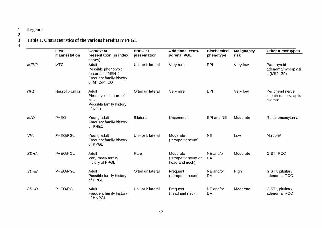
43
Legends 1 2 Table 1. Characteristics of the various hereditary PPGL 3 4
First manifestation
Context at presentation (in index cases)
PHEO at presentation
Additional extra-adrenal PGL
Biochemical phenotype
Malignancy risk
Other tumor types
MEN2 MTC
Adult Possible phenotypic features of MEN-2 Frequent family history of MTC/PHEO
Uni- or bilateral Very rare EPI Very low Parathyroid adenoma/hyperplasia (MEN-2A)
NF1 Neurofibromas Adult Phenotypic feature of NF-1 Possible family history of NF-1
Often unilateral Very rare EPI Very low Peripheral nerve sheath tumors, optic gliomaa
MAX
PHEO Young adult Frequent family history of PHEO
Bilateral Uncommon EPI and NE Moderate Renal oncocytoma
VHL
PHEO/PGL Young adult Frequent family history of PPGL
Uni- or bilateral Moderate (retroperitoneum)
NE Low Multipleb
SDHA PHEO/PGL
Adult Very rarely family history of PPGL
Rare Moderate (retroperitoneum or head and neck)
NE and/or DA
Moderate GIST, RCC
SDHB
PHEO/PGL Adult Possible family history of PPGL
Often unilateral Frequent (retroperitoneum)
NE and/or DA
High GISTc, pituitary adenoma, RCC
SDHD
PHEO/PGL Adult Frequent family history of HNPGL
Uni- or bilateral Frequent (head and neck)
NE and/or DA
Moderate GISTc, pituitary adenoma, RCC
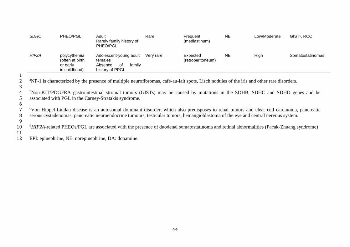
44
SDHC PHEO/PGL Adult
Rarely family history of PHEO/PGL
Rare Frequent (mediastinum)
NE Low/Moderate GISTc, RCC
HIF2A polycythemia (often at birth or early in childhood)
Adolescent-young adult females Absence of family history of PPGL
Very rare Expected (retroperitoneum)
NE High Somatostatinomas
1 aNF-1 is characterized by the presence of multiple neurofibromas, café-au-lait spots, Lisch nodules of the iris and other rare disorders. 2 3 bNon-KIT/PDGFRA gastrointestinal stromal tumors (GISTs) may be caused by mutations in the SDHB, SDHC and SDHD genes and be 4 associated with PGL in the Carney-Stratakis syndrome. 5 6 cVon Hippel-Lindau disease is an autosomal dominant disorder, which also predisposes to renal tumors and clear cell carcinoma, pancreatic 7 serous cystadenomas, pancreatic neuroendocrine tumours, testicular tumors, hemangioblastoma of the eye and central nervous system. 8 9 dHIF2A-related PHEOs/PGL are associated with the presence of duodenal somatostatinoma and retinal abnormalities (Pacak-Zhuang syndrome) 10 11 EPI: epinephrine, NE: norepinephrine, DA: dopamine. 12
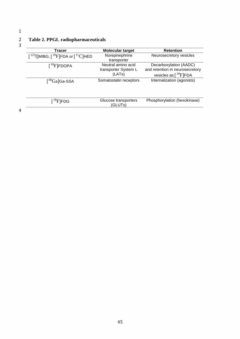
45
1
Table 2. PPGL radiopharmaceuticals 2 3
Tracer Molecular target Retention
123IMIBG, 18FFDA or 11CHED Norepinephrine transporter
Neurosecretory vesicles
18FFDOPA Neutral amino acid transporter System L
(LATs)
Decarboxylation (AADC) and retention in neurosecretory
vesicles as 18FFDA
68GaGa-SSA
Somatostatin receptors Internalization (agonists)
18FFDG Glucose transporters (GLUTs)
Phosphorylation (hexokinase)
4
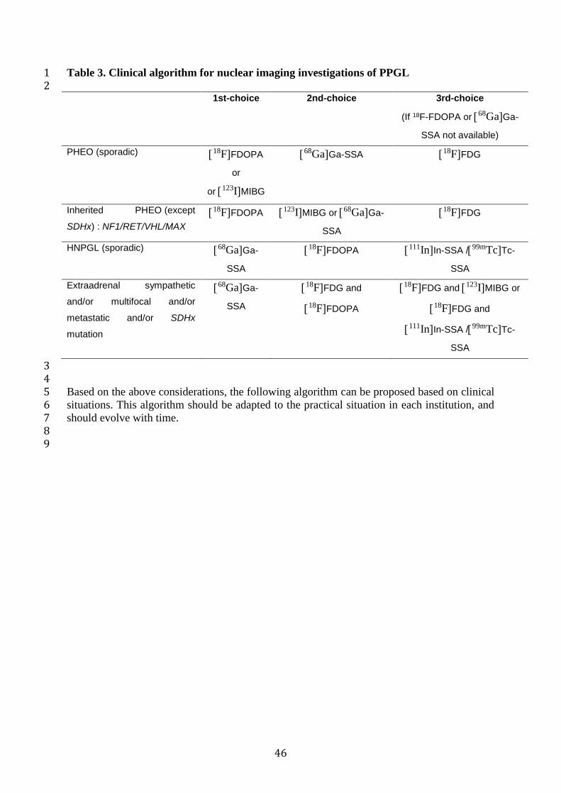
46
Table 3. Clinical algorithm for nuclear imaging investigations of PPGL 1 2 1st-choice 2nd-choice 3rd-choice
(If 18F-FDOPA or 68GaGa-
SSA not available)
PHEO (sporadic) 18FFDOPA
or
or 123IMIBG
68GaGa-SSA 18FFDG
Inherited PHEO (except
SDHx) : NF1/RET/VHL/MAX
18FFDOPA 123IMIBG or 68GaGa-
SSA
18FFDG
HNPGL (sporadic) 68GaGa-
SSA
18FFDOPA 111InIn-SSA /99mTcTc-
SSA
Extraadrenal sympathetic
and/or multifocal and/or
metastatic and/or SDHx
mutation
68GaGa-
SSA
18FFDG and
18FFDOPA
18FFDG and 123IMIBG or
18FFDG and
111InIn-SSA /99mTcTc-
SSA
3 4 Based on the above considerations, the following algorithm can be proposed based on clinical 5 situations. This algorithm should be adapted to the practical situation in each institution, and 6 should evolve with time. 7 8 9
47
References 1
2
1. Lenders JW, Duh QY, Eisenhofer G, Gimenez-Roqueplo AP, Grebe SK, Murad MH, 3
et al. Pheochromocytoma and paraganglioma: an endocrine society clinical practice guideline. 4
J Clin Endocrinol Metab. 2014;99:1915-42. doi:10.1210/jc.2014-1498. 5
2. Lloyd RV, Osamura RY, Kloppel G, Rosai J. WHO Classification of Tumours of 6
Endocrine Organs: IARC Press, Lyon 2017. 7
3. Crona J, Taieb D, Pacak K. New Perspectives on Pheochromocytoma and 8
Paraganglioma: Toward a Molecular Classification. Endocr Rev. 2017;38:489-515. 9
doi:10.1210/er.2017-00062. 10
4. Jafri M, Whitworth J, Rattenberry E, Vialard L, Kilby G, Kumar AV, et al. Evaluation 11
of SDHB, SDHD and VHL gene susceptibility testing in the assessment of individuals with 12
non-syndromic phaeochromocytoma, paraganglioma and head and neck paraganglioma. 13
Clinical endocrinology. 2013;78:898-906. doi:10.1111/cen.12074. 14
5. Neumann HP, Erlic Z, Boedeker CC, Rybicki LA, Robledo M, Hermsen M, et al. 15
Clinical predictors for germline mutations in head and neck paraganglioma patients: cost 16
reduction strategy in genetic diagnostic process as fall-out. Cancer Res. 2009;69:3650-6. 17
doi:10.1158/0008-5472.CAN-08-4057. 18
6. Papathomas TG, Gaal J, Corssmit EP, Oudijk L, Korpershoek E, Heimdal K, et al. 19
Non-pheochromocytoma (PCC)/paraganglioma (PGL) tumors in patients with succinate 20
dehydrogenase-related PCC-PGL syndromes: a clinicopathological and molecular analysis. 21
Eur J Endocrinol. 2014;170:1-12. doi:10.1530/EJE-13-0623. 22
7. Pasini B, McWhinney SR, Bei T, Matyakhina L, Stergiopoulos S, Muchow M, et al. 23
Clinical and molecular genetics of patients with the Carney-Stratakis syndrome and germline 24
mutations of the genes coding for the succinate dehydrogenase subunits SDHB, SDHC, and 25
SDHD. Eur J Hum Genet. 2008;16:79-88. doi:10.1038/sj.ejhg.5201904. 26
8. Casey RT, Warren AY, Martin JE, Challis BG, Rattenberry E, Whitworth J, et al. 27
Clinical and Molecular Features of Renal and Pheochromocytoma/Paraganglioma Tumor 28
Association Syndrome (RAPTAS): Case Series and Literature Review. J Clin Endocrinol 29
Metab. 2017;102:4013-22. doi:10.1210/jc.2017-00562. 30
9. Eisenhofer G, Pacak K, Huynh TT, Qin N, Bratslavsky G, Linehan WM, et al. 31
Catecholamine metabolomic and secretory phenotypes in phaeochromocytoma. Endocrine-32
related cancer. 2011;18:97-111. doi:10.1677/ERC-10-0211. 33

48
10. Leung K, Stamm M, Raja A, Low G. Pheochromocytoma: the range of appearances on 1
ultrasound, CT, MRI, and functional imaging. AJR Am J Roentgenol. 2013;200:370-8. 2
doi:10.2214/AJR.12.9126. 3
11. Heimburger C, Veillon F, Taieb D, Goichot B, Riehm S, Petit-Thomas J, et al. Head-4
to-head comparison between 18F-FDOPA PET/CT and MR/CT angiography in clinically 5
recurrent head and neck paragangliomas. European journal of nuclear medicine and molecular 6
imaging. 2017;44:979-87. doi:10.1007/s00259-016-3605-0. 7
12. Kong G, Grozinsky-Glasberg S, Hofman MS, Callahan J, Meirovitz A, Maimon O, et 8
al. Efficacy of Peptide Receptor Radionuclide Therapy for Functional Metastatic 9
Paraganglioma and Pheochromocytoma. J Clin Endocrinol Metab. 2017;102:3278-87. 10
doi:10.1210/jc.2017-00816. 11
13. Rutgers M, Tytgat GA, Verwijs-Janssen M, Buitenhuis C, Voute PA, Smets LA. 12
Uptake of the neuron-blocking agent meta-iodobenzylguanidine and serotonin by human 13
platelets and neuro-adrenergic tumour cells. International journal of cancer Journal 14
international du cancer. 1993;54:290-5. 15
14. Jaques S, Jr., Tobes MC, Sisson JC. Sodium dependency of uptake of norepinephrine 16
and m-iodobenzylguanidine into cultured human pheochromocytoma cells: evidence for 17
uptake-one. Cancer research. 1987;47:3920-8. 18
15. Bomanji J, Levison DA, Flatman WD, Horne T, Bouloux PM, Ross G, et al. Uptake of 19
iodine-123 MIBG by pheochromocytomas, paragangliomas, and neuroblastomas: a 20
histopathological comparison. Journal of nuclear medicine : official publication, Society of 21
Nuclear Medicine. 1987;28:973-8. 22
16. ICRP. Radiation Dose to Patients from Radiopharmaceuticals. ICRP Publication 53. 23
Annals of the ICRP. 1987;18. 24
17. Sinclair AJ, Bomanji J, Harris P, Ross G, Besser GM, Britton KE. Pre- and post-25
treatment distribution pattern of 123I-MIBG in patients with phaeochromocytomas and 26
paragangliomas. Nuclear medicine communications. 1989;10:567-76. 27
18. Solanki KK, Bomanji J, Moyes J, Mather SJ, Trainer PJ, Britton KE. A 28
pharmacological guide to medicines which interfere with the biodistribution of radiolabelled 29
meta-iodobenzylguanidine (MIBG). Nuclear medicine communications. 1992;13:513-21. 30
19. Jacobson AF, Travin MI. Impact of medications on mIBG uptake, with specific 31
attention to the heart: Comprehensive review of the literature. J Nucl Cardiol. 2015;22:980-32
93. doi:10.1007/s12350-015-0170-z. 33

49
20. Jacobson AF, White S, Travin MI, Tseng C. Impact of concomitant medication use on 1
myocardial 123I-mIBG imaging results in patients with heart failure. Nucl Med Commun. 2
2017;38:141-8. doi:10.1097/MNM.0000000000000619. 3
21. Bombardieri E, Aktolun C, Baum RP, Bishof-Delaloye A, Buscombe J, Chatal JF, et 4
al. 131I/123I-metaiodobenzylguanidine (MIBG) scintigraphy: procedure guidelines for 5
tumour imaging. European journal of nuclear medicine and molecular imaging. 6
2003;30:BP132-9. 7
22. Bombardieri E, Giammarile F, Aktolun C, Baum RP, Bischof Delaloye A, Maffioli L, 8
et al. 131I/123I-metaiodobenzylguanidine (mIBG) scintigraphy: procedure guidelines for 9
tumour imaging. European journal of nuclear medicine and molecular imaging. 10
2010;37:2436-46. doi:10.1007/s00259-010-1545-7. 11
23. Bar-Sever Z, Biassoni L, Shulkin B, Kong G, Hofman MS, Lopci E, et al. Guidelines 12
on nuclear medicine imaging in neuroblastoma. Eur J Nucl Med Mol Imaging. 2018. 13
doi:10.1007/s00259-018-4070-8. 14
24. Khafagi FA, Shapiro B, Fig LM, Mallette S, Sisson JC. Labetalol reduces iodine-131 15
MIBG uptake by pheochromocytoma and normal tissues. Journal of nuclear medicine : 16
official publication, Society of Nuclear Medicine. 1989;30:481-9. 17
25. Apeldoorn L, Voerman HJ, Hoefnagel CA. Interference of MIBG uptake by 18
medication: a case report. Neth J Med. 1995;46:239-43. 19
26. Estorch M, Carrio I, Mena E, Flotats A, Camacho V, Fuertes J, et al. Challenging the 20
neuronal MIBG uptake by pharmacological intervention: effect of a single dose of oral 21
amitriptyline on regional cardiac MIBG uptake. European journal of nuclear medicine and 22
molecular imaging. 2004;31:1575-80. doi:10.1007/s00259-004-1520-2. 23
27. Zaplatnikov K, Menzel C, Dobert N, Hamscho N, Kranert WT, Gotthard M, et al. 24
Case report: drug interference with MIBG uptake in a patient with metastatic paraganglioma. 25
The British journal of radiology. 2004;77:525-7. 26
28. Blake GM, Lewington VJ, Fleming JS, Zivanovic MA, Ackery DM. Modification by 27
nifedipine of 131I-meta-iodobenzylguanidine kinetics in malignant phaeochromocytoma. 28
European journal of nuclear medicine. 1988;14:345-8. 29
29. Taniguchi K, Ishizu K, Torizuka T, Hasegawa S, Okawada T, Ozawa T, et al. 30
Metastases of predominantly dopamine-secreting phaeochromocytoma that did not 31
accumulate meta-iodobenzylguanidine: imaging with whole body positron emission 32
tomography using 18F-labelled deoxyglucose. Eur J Surg. 2001;167:866-70. 33
doi:10.1080/11024150152717733. 34
50
30. Van Der Horst-Schrivers AN, Osinga TE, Kema IP, Van Der Laan BF, Dullaart RP. 1
Dopamine excess in patients with head and neck paragangliomas. Anticancer Res. 2
2010;30:5153-8. 3
31. van Gelder T, Verhoeven GT, de Jong P, Oei HY, Krenning EP, Vuzevski VD, et al. 4
Dopamine-producing paraganglioma not visualized by iodine-123-MIBG scintigraphy. 5
Journal of nuclear medicine : official publication, Society of Nuclear Medicine. 1995;36:620-6
2. 7
32. Hall FT, Perez-Ordonez B, Mackenzie RG, Gilbert RW. Does Catecholamine 8
Secretion from Head and Neck Paragangliomas Respond to Radiotherapy? Case Report and 9
Literature Review. Skull Base. 2003;13:229-34. doi:10.1055/s-2004-817699. 10
33. Ishibashi N, Abe K, Furuhashi S, Fukushima S, Yoshinobu T, Takahashi M, et al. 11
Adverse allergic reaction to 131I MIBG. Ann Nucl Med. 2009;23:697-9. doi:10.1007/s12149-12
009-0282-0. 13
34. ICRP. Radiation dose to patients from radiopharmaceuticals. Addendum 2 to ICRP 14
Publication 53. ICRP Publication 80. Annals of the ICRP. 1998;28. 15
35. Tobes MC, Fig LM, Carey J, Geatti O, Sisson JC, Shapiro B. Alterations of iodine-16
131 MIBG biodistribution in an anephric patient: comparison to normal and impaired renal 17
function. Journal of nuclear medicine : official publication, Society of Nuclear Medicine. 18
1989;30:1476-82. 19
36. Friedman NC, Hassan A, Grady E, Matsuoka DT, Jacobson AF. Efficacy of thyroid 20
blockade on thyroid radioiodine uptake in 123I-mIBG imaging. J Nucl Med. 2014;55:211-5. 21
doi:10.2967/jnumed.113.124826. 22
37. Bombardieri E, Giammarile F, Aktolun C, Baum RP, Bischof Delaloye A, Maffioli L, 23
et al. 131I/123I-metaiodobenzylguanidine (mIBG) scintigraphy: procedure guidelines for 24
tumour imaging. Eur J Nucl Med Mol Imaging. 2010;37:2436-46. doi:10.1007/s00259-010-25
1545-7. 26
38. Beauregard JM, Hofman MS, Pereira JM, Eu P, Hicks RJ. Quantitative (177)Lu 27
SPECT (QSPECT) imaging using a commercially available SPECT/CT system. Cancer 28
Imaging. 2011;11:56-66. doi:10.1102/1470-7330.2011.0012. 29
39. Tran-Gia J, Lassmann M. Characterization of Noise and Resolution for Quantitative 30
(177)Lu SPECT/CT with xSPECT Quant. J Nucl Med. 2018. 31
doi:10.2967/jnumed.118.211094. 32
51
40. Bailey DL, Willowson KP. Quantitative SPECT/CT: SPECT joins PET as a 1
quantitative imaging modality. Eur J Nucl Med Mol Imaging. 2014;41 Suppl 1:S17-25. 2
doi:10.1007/s00259-013-2542-4. 3
41. Ilias I, Chen CC, Carrasquillo JA, Whatley M, Ling A, Lazurova I, et al. Comparison 4
of 6-18F-fluorodopamine PET with 123I-metaiodobenzylguanidine and 111in-pentetreotide 5
scintigraphy in localization of nonmetastatic and metastatic pheochromocytoma. Journal of 6
Nuclear Medicine. 2008;49:1613-9. 7
42. Timmers HJ, Eisenhofer G, Carrasquillo JA, Chen CC, Whatley M, Ling A, et al. Use 8
of 6-[18F]-fluorodopamine positron emission tomography (PET) as first-line investigation for 9
the diagnosis and localization of non-metastatic and metastatic phaeochromocytoma (PHEO). 10
Clin Endocrinol (Oxf). 2009;71:11-7. 11
43. Timmers HJ, Chen CC, Carrasquillo JA, Whatley M, Ling A, Havekes B, et al. 12
Comparison of 18F-Fluoro-L-DOPA, 18F-Fluoro-Deoxyglucose, and 18F-Fluorodopamine 13
PET and 123I-MIBG Scintigraphy in the Localization of Pheochromocytoma and 14
Paraganglioma. Journal of Clinical Endocrinology and Metabolism. 2009;94:4757-67. 15
44. Fiebrich HB, Brouwers AH, Kerstens MN, Pijl ME, Kema IP, de Jong JR, et al. 6-[F-16
18]Fluoro-L-dihydroxyphenylalanine positron emission tomography is superior to 17
conventional imaging with (123)I-metaiodobenzylguanidine scintigraphy, computer 18
tomography, and magnetic resonance imaging in localizing tumors causing catecholamine 19
excess. J Clin Endocrinol Metab. 2009;94:3922-30. 20
45. Fonte JS, Robles JF, Chen CC, Reynolds J, Whatley M, Ling A, et al. False-negative 21
123I-MIBG SPECT is most commonly found in SDHB-related pheochromocytoma or 22
paraganglioma with high frequency to develop metastatic disease. Endocrine-related cancer. 23
2012;19:83-93. doi:10.1530/ERC-11-0243. 24
46. Kaji P, Carrasquillo JA, Linehan WM, Chen CC, Eisenhofer G, Pinto PA, et al. The 25
role of 6-[18F]fluorodopamine positron emission tomography in the localization of adrenal 26
pheochromocytoma associated with von Hippel-Lindau syndrome. Eur J Endocrinol. 27
2007;156:483-7. 28
47. Hoegerle S, Nitzsche E, Altehoefer C, Ghanem N, Manz T, Brink I, et al. 29
Pheochromocytomas: detection with 18F DOPA whole body PET--initial results. Radiology. 30
2002;222:507-12. 31
48. Krenning EP, Bakker WH, Kooij PP, Breeman WA, Oei HY, de Jong M, et al. 32
Somatostatin receptor scintigraphy with indium-111-DTPA-D-Phe-1-octreotide in man: 33
52
metabolism, dosimetry and comparison with iodine-123-Tyr-3-octreotide. J Nucl Med. 1
1992;33:652-8. 2
49. Bombardieri E, Ambrosini V, Aktolun C, Baum RP, Bishof-Delaloye A, Del Vecchio 3
S, et al. 111In-pentetreotide scintigraphy: procedure guidelines for tumour imaging. European 4
journal of nuclear medicine and molecular imaging. 2010;37:1441-8. doi:10.1007/s00259-5
010-1473-6. 6
50. Balon HR, Brown TL, Goldsmith SJ, Silberstein EB, Krenning EP, Lang O, et al. The 7
SNM practice guideline for somatostatin receptor scintigraphy 2.0. J Nucl Med Technol. 8
2011;39:317-24. doi:10.2967/jnmt.111.098277. 9
51. ICRP. Radiation Dose to Patients fromRadiopharmaceuticals: A Compendium of 10
Current Information Related to Frequently Used Substances. ICRP Publication 128. Annals of 11
the ICRP. 2015;44. 12
52. Balon HR. Updated practice guideline for somatostatin receptor scintigraphy. J Nucl 13
Med. 2011;52:1838. doi:10.2967/jnumed.111.098285. 14
53. Bustillo A, Telischi F, Weed D, Civantos F, Angeli S, Serafini A, et al. Octreotide 15
scintigraphy in the head and neck. Laryngoscope. 2004;114:434-40. 16
54. Duet M, Sauvaget E, Petelle B, Rizzo N, Guichard JP, Wassef M, et al. Clinical 17
impact of somatostatin receptor scintigraphy in the management of paragangliomas of the 18
head and neck. J Nucl Med. 2003;44:1767-74. 19
55. Koopmans KP, Jager PL, Kema IP, Kerstens MN, Albers F, Dullaart RP. 111In-20
octreotide is superior to 123I-metaiodobenzylguanidine for scintigraphic detection of head 21
and neck paragangliomas. Journal of Nuclear Medicine. 2008;49:1232-7. 22
56. Muros MA, Llamas-Elvira JM, Rodriguez A, Ramirez A, Gomez M, Arraez MA, et al. 23
111In-pentetreotide scintigraphy is superior to 123I-MIBG scintigraphy in the diagnosis and 24
location of chemodectoma. Nucl Med Commun. 1998;19:735-42. 25
57. Schmidt M, Fischer E, Dietlein M, Michel O, Weber K, Moka D, et al. Clinical value 26
of somatostatin receptor imaging in patients with suspected head and neck paragangliomas. 27
Eur J Nucl Med Mol Imaging. 2002;29:1571-80. 28
58. Telischi FF, Bustillo A, Whiteman ML, Serafini AN, Reisberg MJ, Gomez-Marin O, 29
et al. Octreotide scintigraphy for the detection of paragangliomas. Otolaryngology--head and 30
neck surgery : official journal of American Academy of Otolaryngology-Head and Neck 31
Surgery. 2000;122:358-62. 32
59. Charrier N, Deveze A, Fakhry N, Sebag F, Morange I, Gaborit B, et al. Comparison of 33
[111In]pentetreotide-SPECT and [18F]FDOPA-PET in the localization of extra-adrenal 34
53
paragangliomas: The case for a patient-tailored use of nuclear imaging modalities. Clinical 1
endocrinology. 2011;74:21-9. 2
60. Kaltsas GA, Mukherjee JJ, Grossman AB. The value of radiolabelled MIBG and 3
octreotide in the diagnosis and management of neuroendocrine tumours. Ann Oncol. 2001;12 4
Suppl 2:S47-50. 5
61. van der Harst E, de Herder WW, Bruining HA, Bonjer HJ, de Krijger RR, Lamberts 6
SW, et al. [(123)I]metaiodobenzylguanidine and [(111)In]octreotide uptake in begnign and 7
malignant pheochromocytomas. J Clin Endocrinol Metab. 2001;86:685-93. 8
62. Tenenbaum F, Lumbroso J, Schlumberger M, Mure A, Plouin PF, Caillou B, et al. 9
Comparison of radiolabeled octreotide and meta-iodobenzylguanidine (MIBG) scintigraphy in 10
malignant pheochromocytoma. J Nucl Med. 1995;36:1-6. 11
63. Wild D, Macke HR, Waser B, Reubi JC, Ginj M, Rasch H, et al. 68Ga-DOTANOC: a 12
first compound for PET imaging with high affinity for somatostatin receptor subtypes 2 and 5. 13
Eur J Nucl Med Mol Imaging. 2005;32:724. 14
64. Wild D, Schmitt JS, Ginj M, Macke HR, Bernard BF, Krenning E, et al. DOTA-NOC, 15
a high-affinity ligand of somatostatin receptor subtypes 2, 3 and 5 for labelling with various 16
radiometals. Eur J Nucl Med Mol Imaging. 2003;30:1338-47. 17
65. Reubi JC, Schar JC, Waser B, Wenger S, Heppeler A, Schmitt JS, et al. Affinity 18
profiles for human somatostatin receptor subtypes SST1-SST5 of somatostatin radiotracers 19
selected for scintigraphic and radiotherapeutic use. Eur J Nucl Med. 2000;27:273-82. 20
66. Fani M, Nicolas GP, Wild D. Somatostatin Receptor Antagonists for Imaging and 21
Therapy. Journal of nuclear medicine : official publication, Society of Nuclear Medicine. 22
2017;58:61S-6S. doi:10.2967/jnumed.116.186783. 23
67. Pettinato C, Sarnelli A, Di Donna M, Civollani S, Nanni C, Montini G, et al. 68Ga-24
DOTANOC: biodistribution and dosimetry in patients affected by neuroendocrine tumors. 25
Eur J Nucl Med Mol Imaging. 2008;35:72-9. doi:10.1007/s00259-007-0587-y. 26
68. Cherk MH, Kong G, Hicks RJ, Hofman MS. Changes in biodistribution on (68)Ga-27
DOTA-Octreotate PET/CT after long acting somatostatin analogue therapy in neuroendocrine 28
tumour patients may result in pseudoprogression. Cancer Imaging. 2018;18:3. 29
doi:10.1186/s40644-018-0136-x. 30
69. Sandstrom M, Velikyan I, Garske-Roman U, Sorensen J, Eriksson B, Granberg D, et 31
al. Comparative biodistribution and radiation dosimetry of 68Ga-DOTATOC and 68Ga-32
DOTATATE in patients with neuroendocrine tumors. Journal of nuclear medicine : official 33
publication, Society of Nuclear Medicine. 2013;54:1755-9. doi:10.2967/jnumed.113.120600. 34
54
70. Lasnon C, Hicks RJ, Beauregard JM, Milner A, Paciencia M, Guizard AV, et al. 1
Impact of point spread function reconstruction on thoracic lymph node staging with 18F-FDG 2
PET/CT in non-small cell lung cancer. Clin Nucl Med. 2012;37:971-6. 3
doi:10.1097/RLU.0b013e318251e3d1. 4
71. Castellucci P, Pou Ucha J, Fuccio C, Rubello D, Ambrosini V, Montini GC, et al. 5
Incidence of increased 68Ga-DOTANOC uptake in the pancreatic head in a large series of 6
extrapancreatic NET patients studied with sequential PET/CT. J Nucl Med. 2011;52:886-90. 7
doi:10.2967/jnumed.111.088328. 8
72. Archier A, Varoquaux A, Garrigue P, Montava M, Guerin C, Gabriel S, et al. 9
Prospective comparison of Ga-DOTATATE and F-FDOPA PET/CT in patients with various 10
pheochromocytomas and paragangliomas with emphasis on sporadic cases. European journal 11
of nuclear medicine and molecular imaging. 2015. doi:10.1007/s00259-015-3268-2. 12
73. Hofman MS, Lau WF, Hicks RJ. Somatostatin receptor imaging with 68Ga 13
DOTATATE PET/CT: clinical utility, normal patterns, pearls, and pitfalls in interpretation. 14
Radiographics. 2015;35:500-16. doi:10.1148/rg.352140164. 15
74. Naji M, Al-Nahhas A. (68)Ga-labelled peptides in the management of 16
neuroectodermal tumours. European journal of nuclear medicine and molecular imaging. 17
2012;39 Suppl 1:61-7. doi:10.1007/s00259-011-1990-y. 18
75. Naji M, Zhao C, Welsh SJ, Meades R, Win Z, Ferrarese A, et al. 68Ga-DOTA-TATE 19
PET vs. 123I-MIBG in identifying malignant neural crest tumours. Molecular imaging and 20
biology : MIB : the official publication of the Academy of Molecular Imaging. 2011;13:769-21
75. doi:10.1007/s11307-010-0396-8. 22
76. Sharma P, Thakar A, Suman Kc S, Dhull VS, Singh H, Naswa N, et al. 68Ga-23
DOTANOC PET/CT for Baseline Evaluation of Patients with Head and Neck Paraganglioma. 24
J Nucl Med. 2013;54:841-7. doi:10.2967/jnumed.112.115485. 25
77. Kroiss A, Shulkin BL, Uprimny C, Frech A, Gasser RW, Url C, et al. (68)Ga-26
DOTATOC PET/CT provides accurate tumour extent in patients with extraadrenal 27
paraganglioma compared to (123)I-MIBG SPECT/CT. European Journal of Nuclear Medicine 28
and Molecular Imaging. 2015;42:33-41. doi:10.1007/s00259-014-2892-6. 29
78. Sharma P, Mukherjee A, Karunanithi S, Naswa N, Kumar R, Ammini AC, et al. 30
Accuracy of 68Ga DOTANOC PET/CT Imaging in Patients With Multiple Endocrine 31
Neoplasia Syndromes. Clin Nucl Med. 2015;40:e351-6. 32
doi:10.1097/RLU.0000000000000775. 33
55
79. Han S, Suh CH, Woo S, Kim YJ, Lee JJ. Performance of (68)Ga-DOTA-Conjugated 1
Somatostatin Receptor Targeting Peptide PET in Detection of Pheochromocytoma and 2
Paraganglioma: A Systematic Review and Meta-Analysis. J Nucl Med. 2018. 3
doi:10.2967/jnumed.118.211706. 4
80. Kroiss A, Putzer D, Frech A, Decristoforo C, Uprimny C, Gasser RW, et al. A 5
retrospective comparison between (68)Ga-DOTA-TOC PET/CT and (18)F-DOPA PET/CT in 6
patients with extra-adrenal paraganglioma. European journal of nuclear medicine and 7
molecular imaging. 2013;40:1800-8. doi:10.1007/s00259-013-2548-y. 8
81. Janssen I, Blanchet EM, Adams K, Chen CC, Millo CM, Herscovitch P, et al. 9
Superiority of [68Ga]-DOTATATE PET/CT to Other Functional Imaging Modalities in the 10
Localization of SDHB-Associated Metastatic Pheochromocytoma and Paraganglioma. 11
Clinical cancer research : an official journal of the American Association for Cancer 12
Research. 2015;21:3888-95. doi:10.1158/1078-0432.CCR-14-2751. 13
82. Janssen I, Chen CC, Taieb D, Patronas NJ, Millo CM, Adams KT, et al. 68Ga-14
DOTATATE PET/CT in the localization of head and neck paragangliomas compared to other 15
functional imaging modalities and CT/MRI. Journal of nuclear medicine : official publication, 16
Society of Nuclear Medicine. 2015. doi:10.2967/jnumed.115.161018. 17
83. Hofman MS, Hicks RJ. Moving Beyond "Lumpology": PET/CT Imaging of 18
Pheochromocytoma and Paraganglioma. Clinical cancer research : an official journal of the 19
American Association for Cancer Research. 2015;21:3815-7. doi:10.1158/1078-0432.CCR-20
15-1073. 21
84. Kan Y, Zhang S, Wang W, Liu J, Yang J, Wang Z. (68)Ga-somatostatin receptor 22
analogs and (18)F-FDG PET/CT in the localization of metastatic pheochromocytomas and 23
paragangliomas with germline mutations: a meta-analysis. Acta Radiol. 24
2018:284185118764206. doi:10.1177/0284185118764206. 25
85. Gild ML, Naik N, Hoang J, Hsiao E, McGrath RT, Sywak M, et al. Role of 26
DOTATATE-PET/CT in preoperative assessment of phaeochromocytoma and 27
paragangliomas. Clin Endocrinol (Oxf). 2018. doi:10.1111/cen.13737. 28
86. Jha A, Ling A, Millo C, Gupta G, Viana B, Lin FI, et al. Superiority of (68)Ga-29
DOTATATE over (18)F-FDG and anatomic imaging in the detection of succinate 30
dehydrogenase mutation (SDHx )-related pheochromocytoma and paraganglioma in the 31
pediatric population. Eur J Nucl Med Mol Imaging. 2018;45:787-97. doi:10.1007/s00259-32
017-3896-9. 33
56
87. Darr R, Nambuba J, Del Rivero J, Janssen I, Merino M, Todorovic M, et al. Novel 1
insights into the polycythemia-paraganglioma-somatostatinoma syndrome. Endocr Relat 2
Cancer. 2016;23:899-908. doi:10.1530/ERC-16-0231. 3
88. Hentschel M, Rottenburger C, Boedeker CC, Neumann HP, Brink I. Is there an 4
optimal scan time for 6-[F-18]fluoro-L-DOPA PET in pheochromocytomas and 5
paragangliomas? Clinical nuclear medicine. 2012;37:e24-9. 6
doi:10.1097/RLU.0b013e318238f550. 7
89. Timmers HJ, Hadi M, Carrasquillo JA, Chen CC, Martiniova L, Whatley M, et al. The 8
effects of carbidopa on uptake of 6-18F-Fluoro-L-DOPA in PET of pheochromocytoma and 9
extraadrenal abdominal paraganglioma. J Nucl Med. 2007;48:1599-606. 10
90. Luxen A, Perlmutter M, Bida GT, Van Moffaert G, Cook JS, Satyamurthy N, et al. 11
Remote, semiautomated production of 6-[18F]fluoro-L-dopa for human studies with PET. Int 12
J Rad Appl Instrum A. 1990;41:275-81. 13
91. Namavari M, Bishop A, Satyamurthy N, Bida G, Barrio JR. Regioselective 14
radiofluorodestannylation with [18F]F2 and [18F]CH3COOF: a high yield synthesis of 6-15
[18F]Fluoro-L-dopa. Int J Rad Appl Instrum A. 1992;43:989-96. 16
92. Vernaleken I, Kumakura Y, Cumming P, Buchholz HG, Siessmeier T, Stoeter P, et al. 17
Modulation of [18F]fluorodopa (FDOPA) kinetics in the brain of healthy volunteers after 18
acute haloperidol challenge. Neuroimage. 2006;30:1332-9. 19
doi:10.1016/j.neuroimage.2005.11.014. 20
93. Garnett S, Firnau G, Nahmias C, Chirakal R. Striatal dopamine metabolism in living 21
monkeys examined by positron emission tomography. Brain Res. 1983;280:169-71. 22
94. Beheshti M, Pocher S, Vali R, Waldenberger P, Broinger G, Nader M, et al. The value 23
of 18F-DOPA PET-CT in patients with medullary thyroid carcinoma: comparison with 18F-24
FDG PET-CT. Eur Radiol. 2009;19:1425-34. doi:10.1007/s00330-008-1280-7. 25
95. Soussan M, Nataf V, Kerrou K, Grahek D, Pascal O, Talbot JN, et al. Added value of 26
early 18F-FDOPA PET/CT acquisition time in medullary thyroid cancer. Nucl Med Commun. 27
2012;33:775-9. doi:10.1097/MNM.0b013e3283543304. 28
96. Ginet M, Cuny T, Schmitt E, Marie PY, Verger A. 18F-FDOPA PET Imaging in 29
Prolactinoma. Clin Nucl Med. 2017;42:e383-e4. doi:10.1097/RLU.0000000000001721. 30
97. Berends AMA, Kerstens MN, Bolt JW, Links TP, Korpershoek E, de Krijger RR, et 31
al. False-positive findings on 6-[18F]fluor-l-3,4-dihydroxyphenylalanine PET ((18)F-32
FDOPA-PET) performed for imaging of neuroendocrine tumors. Eur J Endocrinol. 33
2018;179:127-35. doi:10.1530/EJE-18-0321. 34
57
98. Treglia G, Cocciolillo F, de Waure C, Di Nardo F, Gualano MR, Castaldi P, et al. 1
Diagnostic performance of (18)F-dihydroxyphenylalanine positron emission tomography in 2
patients with paraganglioma: a meta-analysis. Eur J Nucl Med Mol Imaging. 2012. 3
doi:10.1007/s00259-012-2087-y. 4
99. Mackenzie IS, Gurnell M, Balan KK, Simpson H, Chatterjee K, Brown MJ. The use of 5
18-fluoro-dihydroxyphenylalanine and 18-fluorodeoxyglucose positron emission tomography 6
scanning in the assessment of metaiodobenzylguanidine-negative phaeochromocytoma. Eur J 7
Endocrinol. 2007;157:533-7. 8
100. Imani F, Agopian VG, Auerbach MS, Walter MA, Imani F, Benz MR, et al. 18F-9
FDOPA PET and PET/CT accurately localize pheochromocytomas. J Nucl Med. 10
2009;50:513-9. 11
101. Kauhanen S, Seppanen M, Ovaska J, Minn H, Bergman J, Korsoff P, et al. The 12
clinical value of [18F]fluoro-dihydroxyphenylalanine positron emission tomography in 13
primary diagnosis, staging, and restaging of neuroendocrine tumors. Endocrine-related 14
cancer. 2009;16:255-65. doi:10.1677/erc-08-0229. 15
102. Luster M, Karges W, Zeich K, Pauls S, Verburg FA, Dralle H, et al. Clinical value of 16
18F-fluorodihydroxyphenylalanine positron emission tomography/computed tomography 17
(18F-DOPA PET/CT) for detecting pheochromocytoma. European Journal of Nuclear 18
Medicine and Molecular Imaging. 2010;37:484-93. 19
103. Fottner C, Helisch A, Anlauf M, Rossmann H, Musholt TJ, Kreft A, et al. 6-18F-20
fluoro-L-dihydroxyphenylalanine positron emission tomography is superior to 123I-21
metaiodobenzyl-guanidine scintigraphy in the detection of extraadrenal and hereditary 22
pheochromocytomas and paragangliomas: correlation with vesicular monoamine transporter 23
expression. Journal of Clinical Endocrinology and Metabolism. 2010;95:2800-10. 24
104. Rufini V, Treglia G, Castaldi P, Perotti G, Calcagni ML, Corsello SM, et al. 25
Comparison of 123I-MIBG SPECT-CT and 18F-DOPA PET-CT in the evaluation of patients 26
with known or suspected recurrent paraganglioma. Nucl Med Commun. 2011;32:575-82. 27
doi:10.1097/MNM.0b013e328345a340. 28
105. Leboulleux S, Deandreis D, Escourrou C, Al Ghuzlan A, Bidault F, Auperin A, et al. 29
Fluorodesoxyglucose uptake in the remaining adrenal glands during the follow-up of patients 30
with adrenocortical carcinoma: do not consider it as malignancy. European journal of 31
endocrinology / European Federation of Endocrine Societies.164:89-94. 32
58
106. Taieb D, Jha A, Guerin C, Pang Y, Adams KT, Chen CC, et al. 18F-FDOPA PET/CT 1
imaging of MAX-related pheochromocytoma. J Clin Endocrinol Metab. 2018. 2
doi:10.1210/jc.2017-02324. 3
107. Charrier N, Deveze A, Fakhry N, Sebag F, Morange I, Gaborit B, et al. Comparison of 4
[(1)(1)(1)In]pentetreotide-SPECT and [(1)F]FDOPA-PET in the localization of extra-adrenal 5
paragangliomas: the case for a patient-tailored use of nuclear imaging modalities. Clinical 6
endocrinology. 2011;74:21-9. doi:10.1111/j.1365-2265.2010.03893.x. 7
108. Fanti S, Ambrosini V, Tomassetti P, Castellucci P, Montini G, Allegri V, et al. 8
Evaluation of unusual neuroendocrine tumours by means of 68Ga-DOTA-NOC PET. 9
Biomedicine and Pharmacotherapy. 2008;62:667-71. 10
109. Hoegerle S, Ghanem N, Altehoefer C, Schipper J, Brink I, Moser E, et al. 18F-DOPA 11
positron emission tomography for the detection of glomus tumours. Eur J Nucl Med Mol 12
Imaging. 2003;30:689-94. 13
110. King KS, Chen CC, Alexopoulos DK, Whatley MA, Reynolds JC, Patronas N, et al. 14
Functional imaging of SDHx-related head and neck paragangliomas: comparison of 18F-15
fluorodihydroxyphenylalanine, 18F-fluorodopamine, 18F-fluoro-2-deoxy-D-glucose PET, 16
123I-metaiodobenzylguanidine scintigraphy, and 111In-pentetreotide scintigraphy. J Clin 17
Endocrinol Metab. 2011;96:2779-85. doi:10.1210/jc.2011-0333. 18
111. King KS, Whatley MA, Alexopoulos DK, Reynolds JC, Chen CC, Mattox DE, et al. 19
The use of functional imaging in a patient with head and neck paragangliomas. J Clin 20
Endocrinol Metab. 2010;95:481-2. 21
112. Weisbrod AB, Kitano M, Gesuwan K, Millo C, Herscovitch P, Nilubol N, et al. 22
Clinical Utility of Functional Imaging with 18F-FDOPA in Von Hippel-Lindau Syndrome. J 23
Clin Endocrinol Metab. 2012. doi:10.1210/jc.2011-2626. 24
113. Taieb D, Tessonnier L, Sebag F, Niccoli-Sire P, Morange I, Colavolpe C, et al. The 25
role of 18F-FDOPA and 18F-FDG-PET in the management of malignant and multifocal 26
phaeochromocytomas. Clin Endocrinol (Oxf). 2008;69:580-6. 27
114. Nambuba J, Darr R, Janssen I, Bullova P, Adams KT, Millo C, et al. Functional 28
imaging experience in a germline fumarate hydratase mutation–positive patient with 29
pheochromocytoma and paraganglioma. AACE Clinical Case Reports. 2016;2:e176-e81. 30
115. Favier J, Briere JJ, Burnichon N, Riviere J, Vescovo L, Benit P, et al. The Warburg 31
effect is genetically determined in inherited pheochromocytomas. PLoS One. 2009;4:e7094. 32
116. Lopez-Jimenez E, Gomez-Lopez G, Leandro-Garcia LJ, Munoz I, Schiavi F, Montero-33
Conde C, et al. Research Resource: Transcriptional Profiling Reveals Different 34
59
Pseudohypoxic Signatures in SDHB and VHL-Related Pheochromocytomas. Mol Endocrinol. 1
2010;24:2382-91. 2
117. Burnichon N, Vescovo L, Amar L, Libe R, de Reynies A, Venisse A, et al. Integrative 3
genomic analysis reveals somatic mutations in pheochromocytoma and paraganglioma. Hum 4
Mol Genet. 2011;20:3974-85. doi:10.1093/hmg/ddr324. 5
118. Pollard PJ, El-Bahrawy M, Poulsom R, Elia G, Killick P, Kelly G, et al. Expression of 6
HIF-1alpha, HIF-2alpha (EPAS1), and their target genes in paraganglioma and 7
pheochromocytoma with VHL and SDH mutations. J Clin Endocrinol Metab. 2006;91:4593-8
8. 9
119. Span PN, Rao JU, Oude Ophuis SB, Lenders JW, Sweep FC, Wesseling P, et al. 10
Overexpression of the natural antisense hypoxia-inducible factor-1alpha transcript is 11
associated with malignant pheochromocytoma/paraganglioma. Endocr Relat Cancer. 12
2011;18:323-31. doi:10.1530/ERC-10-0184. 13
120. Taieb D, Sebag F, Barlier A, Tessonnier L, Palazzo FF, Morange I, et al. 18F-FDG 14
avidity of pheochromocytomas and paragangliomas: a new molecular imaging signature? 15
Journal of Nuclear Medicine. 2009;50:711-7. 16
121. Garrigue P, Bodin-Hullin A, Balasse L, Fernandez S, Essamet W, Dignat-George F, et 17
al. The Evolving Role of Succinate in Tumor Metabolism: An (18)F-FDG-Based Study. J 18
Nucl Med. 2017;58:1749-55. doi:10.2967/jnumed.117.192674. 19
122. Taieb D, Pacak K. New Insights into the Nuclear Imaging Phenotypes of Cluster 1 20
Pheochromocytoma and Paraganglioma. Trends Endocrinol Metab. 2017;28:807-17. 21
doi:10.1016/j.tem.2017.08.001. 22
123. ICRP. Radiation Dose to Patients from Radiopharmaceuticals - Addendum 3 to ICRP 23
Publication 53. ICRP Publication 106. Annals of the ICRP. 2008;38. 24
124. Zanotti-Fregonara P, Jan S, Taieb D, Cammilleri S, Trebossen R, Hindie E, et al. 25
Absorbed 18F-FDG dose to the fetus during early pregnancy. J Nucl Med. 2010;51:803-5. 26
doi:10.2967/jnumed.109.071878. 27
125. Timmers HJ, Chen CC, Carrasquillo JA, Whatley M, Ling A, Eisenhofer G, et al. 28
Staging and functional characterization of pheochromocytoma and paraganglioma by 18F-29
fluorodeoxyglucose (18F-FDG) positron emission tomography. Journal of the National 30
Cancer Institute. 2012;104:700-8. doi:10.1093/jnci/djs188. 31
126. Leboulleux S, Deandreis D, Escourrou C, Al Ghuzlan A, Bidault F, Auperin A, et al. 32
Fluorodesoxyglucose uptake in the remaining adrenal glands during the follow-up of patients 33
60
with adrenocortical carcinoma: do not consider it as malignancy. Eur J Endocrinol. 1
2011;164:89-94. doi:10.1530/EJE-10-0666. 2
127. Guerin C, Pattou F, Brunaud L, Lifante JC, Mirallie E, Haissaguerre M, et al. 3
Performance of 18F-FDG PET/CT in the Characterization of Adrenal Masses in Noncancer 4
Patients: A Prospective Study. J Clin Endocrinol Metab. 2017;102:2465-72. 5
doi:10.1210/jc.2017-00254. 6
128. Timmers HJ, Kozupa A, Chen CC, Carrasquillo JA, Ling A, Eisenhofer G, et al. 7
Superiority of fluorodeoxyglucose positron emission tomography to other functional imaging 8
techniques in the evaluation of metastatic SDHB-associated pheochromocytoma and 9
paraganglioma. Journal of Clinical Oncology. 2007;25:2262-9. 10
129. Tiwari A, Shah N, Sarathi V, Malhotra G, Bakshi G, Prakash G, et al. Genetic status 11
determines (18) F-FDG uptake in pheochromocytoma/paraganglioma. J Med Imaging Radiat 12
Oncol. 2017;61:745-52. doi:10.1111/1754-9485.12620. 13
130. Weisbrod AB, Kitano M, Gesuwan K, Millo C, Herscovitch P, Nilubol N, et al. 14
Clinical utility of functional imaging with 18F-FDOPA in Von Hippel-Lindau syndrome. J 15
Clin Endocrinol Metab. 2012;97:E613-7. doi:10.1210/jc.2011-2626. 16
131. Gabriel M, Decristoforo C, Donnemiller E, Ulmer H, Watfah Rychlinski C, Mather 17
SJ, et al. An intrapatient comparison of 99mTc-EDDA/HYNIC-TOC with 111In-DTPA-18
octreotide for diagnosis of somatostatin receptor-expressing tumors. J Nucl Med. 19
2003;44:708-16. 20
132. Grimes J, Celler A, Birkenfeld B, Shcherbinin S, Listewnik MH, Piwowarska-Bilska 21
H, et al. Patient-specific radiation dosimetry of 99mTc-HYNIC-Tyr3-octreotide in 22
neuroendocrine tumors. J Nucl Med. 2011;52:1474-81. doi:10.2967/jnumed.111.088203. 23
133. Pacak K, Eisenhofer G, Carrasquillo JA, Chen CC, Li ST, Goldstein DS. 6-24
[18F]fluorodopamine positron emission tomographic (PET) scanning for diagnostic 25
localization of pheochromocytoma. Hypertension. 2001;38:6-8. 26
134. Ilias I, Yu J, Carrasquillo JA, Chen CC, Eisenhofer G, Whatley M, et al. Superiority of 27
6-[18F]-fluorodopamine positron emission tomography versus [131I]-28
metaiodobenzylguanidine scintigraphy in the localization of metastatic pheochromocytoma. J 29
Clin Endocrinol Metab. 2003;88:4083-7. 30
135. Mann GN, Link JM, Pham P, Pickett CA, Byrd DR, Kinahan PE, et al. 31
[11C]metahydroxyephedrine and [18F]fluorodeoxyglucose positron emission tomography 32
improve clinical decision making in suspected pheochromocytoma. Ann Surg Oncol. 33
2006;13:187-97. 34

61
136. Shulkin BL, Wieland DM, Schwaiger M, Thompson NW, Francis IR, Haka MS, et al. 1
PET scanning with hydroxyephedrine: an approach to the localization of pheochromocytoma. 2
J Nucl Med. 1992;33:1125-31. 3
137. Trampal C, Engler H, Juhlin C, Bergstrom M, Langstrom B. Pheochromocytomas: 4
detection with 11C hydroxyephedrine PET. Radiology. 2004;230:423-8. 5
138. Franzius C, Hermann K, Weckesser M, Kopka K, Juergens KU, Vormoor J, et al. 6
Whole-body PET/CT with 11C-meta-hydroxyephedrine in tumors of the sympathetic nervous 7
system: feasibility study and comparison with 123I-MIBG SPECT/CT. J Nucl Med. 8
2006;47:1635-42. 9
139. Rahbar K, Kies P, Stegger L, Juergens KU, Weckesser M. Discrepancy between 10
glucose metabolism and sympathetic nerve terminals in a patient with metastatic 11
paraganglioma. Eur J Nucl Med Mol Imaging. 2008;35:687. 12
140. Loc'h C, Mardon K, Valette H, Brutesco C, Merlet P, Syrota A, et al. Preparation and 13
pharmacological characterization of [76Br]-meta-bromobenzylguanidine ([76Br]MBBG). 14
Nucl Med Biol. 1994;21:49-55. 15
141. Vaidyanathan G, Affleck DJ, Zalutsky MR. Validation of 4-[fluorine-18]fluoro-3-16
iodobenzylguanidine as a positron-emitting analog of MIBG. J Nucl Med. 1995;36:644-50. 17
142. Yu M, Bozek J, Lamoy M, Guaraldi M, Silva P, Kagan M, et al. Evaluation of 18
LMI1195, a novel 18F-labeled cardiac neuronal PET imaging agent, in cells and animal 19
models. Circ Cardiovasc Imaging. 2011;4:435-43. doi:10.1161/CIRCIMAGING.110.962126. 20
143. Martiniova L, Perera SM, Brouwers FM, Alesci S, Abu-Asab M, Marvelle AF, et al. 21
Increased uptake of [(1)(2)(3)I]meta-iodobenzylguanidine, [(1)F]fluorodopamine, and 22
[(3)H]norepinephrine in mouse pheochromocytoma cells and tumors after treatment with the 23
histone deacetylase inhibitors. Endocr Relat Cancer. 2011;18:143-57. doi:10.1677/ERC-10-24
0090. 25
144. Chang CA, Pattison DA, Tothill RW, Kong G, Akhurst TJ, Hicks RJ, et al. (68)Ga-26
DOTATATE and (18)F-FDG PET/CT in Paraganglioma and Pheochromocytoma: utility, 27
patterns and heterogeneity. Cancer Imaging. 2016;16:22. doi:10.1186/s40644-016-0084-2. 28
145. Taieb D, Neumann H, Rubello D, Al-Nahhas A, Guillet B, Hindie E. Modern nuclear 29
imaging for paragangliomas: beyond SPECT. Journal of nuclear medicine : official 30
publication, Society of Nuclear Medicine. 2012;53:264-74. doi:10.2967/jnumed.111.098152. 31
146. Taieb D, Rubello D, Al-Nahhas A, Calzada M, Marzola MC, Hindie E. Modern PET 32
imaging for paragangliomas: relation to genetic mutations. Eur J Surg Oncol. 2011;37:662-8. 33
doi:10.1016/j.ejso.2011.05.004. 34
62
147. Havekes B, King K, Lai EW, Romijn JA, Corssmit EP, Pacak K. New imaging 1
approaches to phaeochromocytomas and paragangliomas. Clinical Endocrinology. 2
2010;72:137-45. 3
148. Scholten A, Valk GD, Ulfman D, Borel Rinkes IH, Vriens MR. Unilateral subtotal 4
adrenalectomy for pheochromocytoma in multiple endocrine neoplasia type 2 patients: a 5
feasible surgical strategy. Annals of surgery. 2011;254:1022-7. 6
doi:10.1097/SLA.0b013e318237480c. 7
149. Edstrom E, Grondal S, Norstrom F, Palmer M, Svensson KA, Widell H, et al. Long 8
term experience after subtotal adrenalectomy for multiple endocrine neoplasia type IIa. Eur J 9
Surg. 1999;165:431-5. doi:10.1080/110241599750006659. 10
150. Yip L, Lee JE, Shapiro SE, Waguespack SG, Sherman SI, Hoff AO, et al. Surgical 11
management of hereditary pheochromocytoma. Journal of the American College of Surgeons. 12
2004;198:525-34; discussion 34-5. doi:10.1016/j.jamcollsurg.2003.12.001. 13
151. Hes FJ, Weiss MM, Woortman SA, de Miranda NF, van Bunderen PA, Bonsing BA, 14
et al. Low penetrance of a SDHB mutation in a large Dutch paraganglioma family. BMC Med 15
Genet. 2010;11:92. doi:10.1186/1471-2350-11-92. 16
152. Schiavi F, Milne RL, Anda E, Blay P, Castellano M, Opocher G, et al. Are we 17
overestimating the penetrance of mutations in SDHB? Hum Mutat. 2010;31:761-2. 18
doi:10.1002/humu.21269. 19
153. Solis DC, Burnichon N, Timmers HJ, Raygada MJ, Kozupa A, Merino MJ, et al. 20
Penetrance and clinical consequences of a gross SDHB deletion in a large family. Clin Genet. 21
2009;75:354-63. doi:10.1111/j.1399-0004.2009.01157.x. 22
154. Rijken JA, Niemeijer ND, Jonker MA, Eijkelenkamp K, Jansen JC, van Berkel A, et 23
al. The penetrance of paraganglioma and pheochromocytoma in SDHB germline mutation 24
carriers. Clin Genet. 2017. doi:10.1111/cge.13055. 25
155. Andrews KA, Ascher DB, Pires DEV, Barnes DR, Vialard L, Casey RT, et al. Tumour 26
risks and genotype-phenotype correlations associated with germline variants in succinate 27
dehydrogenase subunit genes SDHB, SDHC and SDHD. J Med Genet. 2018. 28
doi:10.1136/jmedgenet-2017-105127. 29
156. Heesterman BL, de Pont LMH, van der Mey AG, Bayley JP, Corssmit EP, Hes FJ, et 30
al. Clinical progression and metachronous paragangliomas in a large cohort of SDHD 31
germline variant carriers. Eur J Hum Genet. 2018. doi:10.1038/s41431-018-0116-4. 32
63
157. Daniel E, Jones R, Bull M, Newell-Price J. Rapid-sequence MRI for long-term 1
surveillance for paraganglioma and phaeochromocytoma in patients with succinate 2
dehydrogenase mutations. Eur J Endocrinol. 2016;175:561-70. doi:10.1530/EJE-16-0595. 3
4