dynamic changes in dicer levels in adipose tissue control
TRANSCRIPT

Dynamic changes in DICER levels in adipose tissuecontrol metabolic adaptations to exerciseBruna B. Brandãoa,b,c,1
, Søren Madsend,1, Atefeh Rabieed, Matteo Oliverioe,f,g
, Gabriel P. Ruizh,i,Danilo L. Ferruccii, Jéssica L. Branquinhoa,b, Daniela Razollij,k, Silas Pintoh,i, Thomas S. Nielsend
, William T. Festuccial,Adriano S. Martinsa,b,m, Beatriz A. Guerraa,b,i, Thiago L. Knittelh,i, Ditte Søgaardn, Steen Larsenn,o, Jørn W. Helgen,Josef Brandauerp, Lício A. Vellosoj
, Brice Emanuellid, Jan-Wilhelm Kornfeldq, C. Ronald Kahnc,2, Sara G. Vienbergd,
Juleen R. Zierathd,r, Jonas T. Treebakd,2, and Marcelo A. Moria,b,h,i,j,s,2
aProgram in Molecular Biology, Federal University of São Paulo, 04044-020 São Paulo, Brazil; bDepartment of Biophysics, Federal University of São Paulo,04023-062 São Paulo, Brazil; cSection on Integrative Physiology and Metabolism, Joslin Diabetes Center, Harvard Medical School, Boston, MA 02215;dIntegrative Metabolism and Environmental Influences, Novo Nordisk Foundation Center for Basic Metabolic Research, Faculty of Health Sciences, Universityof Copenhagen, DK-2200 Copenhagen, Denmark; eLaboratory of Non-Coding Principles of Energy Homeostasis, Max Planck Institute for MetabolismResearch, 50931 Cologne, Germany; fResearch Area 1, Cologne Cluster of Excellence in Cellular Stress Responses in Ageing-Associated Diseases, 50931Cologne, Germany; gInternal Medicin I, Apoptosis-Signaling and Therapy Resistance Group, University Hospital of Cologne, 50937 Cologne, Germany;hProgram in Genetics and Molecular Biology, Institute of Biology, University of Campinas, 13083-862 São Paulo, Brazil; iDepartment of Biochemistry andTissue Biology, Institute of Biology, University of Campinas, 13083-862 São Paulo, Brazil; jObesity and Comorbidities Research Center, University ofCampinas, 13083-862 Campinas, Brazil; kLaboratory of Cell and Molecular Biology, São Francisco University, 12916-900 Bragança Paulista, Brazil;lDepartment of Physiology, Institute of Biomedical Sciences, University of São Paulo, 05508-000 São Paulo, Brazil; mOrthopedics Department, MaimonidesMedical Center, Brooklyn, NY 11219; nDepartment of Biomedical Sciences, Center of Healthy Aging, University of Copenhagen, DK-2200 Copenhagen,Denmark; oClinical Research Centre, Medical University of Bialystok, 15-089 Bialystok, Poland; pDepartment of Health Sciences, Gettysburg College,Gettysburg, PA 17325; qDepartment for Biochemistry and Molecular Biology, University of Southern Denmark, DK-5230 Odense, Denmark; rDepartment ofMolecular Medicine and Surgery, Karolinska Institutet, 171 77 Stockholm, Sweden; and sExperimental Medicine Research Cluster, University of Campinas,13083-862 Campinas, Brazil
Contributed by C. Ronald Kahn, July 16, 2020 (sent for review June 8, 2020; reviewed by Fernanda G. De Felice and Lei Sun)
DICER is a key enzyme in microRNA (miRNA) biogenesis. Here weshow that aerobic exercise training up-regulates DICER in adiposetissue of mice and humans. This can be mimicked by infusion ofserum from exercised mice into sedentary mice and depends onAMPK-mediated signaling in both muscle and adipocytes. Adipo-cyte DICER is required for whole-body metabolic adaptations toaerobic exercise training, in part, by allowing controlled substrateutilization in adipose tissue, which, in turn, supports skeletal mus-cle function. Exercise training increases overall miRNA expressionin adipose tissue, and up-regulation of miR-203-3p limits glycolysisin adipose under conditions of metabolic stress. We propose thatexercise training-induced DICER-miR-203-3p up-regulation in adi-pocytes is a key adaptive response that coordinates signals fromworking muscle to promote whole-body metabolic adaptations.
microRNA | adipose tissue | exercise | metabolic flexibility | cross-talk
Aerobic exercise training (AET) affects cellular metabolism inan integrative manner, conditioning the organism to changes
in energy homeostasis (1, 2). Among these adaptations, AET in-creases oxidative capacity (3, 4), improves glucose utilization (5),increases insulin sensitivity (6–8), and accelerates lipid turnover atthe whole-body level (9–11). These changes are associated withimproved metabolic flexibility (defined as optimal responsivenessto couple fuel utilization to fuel availability) and increased phys-ical performance, which ultimately protects against metabolicdysfunction (12).Adipose tissue is a key site for the regulation of integrative
metabolism. Adipocytes serve as the main source of substrates forATP synthesis during conditions of continuous negative energybalance (13). This process is mediated, at least in part, by 5′-AMP-activated protein kinase (AMPK), a heterotrimeric energy-sensingprotein complex that is activated in response to increases in theAMP/ATP and ADP/ATP ratios (14, 15). Conditions that con-sume ATP, such as dietary restriction or exercise, often activateAMPK (16–18). In turn, AMPK activation improves oxidativemetabolism and increases fuel utilization (19–21). For example, inadipocytes, exercise-induced elevation of catecholamines signalsthrough the β-adrenergic receptor pathway to induce glycolysis(22) and lipolysis (23, 24). The former is required to maintain ATPlevels in adipocytes, while the latter produces glycerol and free
fatty acids to fuel the exercising body. Approximately 30 to 40% ofall fatty acids released during lipolysis are reesterified by adipo-cytes into triacylglycerol (25). This lipid recycling consumes ATPand activates AMPK (26), which subsequently enhances oxidativemetabolism and inhibits lipolysis to prevent excessive triacylglycerolhydrolysis (27). Hence, adipose tissue serves as an important energysupplier during exercise and in the recovery period after exercise(11). This requires a robust regulatory network to balance substrateutilization, availability, and storage.
Significance
Aerobic exercise elicits an integrated metabolic response thatinvolves multiple tissues and confers beneficial effects to meta-bolic health. Here we found that this integrative response in-volves energy-sensing pathways in muscle and fat and circulatingfactors that lead to the upregulation of the type III endor-ibonuclease DICER in adipose tissue and the consequent increaseof microRNAs. Upon upregulation, DICER and the microRNA-203-3p inhibit glucose utilization by fat cells and favor oxidativemetabolism. In turn, this supports the exercised muscle with ad-equate substrate availability. When this pathway is disrupted,whole-body metabolism is affected, and exercise performance isimpaired. Thus, adipose tissue DICER integrates signals from theexercising muscle to allow a proper metabolic response toexercise training.
Author contributions: B.B.B., S.M., S.G.V., J.T.T., and M.A.M. designed research; B.B.B.,S.M., A.R., M.O., G.P.R., D.L.F., J.L.B., D.R., S.P., T.S.N., W.T.F., A.S.M., B.A.G., and T.L.K.performed research; B.B.B., S.M., W.T.F., D.S., S.L., J.W.H., J.B., L.A.V., J.-W.K., C.R.K.,S.G.V., J.R.Z., J.T.T., and M.A.M. contributed new reagents/analytic tools; B.B.B., S.M.,M.O., G.P.R., D.L.F., J.L.B., S.P., T.S.N., W.T.F., A.S.M., B.A.G., T.L.K., and M.A.M. analyzeddata; and B.B.B., S.M., B.E., C.R.K., S.G.V., J.R.Z., J.T.T., and M.A.M. wrote the paper.
Reviewers: F.G.D.F., Federal University of Rio de Janeiro; and L.S., Whitehead Institute.
The authors declare no competing interest.
Published under the PNAS license.1B.B.B. and S.M. contributed equally to this work.2To whom correspondence may be addressed. Email: [email protected],[email protected], or [email protected].
This article contains supporting information online at https://www.pnas.org/lookup/suppl/doi:10.1073/pnas.2011243117/-/DCSupplemental.
First published September 8, 2020.
23932–23941 | PNAS | September 22, 2020 | vol. 117 | no. 38 www.pnas.org/cgi/doi/10.1073/pnas.2011243117
Dow
nloa
ded
by g
uest
on
Nov
embe
r 20
, 202
1

MicroRNAs (miRNAs) are good candidate molecules to or-chestrate metabolic adaptations in adipose tissue. These smallnoncoding RNAs control complex gene networks by fine-tuningthe translation of multiple target messenger RNAs (mRNAs).The miRNA expression is decreased in adipose tissue of miceupon aging (28) and obesity (29), and this is caused by a down-regulation of the type III endoribonuclease DICER, which is therate-limiting enzyme for the biogenesis of most miRNAs in ad-ipocytes (29). Accordingly, adipose-specific Dicer knockout mice(AdicerKO) develop aggravated obesity- and age-associated in-sulin resistance compared to wild-type (WT) littermates, and asignificant fraction of these mice die prematurely (29–31). Incontrast, dietary restriction up-regulates DICER protein, andthus miRNA expression, in adipose tissue in mice (28), and thisplays a role in improving insulin sensitivity and oxidative me-tabolism (31). While AET affects miRNA expression in skeletalmuscle in both mice and humans, this effect does not appear tobe due to changes in DICER levels in skeletal muscle (32, 33).Since AMPK activation is important for the metabolic effects
of dietary restriction and aerobic exercise (34, 35), we hypothe-sized that exercise training may affect DICER levels and miRNAbiogenesis in adipose tissue in a manner similar to dietary re-striction, and that this may serve as part of a regulatory loop thataffects skeletal muscle function and exercise performance. Herewe found that exercise training increases DICER abundance inadipose tissue of mice and humans. In mice, this up-regulationdepends on AMPK signaling in skeletal muscle and adipocytes,and it leads to an overall increase in miRNA expression in adi-pose tissue. These exercise training-induced changes in adiposetissue miRNA levels are essential to confer an increase inphysical performance, as well as whole-body and adipose tissuemetabolic fitness, and these adaptations are lost in AdicerKOmice. Among the exercise-induced miRNAs, we show that miR-203-3p plays a cell-autonomous role to limit glycolysis in adi-pocytes. Thus, DICER serves as an important factor in adiposetissue to integrate signals from exercising skeletal muscle and, inturn, promotes metabolic flexibility to the organism.
ResultsExercise Training Up-Regulates Components of the miRNA ProcessingPathway in Adipose Tissue of Mice and Humans. To determinewhether key components of the miRNA processing pathway maybe involved in the beneficial effects of aerobic exercise on me-tabolism, we assessed DICER and Argonaute-2 (AGO2; anendoribonuclease required for miRNA action downstream ofDICER) expression in adipose tissue and skeletal muscle of micesubjected to short-term AET (1 h of treadmill running per daywith increasing speed and inclination, for 3 d). Four hours afterthe last exercise bout, this short-term AET induced a twofoldincrease in DICER mRNA and protein in subcutaneous (s.c.)white adipose tissue (sWAT), but not in muscle or other adiposedepots (Fig. 1 A and B), whereas Ago2 was not affected by ex-ercise (SI Appendix, Fig. S1A). Eight weeks of AET resulted inan even more robust and generalized change in the expression/abundance of components of the miRNA-processing pathway inadipose tissues, with up-regulation of DICER in epididymalwhite fat (eWAT, 1.93-fold for mRNA and 3.33-fold for pro-tein), s.c. WAT (sWAT, 2.83-fold for mRNA and 1.81-fold forprotein) and brown adipose tissue (BAT, 1.34-fold for mRNAand 5.53-fold for protein) (Fig. 1 C and D). With chronictraining, AGO2 was also up-regulated in eWAT (3.81-fold formRNA and 1.73-fold for protein) and BAT (2.03-fold for pro-tein), but not in sWAT (SI Appendix, Fig. S1 B and C). Again,neither Dicer nor Ago2 were altered in gastrocnemius muscle(Gas) (Fig. 1 A and C and SI Appendix, Fig. S1B). As expected,these exercise training-induced responses were accompanied byreduced body weight, decreased visceral adiposity, and increased
running capacity, with no change in glucose tolerance (SI Appendix,Fig. S1 D–G).Likewise, DICER abundance was increased by 5.2-fold in s.c.
adipose tissue of humans after 6 wk of high-intensity intervaltraining (Fig. 1E and SI Appendix, Fig. S1H). Exercise trainingincreased DICER levels in both younger (36 ± 11 y of age) andolder (63 ± 6 y of age) adults, although there was a large indi-vidual variation. Some individuals, particularly in the youngercohort, had 10- to 25-fold increases in DICER, while others,especially in the older cohort, did not respond at all. DICERlevels appeared higher in older vs. younger individuals, althoughthe data may not be comparable since the samples were fromdifferent cohorts. Thus, exercise training induces DICER abun-dance in adipose tissue of both mice and humans.
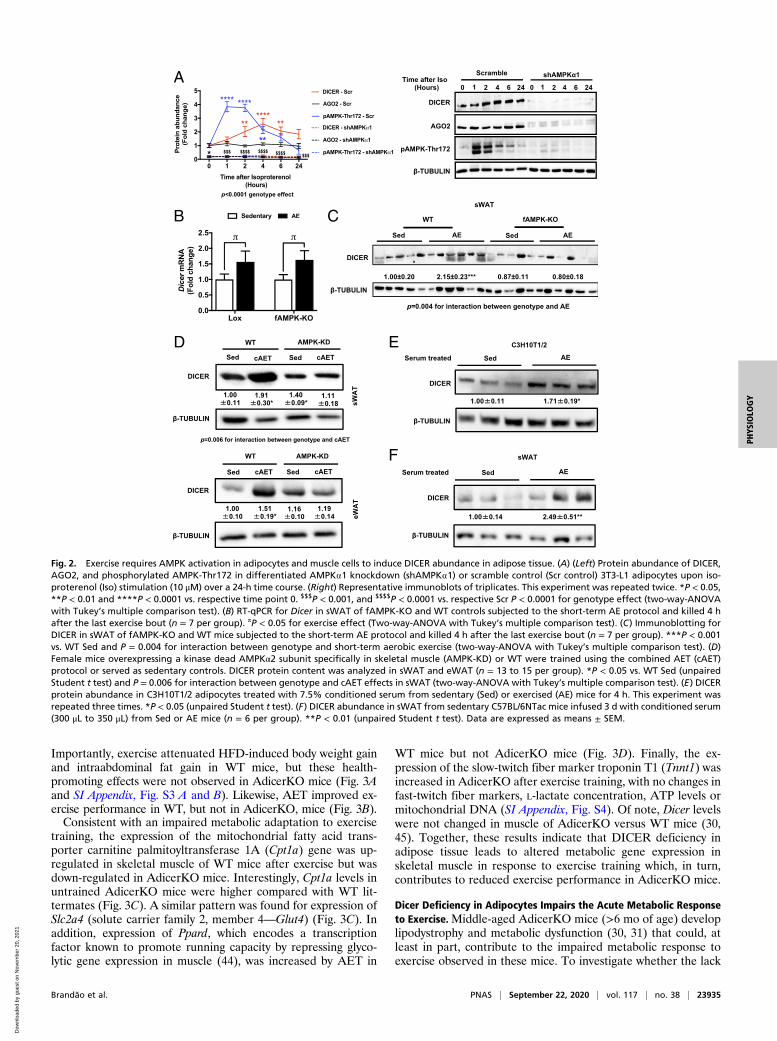
AMPK Signaling Up-Regulates DICER Levels in Adipocytes. Exercise isknown to activate AMPK in skeletal muscle (16, 17, 36, 37), andwe found this is also true in adipose tissue of acutely exercisedmice (SI Appendix, Fig. S2A). Since AMPK has been implicatedin DICER regulation (38, 39), we tested whether AMPK regu-lates DICER levels in adipose tissue, by feeding WT mice theAMPK activator metformin for 30 d. We found that abundance ofDICER in adipose tissue was up-regulated in comparison tovehicle-treated controls (SI Appendix, Fig. S2B). Likewise, treat-ment of 3T3-F442A adipocytes in vitro with metformin or theAMP mimetic AICAR increased Dicer mRNA expression by 2.7-and 3.4-fold, respectively (SI Appendix, Fig. S2C). Treatment of3T3-L1 adipocytes with the β-adrenergic agonist isoproterenolalso resulted in a rapid, transient increase in phosphorylation ofAMPK on Thr172 and its target acetyl-CoA carboxylase on Ser79,as well as a later increase in DICER abundance (Fig. 2A and SIAppendix, Fig. S2D). In contrast, basal and isoproterenol-inducedphosphorylation of ACC-Ser79 was lost in adipocytes withknockdown of AMPKα1 (Fig. 2A and SI Appendix, Fig. S2 D andE). Knockdown of AMPKα1 also markedly reduced DICER andAGO2 abundance and blocked isoproterenol-induced DICER up-regulation (Fig. 2A). Knockdown of AMPK α1 did not affect levelsof the DICER partner protein TRBP (SI Appendix, Fig. S2D).Thus, AMPK regulates DICER mRNA expression and proteinabundance in adipose tissue in a cell autonomous manner.
Exercise-Induced Increase in Adipose Tissue DICER Is Dependent onAMPK Activation in Adipocytes. To determine whether AMPK isrequired for the effect of AET on DICER levels in adipose tissuein vivo, we generated adipose-specific AMPKα1/2 knockout mice(fAMPK-KO) (SI Appendix, Fig. S2F), and subjected these miceto short-term exercise training. As above, DICER protein abun-dance was increased by 2.2-fold in WT mice after exercise, whilethis effect was abrogated in fAMPK-KO mice (Fig. 2C). Inter-estingly, this was associated with a 1.6-fold increase in DicermRNA in sWAT of both control and fAMPK-KO mice (Fig. 2B),indicating that AMPK regulates DICER abundance through aposttranscriptional mechanism. Consistent with the data inFig. 1 A and B, DICER protein abundance was not altered in BATor eWAT in response to short-term aerobic exercise, and the lossof AMPK did not affect DICER levels in these tissues (SI Ap-pendix, Fig. S2G). Together, these results demonstrate thatAMPK is important for exercise-induced DICER up-regulation ins.c. adipocytes in vivo and that this occurs posttranscriptionally.
Exercise-Induced Increase in Adipose Tissue DICER Is Dependent onAMPK Activation in Muscle. AMPK activation in skeletal and car-diac muscle is an important driver of metabolic changes thatoccur during exercise, which includes the release of a variety ofmolecules from muscle into the circulation (e.g., myokines orexerkines) (33, 40). To determine the role of AMPK signaling inmuscle for regulating DICER abundance in adipose tissue, miceoverexpressing a muscle-specific kinase-dead AMPKα2 isoform
Brandão et al. PNAS | September 22, 2020 | vol. 117 | no. 38 | 23933
PHYS
IOLO
GY
Dow
nloa
ded
by g
uest
on
Nov
embe
r 20
, 202
1

(AMPK-KD) in skeletal and cardiac muscles, which has beenshown to reduce AMPK activation by 70 to 99% (41), weresubjected to AET using a combination of running wheel andtreadmill exercise for a period of 6.5 wk. Despite attenuatedmuscle AMPK signaling and impaired metabolic response toexercise (42), running distance was similar between AMPK-KDmice and WT mice throughout the study (43). Consistent withour earlier observations, AET increased DICER 1.9-fold insWAT and 1.5-fold in eWAT in WT mice (Fig. 2D), but thisresponse was absent in AMPK-KD mice (Fig. 2D). There wereno changes in AMPK levels in adipose tissue of AMPK-KDversus WT mice or after exercise training (SI Appendix, Fig. S2 Hand I). Thus, the up-regulation of DICER in adipose tissue inresponse to AET requires AMPK activity in cardiac and skeletalmuscle. The notion that this increase in adipose tissue DICERwas secondary to a circulating factor released by muscle is sup-ported by the observation that differentiated adipocytes treatedin vitro for 4 h with serum from exercised mice exhibited a 1.7-fold increase in DICER protein (Fig. 2E). Moreover, DICERprotein was increased 2.5-fold in sWAT of sedentary WT mice
following infusion with serum from exercised mice for 3 d(Fig. 2F). Collectively, these data indicate that AET up-regulatesDICER in adipose tissue through a mechanism involving releaseof circulating molecules controlled by the muscle in an AMPK-dependent manner.
Lack of Dicer in Adipocytes Limits Performance and Alters MetabolicGene Expression in Skeletal Muscle in Response to AET in Mice. Toinvestigate whether Dicer expression in adipocytes is required forthe metabolic adaptation to exercise, mice with AdicerKO andWT littermates were subjected to AET while challenged with60% high-fat diet (HFD) (SI Appendix, Fig. S3A). As previouslydescribed (30), AdicerKO mice exhibited signs of lipodystrophywith smaller eWAT fat pads, no changes in sWAT, and a hy-pertrophic BAT (SI Appendix, Fig. S3B). AdicerKO mice alsohad higher levels of intramuscular triglycerides (SI Appendix, Fig.S3C). Despite these changes in body composition, daily foodintake was similar among the groups (SI Appendix, Fig. S3D),and there were no changes in resting VO2 consumption or VCO2production (SI Appendix, Fig. S3 E and F) after 8 wk of AET.
A B
C
E
D
Fig. 1. Exercise up-regulates DICER in adipose tissues. (A and B) Twelve-week-old mice were subjected to the short-term aerobic exercise (AE) protocol andkilled 4 h after the last bout. DICER mRNA expression (A) and protein content (B) were measured (n = 8 per group). *P < 0.05, **P < 0.01 (Unpaired Studentt test). (C and D) Twelve-week old mice were subjected to 8 wk of AET and killed at the end of the protocol after overnight fasting. DICER mRNA expression(C) and protein content (D) were measured (n = 7 per group). In (C), *P < 0.05 (unpaired Student t test). In (D), **P < 0.01 and ***P < 0.001 (unpaired Studentt test). (E) DICER protein abundance in human s.c. adipose tissue of seven 36 ± 11-y-old males (younger; black line) and nine 63 ± 6-y-old males (older; red line)pre- and post-6 wk of high-intensity interval training on a bicycle ergometer three times a week. Each high-intensity training session consisted of five 1-minintervals interspersed with 1.5-min rest. *P < 0.05 vs. pre (Paired Student t test). Data are expressed as means ± SEM.
23934 | www.pnas.org/cgi/doi/10.1073/pnas.2011243117 Brandão et al.
Dow
nloa
ded
by g
uest
on
Nov
embe
r 20
, 202
1
Importantly, exercise attenuated HFD-induced body weight gainand intraabdominal fat gain in WT mice, but these health-promoting effects were not observed in AdicerKO mice (Fig. 3Aand SI Appendix, Fig. S3 A and B). Likewise, AET improved ex-ercise performance in WT, but not in AdicerKO, mice (Fig. 3B).Consistent with an impaired metabolic adaptation to exercise
training, the expression of the mitochondrial fatty acid trans-porter carnitine palmitoyltransferase 1A (Cpt1a) gene was up-regulated in skeletal muscle of WT mice after exercise but wasdown-regulated in AdicerKO mice. Interestingly, Cpt1a levels inuntrained AdicerKO mice were higher compared with WT lit-termates (Fig. 3C). A similar pattern was found for expression ofSlc2a4 (solute carrier family 2, member 4—Glut4) (Fig. 3C). Inaddition, expression of Ppard, which encodes a transcriptionfactor known to promote running capacity by repressing glyco-lytic gene expression in muscle (44), was increased by AET in
WT mice but not AdicerKO mice (Fig. 3D). Finally, the ex-pression of the slow-twitch fiber marker troponin T1 (Tnnt1) wasincreased in AdicerKO after exercise training, with no changes infast-twitch fiber markers, L-lactate concentration, ATP levels ormitochondrial DNA (SI Appendix, Fig. S4). Of note, Dicer levelswere not changed in muscle of AdicerKO versus WT mice (30,45). Together, these results indicate that DICER deficiency inadipose tissue leads to altered metabolic gene expression inskeletal muscle in response to exercise training which, in turn,contributes to reduced exercise performance in AdicerKO mice.
Dicer Deficiency in Adipocytes Impairs the Acute Metabolic Responseto Exercise.Middle-aged AdicerKO mice (>6 mo of age) developlipodystrophy and metabolic dysfunction (30, 31) that could, atleast in part, contribute to the impaired metabolic response toexercise observed in these mice. To investigate whether the lack
A
B C
D E
F
Fig. 2. Exercise requires AMPK activation in adipocytes and muscle cells to induce DICER abundance in adipose tissue. (A) (Left) Protein abundance of DICER,AGO2, and phosphorylated AMPK-Thr172 in differentiated AMPK⍺1 knockdown (shAMPK⍺1) or scramble control (Scr control) 3T3-L1 adipocytes upon iso-proterenol (Iso) stimulation (10 μM) over a 24-h time course. (Right) Representative immunoblots of triplicates. This experiment was repeated twice. *P < 0.05,**P < 0.01 and ****P < 0.0001 vs. respective time point 0. $$$P < 0.001, and $$$$P < 0.0001 vs. respective Scr P < 0.0001 for genotype effect (two-way-ANOVAwith Tukey’s multiple comparison test). (B) RT-qPCR for Dicer in sWAT of fAMPK-KO and WT controls subjected to the short-term AE protocol and killed 4 hafter the last exercise bout (n = 7 per group). πP < 0.05 for exercise effect (Two-way-ANOVA with Tukey’s multiple comparison test). (C) Immunoblotting forDICER in sWAT of fAMPK-KO and WT mice subjected to the short-term AE protocol and killed 4 h after the last exercise bout (n = 7 per group). ***P < 0.001vs. WT Sed and P = 0.004 for interaction between genotype and short-term aerobic exercise (two-way-ANOVA with Tukey’s multiple comparison test). (D)Female mice overexpressing a kinase dead AMPKα2 subunit specifically in skeletal muscle (AMPK-KD) or WT were trained using the combined AET (cAET)protocol or served as sedentary controls. DICER protein content was analyzed in sWAT and eWAT (n = 13 to 15 per group). *P < 0.05 vs. WT Sed (unpairedStudent t test) and P = 0.006 for interaction between genotype and cAET effects in sWAT (two-way-ANOVA with Tukey’s multiple comparison test). (E) DICERprotein abundance in C3H10T1/2 adipocytes treated with 7.5% conditioned serum from sedentary (Sed) or exercised (AE) mice for 4 h. This experiment wasrepeated three times. *P < 0.05 (unpaired Student t test). (F) DICER abundance in sWAT from sedentary C57BL/6NTac mice infused 3 d with conditioned serum(300 μL to 350 μL) from Sed or AE mice (n = 6 per group). **P < 0.01 (unpaired Student t test). Data are expressed as means ± SEM.
Brandão et al. PNAS | September 22, 2020 | vol. 117 | no. 38 | 23935
PHYS
IOLO
GY
Dow
nloa
ded
by g
uest
on
Nov
embe
r 20
, 202
1

of Dicer in adipocytes affects the acute response to exercise priorto development of these metabolic derangements, 2-mo-oldcontrol and AdicerKO mice were subjected to a single bout ofstrenuous exercise, and metabolic function of Gas muscle andsWAT—a depot not affected in size or morphology by exerciseor Dicer deletion (SI Appendix, Fig. S3B and ref. 31)—wereassessed. Branched-chain amino acid (i.e., valine) oxidation wasincreased in muscle of WT mice by 11.5-fold in response to ex-ercise, whereas, in AdicerKO mice, this increase was muchsmaller (2.2-fold) and not significant (Fig. 4 A and B). In con-trast, in sWAT, valine oxidation was increased by exercise re-gardless of the genotype (SI Appendix, Fig. S5A). In both tissues,there was no change in palmitate oxidation (Fig. 4C and SI Ap-pendix, Fig. S5A), and glucose oxidation in sWAT was significantlylower in AdicerKOmice than in WTmice after exercise (Fig. 4D).These results demonstrate that Dicer deficiency in adipocytes al-ters the acute metabolic response to exercise in both skeletalmuscle and adipose tissue even in the absence of lipodystrophy.
Lack of Dicer in Adipocytes Promotes Adipose Tissue Glucose Metabolism.In contrast to the decrease in glucose oxidation observed in adiposetissue following acute exercise, AdicerKO mice exhibited increasedglucose uptake in eWAT and sWAT when compared to WT miceafter 8 wk of AET (Fig. 5A); this occurred with no changes inskeletal muscle glucose uptake (SI Appendix, Fig. S5B). There werealso higher L-lactate levels in the sWAT of AdicerKO mice aftertraining compared to WT mice (Fig. 5B) indicating increased an-aerobic glycolysis, but these changes were not sufficient to modify thecirculating levels of L-lactate (SI Appendix, Fig. S5C). ATP content insWAT was unaltered (Fig. 5C).In agreement with a metabolic shift toward anaerobic glycol-
ysis, five of the eight analyzed genes involved in key steps of
glycolysis were up-regulated in sWAT of AdicerKO mice as com-pared with controls (Fig. 5D). These included Slc2a1 (glucosetransporter 1 insulin-independent—GLUT1), Gpi1 (glucose-6-phosphate isomerase1), Pkm2 (pyruvate kinase isozymeM2), Pgam(phosphoglycerate mutase), and Ldha (lactate dehydrogenase A).Only Pkm1 (pyruvate kinase isozyme M1) was down-regulatedafter AET, and this occurred regardless of the genotype. In con-trast, glucose-6-phosphate dehydrogenase mRNA levels werelower in trained AdicerKO mice in comparison to trained WTmice, suggesting that flux through the pentose phosphate pathwayis inhibited in exercised AdicerKO mice (SI Appendix, Fig. S5D).Similarly, genes involved in mitochondrial biogenesis and function(e.g., Ppargc1a, Nrf1, and Mfn1) were down-regulated in sWAT ofAdicerKOmice, particularly in the trained group (SI Appendix, Fig.S5E). Despite these alterations, citrate synthase activity was in-creased in AdicerKO mice (SI Appendix, Fig. S5F), whereas mi-tochondrial DNA was unaltered (SI Appendix, Fig. S5G), indicatingthat the perturbations in mitochondrial function were unrelated tochanges in mitochondrial mass. Together, these results demon-strate that lack of DICER in adipocytes shifts cellular metabolismtoward anaerobic glycolysis, particularly after exercise training.
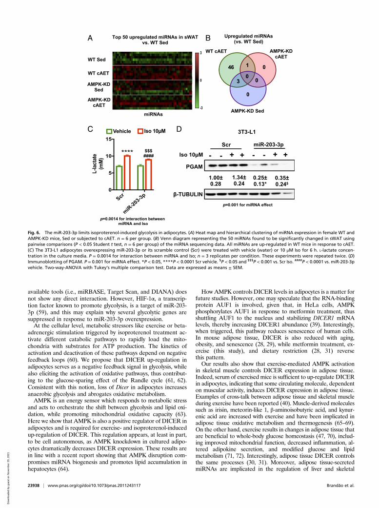
miR-203-3p Limits Glycolysis and Lactate Release in Adipocytes. Toidentify miRNAs that may coordinate these metabolic changes, weperformed small RNA sequencing in sWAT ofWT and AMPK-KDmice without and with AET. We chose this model since DICER isup-regulated by exercise training in sWAT of WT, but not AMPK-KD, mice, enabling us to detect the most DICER-sensitive miRNAs.Pair-wise comparisons revealed 50 differentially expressed miRNAs(P < 0.05) when comparing exercised mice to sedentary controls, ofwhich 46 were up-regulated by exercise in WT mice but not in
A B
C D
Fig. 3. AdicerKO mice are less responsive to AET. Mice were subjected to 13wk of HFD combined with the AET protocol or sedentary condition. (A)Cumulative body weight gain. *P < 0.05 vs. WT Sedentary. (B) Maximumspeed reached in the maximum effort test (Pre, before AET; Post, after AET).**P < 0.01 vs. WT Pre, $P < 0.05 vs. WT Post and P < 0.05 for interactionbetween genotype and AET effect. (C and D) RT-qPCR of mCpt1, Slc2a4, andPpard in Gas. WT mice (n = 6 per group) and AdicerKO (n = 4 per group).*P < 0.05, ***P < 0.001 vs. WT Sedentary. $$P < 0.01 vs. WT AET. #P < 0.05 vs.AdicerKO sedentary. Two-way-ANOVA with Tukey’s multiple comparisontest. Data are expressed as means ± SEM.
A B
WT AdicerKO0
1 10-5
2 10-5
3 10-5
4 10-5
mM
/mg
of ti
ssue
/h
WT AdicerKO0
2 10-5
4 10-5
6 10-5
mM
/mg
of ti
ssue
/h *
WT AdicerKO0.0
5.0 10-7
1.0 10-6
1.5 10-6
2.0 10-6
mM
/mg
of ti
ssue
/h
WT AdicerKO0.0
5.0 10-5
1.0 10-4
1.5 10-4
mM
/mg
of ti
ssue
/h
$
C D
Fig. 4. Skeletal muscle and sWAT metabolism is altered in exercised Adi-cerKO mice. Ex vivo (A and B) valine or (C) palmitate oxidation in gastroc-nemius skeletal muscle (Gas) and (D) glucose oxidation in sWAT of 12-wk oldmice subjected to one maximum effort test and killed at the end of thesession (Exe). Valine incorporation into alpha-ketoisovalerate (KIV), valine,palmitate, or glucose incorporation into CO2 was measured (n = 4 pergroup). *P < 0.05 vs. WT Sedentary and $P < 0.05 vs. WT Exe.Two-way-ANOVA with Tukey’s multiple comparison test. Data are expressedas means ± SEM.
23936 | www.pnas.org/cgi/doi/10.1073/pnas.2011243117 Brandão et al.
Dow
nloa
ded
by g
uest
on
Nov
embe
r 20
, 202
1

AMPK-KD mice (Fig. 6 A and B). One of these miRNAs was miR-203-3p, and we have previously observed that this miRNA is tightlycorrelated with Dicer expression in adipose tissue in the context ofAET (SI Appendix, Fig. S6A), caloric restriction, aging (28), andobesity (29).To further explore the role of miR-203-3p, we transfected
3T3-L1 preadipocytes with miR-203-3p mimic or a scramblecontrol, differentiated these cells into adipocytes, treated themwith isoproterenol for 6 h to activate AMPK, and measured theexpression of glycolytic genes and the level of L-lactate in themedia. Isoproterenol increased L-lactate production in scramblecontrol cells (Fig. 6C). This increase in L-lactate production afterisoproterenol treatment was reduced in adipocytes over-expressing miR-203-3p (Fig. 6D). This was also associated withup-regulation of Slc2a1, Pfkb3, and Ldha, and down-regulationof Gpi and Pgam mRNA (SI Appendix, Fig. S6B). We found a75% (miR-203-3p + Vehicle) and 64% (miR-203-3p + Isopro-terenol) decrease in PGAM protein content in cells over-expressing miR-203-3p (Fig. 6D), suggesting that miR-203-3pup-regulation limits lactate production by, at least in part, de-creasing the protein content of an enzyme present in the distalpart of glycolysis. Taken together, these data indicate that in-duction of DICER and miR-203-3p in adipose tissue in responseto exercise training is a key adaptive response to ensure appro-priate control of energy homeostasis of adipocytes.
DiscussionExercise training has beneficial effects on glucose and energyhomeostasis. Recent studies have shown that these positive ef-fects include both direct effects on muscle (46) and indirect ef-fects via changes in adipose (40, 47) and other tissues (48). In thepresent study, we show that part of these indirect effects involvesan effect of exercise training to up-regulate the miRNA pro-cessing enzyme DICER in adipose tissue of both humans and
mice, and this, in turn, leads to increased miRNA biogenesis,including up-regulation of miR-203-3p. This phenomenon isdependent on AMPK activation in both muscle and adiposetissue and involves circulating factors. Mice lacking the ability toup-regulate miRNAs in adipose tissue (i.e., AdicerKO) havereduced work performance in response to exercise training andexhibit metabolic alterations in skeletal muscle and adipose tis-sue. These results provide mechanistic insights and indicate thatadipose tissue’s ability to produce miRNAs plays a role in themetabolic adaptation to AET.Among the adipose tissue miRNAs affected by exercise
training, miR-203-3p appears to be highly sensitive to changes inthe level of DICER. Thus, in conditions where DICER levels areelevated, such as after exercise training (this study) and dietaryrestriction (28), miR-203-3p is up-regulated. In contrast, miR-203-3p is down-regulated in adipose tissue with aging (28) andobesity (29, 49), which are conditions where DICER is alsodown-regulated. Moreover, miR-203-3p and DICER have beenimplicated in brown fat differentiation and thermogenesis (30,49–51), suggesting that miR-203-3p is a key miRNA downstreamof DICER in adipocytes. The miR-203-3p also plays a role incancers where DICER levels are also low (52–54), and, in thesecells, miR-203 has been shown to suppress cell proliferation andmigration (55–57).Our results provide evidence that high expression of miR-203-
3p also inhibits isoproterenol-induced glycolysis in adipocytes.When miR-203-3p is overexpressed, lactate production and ex-pression of the glycolytic genes Gpi and Pgam are reduced inadipocytes. The impact of miR-203-3p on PGAM protein levelsis particularly robust. PGAM converts 3-phosphoglycerate to2-phosphoglycerate and thus serves as a bottleneck in glycolysisand the pentose phosphate pathway when inhibited (58).Whether PGAM is a direct target of miR-203-3p remains to bedetermined, but computational target prediction using online
WT AdicerKO0.0
5.0 10-9
1.0 10-8
1.5 10-8
ATP
(mM
/mg
of ti
ssue
)
WT
AdicerK
O WT
AdicerK
O WT
AdicerK
O WT
AdicerK
O WT
AdicerK
O WT
AdicerK
O WT
AdicerK
O WT
AdicerK
O0
2
4
6
8
mR
NA
expr
essi
on(F
old
chan
ge)
$$
&&
&&
&
&
Sedentary AET
WT AdicerKO WT AdicerKO WT AdicerKO0
100
200
300
400
In v
ivo
Glu
cose
Upt
ake
(nm
ol/g
of t
issu
e)
eWAT sWAT BAT
**$
&
WT AdicerKO0.0
0.1
0.2
0.3
0.4
L-la
ctat
e(m
M/m
g of
tiss
ue) ####
$$
A B
D
C
Fig. 5. Increased glycolysis in adipose tissue of trained AdicerKO mice. Male mice were subjected to HFD and 8 wk of AET or kept sedentary. WT mice (n = 6per group) and AdicerKO (n = 4 per group). (A) Adipose tissue in vivo glucose uptake. (B) L-lactate concentration in sWAT. (C) ATP concentration in sWAT. (D)RT-qPCR of glycolytic genes in sWAT. Ldha: P = 0.008 for interaction between genotype and AET. **P < 0.01 vs. WT sedentary, ####P < 0.0001 vs. AdicerKOsedentary. $P < 0.05 and $$P < 0.01 vs. WT AET. &P < 0.05 and &&P < 0.01 for genotype effect. πP < 0.05 for exercise effect. Two-way-ANOVA with Tukey’smultiple comparison test. Data are expressed as means ± SEM.
Brandão et al. PNAS | September 22, 2020 | vol. 117 | no. 38 | 23937
PHYS
IOLO
GY
Dow
nloa
ded
by g
uest
on
Nov
embe
r 20
, 202
1
available tools (i.e., miRBASE, Target Scan, and DIANA) doesnot show any direct interaction. However, HIF-1α, a transcrip-tion factor known to promote glycolysis, is a target of miR-203-3p (59), and this may explain why several glycolytic genes aresuppressed in response to miR-203-3p overexpression.At the cellular level, metabolic stressors like exercise or beta-
adrenergic stimulation triggered by isoproterenol treatment ac-tivate different catabolic pathways to rapidly load the mito-chondria with substrates for ATP production. The kinetics ofactivation and deactivation of these pathways depend on negativefeedback loops (60). We propose that DICER up-regulation inadipocytes serves as a negative feedback signal in glycolysis, whilealso eliciting the activation of oxidative pathways, thus contribut-ing to the glucose-sparing effect of the Randle cycle (61, 62).Consistent with this notion, loss of Dicer in adipocytes increasesanaerobic glycolysis and abrogates oxidative metabolism.AMPK is an energy sensor which responds to metabolic stress
and acts to orchestrate the shift between glycolysis and lipid oxi-dation, while promoting mitochondrial oxidative capacity (63).Here we show that AMPK is also a positive regulator of DICER inadipocytes and is required for exercise- and isoproterenol-inducedup-regulation of DICER. This regulation appears, at least in part,to be cell autonomous, as AMPK knockdown in cultured adipo-cytes dramatically decreases DICER expression. These results arein line with a recent report showing that AMPK disruption com-promises miRNA biogenesis and promotes lipid accumulation inhepatocytes (64).
How AMPK controls DICER levels in adipocytes is a matter forfuture studies. However, one may speculate that the RNA-bindingprotein AUF1 is involved, given that, in HeLa cells, AMPKphosphorylates AUF1 in response to metformin treatment, thusshuttling AUF1 to the nucleus and stabilizing DICER1 mRNAlevels, thereby increasing DICER1 abundance (39). Interestingly,when triggered, this pathway reduces senescence of human cells.In mouse adipose tissue, DICER is also reduced with aging,obesity, and senescence (28, 29), while metformin treatment, ex-ercise (this study), and dietary restriction (28, 31) reversethis pattern.Our results also show that exercise-mediated AMPK activation
in skeletal muscle controls DICER expression in adipose tissue.Indeed, serum of exercised mice is sufficient to up-regulate DICERin adipocytes, indicating that some circulating molecule, dependenton muscular activity, induces DICER expression in adipose tissue.Examples of cross-talk between adipose tissue and skeletal muscleduring exercise have been reported (40). Muscle-derived moleculessuch as irisin, meteorin-like 1, β-aminoisobutyric acid, and kynur-enic acid are increased with exercise and have been implicated inadipose tissue oxidative metabolism and thermogenesis (65–69).On the other hand, exercise results in changes in adipose tissue thatare beneficial to whole-body glucose homeostasis (47, 70), includ-ing improved mitochondrial function, decreased inflammation, al-tered adipokine secretion, and modified glucose and lipidmetabolism (71, 72). Interestingly, adipose tissue DICER controlsthe same processes (30, 31). Moreover, adipose tissue-secretedmiRNAs are implicated in the regulation of liver and skeletal
A
C D
B
Fig. 6. The miR-203-3p limits isoproterenol-induced glycolysis in adipocytes. (A) Heat map and hierarchical clustering of miRNA expression in female WT andAMPK-KD mice, Sed or subjected to cAET. n = 6 per group. (B) Venn diagram representing the 50 miRNAs found to be significantly changed in sWAT usingpairwise comparisons (P < 0.05 Student t test, n = 6 per group) of the miRNA sequencing data. All miRNAs are up-regulated in WT mice in response to cAET.(C) The 3T3-L1 adipocytes overexpressing miR-203-3p or its scramble control (Scr) were treated with vehicle (water) or 10 μM Iso for 6 h. L-lactate concen-tration in the culture media. P = 0.0014 for interaction between miRNA and Iso; n = 3 replicates per condition. These experiments were repeated twice. (D)Immunoblotting of PGAM. P = 0.001 for miRNA effect. *P < 0.05, ****P < 0.0001 Scr vehicle. $P < 0.05 and $$$P < 0.001 vs. Scr Iso. ####P < 0.0001 vs. miR-203-3pvehicle. Two-way-ANOVA with Tukey’s multiple comparison test. Data are expressed as means ± SEM.
23938 | www.pnas.org/cgi/doi/10.1073/pnas.2011243117 Brandão et al.
Dow
nloa
ded
by g
uest
on
Nov
embe
r 20
, 202
1
muscle function (73, 74). Although miR-203-3p has been found incirculating exosomes, and brown adipose tissue appears to be animportant contributor to the circulating pool of exosomal miR-203-3p (73), we do not see changes in serum miR-203-3p in WT orAdicerKO mice in response to the acute exercise training protocol,suggesting that circulating miR-203-3p itself does not play a role inintertissue communication after exercise. Whether other exercise-induced adipose tissue miRNAs mediate such communication is asubject for future studies.Here we propose a mechanism where changes in miRNA pro-
cessing in adipocytes affect substrate utilization by the adiposetissue and, in turn, influence skeletal muscle function and its re-sponse to exercise, as well as whole-body metabolism. Fatty acidsderived from adipose tissue normally contribute ∼50% of the totalenergy supply during moderate aerobic exercise in humans (75,76). This mechanism is driven by metabolic changes that modulatenutrient metabolism in adipose and skeletal muscle (5, 44, 77). Forexample, controlled glucose metabolism in muscle helps delayhypoglycemia after strenuous exercise and increases exerciseperformance in mice (44) and humans (5, 77). Moreover, rats withan increased capacity of skeletal muscle to shift from glucose tolipid or amino acid oxidation during exercise can run 3 timeslonger (3). In contrast, exercise performance is impaired in micewith abrogated adipose tissue lipolysis (78–80). Likewise, whenskeletal muscle is forced to use lipids as the exclusive energysource, incomplete organic acid intermediates can accumulate inthe mitochondria, decreasing the energy influx to the trichloro-acetic acid cycle (TCA) (81), which will impair exercise perfor-mance (82). When DICER levels are low in adipocytes, increasedadipose tissue anaerobic glycolysis is expected to increase glucoseuptake from the blood and decrease lipid utilization. The latterwould then remain in circulation to be used as substrate for oxi-dation or accumulation in other tissues. Consistent with this no-tion, free fatty acids are increased in the blood stream ofAdicerKO mice (30). Moreover, AdicerKO mice exhibit ectopicaccumulation of triglycerides in the skeletal muscle (SI Appendix,Fig. S3C). Thus, skeletal muscle of mice lacking DICER in adi-pocytes exhibits changes consistent with an impaired metabolicresponse to exercise training, including reduced capacity forbranched-chain amino acid oxidation. Altogether, these alterationsin skeletal muscle and adipose tissue create a scenario of metabolicinflexibility to limit performance and exercise adaptation.Collectively, our results demonstrate a form of cross-talk be-
tween adipose tissue and muscle during aerobic exercise whichinvolves adipose tissue miRNAs. Under metabolic stress, AMPKis activated in both of these tissues, and, in adipose tissue, thisleads to up-regulation of DICER. When DICER is up-regulated,miRNAs are generated to counterbalance adipose tissue energydemands and metabolism. For example, higher levels of miR-203-3p in adipocytes inhibit glycolysis induced by energy-demanding conditions, such as β-adrenergic stimulus. This will,in part, confer adequate substrate availability for skeletal muscleduring and after exercise, thus allowing the metabolic flexibilityrequired for adaptation to exercise training.
MethodsMouse Models.Male mice were used in all cases unless stated otherwise. Micewere maintained at a 12-h light−dark cycle with ad libitum access to tapwater and chow or 60% HFD (D12492; Research Diets). Protocols for animaluse in Brazil were approved by the IACUC of the Universidade Federal de SãoPaulo (CEP-0218/11, CEP-0237/12, and CEUA4603261015) and UniversidadeEstadual de Campinas (4759-1/2017 and 4749-1/2017) and were in accor-dance with NIH guidelines. For animal use in Denmark, experiments wereperformed in accordance with the European Directive 2010/63/EU of theEuropean Parliament and of the Council for the protection of animals usedfor scientific purposes. Ethical approval was given by the Danish AnimalExperiments Inspectorate (#2012-15-2934-00026). All mice were housed inour animal facilities for at least 1 wk prior to the beginning of the experi-ments. Details are in SI Appendix.
High-Intensity Interval Training (Human Exercise Protocol). Human participantswere exercised as previously described (83). In short, two groups of men, ayounger group (average age 36 ± 11 y, n = 7) and an older group (63 ± 6 y,n = 9) underwent 6 wk of high-intensity interval training on a bicycleergometer three times a week. Each high-intensity training session consistedof five 1-min intervals interspersed with 1.5-min rest. The two groups ofparticipants were exercised in two independent studies (83, 84). The studywas approved by the Ethical Committee of Copenhagen (journal no. H-3-2012-024) and complied with the Danish Data Protection Agency and theguidelines of the Helsinki Declaration.
AET Protocol. Training was performed at 60% of the maximum speedobtained in the maximum effort test, for 1 h·d−1, 5 d per week for 8 wk. Micewere killed at the end of the protocol after overnight fasting, and tissueswere harvested, snap-frozen in liquid nitrogen, and stored at −80 °C untilfurther use. Details are in SI Appendix.
Short-Term Exercise Training Protocol. On day 1, mice ran for 2 min at 8 m/minand 3 min at 10 m/min as warm-up. Then they ran for 40 min at 14 m/min andfinished at 8 m/min for the last 5 min as cooldown (0% inclination). On day 2,the treadmill was raised to a 5% inclination. Mice ran for 5 min at 10 m/min,then 40 min at 14 m/min, and 5 min at 8 m/min. On day 3, the treadmill wasraised to a 10% inclination. Mice ran for 5 min at 10 m/min, then 40 min at16 m/min, and 5 min at 8 m/min. All mice were killed 4 h after the last boutof exercise, and tissues were snap-frozen and stored at −80 °C. Details are inSI Appendix.
Combined AET.Mice were exercised for 6.5 wk in a combination of free accessto running wheel and 1 h·d−1, 5 d per week of 16 m/min treadmill running(Columbus Instruments), as described earlier (43). Details are in SI Appendix.
Single Bout of Exercise Protocol. Eight-week-old mice were subjected to asingle bout of exercise on a treadmill for 1 h at 16 m/min with 5% incline.Mice were killed right after exercise (0 h) or 4 h, 10 h, or 24 h after treadmillrunning. Details are in SI Appendix.
In Vivo Serum Supplementation. Serum (300 μL to 350 μL) was infused at aninfusion rate at 30 μL/min for three consecutive days. On day 1, serum frommice subjected to one bout of exercise or sedentary controls was infused,on day 2, serum from mice subjected to two bouts of exercise or sedentarycontrols was infused, and, on day 3, serum from mice subjected to threebouts of exercise or sedentary controls was infused. Mice were killed 4 hafter the last infusion, and tissues were harvested, snap-frozen in liquid ni-trogen, and stored at −80 °C until further use. Details are in SI Appendix.
In Vitro Serum Supplementation. C3H10T1/2 cells differentiated into adipo-cytes were treated for 4 h with 7.5%mouse serum in complete media. Detailsare in SI Appendix.
Metabolic Phenotyping. Oxygen consumption and carbon dioxide productionwere measured in fed animals through a computer-controlled, open-circuitcalorimeter system (LE405 gas analyzer; Panlab-Harvard Apparatus). Detailsare in SI Appendix.
Glucose Tolerance. Mice were injected intraperitoneally with glucose (1 g/kgbody weight; Sigma-Aldrich) after overnight fasting. Blood samples werecollected at the indicated time points through a small cut at the tail tip, andglucose levels were measured using a glucometer (Accu-Chek, Roche).
Substrate Utilization and Citrate Synthase Activity. Citrate synthase activityand valine, glucose, and palmitic acid conversion into CO2 were performed aspreviously described (31).
In Vivo Glucose Uptake. Eight-week-old mice were given a 60% HFD 4 wkprior to the beginning of the training protocol and were maintained on thesame diet until sacrifice. When 12 wk old, mice were subjected to the AETprotocols as described previously. After overnight fasting, mice were intra-peritoneally injected with [3H]-2-deoxy-D-glucose in phosphate-bufferedsaline solution (0.15 μCi per gram of body weight), and, 30 min later, ani-mals were killed by decapitation, blood samples were collected, and tissueswere weighted and snap-frozen. Tissues were digested in 5 M NaOH solutionfor 1 h at 70 °C, and a sample was used to determine [3H]-2-deoxy-D-glucoselevels using the Tri-Carb 4910TR liquid scintillation counter. Glucose uptake
Brandão et al. PNAS | September 22, 2020 | vol. 117 | no. 38 | 23939
PHYS
IOLO
GY
Dow
nloa
ded
by g
uest
on
Nov
embe
r 20
, 202
1
was calculated using blood glucose and [3H]-2-deoxy-D-glucose levels as areference and normalized by grams of tissue.
Lactate and ATP Analysis. Tomeasure the level of circulating lactate, 24 h afterthe last training section, we collected 10 μL of blood in a tube containing150 μL of 4% trichloroacetic acid. Details on lactate and ATP quantificationin blood and cells are in SI Appendix.
Triglycerides Analysis. For tissue triglyceride measurement, we used the Abcamtriglyceride quantification kit (ab65336) according to the manufacturer’s protocol.
Cell Culture.Cell maintenance. Cells were maintained in Dulbecco’s modified Eagle’s me-dium (DMEM—4.5g/L glucose) growth medium supplemented with 10%fetal bovine serum (FBS) and 1% of penicillin/streptomycin (P/S) at 37 °C and5% CO2. Stable AMPKα1 knockdown cell generated by lentivirus.Adipocyte differentiation. Confluent preadipocytes were given DMEM con-taining 10% FBS, isobutylmethylxanthine (500 μM), dexamethasone (1 μM),insulin (500 nM), and rosiglitazone (1 μM). Two days after induction, cellswere switched to the maintenance medium containing 10% FBS, insulin(500 nM), and rosiglitazone (1 μM) for 4 d. C3H10T1/2 adipocyte differen-tiation was induced according to ref. 85.AMPK activation and inhibition. To activateAMPK, cellswere treatedwith 1mMofAICAR (5-Aminoimidazole-4-carboxamide 1-β-D-ribofuranoside, Acadesine, N1-(β-D-Ribofuranosyl)-5-aminoimidazole-4-carboxamide; Sigma A9978) diluted inDMSO, or 0.5 mM metformin (Sigma, D150959) or 10 μM isoproterenol (Sigma,I6504) diluted in autoclaved MilliQ water. For AMPK inhibition, cells weretreated with 50 μM of dorsomorphin/compound C (Abcam, ab120843).miRNA-203-3p overexpression. The 3T3-L1 preadipocytes were transfected at100% confluence with 50 nM of mouse miR-203-3p mimic or the scramblecontrol (abmGood; Applied Biological Materials). Details are in SI Appendix.
miRNA Sequencing.NEBNext Multiplex Small RNA library Prep Set for Illuminakit (#E7300) was used for small RNA library preparation after the manu-facturer’s instruction. Raw data are deposited at Gene Expression Omnibus(GEO) (accession number GSE115408). Details are in SI Appendix.
Western Blot Analyses.Western blot analyseswere performed as described (86).The following primary antibodies were used: DICER (Ab13502 for detection of
mouse DICER and Ab14601 for human DICER) and TRBP (ab157812) fromAbcam; AGO2 (2997), total AMPKα1/2 (2532), pAMPK (2532), and β-tubulin(2146) from Cell Signaling; total ACC (streptavidin/HRP, P039701-2) from Agi-lent; pACC-Ser79 (07-303) from Millipore; and PGAM1/4 (D-5) (sc-365677) fromSanta Cruz. Mini-Protean TGX Stain-Free Gels (#456–8086, Bio-Rad) were usedfor quantification of total protein in human adipose tissue samples.
Gene Expression. Gene expression analysis was performed by qPCR as de-scribed (30). Details are in SI Appendix.
Statistics. Results are expressed as the mean ± SEM (SEM) unless indicatedotherwise. Normal distribution was tested using a Shapiro-Wilk test. Weused Student’s t test to compare two independent groups, one-way ANOVAwas used to compare more than two groups, and two-way ANOVA followedby Tukey’s multiple comparison test was used when data had more than onecategorical independent variable (such as genotype, time and/or exercise).Statistical analyses were run in Graph Prism and R. Statistical significance wasconsidered when P < 0.05.
Data Availability. Raw sequencing data are deposited at GEO (accession no.GSE115408). All study data are included in the text and SI Appendix.
ACKNOWLEDGMENTS. We thank Elzira E. Saviani (University of Campinas)for technical support, Erik A. Richter and Jørgen F. P. Wojtaszewski (Univer-sity of Copenhagen) for providing access to AMPK-KD mice and for criticalintellectual advice, and Arthur F. Gáspari (University of Campinas) for valu-able discussions on exercise physiology. This work was, in part, funded byFundação de Amparo à Pesquisa do Estado de São Paulo (FAPESP) (Grants2017/01184-9, 2017/07975-8, 2018/21635-8, 2017/03423-0, 2017/04377-2,2015/01316-7, 2015/03292-8, 2012/04079-8, 2012/06238-6, and 2010/52557-0) and Conselho Nacional de Desenvolvimento Científico e Tecnológico(CNPq) (Grants 444424/2014-8 and 207365/2014-8) and NIH GrantR01DK082659. The Lundbeck Foundation (Grant R170-2014-1316) and theDanish Diabetes Academy provided funding for S.M., and J.T.T. was sup-ported by Novo Nordisk Foundation (NNF) Excellence Project AwardNNF14OC0009315. J.L.B. was supported by a Research and Professional De-velopment Grant by Gettysburg College. Support for this study was alsoprovided by the NNF Center for Basic Metabolic Research (NNF-CBMR).NNF-CBMR is an independent Research Center (cbmr.ku.dk) at the Universityof Copenhagen and partially funded by an unrestricted donation (GrantNNF18CC0034900) from the NNF.
1. B. H. Goodpaster, L. M. Sparks, Metabolic flexibility in health and disease. Cell Metab.25, 1027–1036 (2017).
2. G. D. Cartee, R. T. Hepple, M. M. Bamman, J. R. Zierath, Exercise promotes healthyaging of skeletal muscle. Cell Metab. 23, 1034–1047 (2016).
3. K. A. Overmyer et al., Maximal oxidative capacity during exercise is associated withskeletal muscle fuel selection and dynamic changes in mitochondrial protein acety-lation. Cell Metab. 21, 468–478 (2015).
4. T. S. Higa, A. V. Spinola, M. H. Fonseca-Alaniz, F. S. Evangelista, Remodeling of whiteadipose tissue metabolism by physical training prevents insulin resistance. Life Sci.103, 41–48 (2014).
5. J. O. Holloszy, F. W. Booth, Biochemical adaptations to endurance exercise in muscle.Annu. Rev. Physiol. 38, 273–291 (1976).
6. E. A. Richter, L. P. Garetto, M. N. Goodman, N. B. Ruderman, Muscle glucose me-tabolism following exercise in the rat: Increased sensitivity to insulin. J. Clin. Invest. 69,785–793 (1982).
7. K. J. Mikines, B. Sonne, P. A. Farrell, B. Tronier, H. Galbo, Effect of training on thedose-response relationship for insulin action in men. J. Appl. Physiol. 66, 695–703(1989).
8. K. J. Rodnick, W. L. Haskell, A. L. Swislocki, J. E. Foley, G. M. Reaven, Improved insulinaction in muscle, liver, and adipose tissue in physically trained human subjects. Am.J. Physiol. 253, E489–E495 (1987).
9. M. L. Johnson et al., Twelve weeks of endurance training increases FFA mobilizationand reesterification in postmenopausal women. J. Appl. Physiol. 109, 1573–1581(2010).
10. R. R. Wolfe, S. Klein, F. Carraro, J. M. Weber, Role of triglyceride-fatty acid cycle incontrolling fat metabolism in humans during and after exercise. Am. J. Physiol. 258,E382–E389 (1990).
11. B. Kiens, Skeletal muscle lipid metabolism in exercise and insulin resistance. Physiol.Rev. 86, 205–243 (2006).
12. D. E. Kelley, L. J. Mandarino, Fuel selection in human skeletal muscle in insulin re-sistance: A reexamination. Diabetes 49, 677–683 (2000).
13. S. V. Ramos, P. C. Turnbull, R. E. MacPherson, Adipose tissue depot specific differencesof PLIN protein content in endurance trained rats. Adipocyte 5, 212–223 (2016).
14. S. J. Mancini et al., Activation of AMP-activated protein kinase rapidly suppressesmultiple pro-inflammatory pathways in adipocytes including IL-1 receptor-associatedkinase-4 phosphorylation. Mol. Cell. Endocrinol. 440, 44–56 (2017).
15. D. G. Hardie, AMPK − Sensing energy while talking to other signaling pathways. CellMetab. 20, 939–952 (2014).
16. M. J. Watt et al., Regulation of HSL serine phosphorylation in skeletal muscle andadipose tissue. Am. J. Physiol. Endocrinol. Metab. 290, E500–E508 (2006).
17. H. Park et al., Coordinate regulation of malonyl-CoA decarboxylase, sn-glycerol-3-phosphate acyltransferase, and acetyl-CoA carboxylase by AMP-activated protein ki-nase in rat tissues in response to exercise. J. Biol. Chem. 277, 32571–32577 (2002).
18. N. J. Hoffman et al., Global phosphoproteomic analysis of human skeletal musclereveals a network of exercise-regulated kinases and AMPK substrates. Cell Metab. 22,922–935 (2015).
19. H. Yun et al., AMP-activated protein kinase mediates the antioxidant effects of re-sveratrol through regulation of the transcription factor FoxO1. FEBS J. 281, 4421–4438(2014).
20. Y. C. Long, J. R. Zierath, AMP-activated protein kinase signaling in metabolic regu-lation. J. Clin. Invest. 116, 1776–1783 (2006).
21. C. Cantó et al., AMPK regulates energy expenditure by modulating NAD+ metabolismand SIRT1 activity. Nature 458, 1056–1060 (2009).
22. C. N. Miller et al., Isoproterenol increases uncoupling, glycolysis, and markers ofbeiging in mature 3T3-L1 adipocytes. PLoS One 10, e0138344 (2015).
23. M. Schweiger et al., Adipose triglyceride lipase and hormone-sensitive lipase are themajor enzymes in adipose tissue triacylglycerol catabolism. J. Biol. Chem. 281,40236–40241 (2006).
24. T. S. Nielsen, N. Jessen, J. O. Jørgensen, N. Møller, S. Lund, Dissecting adipose tissuelipolysis: Molecular regulation and implications for metabolic disease. J. Mol. Endo-crinol. 52, R199–R222 (2014).
25. L. Reshef et al., Glyceroneogenesis and the triglyceride/fatty acid cycle. J. Biol. Chem.278, 30413–30416 (2003).
26. M. S. Gauthier et al., AMP-activated protein kinase is activated as a consequence oflipolysis in the adipocyte: Potential mechanism and physiological relevance. J. Biol.Chem. 283, 16514–16524 (2008).
27. B. Omar, E. Zmuda-Trzebiatowska, V. Manganiello, O. Göransson, E. Degerman,Regulation of AMP-activated protein kinase by cAMP in adipocytes: Roles for phos-phodiesterases, protein kinase B, protein kinase A, Epac and lipolysis. Cell. Signal. 21,760–766 (2009).
28. M. A. Mori et al., Role of microRNA processing in adipose tissue in stress defense andlongevity. Cell Metab. 16, 336–347 (2012).
29. M. Oliverio et al., Dicer1-miR-328-Bace1 signalling controls brown adipose tissuedifferentiation and function. Nat. Cell Biol. 18, 328–336 (2016).
30. M. A. Mori et al., Altered miRNA processing disrupts brown/white adipocyte deter-mination and associates with lipodystrophy. J. Clin. Invest. 124, 3339–3351 (2014).
23940 | www.pnas.org/cgi/doi/10.1073/pnas.2011243117 Brandão et al.
Dow
nloa
ded
by g
uest
on
Nov
embe
r 20
, 202
1

31. F. C. Reis et al., Fat-specific Dicer deficiency accelerates aging and mitigates severaleffects of dietary restriction in mice. Aging (Albany NY) 8, 1201–1222 (2016).
32. A. P. Russell et al., Regulation of miRNAs in human skeletal muscle following acuteendurance exercise and short-term endurance training. J. Physiol. 591, 4637–4653(2013).
33. A. Safdar, A. Saleem, M. A. Tarnopolsky, The potential of endurance exercise-derivedexosomes to treat metabolic diseases. Nat. Rev. Endocrinol. 12, 504–517 (2016).
34. H. M. O’Neill et al., AMP-activated protein kinase (AMPK) beta1beta2 muscle nullmice reveal an essential role for AMPK in maintaining mitochondrial content andglucose uptake during exercise. Proc. Natl. Acad. Sci. U.S.A. 108, 16092–16097 (2011).
35. O. M. Palacios et al., Diet and exercise signals regulate SIRT3 and activate AMPK andPGC-1alpha in skeletal muscle. Aging (Albany NY) 1, 771–783 (2009).
36. N. B. Ruderman et al., AMPK as a metabolic switch in rat muscle, liver and adiposetissue after exercise. Acta Physiol. Scand. 178, 435–442 (2003).
37. J. B. Birk, J. F. Wojtaszewski, Predominant alpha2/beta2/gamma3 AMPK activationduring exercise in human skeletal muscle. J. Physiol. 577, 1021–1032 (2006).
38. G. Blandino et al., Metformin elicits anticancer effects through the sequential mod-ulation of DICER and c-MYC. Nat. Commun. 3, 865 (2012).
39. N. Noren Hooten et al., Metformin-mediated increase in DICER1 regulates microRNAexpression and cellular senescence. Aging Cell 15, 572–581 (2016).
40. K. I. Stanford, L. J. Goodyear, Muscle-adipose tissue cross talk. Cold Spring Harb.Perspect. Med. 8, a029801 (2018).
41. J. Mu, J. T. Brozinick Jr., O. Valladares, M. Bucan, M. J. Birnbaum, A role for AMP-activated protein kinase in contraction- and hypoxia-regulated glucose transport inskeletal muscle. Mol. Cell 7, 1085–1094 (2001).
42. R. S. Lee-Young et al., Skeletal muscle AMP-activated protein kinase is essential forthe metabolic response to exercise in vivo. J. Biol. Chem. 284, 23925–23934 (2009).
43. J. Brandauer et al., AMP-activated protein kinase regulates nicotinamide phosphor-ibosyl transferase expression in skeletal muscle. J. Physiol. 591, 5207–5220 (2013).
44. W. Fan et al., PPARδ promotes running endurance by preserving glucose. Cell Metab.25, 1186–1193.e4 (2017).
45. K. Y. Lee et al., Lessons on conditional gene targeting in mouse adipose tissue. Dia-betes 62, 864–874 (2013).
46. J. A. Hawley, M. Hargreaves, M. J. Joyner, J. R. Zierath, Integrative biology of exercise.Cell 159, 738–749 (2014).
47. H. Takahashi et al., TGF-β2 is an exercise-induced adipokine that regulates glucoseand fatty acid metabolism. Nat. Metab. 1, 291–303 (2019).
48. B. K. Pedersen, Physical activity and muscle-brain crosstalk. Nat. Rev. Endocrinol. 15,383–392 (2019).
49. X. Guo et al., cAMP-MicroRNA-203-IFNγ network regulates subcutaneous white fatbrowning and glucose tolerance. Mol. Metab. 28, 36–47 (2019).
50. H. J. Kim et al., MicroRNAs are required for the feature maintenance and differen-tiation of brown adipocytes. Diabetes 63, 4045–4056 (2014).
51. B. A. Guerra et al., Dietary sulfur amino acid restriction upregulates DICER to conferbeneficial effects. Mol. Metab. 29, 124–135 (2019).
52. Y. Karube et al., Reduced expression of Dicer associated with poor prognosis in lungcancer patients. Cancer Sci. 96, 111–115 (2005).
53. R. Ueda et al., Dicer-regulated microRNAs 222 and 339 promote resistance of cancercells to cytotoxic T-lymphocytes by down-regulation of ICAM-1. Proc. Natl. Acad. Sci.U.S.A. 106, 10746–10751 (2009).
54. V. Swahari, A. Nakamura, M. Deshmukh, The paradox of dicer in cancer. Mol. Cell.Oncol. 3, e1155006 (2016).
55. G. Zhao et al., miR-203 functions as a tumor suppressor by inhibiting epithelial tomesenchymal transition in ovarian cancer. J. Cancer Sci. Ther. 7, 34–43 (2015).
56. D. Pal et al., Regulation of cell proliferation and migration by miR-203 via GAS41/miR-10b Axis in human glioblastoma cells. PLoS One 11, e0159092 (2016).
57. Y. Zhou et al., miR-203 enhances let-7 biogenesis by targeting LIN28B to suppresstumor growth in lung cancer. Sci. Rep. 7, 42680 (2017).
58. T. Hitosugi et al., Phosphoglycerate mutase 1 coordinates glycolysis and biosynthesisto promote tumor growth. Cancer Cell 22, 585–600 (2012).
59. N. Han, H. Xu, N. Yu, Y. Wu, L. Yu, MiR-203a-3p inhibits retinal angiogenesis andalleviates proliferative diabetic retinopathy in oxygen-induced retinopathy (OIR) ratmodel via targeting VEGFA and HIF-1α. Clin. Exp. Pharmacol. Physiol. 47, 85–94 (2020).
60. D. M. Muoio, P. D. Neufer, Lipid-induced mitochondrial stress and insulin action inmuscle. Cell Metab. 15, 595–605 (2012).
61. L. Hue, H. Taegtmeyer, The Randle cycle revisited: A new head for an old hat. Am.J. Physiol. Endocrinol. Metab. 297, E578–E591 (2009).
62. N. M. Held et al., Pyruvate dehydrogenase complex plays a central role in brownadipocyte energy expenditure and fuel utilization during short-term beta-adrenergicactivation. Sci. Rep. 8, 9562 (2018).
63. M. D. Chau, J. Gao, Q. Yang, Z. Wu, J. Gromada, Fibroblast growth factor 21 regulatesenergy metabolism by activating the AMPK-SIRT1-PGC-1alpha pathway. Proc. Natl.Acad. Sci. U.S.A. 107, 12553–12558 (2010).
64. J. Latorre et al., Compounds that modulate AMPK activity and hepatic steatosis im-pact the biosynthesis of microRNAs required to maintain lipid homeostasis in hepa-tocytes. EBioMedicine 53, 102697 (2020).
65. L. D. Roberts et al., β-Aminoisobutyric acid induces browning of white fat and hepaticβ-oxidation and is inversely correlated with cardiometabolic risk factors. Cell Metab.19, 96–108 (2014).
66. P. Boström et al., A PGC1-α-dependent myokine that drives brown-fat-like develop-ment of white fat and thermogenesis. Nature 481, 463–468 (2012).
67. L. Z. Agudelo et al., Skeletal muscle PGC-1α1 modulates kynurenine metabolism andmediates resilience to stress-induced depression. Cell 159, 33–45 (2014).
68. L. Z. Agudelo et al., Kynurenic acid and Gpr35 regulate adipose tissue energy ho-meostasis and inflammation. Cell Metab. 27, 378–392.e5 (2018).
69. R. R. Rao et al., Meteorin-like is a hormone that regulates immune-adipose interac-tions to increase beige fat thermogenesis. Cell 157, 1279–1291 (2014).
70. K. I. Stanford et al., A novel role for subcutaneous adipose tissue in exercise-inducedimprovements in glucose homeostasis. Diabetes 64, 2002–2014 (2015).
71. F. J. May et al., Lipidomic adaptations in white and brown adipose tissue in responseto exercise demonstrate molecular species-specific remodeling. Cell Rep. 18,1558–1572 (2017).
72. A. C. Lehnig, K. I. Stanford, Exercise-induced adaptations to white and brown adiposetissue. J. Exp. Biol. 221 (Pt, suppl. 1), 221 (2018).
73. T. Thomou et al., Adipose-derived circulating miRNAs regulate gene expression inother tissues. Nature 542, 450–455 (2017).
74. W. Ying et al., Adipose tissue macrophage-derived exosomal miRNAs can modulatein vivo and in vitro insulin sensitivity. Cell 171, 372–384.e12 (2017).
75. B. Mittendorfer, J. F. Horowitz, S. Klein, Effect of gender on lipid kinetics duringendurance exercise of moderate intensity in untrained subjects. Am. J. Physiol. En-docrinol. Metab. 283, E58–E65 (2002).
76. J. A. Romijn et al., Regulation of endogenous fat and carbohydrate metabolism inrelation to exercise intensity and duration. Am. J. Physiol. 265, E380–E391 (1993).
77. J. O. Holloszy, Adaptations of muscular tissue to training. Prog. Cardiovasc. Dis. 18,445–458 (1976).
78. E. Huijsman et al., Adipose triacylglycerol lipase deletion alters whole body energymetabolism and impairs exercise performance in mice. Am. J. Physiol. Endocrinol.Metab. 297, E505–E513 (2009).
79. J. J. Dubé et al., Adipose triglyceride lipase deletion from adipocytes, but not skeletalmyocytes, impairs acute exercise performance in mice. Am. J. Physiol. Endocrinol.Metab. 308, E879–E890 (2015).
80. N. J. Zachwieja et al., Loss of adipocyte VEGF impairs endurance exercise capacity inmice. Med. Sci. Sports Exerc. 47, 2329–2339 (2015).
81. T. R. Koves et al., Mitochondrial overload and incomplete fatty acid oxidation con-tribute to skeletal muscle insulin resistance. Cell Metab. 7, 45–56 (2008).
82. T. R. Koves et al., Peroxisome proliferator-activated receptor-gamma co-activator1alpha-mediated metabolic remodeling of skeletal myocytes mimics exercise train-ing and reverses lipid-induced mitochondrial inefficiency. J. Biol. Chem. 280,33588–33598 (2005).
83. D. Søgaard et al., High-intensity interval training improves insulin sensitivity in olderindividuals. Acta Physiol. (Oxf.) 222, e13009 (2018).
84. S. Larsen et al., The effect of high-intensity training on mitochondrial fat oxidation inskeletal muscle and subcutaneous adipose tissue. Scand. J. Med. Sci. Sports 25,e59–e69 (2015).
85. M. Lundh, K. Pluciñska, M. S. Isidor, P. S. S. Petersen, B. Emanuelli, Bidirectional ma-nipulation of gene expression in adipocytes using CRISPRa and siRNA. Mol. Metab. 6,1313–1320 (2017).
86. J. Brandauer et al., AMP-activated protein kinase controls exercise training- andAICAR-induced increases in SIRT3 and MnSOD. Front. Physiol. 6, 85 (2015).
Brandão et al. PNAS | September 22, 2020 | vol. 117 | no. 38 | 23941
PHYS
IOLO
GY
Dow
nloa
ded
by g
uest
on
Nov
embe
r 20
, 202
1