Triggering of erythropoietin production by hypoxia is inhibited by respiratory and metabolic acidosis
KAI-UWE ECKARDT, ARMIN KURTZ, AND CHRISTIAN BAUER Department of Physiology, University of Zurich, CH-8057 Zurich, Switzerland
ECKARDT, KAI-UWE, ARMIN KURTZ, AND CHRISTIAN BAUER. Triggering of erythropoietin production by hypoxia is inhibited by respiratory and metabolic acidosis. Am. J. Physiol. 258 (Regulatory Integrative Comp. Physiol. 27): R678-R683, 1990.-Erythropoietin (EPO) production in response to hy- poxic hypoxia is known to be attenuated by simultaneous hypercapnia. This study aimed to investigate whether this inhibitory effect of hypercapnia is 1) a direct effect of carbon dioxide or mediated by changes in pH or bicarbonate, 2) affects also carbon monoxide hypoxia, and 3) influences either the synthesis and release of EPO or the mechanisms by which hypoxia triggers an increase in EPO production rate. We found that EPO formation in mice exposed to normobaric hypoxia (8% 02) or to carbon monoxide (0.1%) was reduced by 30 and 42% when animals were simultaneously exposed to hypercapnia (7% CO& by 35 and 38% when subjected to metabolic acidosis (NH&l), and unchanged when subjected to metabolic alkalosis (NaHC03). In animals exposed to brief hypoxia (15 min) and subsequent normoxia (2 h), metabolic acidosis did not affect EPO levels when initiated after the hypoxic period. The results indicate that acidosis inhibits hypoxia-induced triggering of EPO formation independently of PCO~ and HC03 levels. Be- cause this inhibitory effect is also present during carbon mon- oxide hypoxia, it appears not solely due to potentiated hyper- pnea. Alternatively, it may result from a facilitated intrarenal oxygen release or a direct effect at the EPO production sites.
kidney; normobaric metabolic alkalosis
hypoxia; functional anemia; hypercapnia;
ERYTHROPOIETIN (EPO) is produced by renal peritubular cells in an inverse correlation with the oxygen content of arterial blood. This adaptation of EPO formation to oxygen availability appears to occur in the kidney itself and involves alterations in EPO gene transcription (21). However, the mechanisms by which changes in renal oxygen supply affect the production rate of EPO are largely unknown.
Interestingly, the increase in serum EPO levels in response to diminished inspiratory oxygen tension is attenuated when inspiratory carbon dioxide tensions are simultaneously increased (1, 3, 8, 14, 20, 25, 26). This inhibitory effect of hypercapnia has not been fully clar- ified, and a number of possibilities have to be considered. The primary alteration in acid-base balance comprises an increase in PCO~ and bicarbonate levels and a decrease in pH, and each of these changes might affect the amount of circulating EPO. The increase in PCO~ and decrease in pH diminish the oxygen affinity of hemoglobin, and it has been suggested that this results in increased pe-
ripheral oxygenation, thereby reducing the signal for EPO formation (14, 16, 20, 25). In addition, hypercarbia improves oxygen availability by increasing the ventila- tory response to hypoxia, which in turn enhances arterial Paz and oxygen saturation (3, 14, 23, 25).
Furthermore, in a variety of models, including the isolated perfused kidney (22), renal cortical slices (IS), and isolated tubule preparations (2, 24), a protective effect of acidosis against hypoxic injury has been dem- onstrated. Although the subcellular mechanisms of this effect remain elusive, it is conceivable that acidosis may render the cells that govern EPO production less suscep- tible toward hypoxia. Alternatively, not only t.he signals inducing EPO formation but also the synthesis of the hormone itself or EPO clearance may be sensitive to acid-base changes, thereby affecting serum levels.
In the present study, we investigated the effect of acid- base changes on EPO production in mice to confine these possible mechanisms. First, the effects of respiratory and metabolic acidosis were compared to evaluate whether EPO responds directly to changes in systemic pH or is related to specific effects of changes in Pco~. Miller and co-workers (16) and Schooley and Mahlmann (20) pre- viously reported an inhibition of EPO formation after treatment with acetazolamide, which is associated with metabolic acidosis. However, we have recently demon- strated that acetazolamide can inhibit EPO formation independent of changes in systemic acid-base status, presumably by inhibition of proximal tubular sodium reabsorption (6). Therefore, the effect of a systemic metabolic acidosis on EPO production had to be reas- sessed. Second, to evaluate the role of an acidosis-in- duced increase in ventilation for the suppression of EPO levels, we examined the effect of acid-base changes on hypoxia induced by carbon monoxide inhalation. Under this condition of diminished oxygen transport capacity, an increased ventilation cannot significantly enhance peripheral oxygenation. However, previous reports on the effect of hypercapnia on increases in EPO levels after reductions in oxygen-carrying capacity are conflicting; whereas Faura and co-workers were unable to demon- strate a suppression of EPO levels in anemia (8), Miller and Howard found a significant reduction (14). Third, we used a combination of short-term hypoxia and sub- sequent normoxia to separate an initial phase of signal transduction from subsequent hormone synthesis, to evaluate the effect of acid-base alteration on each phase.
Our results indicate that hypoxia-induced EPO for- mation is inhibited by a decrease in systemic pH, irre-
R678 0363-6119/90 $1.50 Copyright) 0 1990 the American Physiological Society 12 http://ajpregu.physiology.org/
Dow
nloaded from
ERYTHROPOIETIN FORMATION AND ACID-BASE STATUS R679
spective of the way by which it is brought about, and that this inhibition affects the mechanisms triggering an increase in EPO formation but not hormone synthesis itself.
MATERIALS AND METHODS
Institute of Cancer Research (ICR) strain male mice (body wt 27-34 g) were used in this study. Animals had free access to standard laboratory chow and tap water before and during the experiments.
Hypoxic stimulation of EPO formation. Normobaric arterial hypoxia or functional anemia caused by carbon monoxide inhalation were used to stimulate EPO for- mation, as described (6). In brief, animals were exposed for 3 h to atmospheres low in oxygen (8% 0, and balance N2) or normal air with added carbon monoxide (0.1%). Within 15 min after termination of hypoxia, animals were bled from the retroorbital sinus for determination of serum EPO levels.
In a separate set of experiments, designed to distin- guish the induction of EPO formation from a subsequent phase of hormone synthesis, animals were exposed to 8cT 0
0, for 5-40 min a .nd thereafter expo lsed to normoxia for an additional l-2.5 h before blood sampling.
Induction of alterations in acid-base status. To study the effect of respiratory acidosis on hypoxia-induced EPO levels, animals were exposed to atmospheres of 1) 8% 0, together with 5, 7, or 10% CO2 and balance N2 or 2) 0.1% CO, 20% 02, and 7% COZ. Previous studies employing hypoxia of comparable duration and severity in rats or rabbits (7.5~8.8% 02) have shown that the addition of 5-5.6% CO2 or 10% CO2 increases arterial PCO~ from 15.9-27.9 to 41.4-48.1 and 70.8 mmHg and reduces blood pH from 7.43-7.51 to 7.30-7.37 and 7.28, respectively (3, 14, 25).
Metabolic acid-base disturbances were induced by ap- plication of ammonium chloride (metabolic acidosis) or sodium bicarbonate (metabolic alkalosis). One-molar so- lutions of ammonium chloride and sodium bicarbonate were injected 50% intraperitoneally and 50% subcuta- neously at a dose of 10 and 20 mmol/kg body wt, respec- tively, immediatly before the onset of hypoxia. In those experiments, comprising a first interval of hypoxia and a second interval of normoxia, ammonium chloride was injected immediately before or after termination of hy- poxia. Control animals were treated with identical vol- umes of equimolar sodium chloride and simultaneously exposed to the respective hypoxic stimuli.
Determination of EPO. EPO was measured in serum by radioimmunoassay as described (7), using a rabbit antiserum raised against recombinant human EPO and iodinated recombinant EPO (Amersham International, Amersham, UK) as tracer. A mouse serum pool enriched in EPO, prepared and calibrated by in vivo bioassay as described (6), was used as a standard.
Determination of acid-base parameters. To assess the effect of ammonium chloride and sodium bicarbonate treatment on acid-base status, pH and standard bicar- bonate concentrations in peripheral venous blood were determined. Animals were anesthetized with 400 mg/kg bodv wt 5-ethyl-(1’-methvlpropvl)-Z-thiobarbituric acid
(Inactin, Byk-Gulden, Constance, FRG) injected intra- peritoneally in a concentration of 100 mg/ml. Immedi- ately after onset of anesthesia they were bled from the retroorbital sinus into heparinized glass capillary tubes that were then instantly transferred to a computerized blood gas analyzer (IL 1304, Instrumentation Labora- tory, Lexington, MA) for determination of pH, Paz, and PCO~ with the appropriate electrodes. Oxygen saturation was estimated from Paz using an oxygen dissociation curve that was previously determined by tonometry of pooled mouse blood. Standard bicarbonate concentra- tions were calculated employing the values for pH, Pco2, and oxygen saturation in standard formulas.
Statistics. Statistical analysis was performed by use of Student’s t test with Bonferroni’s reduction for multiple comparisons. P c 0.05 was considered significant.
RESULTS
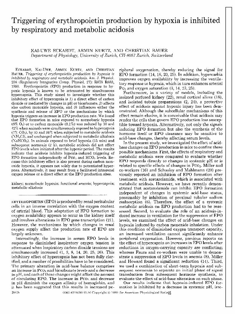
Effect of acid-base alterations on EPOproduction under hypoxic hypoxia. The effect of increasing concentrations of carbon dioxide in the inhaled gas mixture on serum EPO levels induced by 3 h of normobaric arterial hypoxia (8% Ox) is shown in Fig. 1. Carbon dioxide led to a concentration-dependent suppression of EPO values. To investigate whether the inhibitory effect of hypercapnia was related to the decrease in pH or the increase in PCO~ or bicarbonate, we intended to 1) decrease pH without increasing PCO~ and bicarbonate and 2) elevate bicarbon- ate without decreasing pH, by inducing a metabolic aci- dosis or alkalosis. Therefore animals were treated with
350
300
n
z 3
200
E v
i? w L .- E 3 ; 100 cl3
0
(30)
I (30)
\ (10)
;;_
ttp://ajpregu.physiology.org/m
R680 ERYTHROPOIETIN FORMATION AND ACID-BASE STATUS
TABLE 1. Peripheral venous pH and standard sodium bicarbonate concentrations in mice 1 and 3 h after injection of NH&l or NaHCOz
Controls NH&l NaHC03
lh 3h lh 3h
PH
Standard bicarbonate
7.342kO.04 7.207t0.03 7.248t0.05 7.467t0.03 7.431t0.02 03) 63) (7) (5) (5)
23.7t1.6 15.8t1.2 17.2t4.5 32.3t1.4 31.4t1.7 (7) (6) (6) (5) (5)
Values are means ~fr SD; nos. in parentheses are nos. of animals. Values in all 4 treatment groups were significantly different from controls.
350
300
15 10
*I *I
19 20 - 12 U
Control NH&l 7% co,
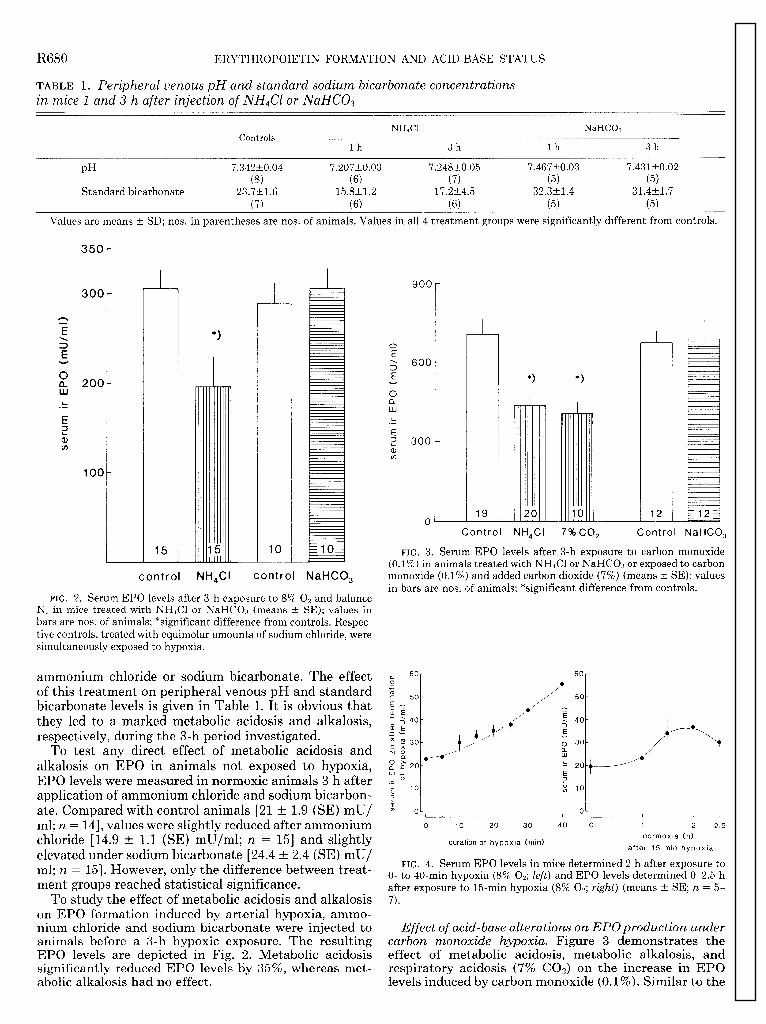
monoxide (0.1%) and added carbon dioxide (7%) (means -+ SE); values
Control NaHCO,
in bars are nos. of animals; *significant difference from controls.
FIG. 3. Serum EPO levels after 3-h exposure to carbon monoxide (0.1%) in animals treated with NH&l or NaHC03 or exposed to carbon
control NH&I control NaHCO,
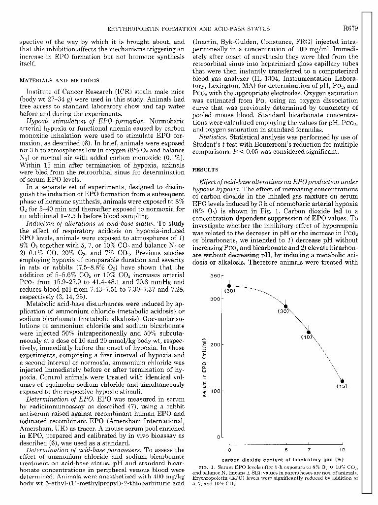
FIG. 2. Serum EPO levels after 3-h exposure to 8% 0, and balance NP in mice treated with NH&l or NaHC03 (means t SE); values in bars are nos. of animals; *significant difference from controls. Respec- tive controls, treated with equimolar amounts of sodium chloride, were simultaneously exposed to hypoxia.
ammonium chloride or sodium bicarbonate. The effect of this treatment on peripheral venous pH and standard bicarbonate levels is given in Table 1. It is obvious that they led to a marked metabolic acidosis and alkalosis, respectively, during the 3-h period investigated.
To test any direct effect of metabolic acidosis and alkalosis on EPO in animals not exposed to hypoxia, EPO levels were measured in normoxic animals 3 h after application of ammonium chloride and sodium bicarbon- ate. Compared with control animals [2l t 1.9 (SE) mU/ ml; n = 14, values were slightly reduced after ammonium chloride [ 14.9 t 1.1 (SE) mu/ml; n = 151 and slightly elevated under sodium bicarbonate [24.4 t 2.4 (SE) mU/ ml; n = 151. However, only the difference between treat- ment groups reached statistical significance.
To study the effect of metabolic acidosis and alkalosis on EPO formation induced by arterial hypoxia, ammo- nium chloride and sodium bicarbonate were injected to animals before a 3-h hypoxic exposure. The resulting EPO levels are depicted in Fig. 2. Metabolic acidosis significantly reduced EPO levels by 35%, whereas met-
E 60
.- +
60 r 50
1
O-
0
I 1 1
1 2 2.5
normoxia (h) duration of hypoxia (min)
after 15 min hypoxia
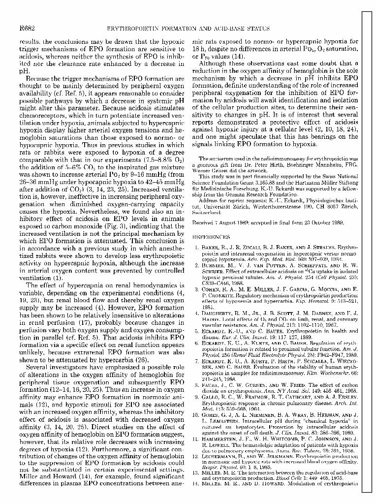
FIG. 4. Serum EPO levels in mice determined 2 h after exposure to 0- to 40-min hypoxia (8% 02; left) and EPO levels determined O-2.5 h after exposure to 15-min hypoxia (8% 02; right) (means t SE; n = 5- 7).
Effect of acid-base alterations on EPOproduction under carbon monoxide hypoxia. Figure 3 demonstrates the effect of metabolic acidosis, metabolic alkalosis, and respiratory acidosis (7% COa) on the increase in EPO
abolic alkalosis had no effect. levels induced by carbon monoxide (0.1%). Similar to the

ERYTHROPOIETIN FORMATION AND ACID-BASE STATUS R681
15
control NH&l NH&I inj. before inj. after hypoxia hypoxia
FIG. 5. Serum EPO levels determined 2 h after a hypoxic exposure of 15 min (8% 0,) in animals injected with NH&l before onset of hypoxia or immediately after termination of hypoxia (means t SE); values in bars are nos. of animals; *significant difference from controls.
results obtained with hypoxic hypoxia, respiratory as well as metabolic acidosis led to significantly diminished serum EPO levels (58 and 62% of control values, respec- tively), whereas metabolic alkalosis did not affect EPO values.
So far these results indicate that the observed suppres- sion of EPO levels in response to hypoxic or carbon monoxide hypoxia is related to a decrease in pH, irre- spective of the way by which it has been brought about, and is independent of changes in PCO~ or serum bicar- bonate levels.
Effect of acidosis on induction of EPO formation and EPO synthesis. To assess whether the triggering of EPO formation and/or EPO synthesis is sensitive to a de- crease in pH, we aimed at roughly separating both pe- riods. We thereby define the trigger phase as time period during which hypoxia initiates the subsequent synthesis and release of EPO from the kidney. Under continuous hypoxia, when triggering and hormone synthesis overlap, EPO levels are known to be first elevated afer l-2 h (3, 5, 6, 13, 25). To separate an initial phase of triggering, animals were exposed to shorter periods of hypoxia (8% 0,; 5-40 min) and then returned into normoxic atmos- phere before determination of EPO levels to allow suffi- cient time for hormone production. Figure 4 (left) illus- trates that EPO levels determined 2 h after the end of hypoxic exposures are slightly elevated when hvpoxia had lasted for 10 min and increase with further prolon- gation of the hypoxic period. When EPO levels were determined between 1 and 2.5 h after a hypoxic stimu- lation of 15 min, mean EPO levels were maximally elevated after 2 h of normoxia and thereafter tended to decline again (Fig. 4, right). A combination of 15-min hvpoxia and 2-h normoxia was therefore used in the
following experiment, designed to determine the possible effect of a decrease in pH on the different phases of hypoxia-induced EPO formation. As illustrated in Fig. 5, ammonium chloride led to a significant reduction of EPO levels when injected before the onset of hypoxia and was ineffective when injected immediately after the end of hypoxic exposure.
DISCUSSION
Following the observation that patients with carbon dioxide retention due to pulmonary insufficiency may fail to develop polycycthemia to a degree expected from the extent of their hypoxia (9, II), Faura and co-workers were, to our knowledge, the first to demonstrate a suppression of erythropoietic bioactivity when hypoxia was combined with hypercapnia (8). This effect was confirmed by several investigators (1, 3, 14, 20, 25, 26), and the concentration-dependent suppression of immu- noreactive EPO levels with increasing carbon dioxide tension found in the present study (Fig. 1) is qualitatively in accordance with these findings. However, quantita- tively, the effect of an increasing carbon dioxide concen- tration in the inhaled gas mixture appears more variable. In this study, we observed only a slight reduction by adding 5% COs, whereas other investigators reported more pronounced effects (25, 26), which may be related, for example, to different changes in ventilation.
More important, the question arises about the mech- anisms by which hypercapnia may suppress EPO levels. From our finding that both respiratory and metabolic acidosis result in diminished EPO levels in response to hypoxia (Figs. 2 and 3), it may be concluded that this effect is directly related to a decrease in systemic pH rather than an increase in Pco~. Also, changes in plasma bicarbonate levels appear not to mediate the inhibitory effect of hypercapnia, since they are oppositely affected in respiratory and metabolic acidosis, and, in addition, metabolic alkalosis due to an increase in bicarbonate levels had no effect on EPO values (Figs. 2 and 3). This latter finding indicates furthermore that the relationship between EPO levels and pH is not consistent over the whole biological range of hydrogen ion concentrations. The respiratory alkalosis normally occuring under hy- poxia appears to enhance EPO levels, since its prevention by addition of 5% COZ reduces EPO titers, whereas any further alkalinization does not further enhance hypoxia- induced EPO values.
The increase of serum EPO after hypoxia is deter- mined by as-yet-undefined mechanisms triggering an enhanced rate of EPO gene transcription, the subsequent increase in EPO synthesis, and, finally, the rate of hor- mone clearance. To determine, which of these parame- ters is sensitive to a decrease in pH, we attempted to roughly separate an initial trigger phase from a subse- quent phase of hormone synthesis and metabolism. An- imals were exposed to hypoxia for 15 min and allowed another 2 h in normoxia before determination of EPO levels (Fig. 4). Acidosis did only reduce EPO levels when initiated before the hypoxic pulse. When acidosis was applied immediately after termination of the hypoxic period. there was no change in EPO levels. From these
R682 ERYTHROPOIETIN FORMATION AND ACID-BASE STATUS
results, the conclusions may be drawn that the hypoxic trigger mechanisms of EPO formation are sensitive to acidosis, whereas neither the synthesis of EPO is inhib- ited nor the clearance rate enhanced by a decrease in PH.
Because the trigger mechanisms of EPO formation are thought to be mainly determined by peripheral oxygen availability (cf. Ref. 5), it appears reasonable to consider possible pathways by which a decrease in systemic pH might alter this parameter. Because acidosis stimulates chemoreceptors, which in turn potentiate increased ven- tilation under hypoxia, animals subjected to hypercapnic hypoxia display higher arterial oxygen tensions and he- moglobin saturations than those exposed to normo- or hypocapnic hypoxia. Thus in previous studies in which rats or rabbits were exposed to hypoxia of a degree comparable with that in our experiments (7.58.8% O& the addition of 56% CO2 to the inspirated gas mixture was shown to increase arterial Paz by 9-16 mmHg (from 26-36 mmHg under hypocapnic hypoxia to 42-45 mmHg after addition of COZ) (3, 14, 23, 25). Increased ventila- tion is, however, ineffective in increasing peripheral oxy- genation when diminished oxygen-carrying capacity causes the hypoxia. Nevertheless, we found also an in- hibitory effect of acidosis on EPO levels in animals exposed to carbon monoxide (Fig. 3), indicating that the increased ventilation is not the principal mechanism by which EPO formation is attenuated. This conclusion is in accordance with a previous study in which anesthe- tized rabbits were shown to develop less erythropoietic activity on hypercapnic hypoxia, although the increase in arterial oxygen content was prevented by controlled ventilation (1).
The effect of hypercapnia on renal hemodynamics is variable, depending on the experimental conditions (4, 19, 23), but renal blood flow and thereby renal oxygen supply may be increased (4). However, EPO formation has been shown to be relatively insensitive to alterations in renal perfusion (17), probably because changes in perfusion vary both oxygen supply and oxygen consump- tion in parallel (cf. Ref. 5). That acidosis inhibits EPO formation via a specific effect on renal function appears unlikely, because extrarenal EPO formation was also shown to be attenuated by hypercarbia (26).
Several investigators have emphasized a possible role of alterations in the oxygen affinity of hemoglobin for peripheral tissue oxygenation and subsequently EPO formation (12-14,16,20,25). Thus an increase in oxygen affinity may enhance EPO formation in normoxic ani- mals (IZ), and hypoxic stimuli for EPO are associated with an increased oxygen affinity, whereas the inhibitory effect of acidosis is associated with decreased oxygen affinity (3, 14, 20, 25). Direct studies on the effect of oxygen affinity of hemoglobin on EPO formation suggest, however, that its relative role decreases with increasing degrees of hypoxia (12). Furthermore, a significant con- tribution of changes of the oxygen affinity of hemoglobin to the suppression of EPO formation by acidosis could not be substantiated in certain experimental settings. Miller and Howard (14), for example, found significant differences in plasma EPO concentrations between ane-
mic rats exposed to normo- or hypercapnic hypoxia for 18 h, despite no differences in arterial Paz, 0, saturation, or PSO values (14).
Although these observations cast some doubt that a reduction in the oxygen affinity of hemoglobin is the sole mechanism by which a decrease in pH inhibits EPO formation, definite understanding of the role of increased peripheral oxygenation for the inhibition of EPO for- mation by acidosis will await identification and isolation of the cellular production sites, to determine their sen- sitivity to changes in pH. It is of interest that several reports demonstrated a protective effect of acidosis against hypoxic injury at a cellular level (2, 10, 18, 24), and one might speculate that this has bearings on the signals linking EPO formation to hypoxia.
The antiserum used in the radioimmunoassay for erythropoietin was a generous gift from Dr. Peter Hirth, Boehringer Mannheim, FRG. Werner Gehret did the artwork.
This study was in part financially supported by the Swiss National Science Foundation Grant 3.16588 and the Hartmann Muller Stiftung fur Medizinische Forschung. K.-U. Eckardt was supported by a fellow- ship from the German Research Foundation.
Address for reprint requests: K.-U. Eckardt, Physiologisches Insti- tut, Universitat Zurich, Winterthurerstrasse 190, CH 8057 Zurich, Switzerland.
Received 7 August 1989; accepted in final form 23 October 1989.
REFERENCES
1.
2.
3.
4.
5.
6.
7.
8.
9.
10.
11.
12.
13.
14.
BAKER, R., J. R. ZUCALI, B. J. BAKER, AND J. STRAUSS. Erythro- poietin and intrarenal oxygenation in hypercapnic versus normo- capnic hypoxemia. Adu. Exp. Med. Biol. 169: 597-609, 1984. BURNIER, M., V. J. VAN PUTTEN, A. SCHIEPPATI, AND R. W. SCHRIER. Effect of extracellular acidosis on 45Ca uptake in isolated hypoxic proximal tubules. Am. J. PhysioZ. 254 (Cell Physiol. 23): C839-C846,1988. COHEN, R. A., M. E. MILLER, J. F. GARCIA, G. MOCCIA, AND E. P. CRONKITE. Regulatory mechanism of erythropoietin production: effects of hypoxemia and hypercarbia. Exp. Hematol. 9: 513-521, 1981. DAUGHERTY, R. M., JR., J. B. SCOTT, J. M. DABNEY, AND F. J. HADDY. Local effects of 0, and CO, on limb, renal, and coronary vascular resistance. Am. J. Physiol. 213: 1102-1110, 1967. ECKARDT, K.-U., AND C. BAUER. Erythropoietin in health and disease. Eur. J. CZin. Inuest. 19: 117-127, 1989. ECKARDT, K.-U., A. KURTZ, AND C. BAUER. Regulation of eryth- ropoietin formation is related to proximal tubular function. Am. J. Physiol. 256 (Renal Fluid Electrolyte Physiol. 25): F942-F947,1989. ECKARDT, K.-U., A. KURTZ, P. HIRTH, P. SCIGALLA, L. WIECZO- REK, AND C. BAUER. Evaluation of the stability of human eryth- ropoietin in samples for radioimmunoassay. KZin. Wochenschr. 66: 241-245,1988. FAURA, J., C. W. GURNEY, AND W. FRIED. The effect of carbon dioxide on erythropoiesis. Ann. NY Acad. Sci. 149: 456-461, 1968. GALLO, R. C., W. FRAIMOW, R. T. CATHCART, AND A. J. ERSLEV. Erythropoietic response in chronic pulmonary disease. Arch. Int. Med. 113: 559-568,1964. GORES, G. J, A.-L. NIEMINEN, B. A. WRAY, B. HERMAN, AND J. L. LEMASTERS. Intracellular pH during “chemical hypoxia” in cultured rat hepatocytes. Protection by intracellular acidosis against the onset of cell death. J. CZin. Inuest. 83: 386-396, 1989. HAMMERSTEN, J. F., W. H. WHITCOMB, P. C. JOHNSON, AND J. R. LOWELL. The hematololgic adaptation of patients with hypoxia due to pulmonary emphysema. Annu. Rev. Tuberc. 78: 391, 1958. LECHERMANN, B., AND W. JELKMANN. Erythropoietin production in normoxic and hypoxic rats with increased blood oxygen affinity. Respir. Physiol. 60: l-8, 1985. MILLER, M. E. The interaction between the regulation of acid-base and erythropoietin production. Blood CeZZs 1: 449-465, 1975. MILLER, M. E., AND D. HOWARD. Modulation of erythropoietin
ERYTHROPOIETIN FORMATION AND ACID-BASE STATUS R683
15.
16.
17.
18.
19.
20.
concentrations by manipulation of hypercarbia. Blood Cells 5: 389- 21. 403, 1979. MILLER, M. E., D. HOWARD, F. STOHLMAN, AND P. FLANAGAN. Mechanism of erythropoietin production by cobaltous chloride. BZood 44: 339-346,1974. 22. MILLER, M. E., M. R@RTH, H. H. PARVING, D. HOWARD, I. REDDINGTON, C. R. VALERI, AND F. STOHLMAN, JR. pH effect on erythropoietin response to hypoxia. N. Engl. J. Med. 288: 706-710, 1973.
23 .
PAGEL, H., W. JELKMANN, AND C. WEISS. A comparison of the effects of renal artery constriction and anemia on the production of erythropoietin. Pfluegers Arch. 413: 62-66, 1988. PENTTILA, A., AND B. J. TRUMP. Extracellular acidosis protects 24.
ehrlich ascites tumor cells and rat renal cortex against anoxic injury. Science Wash. DC 185: 277-278, 1974. 25.
ROSE, C. E., JR., D. P. KIMMEL, R. L. GODINE, JR., D. L. KAISER, AND R. M. CAREY. Synergistic effects of acute hypoxemia and hypercapnic acidosis in conscious dogs. Circ. Res. 53: 202-213, 1983. 26. SCHOOLEY, J. C., AND L.-J. MAHLMANN. Hypoxia and the initia- tion of erythropoietin production. Blood Cells 1: 429-448, 1975.
SCHUSTER, S. J, E. V. BADIAVAS , P. COSTA-GIOMI, R. WEINMANN, A. J. ERSLEV, AND J. CARO. Stimulation of erythropietin gene transcription during hypoxia and cobalt exposure. Blood 73: 13-16, 1989. SHANLEY, P. F., J. I. SHAPIRO, L. CHAN, T. J. BURKE, AND G. C. JOHNSON. Acidosis and hypoxic medullary injury in the isolated perfused kidney. Kidney Int. 34: 791-796, 1988. WALKER, B. R., AND B. L. BRIZZEE. Renal vascular response to combined hypoxia and hypercapnia in conscious rats. Am. J. Phys- iol. 254 (Regulatory Integrative Cornp. Physiol. 23): R552-R558, 1988. WEINBERG, J. M. Oxygen deprivation-induced injury to isolated rabbit kidney tubules. J. CZin. Inuest. 76: 1193-1208, 1985. WOLF-PRIESSNITZ, J., J. C. SCHOOLEY, AND L.-J. MAHLMANN. Inhibition of erythropoietin production in unanesthetized rabbits exposed to an acute hypoxic-hypercapnic environment. Blood 52: 153-162,1978. ZUCALI, J. R., M. LEE, AND E. A. MIRAND. Carbon dioxide effects on erythropoietin and erythropoiesis. J. Lab. CZin. Med. 92: 648- 655, 1978.


