LUND UNIVERSITY
PO Box 117221 00 Lund+46 46-222 00 00
The effect of endogenously released glucose, insulin, glucagon-like peptide 1, ghrelinon cardiac output, heart rate, stroke volume, and blood pressure
Hlebowicz, Joanna; Lindstedt Ingemansson, Sandra; Björgell, Ola; Dencker, Magnus
Published in:Cardiovascular Ultrasound
DOI:10.1186/1476-7120-9-43
Published: 2011-01-01
Link to publication
Citation for published version (APA):Hlebowicz, J., Lindstedt Ingemansson, S., Björgell, O., & Dencker, M. (2011). The effect of endogenouslyreleased glucose, insulin, glucagon-like peptide 1, ghrelin on cardiac output, heart rate, stroke volume, andblood pressure. Cardiovascular Ultrasound, 9(43). DOI: 10.1186/1476-7120-9-43
General rightsCopyright and moral rights for the publications made accessible in the public portal are retained by the authorsand/or other copyright owners and it is a condition of accessing publications that users recognise and abide by thelegal requirements associated with these rights.
• Users may download and print one copy of any publication from the public portal for the purpose of privatestudy or research. • You may not further distribute the material or use it for any profit-making activity or commercial gain • You may freely distribute the URL identifying the publication in the public portal

Take down policyIf you believe that this document breaches copyright please contact us providing details, and we will removeaccess to the work immediately and investigate your claim.
Download date: 02. Aug. 2018
RESEARCH Open Access
The effect of endogenously released glucose,insulin, glucagon-like peptide 1, ghrelin oncardiac output, heart rate, stroke volume, andblood pressureJoanna Hlebowicz1*, Sandra Lindstedt2, Ola Björgell3 and Magnus Dencker4
Abstract
Background: Ingestion of a meal increases the blood flow to the gastrointestinal organs and affects the heart rate(HR), blood pressure and cardiac output (CO), although the mechanisms are not known. The aim of this study wasto evaluate the effect of endogenously released glucose, insulin, glucagon-like peptide 1 (GLP-1), ghrelin on CO,HR, stroke volume (SV), and blood pressure.
Methods: Eleven healthy men and twelve healthy women ((mean ± SEM) aged: 26 ± 0.2 y; body mass index: 21.8± 0.1 kg/m2)) were included in this study. The CO, HR, SV, systolic and diastolic blood pressure, antral area, gastricemptying rate, and glucose, insulin, GLP-1 and ghrelin levels were measured.
Results: The CO and SV at 30 min were significantly higher, and the diastolic blood pressure was significantlylower, than the fasting in both men and women (P < 0.05). In men, significant correlations were found betweenGLP-1 level at 30 min and SV at 30 min (P = 0.015, r = 0.946), and between ghrelin levels and HR (P = 0.013, r =0.951) at 110 min. Significant correlations were also found between the change in glucose level at 30 min and thechange in systolic blood pressure (P = 0.021, r = -0.681), and the change in SV (P = 0.008, r = -0.748) relative to thefasting in men. The insulin 0-30 min AUC was significantly correlated to the CO 0-30 min AUC (P = 0.002, r =0.814) in men. Significant correlations were also found between the 0-120 min ghrelin and HR AUCs (P = 0.007, r =0.966) in men. No statistically significant correlations were seen in women.
Conclusions: Physiological changes in the levels of glucose, insulin, GLP-1 and ghrelin may influence the activity ofthe heart and the blood pressure. There may also be gender-related differences in the haemodynamic responsesto postprandial changes in hormone levels. The results of this study show that subjects should not eat immediatelyprior to, or during, the evaluation of cardiovascular interventions as postprandial affects may affect the results,leading to erroneous interpretation of the cardiovascular effects of the primary intervention.
Trial registration number: NCT01027507
BackgroundSeveral kinds of postprandial cardiovascular changeshave been reported in the literature. The postprandialblood flow in the superior mesenteric artery (SMA)seems to be approximately double the fasting value, andinitiates an increase in cardiac output (CO) [1].
Postprandial CO increase has been suggested to resultfrom increases in the heart rate (HR) and stroke volume(SV). The postprandial blood flow in the SMA increasessimultaneously with the gradual increase in the CO,reaching a maximum 30 to 60 min after eating. A largemeal increases CO more, and for a longer time, than asmall meal [2]. However, the composition of the mealseems to be less important in this respect [3]. Theingestion of food has also been shown to decrease thediastolic blood pressure [4,5].
* Correspondence: [email protected] for Emergency, Department of Cardiology, Skåne University Hospital,Malmö, Lund University, SwedenFull list of author information is available at the end of the article
Hlebowicz et al. Cardiovascular Ultrasound 2011, 9:43http://www.cardiovascularultrasound.com/content/9/1/43
CARDIOVASCULAR ULTRASOUND
© 2011 Hlebowicz et al; licensee BioMed Central Ltd. This is an Open Access article distributed under the terms of the CreativeCommons Attribution License (http://creativecommons.org/licenses/by/2.0), which permits unrestricted use, distribution, andreproduction in any medium, provided the original work is properly cited.
The autonomic innervation of the stomach and theheart is divided into the parasympathetic and sympa-thetic systems. The parasympathetic innervation is con-trolled by the vagus nerve, whose cell bodies are foundin the brainstem. The vagus nerve consists of 20% effer-ent fibres and 80% afferent sensory fibres, which trans-mit information to the brain [6]. The vagus nervemediates the adaptive relaxation of the proximal sto-mach, the fundic-antral co-ordination (by controlleddelivery of the food from the fundus to the antrum) andthe peristaltic contractions of the distal stomach after ameal [7]. Sympathetic stimulation increases the HR(positive chronotropy), inotropy and conduction velocity(positive dromotropy), whereas parasympathetic stimula-tion of the heart has the opposite effects [6]. Changes inHR, heart rate variability (HRV), and blood pressure aresome of the factors that may reflect the balance betweenthe sympathetic and parasympathetic nervous systems. Itappears that neural signals are less likely to be responsi-ble for the increase in CO [8]. In order to investigatedenervated hearts, Waaler at al. investigated subjectswith transplanted hearts and found that the CO, HRand SV increased in a similar way to that of controlsubjects [4,8]. It is still not known what causes thechanges in the pumping activity of the heart postpran-dially. Our hypothesis was that the intake of food wouldchange the activity of the heart due to postprandialchanges in the antral area, or levels of glucose, insulin,GLP-1 and ghrelin. It is known that insulin has positivechronotropic and inotropic effects on the heart [9], andthe hormone glucagon-like peptide 1 (GLP-1) has beenshown to improve left ventricular function [10,11]. Thehormone ghrelin has been shown to increase CO andstroke volume (SV) [12-15]. Postprandial haemodynamicchanges have been shown to resemble the effects ofvasodilator drugs [16,17]. The evaluation of cardiovascu-lar interventions postprandially may affect the results,leading to erroneous interpretation of the cardiovasculareffects of the primary intervention. The aim of thisstudy was to evaluate the effect of endogenouslyreleased glucose, insulin, GLP-1, ghrelin on CO, HR,stroke volume (SV), and blood pressure.
MethodsTwenty-three healthy subjects (11 men, 12 women;(mean ± SEM) aged: 26 ± 0.2 y (range: 18-33 y); bodymass index: 21.8 ± 0.1 kg/m2 (range: 17.0-25.9 kg/m2))without symptoms or a history of gastrointestinal dis-ease, abdominal surgery or diabetes mellitus, wereincluded in this observational study. The mean waist:hipratio of the women was 0.74 ± 0.02 and of the men 0.87± 0.01. The subjects had no connective tissue disease orcerebrovascular or endocrine disease, and none was tak-ing any medication, except four women who were taking
oral contraceptives. Three men were snuff users and onewas a smoker, while two of the women were snuff users.All subjects were recruited from the population ofsouthern Sweden.All subjects gave their written informed consent. The
study was approved by the Ethics Committee of LundUniversity, and performed according to the HelsinkiDeclaration. The study started on 13 January 2009 andended on 18 September 2009. The trial is registered inthe US National Library of Medicine with the trial regis-tration number NCT01027507.The subjects were examined between 7.30 and 11.00 a.
m. after an 8-h fast. Smoking and snuff-taking were pro-hibited 8 h prior to and during the examination. Thefasting blood glucose concentration of each subject wasmeasured on the day of the examination to ensure thatit was normal (≤ 7.0 mmol/L). If the subjects reportedgastrointestinal symptoms (diarrhoea or constipation)on the day of the study, the examination was postponed.The test meal consisted of 300 g rice pudding (AXA
Goda Gröten Risgrynsgröt; Lantmännen AXA, Järna,Sweden). The total caloric value of the meal was 330kcal: 10% from protein (9 g), 58% from carbohydrates(48 g) and 32% from fat (12 g). The meal was ingestedwithin 5 minutes. The gastric emptying rate (GER) wasestimated using an ultrasound method described pre-viously [18]. The sonographic examination was per-formed with a 3.5-MHz abdominal transducer and animaging system ((Acusone Sequioa 512, Mountain View,CA), (Siemens Elegra, Siemens Medical Solutions,Mountain View, CA)). Measurements were made 15 and90 min after the meal had been consumed, and thedegree of gastric emptying was expressed as the percen-tage change in the antral cross-sectional area betweenthese two measurements. At each examination, the long-itudinal and anteroposterior diameters were measuredthree times, and the mean values were used to calculatethe cross-sectional area of the gastric antrum. The GER(%) was calculated using the following equation:
GER =(1 − (
Antrum area 90 min/Antrum area 15 min)) × 100
Measurements of the gastric antrum were performedby the same radiologist. The abdominal aorta and theleft lobe of the liver were used as internal landmarks inthe measurements on the gastric antrum. The subjectswere examined in the supine position, and were notallowed to sit up between examinations.Blood pressure was measured in the supine position
using a conventional (mechanical) sphygmomanometerwith an aneroid manometer and a stethoscope. Systolicpressure (first phase) was identified by the first of thecontinuous Korotkoff sounds. Diastolic pressure wasidentified as the moment the Korotkoff sounds
Hlebowicz et al. Cardiovascular Ultrasound 2011, 9:43http://www.cardiovascularultrasound.com/content/9/1/43
Page 2 of 11
disappeared (fifth phase). Transthoracic echocardiogra-phy examinations were performed with a Sonos 5500ultrasound system (Philips, Andover, MA, USA) in theleft lateral position, after 15 minutes’ rest. A singleobserver performed all the echocardiography measure-ments. The left ventricular SV was measured, and theleft ventricular CO was calculated according to currentguidelines [19]. The Doppler method was used SV = D2
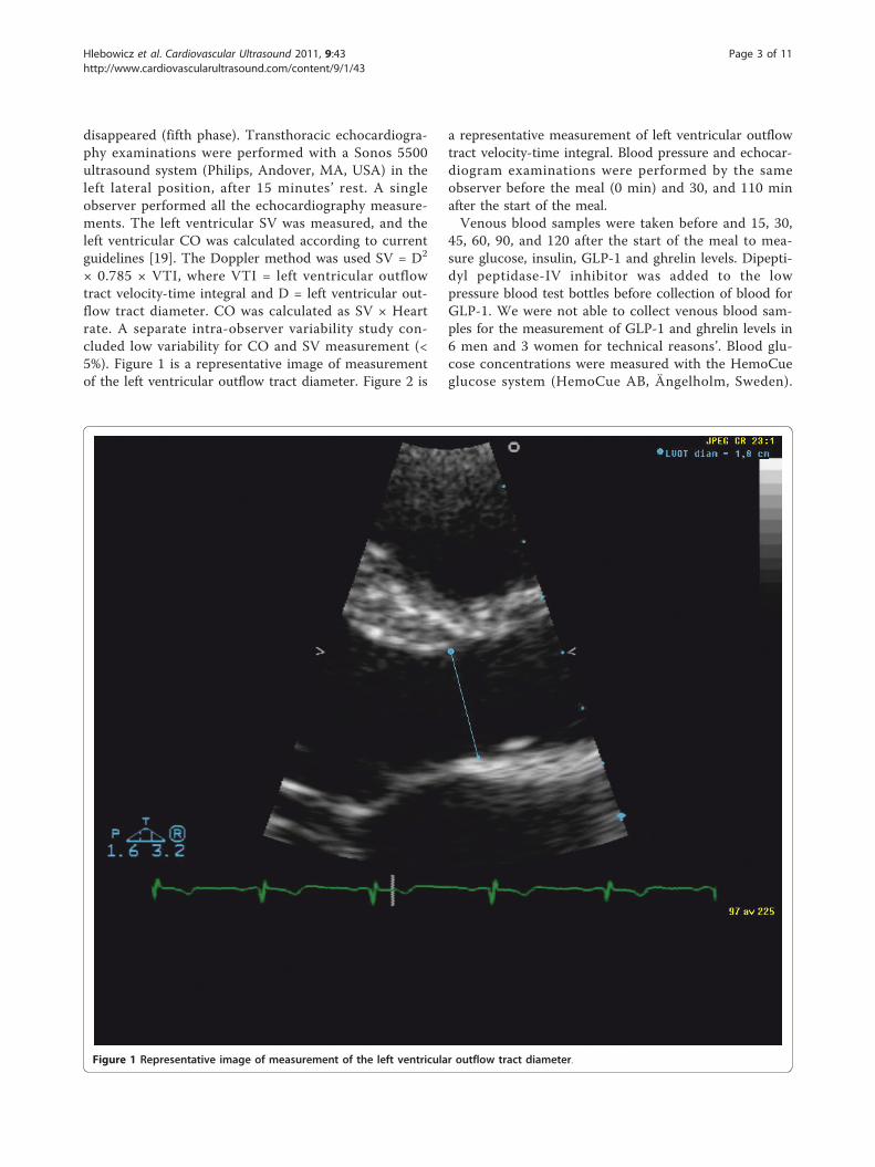
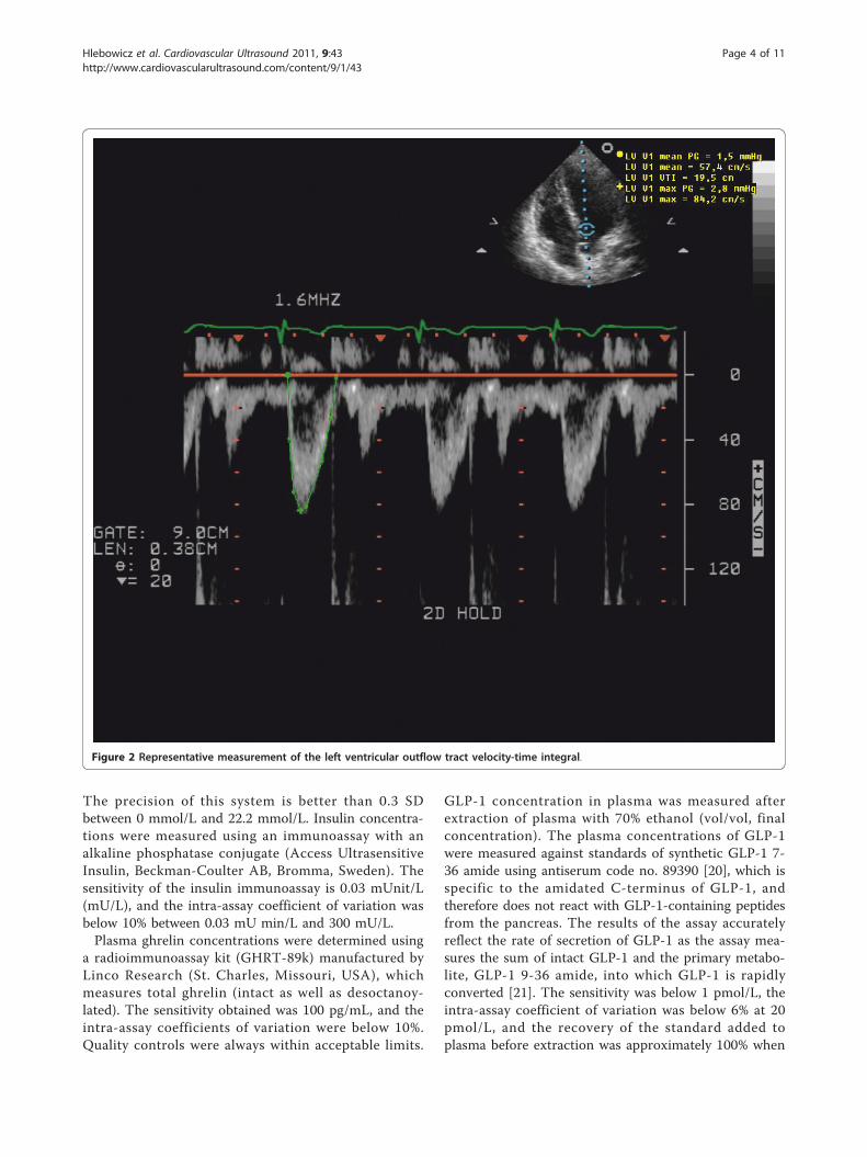
× 0.785 × VTI, where VTI = left ventricular outflowtract velocity-time integral and D = left ventricular out-flow tract diameter. CO was calculated as SV × Heartrate. A separate intra-observer variability study con-cluded low variability for CO and SV measurement (<5%). Figure 1 is a representative image of measurementof the left ventricular outflow tract diameter. Figure 2 is
a representative measurement of left ventricular outflowtract velocity-time integral. Blood pressure and echocar-diogram examinations were performed by the sameobserver before the meal (0 min) and 30, and 110 minafter the start of the meal.Venous blood samples were taken before and 15, 30,
45, 60, 90, and 120 after the start of the meal to mea-sure glucose, insulin, GLP-1 and ghrelin levels. Dipepti-dyl peptidase-IV inhibitor was added to the lowpressure blood test bottles before collection of blood forGLP-1. We were not able to collect venous blood sam-ples for the measurement of GLP-1 and ghrelin levels in6 men and 3 women for technical reasons’. Blood glu-cose concentrations were measured with the HemoCueglucose system (HemoCue AB, Ängelholm, Sweden).
Figure 1 Representative image of measurement of the left ventricular outflow tract diameter.
Hlebowicz et al. Cardiovascular Ultrasound 2011, 9:43http://www.cardiovascularultrasound.com/content/9/1/43
Page 3 of 11
The precision of this system is better than 0.3 SDbetween 0 mmol/L and 22.2 mmol/L. Insulin concentra-tions were measured using an immunoassay with analkaline phosphatase conjugate (Access UltrasensitiveInsulin, Beckman-Coulter AB, Bromma, Sweden). Thesensitivity of the insulin immunoassay is 0.03 mUnit/L(mU/L), and the intra-assay coefficient of variation wasbelow 10% between 0.03 mU min/L and 300 mU/L.Plasma ghrelin concentrations were determined using
a radioimmunoassay kit (GHRT-89k) manufactured byLinco Research (St. Charles, Missouri, USA), whichmeasures total ghrelin (intact as well as desoctanoy-lated). The sensitivity obtained was 100 pg/mL, and theintra-assay coefficients of variation were below 10%.Quality controls were always within acceptable limits.
GLP-1 concentration in plasma was measured afterextraction of plasma with 70% ethanol (vol/vol, finalconcentration). The plasma concentrations of GLP-1were measured against standards of synthetic GLP-1 7-36 amide using antiserum code no. 89390 [20], which isspecific to the amidated C-terminus of GLP-1, andtherefore does not react with GLP-1-containing peptidesfrom the pancreas. The results of the assay accuratelyreflect the rate of secretion of GLP-1 as the assay mea-sures the sum of intact GLP-1 and the primary metabo-lite, GLP-1 9-36 amide, into which GLP-1 is rapidlyconverted [21]. The sensitivity was below 1 pmol/L, theintra-assay coefficient of variation was below 6% at 20pmol/L, and the recovery of the standard added toplasma before extraction was approximately 100% when
Figure 2 Representative measurement of the left ventricular outflow tract velocity-time integral.
Hlebowicz et al. Cardiovascular Ultrasound 2011, 9:43http://www.cardiovascularultrasound.com/content/9/1/43
Page 4 of 11
corrected for losses inherent in the plasma extractionprocedure.
Statistical analysisAll analyses were performed for men and women sepa-rately. Results are given as mean values and SEM, unlessotherwise stated. The total areas under the curves(AUCs) were calculated for blood glucose, insulin, GLP-1, ghrelin, CO, HR, SV, and systolic and diastolic bloodpressure in each subject using GraphPad Prism software(version 4; GraphPad, San Diego, CA, USA). All otherstatistical calculations were performed in SPSS for Win-dows (SPSS, Version 14.0, 2005; Chicago, IL, USA).Changes in the values of blood glucose, insulin, GLP-1and ghrelin levels were calculated as the differencebetween the levels before the meal (fasting value) and30 and 120 min after the end of the meal. Changes inCO, HR, SV, and systolic and diastolic blood pressurewere calculated as the difference between levels beforethe meal (fasting value) and 30 and 110 min after theend of the meal. To determine whether the mealaffected a given parameter, the fasting value was com-pared with the values 30 min and 120 min after themeal, using the Wilcoxon t-test. The values at 30 minand 120 min postprandially were also compared usingthe Wilcoxon t-test. The significance in differencesbetween a given parameter in women and men wasdetermined using the Mann-Whitney U test. Possiblecorrelations between blood pressure, CO, HR, SV andlevels of blood glucose, plasma insulin, GLP-1, ghrelin,or antral area were analysed with the Pearson correla-tion. The level of statistical significance was set at P <0.025 after Bonferroni correction.
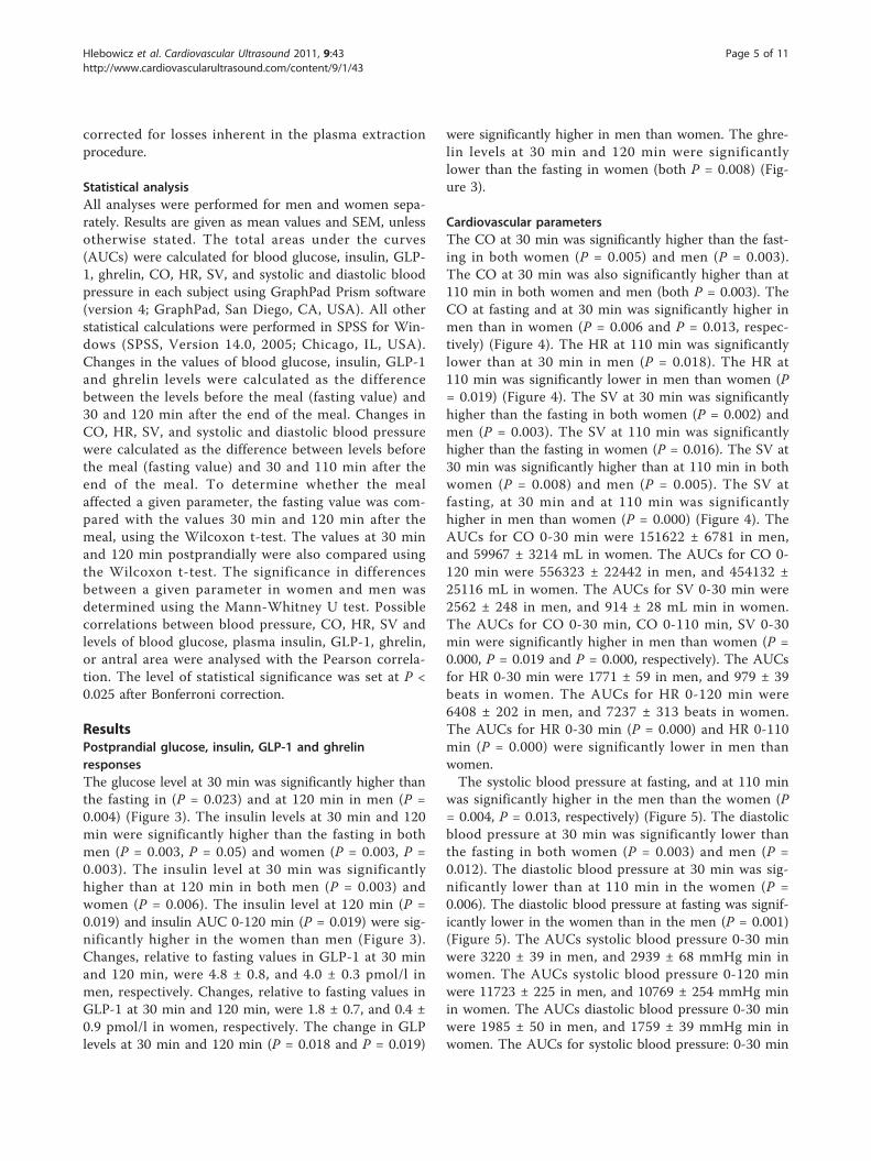
ResultsPostprandial glucose, insulin, GLP-1 and ghrelinresponsesThe glucose level at 30 min was significantly higher thanthe fasting in (P = 0.023) and at 120 min in men (P =0.004) (Figure 3). The insulin levels at 30 min and 120min were significantly higher than the fasting in bothmen (P = 0.003, P = 0.05) and women (P = 0.003, P =0.003). The insulin level at 30 min was significantlyhigher than at 120 min in both men (P = 0.003) andwomen (P = 0.006). The insulin level at 120 min (P =0.019) and insulin AUC 0-120 min (P = 0.019) were sig-nificantly higher in the women than men (Figure 3).Changes, relative to fasting values in GLP-1 at 30 minand 120 min, were 4.8 ± 0.8, and 4.0 ± 0.3 pmol/l inmen, respectively. Changes, relative to fasting values inGLP-1 at 30 min and 120 min, were 1.8 ± 0.7, and 0.4 ±0.9 pmol/l in women, respectively. The change in GLPlevels at 30 min and 120 min (P = 0.018 and P = 0.019)
were significantly higher in men than women. The ghre-lin levels at 30 min and 120 min were significantlylower than the fasting in women (both P = 0.008) (Fig-ure 3).
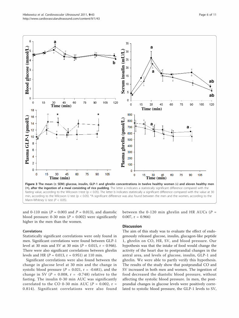
Cardiovascular parametersThe CO at 30 min was significantly higher than the fast-ing in both women (P = 0.005) and men (P = 0.003).The CO at 30 min was also significantly higher than at110 min in both women and men (both P = 0.003). TheCO at fasting and at 30 min was significantly higher inmen than in women (P = 0.006 and P = 0.013, respec-tively) (Figure 4). The HR at 110 min was significantlylower than at 30 min in men (P = 0.018). The HR at110 min was significantly lower in men than women (P= 0.019) (Figure 4). The SV at 30 min was significantlyhigher than the fasting in both women (P = 0.002) andmen (P = 0.003). The SV at 110 min was significantlyhigher than the fasting in women (P = 0.016). The SV at30 min was significantly higher than at 110 min in bothwomen (P = 0.008) and men (P = 0.005). The SV atfasting, at 30 min and at 110 min was significantlyhigher in men than women (P = 0.000) (Figure 4). TheAUCs for CO 0-30 min were 151622 ± 6781 in men,and 59967 ± 3214 mL in women. The AUCs for CO 0-120 min were 556323 ± 22442 in men, and 454132 ±25116 mL in women. The AUCs for SV 0-30 min were2562 ± 248 in men, and 914 ± 28 mL min in women.The AUCs for CO 0-30 min, CO 0-110 min, SV 0-30min were significantly higher in men than women (P =0.000, P = 0.019 and P = 0.000, respectively). The AUCsfor HR 0-30 min were 1771 ± 59 in men, and 979 ± 39beats in women. The AUCs for HR 0-120 min were6408 ± 202 in men, and 7237 ± 313 beats in women.The AUCs for HR 0-30 min (P = 0.000) and HR 0-110min (P = 0.000) were significantly lower in men thanwomen.The systolic blood pressure at fasting, and at 110 min
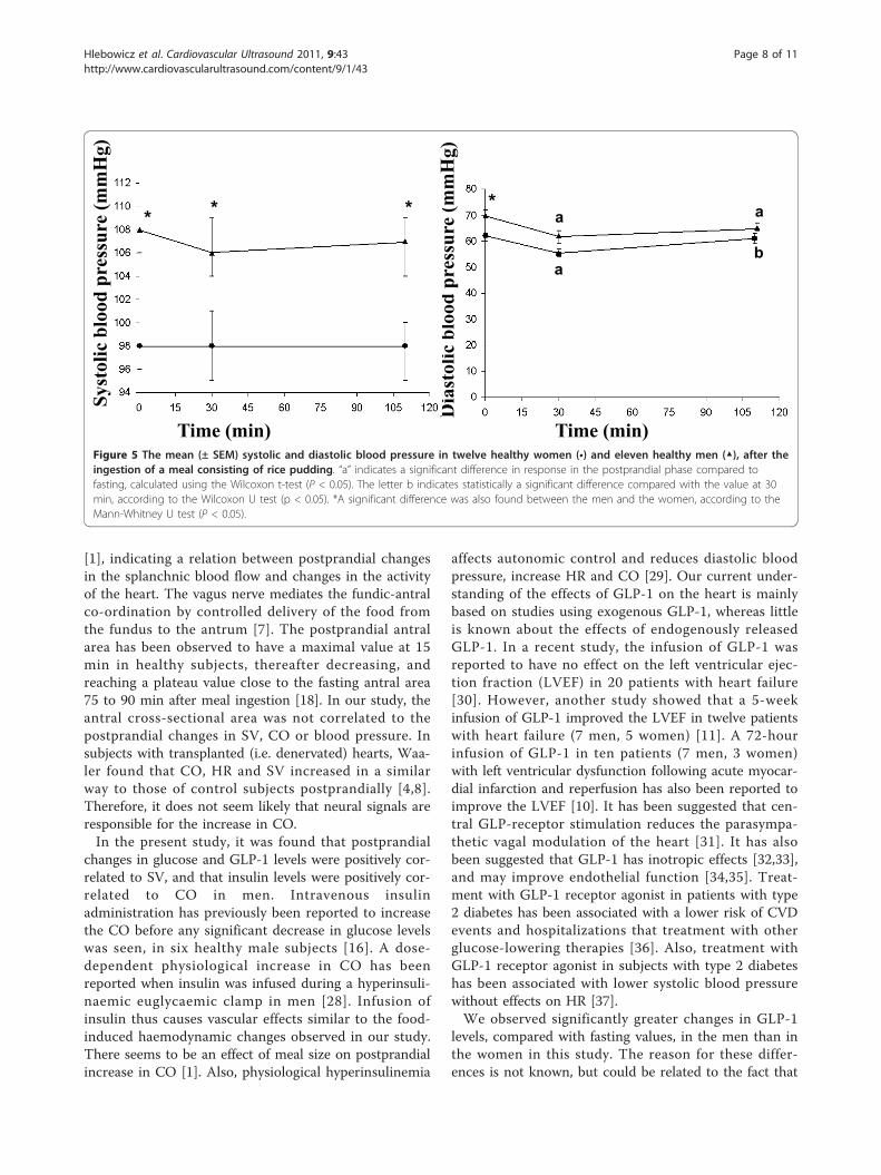
was significantly higher in the men than the women (P= 0.004, P = 0.013, respectively) (Figure 5). The diastolicblood pressure at 30 min was significantly lower thanthe fasting in both women (P = 0.003) and men (P =0.012). The diastolic blood pressure at 30 min was sig-nificantly lower than at 110 min in the women (P =0.006). The diastolic blood pressure at fasting was signif-icantly lower in the women than in the men (P = 0.001)(Figure 5). The AUCs systolic blood pressure 0-30 minwere 3220 ± 39 in men, and 2939 ± 68 mmHg min inwomen. The AUCs systolic blood pressure 0-120 minwere 11723 ± 225 in men, and 10769 ± 254 mmHg minin women. The AUCs diastolic blood pressure 0-30 minwere 1985 ± 50 in men, and 1759 ± 39 mmHg min inwomen. The AUCs for systolic blood pressure: 0-30 min
Hlebowicz et al. Cardiovascular Ultrasound 2011, 9:43http://www.cardiovascularultrasound.com/content/9/1/43
Page 5 of 11
and 0-110 min (P = 0.003 and P = 0.013), and diastolicblood pressure: 0-30 min (P = 0.002) were significantlyhigher in the men than the women.
CorrelationsStatistically significant correlations were only found inmen. Significant correlations were found between GLP-1level at 30 min and SV at 30 min (P = 0.015, r = 0.946).There were also significant correlations between ghrelinlevels and HR (P = 0.013, r = 0.951) at 110 min.Significant correlations were also found between the
change in glucose level at 30 min and the change insystolic blood pressure (P = 0.021, r = -0.681), and thechange in SV (P = 0.008, r = -0.748) relative to thefasting. The insulin 0-30 min AUC was significantlycorrelated to the CO 0-30 min AUC (P = 0.002, r =0.814). Significant correlations were also found
between the 0-120 min ghrelin and HR AUCs (P =0.007, r = 0.966)
DiscussionThe aim of this study was to evaluate the effect of endo-genously released glucose, insulin, glucagon-like peptide1, ghrelin on CO, HR, SV, and blood pressure. Ourhypothesis was that the intake of food would change theactivity of the heart due to postprandial changes in theantral area, and levels of glucose, insulin, GLP-1 andghrelin. We were able to partly verify this hypothesis.The results of the study show that postprandial CO andSV increased in both men and women. The ingestion offood decreased the diastolic blood pressure, withoutaffecting the systolic blood pressure. In men, the post-prandial changes in glucose levels were positively corre-lated to systolic blood pressure, the GLP-1 levels to SV,
Time (min)
Blo
od g
luco
se (
mm
ol/L
)
Seru
m in
sulin
(m
U/L
)
Time (min)
a
aa
b
ab
Pla
sma
GL
P-1
(pm
ol/L
)
Time (min) Time (min)
Pla
sma
ghre
lin (
pmol
/L)
a a
* *
ab
Figure 3 The mean (± SEM) glucose, insulin, GLP-1 and ghrelin concentrations in twelve healthy women (•) and eleven healthy men(▲), after the ingestion of a meal consisting of rice pudding. The letter a indicates a statistically significant difference compared with thefasting value, according to the Wilcoxon t-test (p < 0.05). The letter b indicates statistically a significant difference compared with the value at 30min, according to the Wilcoxon U test (p < 0.05). *A significant difference was also found between the men and the women, according to theMann-Whitney U test (P < 0.05).
Hlebowicz et al. Cardiovascular Ultrasound 2011, 9:43http://www.cardiovascularultrasound.com/content/9/1/43
Page 6 of 11
the insulin levels to CO and the ghrelin level to HR. Toour best knowledge, this is the first study to examine atthe effect of, endogenously released insulin, glucagon-like peptide 1, ghrelin levels on CO, SV, HR, systolicand diastolic blood pressure in both men and women.In our study, the ingestion of food led to a decrease in
the diastolic blood pressure, without affecting the systo-lic blood pressure. Previous studies have shown that gas-tric distension influences blood pressure, probably dueto activation of the gastrovascular reflex in patients withautonomic failure [22-24], and in older healthy subjects[25]. However, the systolic blood pressure was notaffected in healthy adolescents [24]. Gastric distensionhas been shown to be attenuated in the elderly [26]. Inthe present study, the systolic blood pressure was notaffected postprandially. The reason why we did notobserve any correlation between blood pressure and
antral area may be because we evaluated only younghealthy subjects with a normal gastric emptying rateand normal autonomic innervation of the stomach andheart. The gastric distension was not quantified by mea-surements of the intragastric volume.In a previous study on two men and two women,
Waaler reported postprandial increases in CO and SV,which were explained by an increase in HR [1]. In aslightly larger study, on seven men and one woman,Kelbaek found an increase in postprandial CO, HR andSV [27]. Our results suggest slightly greater early post-prandial HR, but no statistically significant differenceswere found. A gradual increase in CO 30 min to 1 hafter eating, and a relationship between the CO andmeal size has been suggested to reflect the postprandialchanges in the gastrointestinal tract [1]. The increases inCO and splanchnic blood flow seem to be synchronous
Time (min)
Car
diac
out
put
(ml/m
in)
Hea
rt r
ate
(bea
ts/m
in)
Time (min)
Stro
ke v
olum
e (m
l)
Time (min)
a
a*
aab
b*
a* ab*
b
b
*
*
Figure 4 The mean (± SEM) cardiac output (CO), heart rate (HR), and stroke volume (SV) in twelve healthy women (•) and elevenhealthy men (▲), after the ingestion of a meal consisting of rice pudding. The letter a indicates a significant difference in response in thepostprandial phase compared to fasting, calculated using the Wilcoxon t-test (P < 0.05). The letter b indicates statistically a significant differencecompared with the value at 30 min, according to the Wilcoxon U test (p < 0.05). *A significant difference was also found between the men andthe women, according to the Mann-Whitney U test (P < 0.05).
Hlebowicz et al. Cardiovascular Ultrasound 2011, 9:43http://www.cardiovascularultrasound.com/content/9/1/43
Page 7 of 11
[1], indicating a relation between postprandial changesin the splanchnic blood flow and changes in the activityof the heart. The vagus nerve mediates the fundic-antralco-ordination by controlled delivery of the food fromthe fundus to the antrum [7]. The postprandial antralarea has been observed to have a maximal value at 15min in healthy subjects, thereafter decreasing, andreaching a plateau value close to the fasting antral area75 to 90 min after meal ingestion [18]. In our study, theantral cross-sectional area was not correlated to thepostprandial changes in SV, CO or blood pressure. Insubjects with transplanted (i.e. denervated) hearts, Waa-ler found that CO, HR and SV increased in a similarway to those of control subjects postprandially [4,8].Therefore, it does not seem likely that neural signals areresponsible for the increase in CO.In the present study, it was found that postprandial
changes in glucose and GLP-1 levels were positively cor-related to SV, and that insulin levels were positively cor-related to CO in men. Intravenous insulinadministration has previously been reported to increasethe CO before any significant decrease in glucose levelswas seen, in six healthy male subjects [16]. A dose-dependent physiological increase in CO has beenreported when insulin was infused during a hyperinsuli-naemic euglycaemic clamp in men [28]. Infusion ofinsulin thus causes vascular effects similar to the food-induced haemodynamic changes observed in our study.There seems to be an effect of meal size on postprandialincrease in CO [1]. Also, physiological hyperinsulinemia
affects autonomic control and reduces diastolic bloodpressure, increase HR and CO [29]. Our current under-standing of the effects of GLP-1 on the heart is mainlybased on studies using exogenous GLP-1, whereas littleis known about the effects of endogenously releasedGLP-1. In a recent study, the infusion of GLP-1 wasreported to have no effect on the left ventricular ejec-tion fraction (LVEF) in 20 patients with heart failure[30]. However, another study showed that a 5-weekinfusion of GLP-1 improved the LVEF in twelve patientswith heart failure (7 men, 5 women) [11]. A 72-hourinfusion of GLP-1 in ten patients (7 men, 3 women)with left ventricular dysfunction following acute myocar-dial infarction and reperfusion has also been reported toimprove the LVEF [10]. It has been suggested that cen-tral GLP-receptor stimulation reduces the parasympa-thetic vagal modulation of the heart [31]. It has alsobeen suggested that GLP-1 has inotropic effects [32,33],and may improve endothelial function [34,35]. Treat-ment with GLP-1 receptor agonist in patients with type2 diabetes has been associated with a lower risk of CVDevents and hospitalizations that treatment with otherglucose-lowering therapies [36]. Also, treatment withGLP-1 receptor agonist in subjects with type 2 diabeteshas been associated with lower systolic blood pressurewithout effects on HR [37].We observed significantly greater changes in GLP-1
levels, compared with fasting values, in the men than inthe women in this study. The reason for these differ-ences is not known, but could be related to the fact that
Time (min) Time (min)
Sys
tolic
blo
od p
ress
ure
(mm
Hg)
Dia
stol
ic b
lood
pre
ssur
e (m
mH
g)
a b
a a *
*
*
*
Figure 5 The mean (± SEM) systolic and diastolic blood pressure in twelve healthy women (•) and eleven healthy men (▲), after theingestion of a meal consisting of rice pudding. “a” indicates a significant difference in response in the postprandial phase compared tofasting, calculated using the Wilcoxon t-test (P < 0.05). The letter b indicates statistically a significant difference compared with the value at 30min, according to the Wilcoxon U test (p < 0.05). *A significant difference was also found between the men and the women, according to theMann-Whitney U test (P < 0.05).
Hlebowicz et al. Cardiovascular Ultrasound 2011, 9:43http://www.cardiovascularultrasound.com/content/9/1/43
Page 8 of 11

we did not observe any correlations between postpran-dial GLP levels and haemodynamic responses in women.We observed no differences in ghrelin levels betweenthe genders, but the ghrelin levels were correlated to theHR in the men in this study. It has been suggested thatghrelin suppresses sympathetic activity [38]. Intravenousand subcutaneous ghrelin injections have been reportedto increase CO and improve cardiac contractility inhealthy subjects [12]. Treatment with ghrelin has alsobeen shown to decrease systemic vascular resistance andincrease CO, cardiac index, and SV in patients withheart failure [13].Postprandial haemodynamic responses in men and
women were analysed separately in this study as thereare known gender differences in heart function [39-45].There are also gender-related differences in cardiovascu-lar regulation. Females have a higher HR and lowerHRV than men [46-48]. Reports on these autonomicresponses are not always in agreement, for example, ithas been claimed that women show higher [49] andlower [48] parasympathetic effects, and lower sympa-thetic activity [47], and that men have higher parasym-pathetic activity [46]. Overnight fasting has beensuggested to be associated with an increase in parasym-pathetic activity that is counteracted by eating breakfast[50]. The response has been reported to differ in thatmen show greater parasympathetic activity than women[50]. Thus, it appears that there are also gender-relateddifferences in the haemodynamic responses to postpran-dial changes in hormone levels. However, the genderdifferences might be due to differences in the level ofinsulin and GLP-1. The difference the hemodynamicresponse to glucose and ghrelin remain unclear. Thereare also known gender differences in cardiovascular dis-eases and this may be related to sex hormones, estrogenand testosterone [51].The present study had some limitations: the small
sample size of healthy young subjects and the fact thatit was not possible to perform the echocardiography orgastric ultrasound examinations at the same time. How-ever, the measurements of the gastric antrum and theheart were performed by the same radiologist. Theresults of the echocardiography examinations werestored digitally and analysed at a later date in anattempt to avoid bias.
ConclusionsThis study shows that postprandial CO and SV increasein both men and women. Ingestion of food decreasedthe diastolic blood pressure without affecting the systolicblood pressure or heart rate in healthy subjects. Physio-logical changes in the levels of glucose, insulin, GLP-1and ghrelin may influence the activity of the heart andthe blood pressure. There may also be gender-related
differences in the haemodynamic responses to postpran-dial changes in hormone levels. Subjects undergoingevaluation of cardiovascular interventions should there-fore refrain from eating before or during such examina-tions, as postprandial affects may affect the results,leading to erroneous interpretation of the cardiovasculareffects of the primary intervention.
AbbreviationsAUCs: areas under the curves; CO: in cardiac output; GER: gastric emptyingrate; GLP-1: glucagon-like peptide 1; HR: heart rate; HRV: heart rate variability;LVEF: left ventricular ejection fraction; SMA: superior mesenteric artery; SV:stroke volume.
AcknowledgementsThis study was financially supported by Foundations-Research, SkåneUniversity Hospital, Malmö, and Swedish government funds for clinicalresearch (ALF), funds from the Skåne University Hospital in Malmö, Sweden.
Author details1Center for Emergency, Department of Cardiology, Skåne University Hospital,Malmö, Lund University, Sweden. 2Department of Cardiothoracic Surgery,Skåne University Hospital, Lund, Lund University, Sweden. 3Department ofRadiology, Skåne University Hospital, Malmö, Lund University, Sweden.4Department of Clinical Physiology and Nuclear Medicine, Skåne UniversityHospital, Malmö, Lund University, Sweden.
Authors’ contributionsThe authors’ contributions were as follows: JH and MD designed the study;JH was responsible for recruiting the subjects. OB performed the ultrasoundexaminations; MD performed the echocardiographic examinations; JH, SL,and MD carried out the statistical calculations. JH and MD wrote the firstdraft of the manuscript, and SL and OB made critical revisions of themanuscript. All authors read and approved the final manuscript.
Competing interestsThe authors declare that they have no competing interests.
Received: 22 August 2011 Accepted: 29 December 2011Published: 29 December 2011
References1. Waaler BA, Eriksen M, Toska K: The effect of meal size on postprandial
increase in cardiac output. Acta Physiol Scand 1991, 142(1):33-39.2. Waaler BA, Eriksen M: Post-prandial cardiovascular responses in man after
ingestion of carbohydrate, protein or fat. Acta Physiol Scand 1992,146(3):321-327.
3. Fagan TC, Sawyer PR, Gourley LA, Lee JT, Gaffney TE: Postprandialalterations in hemodynamics and blood pressure in normal subjects. AmJ Cardiol 1986, 58:636-641.
4. Waaler BA, Hisdal J, Eriksen M: Circulatory responses to a meal in patientswith a newly transplanted heart. Acta Physiol Scand 2002, 174:101-108.
5. Hlebowicz J, Lindstedt S, Björgell O, Dencker M: Relationship betweenpostprandial changes in cardiac left ventricular function, glucose andinsulin concentrations, gastric emptying, and satiety in healthy subjects.Nutr J 2011, 10:26.
6. Smout AJMP, Akkermans LMA: Normal and disturbed motility of thegastrointestinal tract. Wrightson Biomedical Publishing LTD Petersfield; 1992.
7. Sheiner HJ, Quinlan MF, Thompson IJ: Gastric motility and emptying innormal and post-vagotomy subjects. Gut 1980, 21(9):753-759.
8. Waaler BA, Hisdal J, Ihlen H, Kjekshus J: Mechanisms behind thepostprandial increase in cardiac output: a clue obtained fromtransplanted hearts. Eur J Appl Physiol 2006, 97:516-520.
9. Baron AD: Hemodynamic actions of insulin. Am J Physiol 1994, 267:E187-202.
10. Nikolaidis LA, Mankad S, Sokos GG, Miske G, Shah A, Elahi D, Shannon RP:Effects of glucagon-like peptide-1 in patients with acute myocardial
Hlebowicz et al. Cardiovascular Ultrasound 2011, 9:43http://www.cardiovascularultrasound.com/content/9/1/43
Page 9 of 11
infarction and left ventricular dysfunction after successful reperfusion.Circulation 2004, 109(8):962-925.
11. Sokos GG, Nikolaidis LA, Mankad S, Elahi D, Shannon RP: Glucagon-likepeptide-1 infusion improves left ventricular ejection fraction andfunctional status in patients with chronic heart failure. J Card Fail 2006,212(9):694-699.
12. Nagaya N, Kojima M, Uematsu M, Yamagishi M, Hosoda H, Oya H,Hayashi Y, Kangawa K: Hemodynamic and hormonal effects of humanghrelin in healthy volunteers. Am J Physiol Regul Integr Comp Physiol 2010,280(5):R1483-1487.
13. Nagaya N, Miyatake K, Uematsu M, Oya H, Shimizu W, Hosoda H, Kojima M,Nakanishi N, Mori H, Kangawa K: Hemodynamic, renal, and hormonaleffects of ghrelin infusion in patients with chronic heart failure. J ClinEndocrinol Metab 2010, 86(12):5854-5859.
14. Nagaya N, Moriya J, Yasumura Y, Uematsu M, Ono F, Shimizu W, Ueno K,Kitakaze M, Miyatake K, Kangawa K: Effects of ghrelin administration onleft ventricular function, exercise capacity, and muscle wasting inpatients with chronic heart failure. Circulation 2004, 110(24):3674-3679.
15. Nagaya N, Moriya J, Yasumura Y, Uematsu M, Ono F, Shimizu W, Ueno K,Kitakaze M, Miyatake K, Kangawa K: Effects of glucagon-like peptide-1 inpatients with acute myocardial infarction and left ventriculardysfunction after successful reperfusion. Circulation 2004, 109(8):962-965.
16. Fisher BM, Gillen G, Dargie HJ, Inglis GC, Frier BM: The effects of insulininduced hypoglycemia on cardiovascular function in normal man:studies using radionuclide ventriculography. Diabetologia 1987,30:841-845.
17. Packer M, Medina N, Yushak M: Hemodynamic changes mimicking avasodilator drug response in the absence of drug therapy after rightheart catheterization in patients with chronic heart failure. Circulation1985, 71:761-766.
18. Darwiche G, Almér LO, Björgell O, Cederholm C, Nilsson P: Measurement ofgastric emptying by standardized real-time ultrasonography in healthysubjects and diabetic patients. J Ultrasound Med 1999, 18(10):673-682.
19. Quiñones MA, Otto CM, Stoddard M, Waggoner A, Zoghbi WA:Recommendations for quantification of Doppler echocardiography: areport from the Doppler Quantification Task Force of the Nomenclatureand Standards Committee of the American Society of Echocardiography.J Am Soc Echocardiogr 2002, 15(2):167-184.
20. Ørskov C, Rabenhøj L, Kofod H, Wettergren A, Holst JJ: Production andsecretion of amidated and glycine-extended glucagon-like peptide-1(GLP-1) in man. Diabetes 1994, 43:535-539.
21. Deacon CF, Pridal L, Klarskov L, Olesen M, Holst JJ: Glucagon-like peptide-1under-goes differential tissue-specific metabolism in the anaesthetisedpig. Am J Physiol 1996, 271:E458-464.
22. Cariga P, Mathias CJ: Haemodynamics of the pressor effect of oral waterin human sympathetic denervation due to autonomic failure. Clin Sci(Lond) 2001, 101(3):313-319.
23. Jordan J, Shannon JR, Black BK, Ali Y, Farley M, Costa F, Diedrich A,Robertson RM, Biaggioni I, Robertson D: The pressor response to waterdrinking in humans: a sympathetic reflex? Circulation 2002,101(5):504-509.
24. Jordan J, Shannon JR, Grogan E, Biaggioni I, Robertson D: A potent pressorresponse elicited by drinking water. Lancet 1999, 353(9154):723.
25. Gentilcore D, Meyer JH, Rayner CK, Horowitz M, Jones C: Gastric distensionattenuates the hypotensive effect of intraduodenal glucose in healthyolder subjects. Am J Physiol Regul Integr Comp Physiol 2008, 295(2):R472-477.
26. van Orshoven NP, Oey PL, van Schelven LJ, Roelofs JM, Jansen PA,Akkermans LM: Effect of gastric distension on cardiovascular parameters:gastrovascular reflex is attenuated in the elderly. J Physiol 2004, 555(Pt2):573-583.
27. Kelbaek H, Muck O, Christensen NJ, Godtfredsen J: Central haemodynamicchanges after a meal. Br Heart J 1990, 61:506-559.
28. Baron AD, Brechtel G: Insulin differentially regulates systemic and skeletalmuscle vascular resistance. Am J Physiol 1993, 265:E61-67.
29. Regitz-Zagrosek V, Seeland U: Sex and gender differences in myocardialhypertrophy and heart failure. Wien Med Wochenschr. 2011, 161(5-6):109-116.
30. Halbirk M, Nørrelund H, Møller N, Holst JJ, Schmitz O, Nielsen R, Nielsen-Kudsk JE, Nielsen SS, Nielsen TT, Eiskjaer H, Bøtker HE, Wiggers H:Cardiovascular and metabolic effects of 48-h glucagon-like peptide-1
infusion in compensated chronic patients with heart failure. Am J PhysiolHeart Circ Physiol 2010, 298(3):H1096-1102.
31. Griffioen KJ, Wan R, Okun E, Wang X, Lovett-Barr MR, Li Y, Mughal MR,Mendelowitz D, Mattson MP: GLP-1 receptor stimulation depresses heartrate variability and inhibits neurotransmission to cardiac vagal neurons.Cardiovasc Res 2001, 89(1):72-78.
32. Barragán JM, Rodríguez RE, Blázquez E: Changes in arterial blood pressureand heart rate induced by glucagon-like peptide-1-(7-36) amide in rats.Am J Physiol 1994, 266(3 Pt 1):E459-466.
33. Edwards CM, Edwards AV, Bloom SR: Cardiovascular and pancreaticendocrine responses to glucagon-like peptide-1(7-36) amide in theconscious calf. Exp Physiol 1997, 82(4):709-716.
34. Nyström T, Gutniak MK, Zhang Q, Zhang F, Holst JJ, Ahrén B, Sjöholm A:Effects of glucagon-like peptide-1 on endothelial function in type 2diabetes patients with stable coronary artery disease. Am J PhysiolEndocrinol Metab 2004, 287(6):E1209-1215.
35. Vila Petroff MG, Egan JM, Wang X, Sollott SJ: Glucagon-like peptide-1increases cAMP but fails to augment contraction in adult rat cardiacmyocytes. Circ Res 2001, 89(5):445-452.
36. Best JH, Hoogwerf BJ, Herman WH, Pelletier EM, Smith DB, Wenten M,Hussein MA: Risk of cardiovascular disease events in patients with type 2diabetes prescribed the glucagon-like peptide 1 (GLP-1) receptoragonist exenatide twice daily or other glucose-lowering therapies: aretrospective analysis of the LifeLink database. Diabetes Care 2011,34(1):90-905.
37. Gill A, Hoogwerf BJ, Burger J, Bruce S, Macconell L, Yan P, Braun D,Giaconia J, Malone J: Effect of exenatide on heart rate and bloodpressure in subjects with type 2 diabetes mellitus: a double-blind,placebo-controlled, randomized pilot study. Cardiovasc Diabetol 2010, 9:6.
38. García EA, Korbonits M: Ghrelin and cardiovascular health. Curr OpinPharmacol 2006, 6(2):142-147.
39. Bella JN, Palmieri V, Roman MJ, Paranicas MF, Welty TK, Lee ET, Fabsitz RR,Howard BV, Devereux RB: Gender differences in left ventricular systolicfunction in American Indians (from the Strong Heart Study). Am J Cardiol2006, 98(6):834-837.
40. Buonanno C, Arbustini E, Rossi L, Dander B, Vassanelli C, Paris B, Poppi A:Left ventricular function in men and women: another differencebetween sexes. Eur Heart J 1982, 3(6):525-528.
41. Devereux RB, Roman MJ, Paranicas M, Lee ET, Welty TK, Fabsitz RR,Robbins D, Rhoades ER, Rodeheffer RJ, Cowan LD, Howard BV: Apopulation-based assessment of left ventricular systolic dysfunction inmiddle-aged and older adults: the Strong Heart Study. Am Heart J 2001,141(3):439-446.
42. Chung AK, Das SR, Leonard D, Peshock RM, Kazi F, Abdullah SM,Canham RM, Levine BD, Drazner MH: Women have higher left ventricularejection fractions than men independent of differences in leftventricular volume: the Dallas Heart Study. Circulation 2006,113(12):1597-1604.
43. Gerdts E, Zabalgoitia M, Bjornstad H, Svendsen TL, Devereux RB: Genderdifferences in systolic left ventricular function in hypertensive patientswith electrocardiographic left ventricular hypertrophy. Am J Cardiol 2001,87:980-983.
44. Luchner A, Bröckel U, Muscholl M, Hense HW, Döring A, Riegger GA,Schunkert H: Gender-specific differences of cardiac remodeling insubjects with left ventricular dysfunction: a population-based study.Cardiovasc Re 2002, 53(3):720-727.
45. Wong ND, Gardin JM, Kurosaki T, Anton-Culver H, Sidney S, Roseman J,Gidding S: Echocardiographic left ventricular systolic function andvolumes in young adults: distribution and factors influencing variability.Am Heart J 1995, 129(3):571-577.
46. Bonnemeier H, Richardt G, Potratz J, Wiegand UK, Brandes A, Kluge N,Katus HA: Circadian profile of cardiac autonomic nervous modulation inhealthy subjects: differing effects of aging and gender on heart ratevariability. J Cardiovasc Electrophysiol 2003, 14(8):791-799.
47. Ramaekers D, Ector H, Aubert AE, Rubens A, Van de Werf F: Heart ratevariability and heart rate in healthy volunteers. Is the female autonomicnervous system cardioprotective? Eur Heart J 1998, 19(9):1334-1341.
48. Umetani K, Singer DH, McCraty R, Atkinson M: Twenty-four hour timedomain heart rate variability and heart rate: relations to age and genderover nine decades. J Am Coll Cardiol 1998, 31(3):593-601.
Hlebowicz et al. Cardiovascular Ultrasound 2011, 9:43http://www.cardiovascularultrasound.com/content/9/1/43
Page 10 of 11
49. Evans JM, Ziegler MG, Patwardhan AR, Ott JB, Kim CS, Leonelli FM,Knapp CF: Gender differences in autonomic cardiovascular regulation:spectral, hormonal, and hemodynamic indexes. J Appl Physiol 2001,91(6):2611-2618.
50. Pivik RT, Dykman RA, Tennal K, Gu Y: Skipping breakfast: gender effectson resting heart rate measures in preadolescents. Physiol Behav 2006,89(2):270-280.
51. Muscelli E, Emdin M, Natali A, Pratali L, Camastra S, Gastaldelli A, Baldi S,Carpeggiani C, Ferrannini E: Autonomic and hemodynamic responses toinsulin in lean and obese humans. J Clin Endocrinol Metab 1998,83(6):2084-2090.
doi:10.1186/1476-7120-9-43Cite this article as: Hlebowicz et al.: The effect of endogenouslyreleased glucose, insulin, glucagon-like peptide 1, ghrelin on cardiacoutput, heart rate, stroke volume, and blood pressure. CardiovascularUltrasound 2011 9:43.
Submit your next manuscript to BioMed Centraland take full advantage of:
• Convenient online submission
• Thorough peer review
• No space constraints or color figure charges
• Immediate publication on acceptance
• Inclusion in PubMed, CAS, Scopus and Google Scholar
• Research which is freely available for redistribution
Submit your manuscript at www.biomedcentral.com/submit
Hlebowicz et al. Cardiovascular Ultrasound 2011, 9:43http://www.cardiovascularultrasound.com/content/9/1/43
Page 11 of 11


