Download - Tertiary Health Institution

Sinonasal Malignancies: A 10-Year Review in aTertiary Health InstitutionAyotunde J. Fasunla, MBChB and Akeem 0. Lasisi, FWACS
Sinonasal malignancy is a cause of otorhinolaryngologicmorbidity and mortality in West Africa. However, there is adearth of information in the literature on its clinicopatho-logic presentation in West Africa. It is our aim to determinethe prevalence of sinonasal malignancy and highlight theclinicopathologic features in our environment. A 10-yearretrospective review of cases with histologically diagnosedmalignant sinonasal tumors in University College Hospital,Ibadan, Oyo State, Nigeria was carried out. There were 82cases-56 (68.29%) males and 26 (31.71%) females-whoseages ranged from 4-69 years. Epistaxis, rhinorrhea andnasal blockage were seen in all patients; other symptomswere facial [76 (93%)], oral cavity [48 (59%)], ophthalmic [33(40%)] and [otologic 21 (25%)]. Squamous cell carcinomaaccounted for 69/75 (92%) of epithelial tumors, and malig-nant lymphoma accounted for 4/7 (57%) of nonepithelialtumors. Advanced disease was the predominant presenta-tion in our series, stage 3 in 59 (79%) and stage 4 in 12 (16%)cases. Therefore, health education on early presentation tohospitals and efforts at early detection of the disease areneeded in order to achieve cure. Industrial workers shouldalways wear face masks to protect their nasal cavity.
Key words: tumor l pathology * sinonasal malignancy Uclinicopathologic U Nigeria U epithelial
© 2007. From the Department of Otorhinolaryngology, University CollegeHospital, Ibadan. Oyo State, Nigeria. Send correspondence and reprintrequests for J NotI Med Assoc. 2007;99:1407-141 0 to: Dr. Ayotunde J. Fasun-la, Senior Registrar, Department of Otorhinolaryngology, College of Medi-cine of University of Ibadan, University College Hospital, PMB 5116, Ibadan,Nigeria; e-mail: [email protected]
INTRODUCTIONM , ralignant tumors constitute a small percentage
of sinonasal pathologies;' nevertheless, theyconstitute a significant cause of otorhinolar-
yngologic morbidity among Africans. Most of thesetumors arise from the maxillary sinus and are predomi-nantly squamous cell carcinoma.",2
Exposure to substances such as wood dust, textile orleather dusts, nickel, isopropyl oils, among others, hasbeen implicated as a predisposing factor to sinonasal
malignancies.3'4 The symptoms depend on the site andextent of tumor involvement. Epistaxis often accountsfor hospital presentation, though there could be orofa-cial, ophthalmic and cerebral symptoms in advanceddisease. However, the dearth of literature on the clini-copathologic behavior of this disease in West Africa isremarkable. Our aim is to report the prevalence and clin-icopathologic features of malignant sinonasal tumorsseen in our center.
MATERIALS AND METHODSThis was a 10-year retrospective study of all patients
managed for malignant sinonasal lesions in Univer-sity College Hospital, Ibadan, Oyo State, Nigeria from1996-2006. Patients' data collected from their casenotes and Ibadan cancer registry records included demo-graphic data (age, sex), presenting symptoms and theirduration, and tumor characteristics (site, stage and his-tologic subtypes).
Those with incomplete clinical entries/histologicalreports were excluded from this study. The data wereanalyzed using simple descriptive method and the resultspresented in tabular forms.
RESULTSMalignant sinonasal tumors constituted 59.42%
of the 138 sinonasal neoplasms seen during the studyperiod. This formed 1.57% of the total 5,224 rhinologiccases. There were 56 (68.29%) males and 26 (31.71%)females with a male:female sex ratio of 2.15:1. The ageranged from 4-69 years with a mean age of 43.91 yearsand with the peak age at the fifth decade for epithelialsinonasal malignancies and second and third decadesfor nonepithelial sinonasal malignancies. However, themean age of patients with squamous cell carcinoma was48.98 ± 17.30 years.
Histologic analysis revealed that 75 (91.46%) wereepithelial tumors, while seven (8.54%) were nonepi-thelial tumors. Squamous cell carcinoma accountedfor 90.67% of the epithelial tumors and 82.93% ofthe malignant sinonasal tumors. Rhabdomyosarcoma(14.29%) accounted for the least percentage of the non-
JOURNAL OF THE NATIONAL MEDICAL ASSOCIATION VOL. 99, NO. 12, DECEMBER 2007 1407

SINONASAL MALIGNANCIES IN WEST AFRICA
epithelial tumors (Table 1).The duration of symptom at presentation ranged
between 3-15 months, with a mean of 8.5 months. Mostof these patients (82.93%) belonged to low socioeco-nomic class, while 4.89% belonged to high socioeco-nomic class. The right sinonasal region only was involvedin 40 (48.78%) patients and left sinonasal region only in38 (46.34%) patients, while four (4.88%) patients hadbilateral involvement.
The various clinical presentations were as shown inTable 2, and epistaxis was seen in 100% of the patients.
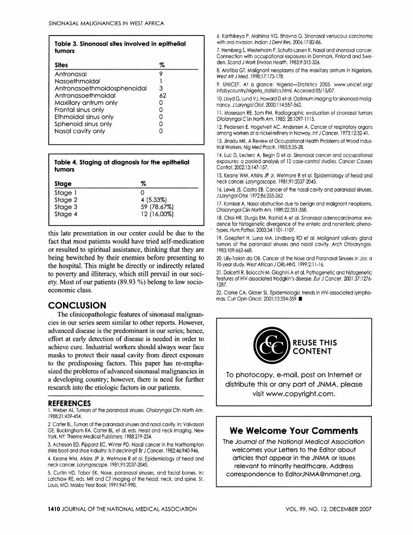
The site of origin of the tumor could not be deter-mined in our patients because at presentation, the tumorhad spread to involve more than one anatomic site oftheparanasal sinuses and nose in the majority of the cases(Table 3).
Seven of our patients were wood workers, two wereinvolved in welding jobs, four were electronic techni-cians that solder, and one had worked in a textile factory.Other patients were traders, civil servants and students.
Fifty-nine (78.67%) of our 75 patients with epithe-lial tumors were at stage 3 at diagnosis, while none pre-sented at stage 1 (Table 4).
DISCUSSIONSinonasal carcinoma represents 3% of all head and
neck malignancies and 0.2-0.8% of all malignancies inthe body.5- In this study, sinonasal malignancies accountedfor 1.57% of all rhinologic diseases. The predominance ofmales seen in this study is similar to other reports in theliterature." 2 Our study showed that the squamous cell car-cinoma was found at a younger age compared to previousreports.478 This may be attributable to the lower life expec-tancy in Nigeria, which is 44 years when compared to othercountries.9 The average duration at presentation in this studywas 8.5 months. The delay in presentation could be attrib-uted, not only to the nonspecific symptoms of the lesion atan early stage but also to the sociocultural beliefs and prac-tices ofthe people in this environment, which lead to delayin presenting to the hospital (Figure 1). In addition, contem-
Table 1. Histologic types of malignant sinonasalneoplasm
Types IncidenceEpithelial 75 (100.00%)
Squamous cell carcinoma 69 (92.00%)Adenocarcinoma 4 (5.33%)Adenoid cystic carcinoma 2 (2.67%)
Nonepithelial 7 (100.00%)
Rhabdomyosarcoma 1 (14.29%)Lymphoma 4 (57.14%)Osteogenic sarcoma 2 (28.57%)
porary radiologic examination tools such as computerizedtomographic (CT) scan and magnetic resonance imaging(MRI), which are effective tools for early detection ofthesesinonasal lesions, are not readily available and affordable inmost practices in the subregion.'0"' Various environmentalfactors, especially the industrial agents, have been reportedas known predisposing factors to sinonasal malignancies.'215 In this study, exposure to environmental risk factors forsinonasal neoplasms was seen in only 14 (17.07%) cases;hence, our impression was that the factors that predisposeour patients to the disease were yet to be identified. Furtherstudies may be indicated in order to find out these factorsin our environment. Exposure to wood dust was found intwo (50%) of our patients with adenocarcinoma, and thisagrees with previous studies that had implicated wood dustin the etiology of adenocarcinoma.7"'3 The offending sub-stances in the wood dusts have been reported to be formicacid and hydrocarbon produced by pyrolysis ofwood. Thefurniture makers who are likely to be exposed to the finewood dusts ofthreshold >Smg/m3/day are at greater risk.'3
The presentation of sinonasal malignant tumordepends on the site involved and direction of spread.Approximately 55% of sinonasal tumors originate fromthe maxillary sinus, 35% from the nasal cavity, 9% fromthe ethmoid sinus and the remainder from the frontaland sphenoid sinuses.'6 In our study, the site of originof the tumor could not be determined, as most of ourpatients had large tumors which had spread to involveadjacent structures before presentation (Figure 2). Sixty-two (82.67%) of our patients with sinonasal carcinomahad involvement of maxillary antrum, ethmoid and nasalcavity at presentation. Bone erosion occurs when thetumor spreads and involves adjacent structures, causingdysfunction and resulting in symptoms. Our patients pre-sented with similar symptoms documented in the litera-
Figure 1. A patient with fungating left sinonasaltumor
1408 JOURNAL OF THE NATIONAL MEDICAL ASSOCIATION VOL. 99, NO. 12, DECEMBER 2007

SINONASAL MALIGNANCIES IN WEST AFRICA
ture. However, all our patients had nasal symptoms thatincluded nasal obstruction, epistaxis or blood-stainednasal discharge. Early complaints are often minimaland can mimic those of chronic sinusitis.6"7 When painoccurs, it is an indicator of perineural extension of themalignancy or tumor infection.6 Pain over the cheek is anearly symptom of adenoid cystic carcinoma, which has ahigher predilection for neural involvement and spread."
Squamous cell carcinoma had been reported to bethe predominant epithelial cell type. In this environ-ment, squamous cell carcinoma appears relatively morecommon as 92.00% of our patients had squamous cellcarcinoma; an earlier similar study had reported 80%.'1Adenocarcinoma represents approximately 10-20% ofall sinonasal malignancies. 8,19 However, in our study, itrepresented 5.33%. The adenocarcinoma is far less com-mon in our environment than what had been reportedelsewhere. Since the histopathologic features of bothare different, the case of misdiagnosis cannot be enter-tained. Lymphoma accounted for 57.14% of all the non-epithelial malignant sinonasal tumors in our center. Thisis different to what had been reported earlier, whererhabdomyosarcoma was reported has having the high-est frequency.9'20 The relatively increased incidence oflymphoma of the sinonasal region might not be uncon-nected with increased HIV infection, which is one ofthe predisposing factors to malignant lymphoma.2"22About 95% of our patients presented at a late stage; 59(78.67%) patients presented at stage 3. The reason for
Table 2. Symptomatology of malignantsinonasal tumors
Symptoms IncidenceNasal
Epistaxis 82 (100.00%)Nasal blockage 82 (100.00%)Nasal discharge 82 (100.00%)Hyponasal speech 74 (90.24%)Anosmia/hyposmia 24 (29.27%)
FacialCheek swelling 76 (92.68%)Cheek pain/paraesthesia 57 (69.51%)
Oral CavityTrismus 28 (34.15%)Loosening/lost tooth 32 (39.02%)Tooth ache 48 (58.54%)Palatal swelling 21 (25.61%)Palatal ulceration 8 (9.76%)
OphthalmicProptosis 25 (30.49%)Double vision 29 (35.37%)Epiphora 33 (40.24%)Visual impairment 16 (19.51%)
OtologicTinnitus 16 (19.51)Aural blockage 12 (14.63%)Hearing impairment 21 (25.61%)
Cerebral/Cranial NerveSeizure 2 (2.44%)Headache 1 (1.22%)
Figure 2. Coronal CT scan showing tumor involving the left nasal cavity, maxillary antrum, ethmoid andorbit
JOURNAL OF THE NATIONAL M A S TV 9 . 12, DCME20L*3-~~~~~~~~~~~~~~~~~~s
JOURNAL OF THE NATIONAL MEDICAL ASSOCIATION VOL. 99, NO. 12. DECEMBER 2007 1409
SINONASAL MALIGNANCIES IN WEST AFRICA
Table 3. Sinonasal sites involved in epithelialtumors
SitesAntronasal 9Nasoethmoidal 1Antronasoethmoidosphenoidal 3Antronasoethmoidal 62Maxillary antrum only 0Frontal sinus only 0Ethmoidal sinus only 0Sphenoid sinus only 0Nasal cavity only 0
Table 4. Staging at diagnosis for the epithelialtumors
StageStage 1 0Stage 2 4 (5.33%)Stage 3 59 (78.67%)Stage 4 12 (16.00%)
this late presentation in our center could be due to thefact that most patients would have tried self-medicationor resulted to spiritual assistance, thinking that they arebeing bewitched by their enemies before presenting tothe hospital. This might be directly or indirectly relatedto poverty and illiteracy, which still prevail in our soci-ety. Most of our patients (89.93 %) belong to low socio-economic class.
CONCLUSIONThe clinicopathologic features of sinonasal malignan-
cies in our series seem similar to other reports. However,advanced disease is the predominant in our series; hence,effort at early detection of disease is needed in order toachieve cure. Industrial workers should always wear facemasks to protect their nasal cavity from direct exposureto the predisposing factors. This paper has re-empha-sized the problems ofadvanced sinonasal malignancies ina developing country; however, there is need for furtherresearch into the etiologic factors in our patients.
REFERENCES1. Weber AL. Tumors of the paranasal sinuses. Otoloryngol Clin North Am.1 988;21 :439-454.2. Carter BL. Tumors of the paronasal sinuses and nasal cavity. In: ValvassoriGE, Buckingham RA, Carter BL, et al, eds. Head and neck imaging. NewYork, NY: Thieme Medical Publishers; 1988:219-234.3. Acheson ED, Pippard EC, Winter PD. Nasal cancer in the Northamptonshire boot and shoe industry: Is it declining? BrJ Cancer. 1982;46:940-946.4. Keane WM, Atkins JP Jr, Wetmore R et al. Epidemiology of head andneck cancer. Laryngoscope. 1981;91:2037-2045.5. Curtin HD, Tabor EK. Nose, paranasal sinuses, and facial bones. In:Latchaw RE. eds. MR and CT imaging of the head, neck, and spine. St.Louis, MO: Mosby Year Book: 1991 :947-990.
6. Karthikeya P, Mahima VG, Bhavna G. Sinonasal verrucous carcinomawith oral invasion. Indian J Dent Res. 2006;1 7:82-86.7. Hemberg S, Westerholm P, Schultz-Larsen K. Nasal and sinonasal cancer.Connection with occupational exposures in Denmark, Finland and Swe-den. Scand J Work Environ Health. 1983;9:315-326.8. Arotiba GT. Malignant neoplasms of the maxillary antrum in Nigerians.West Afr J Med. 1998;1 7:173-178.9. UNICEF. At a glance: Nigeria-Statistics 2005. www.unicef.org/infobycountry/nigeria-statistics.html. Accessed 05/15/07.10. Lloyd G, Lund VJ, Howard D et al. Optimum imaging for sinonasal malig-nancy. J Laryngol Otol. 2000; 114:557-562.11. Mosesson RE, Som PM. Radiographic evaluation of sinonasal tumorsOtolaryngol C 1in North Am. 1985; 28:1097-1115.12. Pedersen E, Hogetveit AC, Andersen A. Cancer of respiratory organsamong workers at a nickel refinery in Norway. Int J Cancer. 1973;12:32-4 1.13. Jinadu MK. A Review of Occupational Health Problems of Wood Indus-trial Workers. Nig Med Practr. 1983;5:25-28.14. Luc D, Leclerc A, Begin D et al. Sinonasal cancer and occupationalexposures: a pooled analysis of 12 case-control studies. Cancer CausesControl. 2002;1 3:147-157.15. Keane WM, Atkins JP Jr, Wetmore R et al. Epidemiology of head andneck cancer. Laryngoscope. 1981;91:2037-2045.16. Lewis JS, Castro EB. Cancer of the nasal cavity and paranasal sinuses.J Laryngol Otol. 1972;86:255-262.17. Komisar A. Nasal obstruction due to benign and malignant neoplasms,Otolaryngol Clin North Am. 1989;22:351-358.18. Choi HR, Sturgis EM, Rashid A et al. Sinonasal adenocarcinoma: evi-dence for histogenetic divergence of the enteric and nonenteric pheno-types. Hum Pathol. 2003;34:1101-1107.19. Goepfert H, Luna MA, Lindberg RD et al. Malignant salivary glandtumors of the paranasal sinuses and nasal cavity. Arch Otoloryngol.1983; 1 09:662-668.20. Lilly-Tariah da OB. Cancer of the Nose and Paranasal Sinuses in Jos: a1 0-year study. West African J ORL-HNS. 1999;2:1 1-16.21. Dolcetti R, Boiocchi M, Gloghini A et al. Pathogenetic and histogeneticfeatures of HIV-associated Hodgkin's disease. Eur J Cancer. 2001;37:1276-1287.22. Clarke CA, Glaser SL. Epidemiologic trends in HIV-associated lympho-mas. Curr Opin Oncol. 2001;13:354-359. 1
*iK-ffi REUSE THISCONTENT
To photocopy, e-mail, post on Internet ordistribute this or any part of JNMA, please
visit www.copyright.com.
We Welcome Your CommentsThe Joumal of the National Medical Association
welcomes your Letters to the Editor aboutarticles that appear in the JNMA or issuesrelevant to minority healthcare. Address
correspondence to [email protected].
1410 JOURNAL OF THE NATIONAL MEDICAL ASSOCIATION VOL. 99, NO. 12, DECEMBER 2007


