
1
Supraventricular Tachycardia
Topics in Cardiovascular CareLancaster General Health
Sandeep Bansal, MD
Cardiac Electrophysiology
The Heart Group
February 6, 2015
Disclosures
• Honoraria for Educational Programs:
– Medtronic
– St. Jude Medical
– Biotronik
– Aurora Health Care
24 year old woman with recurrent palpitations. You should give:A. IV MetoprololB. IV AdenosineC. ReassuranceD. IV fluidsE. DC cardioversion
Tachycardia
• Heart rate > 100 bpm
• Narrow Complex = QRS < 120ms
• Wide Complex = QRS ≥ 120ms
Tachycardia
Narrow QRS
Wide QRS
SVT
• Narrow Complex Tachycardia (NCT)
– Mechanisms
– Diagnostic approach
– Management
Normal Cardiac Conduction System

2
Normal Sinus Rhythm
Narrow Complex Tachycardia
• Must involve the atrium or AV node (supraventricular)
• Must use normal conduction system of the heart
• Can arise from:
– Abnormal automaticity
– Triggered activity
– Re-entry circuit
Mechanisms of NCT
Location Rapidly Firing Focus Re-entry Circuit
SA Node Sinus Tachycardia SA Nodal Reentry Tachycardia
Atrium PACs, Atrial tachycardia, MAT, AF
Atrial tachycardia, Atrial Flutter
AV Node Junctional ectopic tachycardia
AVNRT, ORT
Inappropriate Sinus Tachycardia
• Accelerated baseline sinus rate in the absence of a physiological stressor
• Most commonly in young women without structural heart disease
• Other causes of sinus tachycardia must be excluded
Mechanisms of NCT
Location Rapidly Firing Focus Re-entry Circuit
SA Node Sinus Tachycardia SA Nodal Reentry Tachycardia
Atrium PACs, Atrial tachycardia, MAT, AF
Atrial tachycardia, Atrial Flutter
AV Node Junctional ectopic tachycardia
AVNRT, ORT
Premature Atrial Contractions (PAC)
• Atrial bigeminy

3
Atrial Tachycardia
• Can be single rapidly firing focus in atrium, or small re-entry circuit in atrium
• Heart rates range from 120-200 bpm
• Can be seen with prior surgery or catheter ablation
• Commonly seen with digitalis toxicity
Atrial Tachycardia (ECG)
• Single P wave morphology that looks different than sinus P wave (unless originating near the SA node)
• Abrupt onset and termination
• Discrete P waves with isoelectric period between them (as opposed to atrial flutter)
Atrial Tachycardia Multifocal Atrial Tachycardia
• Multiple (3) competing loci in atrium
• Irregularly irregular (no pattern)
• Atrial rate usually 100 – 180 bpm
• Visible P waves with different morphologies, different P-P and P-R intervals
• Often seen with chronic lung conditions
Multifocal Atrial Tachycardia Atrial Fibrillation
• Most common cardiac arrhythmia
• Rapid and disorganized atrial impulses
• Irregular conduction through AV node leads to RR intervals with no pattern = “irregularly irregular”

4
Atrial Fibrillation
Fibrillatory activity
Sinus P wave
Mechanisms of NCT
Location Rapidly Firing Focus Re-entry Circuit
SA Node Sinus Tachycardia SA Nodal Reentry Tachycardia
Atrium PACs, Atrial tachycardia, MAT, AF
Atrial tachycardia, Atrial Flutter
AV Node Junctional ectopic tachycardia
AVNRT, ORT
• Caused by sudden rapid pacing of an automatic focus in the AV Junction
• 150-250 Beats per minute
• May also see retrograde P-waves immediately before, after, or buried in QRS
Junctional Ectopic Tachycardia Mechanisms of NCT
Location Rapidly Firing Focus Re-entry Circuit
SA Node Sinus Tachycardia SA Nodal Reentry Tachycardia
Atrium PACs, Atrial tachycardia, MAT, AF
Atrial tachycardia, Atrial Flutter
AV Node Junctional ectopic tachycardia
AVNRT, ORT
Re-Entry Circuits
• Electrical impulse travels in a tight circle within the heart, leading to fast heart rates
• Requires:
– 2 different refractory periods
– Zone of slow conduction
• Premature beat typically initiates the cycle
Sinus Node Reentrant Tachycardia (SNRT)
• A subset of focal atrial tachycardia due to reentry arising within the sinus node
• The P waves are identical to sinus tachycardia
• Abrupt onset and termination
• Atrial rate usually 100-150 bpm
• Unable to distinguish from sinus tachycardia on ECG unless onset/termination seen

5
Mechanisms of NCT
Location Rapidly Firing Focus Re-entry Circuit
SA Node Sinus Tachycardia SA Nodal Reentry Tachycardia
Atrium PACs, Atrial tachycardia, MAT, AF
Atrial tachycardia, Atrial Flutter
AV Node Junctional ectopic tachycardia
AVNRT, ORT
Atrial Flutter
• Caused by re-entrant rhythm, typically in the right atrium around cavo-tricuspid isthmus
• Typical atrial rate is 240-300 bpm
• “F waves” are seen on ECG: rapid, negative sawtooth atrial waves without isoelectric intervals between them
F-waves
Atrial Flutter
•Typical counter-clockwise flutter around cavo-tricuspid isthmus
•“Saw-tooth” F waves, negative in leads II, III, and aVF
Typical Atrial Flutter
Mechanisms of NCT
Location Rapidly Firing Focus Re-entry Circuit
SA Node Sinus Tachycardia SA Nodal Reentry Tachycardia
Atrium PACs, Atrial tachycardia, MAT, AF
Atrial tachycardia, Atrial Flutter
AV Node Junctional ectopic tachycardia
AVNRT, ORT
AV Nodal Reentrant Tachycardia (AVNRT)
• Most common form of reentrant SVT; 60% of all paroxysmal SVT patients; more common in women
• Usually between 150-250 bpm
• Requires “dual AV nodal physiology”
6
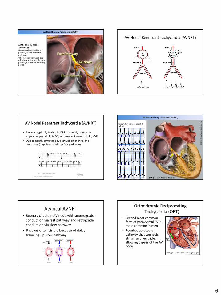
AV Nodal Reentry Tachycardia (AVNRT)
AVNRT Dual AV node physiology
•Functionally divided into 2 pathways – fast and slowpathways
•The fast pathway has a long refractory period and the slow pathway has a short refractory period
AV Nodal Reentrant Tachycardia (AVNRT)
AV Nodal Reentrant Tachycardia (AVNRT)
• P waves typically buried in QRS or shortly after (can appear as pseudo R’ in V1, or pseudo S wave in II, III, aVF)
• Due to nearly simultaneous activation of atria and ventricles (impulse travels up fast pathway)
AV Nodal Re-entry Tachycardia (AVNRT)
Retrograde P-waves in leads I, II, V1-V3
Atypical AVNRT• Reentry circuit in AV node with anterograde
conduction via fast pathway and retrograde conduction via slow pathway
• P waves often visible because of delay traveling up slow pathway
Orthodromic Reciprocating Tachycardia (ORT)
• Second most common form of paroxysmal SVT; more common in men
• Requires accessory pathway that connects atrium and ventricle, allowing bypass of the AV node

7
Accessory Pathway
• Atrioventicular bypass tract is known as the Bundle of Kent (conduction can occur anterograde, retrograde, or both)
• During sinus rhythm, anterograde conduction down accessory pathway leads to ventricular “pre-excitation”
– Short PR interval
– Delta wave
– Widened QRS
Wolff-Parkinson-White pattern: Pre-excitation
ECG requirements for diagnosis of WPW pattern
•Short PR interval (< 120 ms)
•Normal P wave vector (to exclude junctional rhythm)
•Presence of a delta wave
•QRS duration > 100 ms
Orthodromic Reciprocating Tachycardia (ORT)
• Reentry circuit is established by conduction traveling anterograde through the AV node and retrograde through the accessory pathway
• Retrograde P wave usually falls after QRS complex
Orthodromic Reciprocating Tachycardia (ORT)
Supraventricular tachycardia•can be initiated by a closely
coupled premature atrial complex (PAC)
•blocks in accessory pathway, but conducts through AV node
•retrograde conduction via accessory pathway
•inverted P wave produced by retrograde conduction visible in the inferior ECG leads
Orthodromic Reciprocating Tachycardia (ORT)
Retrograde P wave
Retrograde P wave
Mechanisms of NCT
Location Rapidly Firing Focus Re-entry Circuit
SA Node Sinus Tachycardia SA Nodal Reentry Tachycardia
Atrium PACs, Atrial tachycardia, MAT, AF
Atrial tachycardia, Atrial Flutter
AV Node Junctional ectopic tachycardia
AVNRT, ORT

8
SVT
• Narrow Complex Tachycardia (NCT)
– Mechanisms
– Diagnostic approach
– Management
NCT Evaluation
Key: Is the rhythm regular?
Tachycardia
Narrow QRS
Irregular Rhythm
Regular Rhythm
Wide QRS
Regular NCT
• If regular, can be due to
– Single rapidly firing focus
– Reentry rhythm
• Find the P-waves!
• Key point: determine if sinus tachycardia
Regular NCT
Location Rapidly Firing Focus Re-entry Circuit
SA Node Sinus Tachycardia SA Nodal Reentry Tachycardia
Atrium PACs, Atrialtachycardia, MAT, AF
Atrial tachycardia, Atrial Flutter
AV Node Junctional ectopic tachycardia
AVNRT, ORT
Irregular NCT
• If QRS’s are irregular, can be due to:
– Multiple rapidly firing foci (MAT, AF)
– Any regular NCT with variable AV conduction
• Find the P-waves!
• Key point: determine if atrial fibrillation or MAT
Irregular NCT
• Atrial rhythm is regular (300 bpm)
• Atrial flutter with variable conduction

9
NCT Diagnosis Approach
• If either: – Regular rhythm, but not sinus tachycardia– Irregular rhythm, but not AF/MAT
Slow conduction through AV node to help with diagnosis
– Atrial activity can be better visualized– Re-entry circuits that involve the AV node will be
terminated
• Vagal maneuvers:– Carotid sinus massage – Valsalva
• Pharmacologic: adenosine
Adenosine• Most potent AV nodal blocker• Very short half-life (10-20 seconds)• Give 6mg, 12mg, 12mg as rapid push with saline
flush• Typical side effects include flushing, chest pain, and
dizziness – warn the patient!• Contraindicated in asthma or severe COPD• You should always get transient AV block – if not,
enough adenosine did not get to the AV node• Will terminate AVNRT or AVRT 90% of the time• Does not terminate atrial fibrillation or MAT
SVT
• Narrow Complex Tachycardia (NCT)
– Mechanisms
– Diagnostic approach
– Management
Initial Triage
Step 1:
Stable or Unstable?
Tachycardia
Unstable
Due to Rhythm
Cardioversion
Due to Other Causes (e.g.
sepsis)
Treat Underlying
Cause
Stable
Initial TriageAV Block:
– Carotid Sinus Massage
– Valsalva
– Adenosine
Rate Control:
– Beta-blockers
– Calcium channel blockers
– Digoxin
– Amiodarone
Tachycardia
Unstable Stable
Narrow Complex
Irregular
AF/MAT
Rate Control
Regular
All others
AV block
Sinus Tachycardia
Treat underlying
Wide Complex
Work-up of NCT
• Comprehensive history and physical exam
• Consider:
– Thyroid function testing (hyper or hypothyroidism)
– Electrolyte testing (potassium and magnesium)
– Hemoglobin measurement (anemia)
– Cardiac biomarkers (in at risk patients)
– Digoxin level (if prescribed)
– Echocardiogram (to rule out structural heart disease)
– Chest x-ray (cardiomegaly, pulmonary disease, edema)

10
Chronic Management:Re-entry Circuits
• For large reentry circuits, catheter ablation can be curative:– Atrial flutter
– AVNRT
– Accessory pathway (AVRT)
• Anti-arrhythmic therapy may be useful as well
• For infrequent AVNRT, teaching patient vagal maneuvers may be adequate
Chronic Management:Rapidly Firing Foci
• For rapidly firing foci:
– Treat underlying cause
– Catheter ablation
– Antiarrhythmics can be useful
– Rate control
24 year old woman with recurrent palpitations. You should give:A. IV MetoprololB. IV AdenosineC. ReassuranceD. IV fluidsE. DC cardioversion
54 year old man with no PMH and new onset dyspnea, fatigue. You suggest:
A. Cavotricuspid isthmus ablation
B. Pulmonary vein isolation
C. Amiodarone/cardioversion
D. Flecainide 100mg BID with Metoprolol ER 25mg daily
E. Metoprolol XL 100mg
84 year old man with recent long hospitalization for sacral decubitus ulcers,
presents with acute onset dyspnea and chest pain. BP 80/50. You suggest:
A. Adenosine
B. Emergent cardioversion
C. Amiodarone
D. Metoprolol 5mg IV push x 3
E. IV fluids


