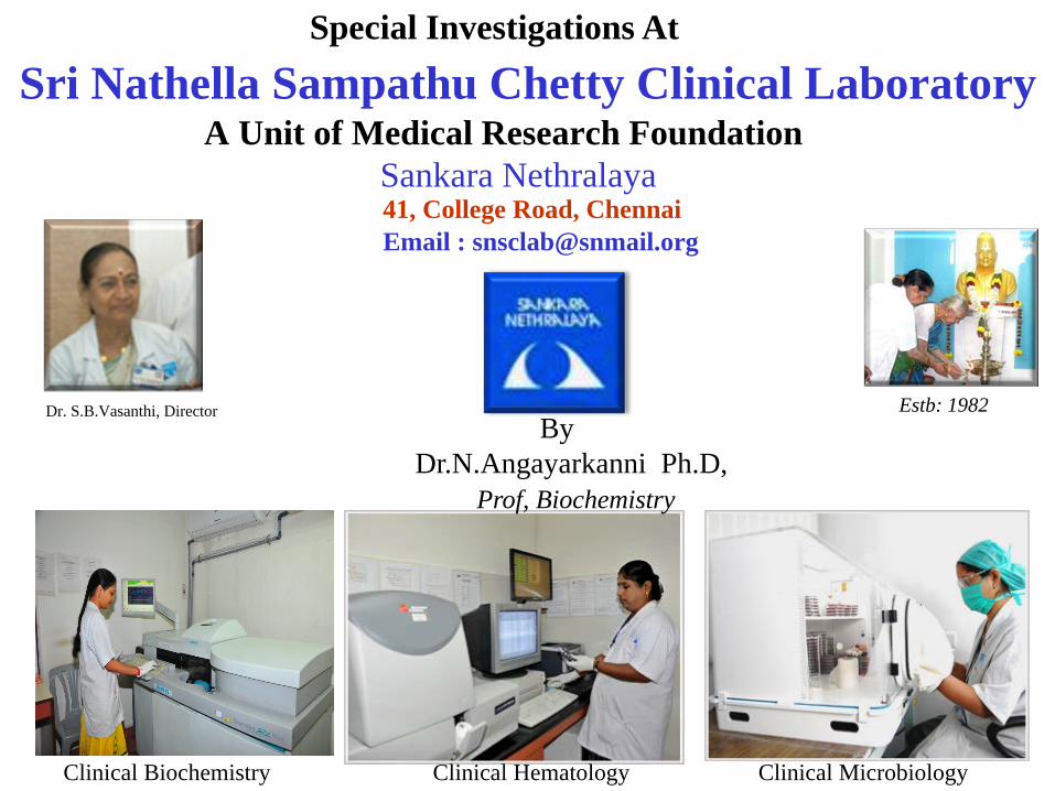
Sri Nathella Sampathu Chetty Clinical LaboratoryA Unit of Medical Research Foundation
Sankara Nethralaya 41, College Road, Chennai
Clinical Microbiology
Special Investigations At
Estb: 1982Dr. S.B.Vasanthi, Director By
Dr.N.Angayarkanni Ph.D, Prof, Biochemistry
Email : [email protected]
Clinical HematologyClinical Biochemistry
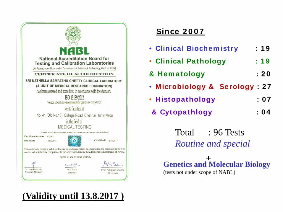
Since 2007
• Clinical Biochemistry : 19
• Clinical Pathology : 19
& Hematology : 20
• Microbiology & Serology : 27
• Histopathology : 07
& Cytopathlogy : 04
Total : 96 TestsRoutine and special
Genetics and Molecular Biology(tests not under scope of NABL)
+
(Validity until 13.8.2017 )
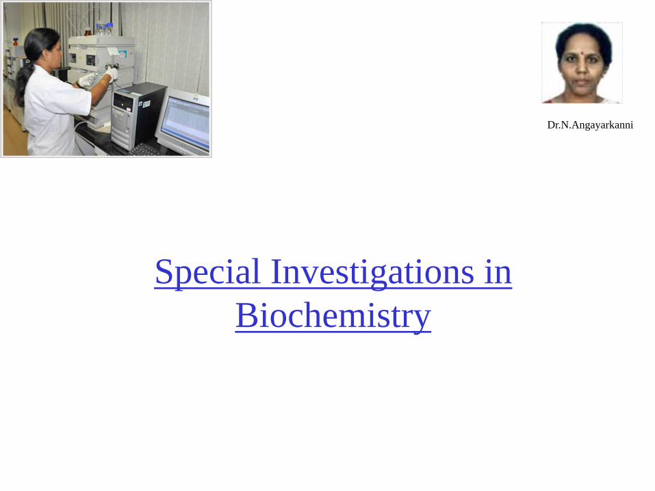
Special Investigations in Biochemistry
Dr.N.Angayarkanni
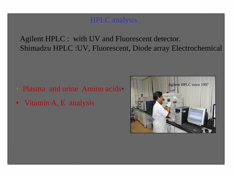
HPLC analysis
Agilent HPLC : with UV and Fluorescent detector.Shimadzu HPLC :UV, Fluorescent, Diode array Electrochemical
Agilent HPLC since 1997• Plasma and urine Amino acids*
• Vitamin A, E analysis
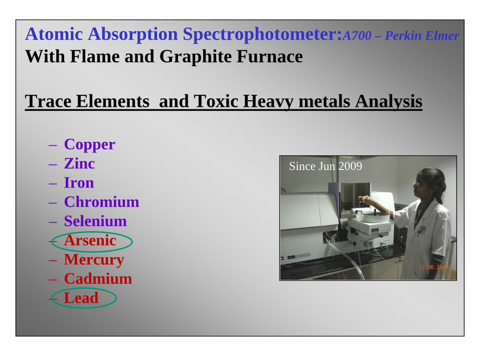
Atomic Absorption Spectrophotometer:A700 – Perkin Elmer
With Flame and Graphite Furnace
Trace Elements and Toxic Heavy metals Analysis
– Copper– Zinc– Iron – Chromium– Selenium– Arsenic– Mercury– Cadmium– Lead
Since Jun 2009
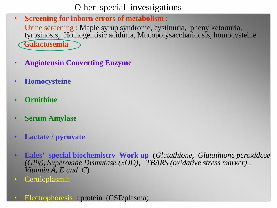
• Screening for inborn errors of metabolism : Urine screening : Maple syrup syndrome, cystinuria, phenylketonuria, tyrosinosis, Homogentisic aciduria, Mucopolysaccharidosis, homocysteineGalactosemia
• Angiotensin Converting Enzyme
• Homocysteine
• Ornithine
• Serum Amylase
• Lactate / pyruvate
• Eales’ special biochemistry Work up (Glutathione, Glutathione peroxidase (GPx), Superoxide Dismutase (SOD), TBARS (oxidative stress marker) , Vitamin A, E and C)
• Ceruloplasmin
• Electrophoresis : protein (CSF/plasma)
Other special investigations
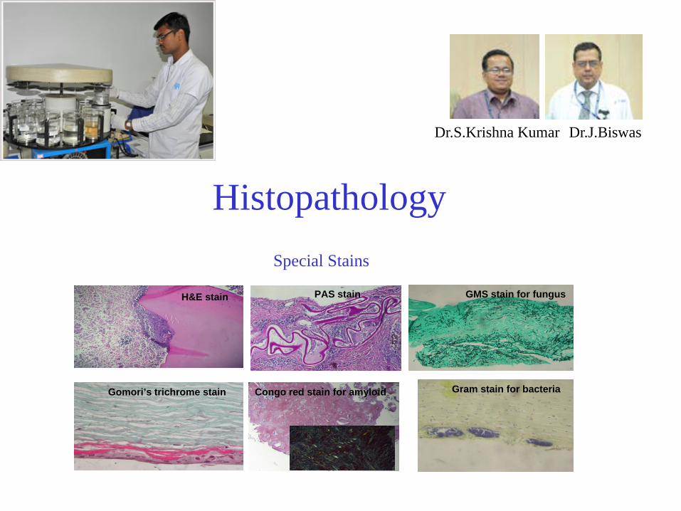
HistopathologySpecial Stains
H&E stain PAS stain
Gomori’s trichrome stain Congo red stain for amyloid Gram stain for bacteria
GMS stain for fungus
Dr.S.Krishna Kumar Dr.J.Biswas
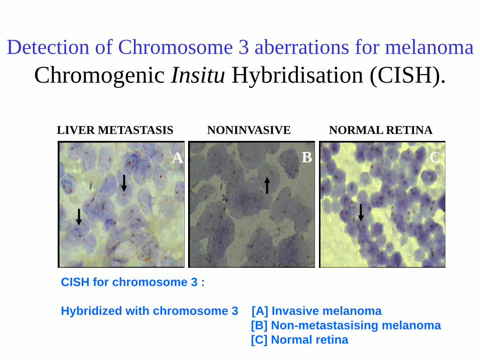
Detection of Chromosome 3 aberrations for melanomaChromogenic Insitu Hybridisation (CISH).
NONINVASIVE NORMAL RETINA
A B C
LIVER METASTASIS
CISH for chromosome 3 :
Hybridized with chromosome 3 [A] Invasive melanoma [B] Non-metastasising melanoma[C] Normal retina
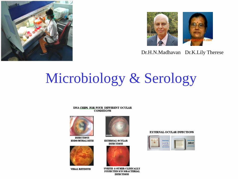
Microbiology & Serology
Dr.H.N.Madhavan Dr.K.Lily Therese
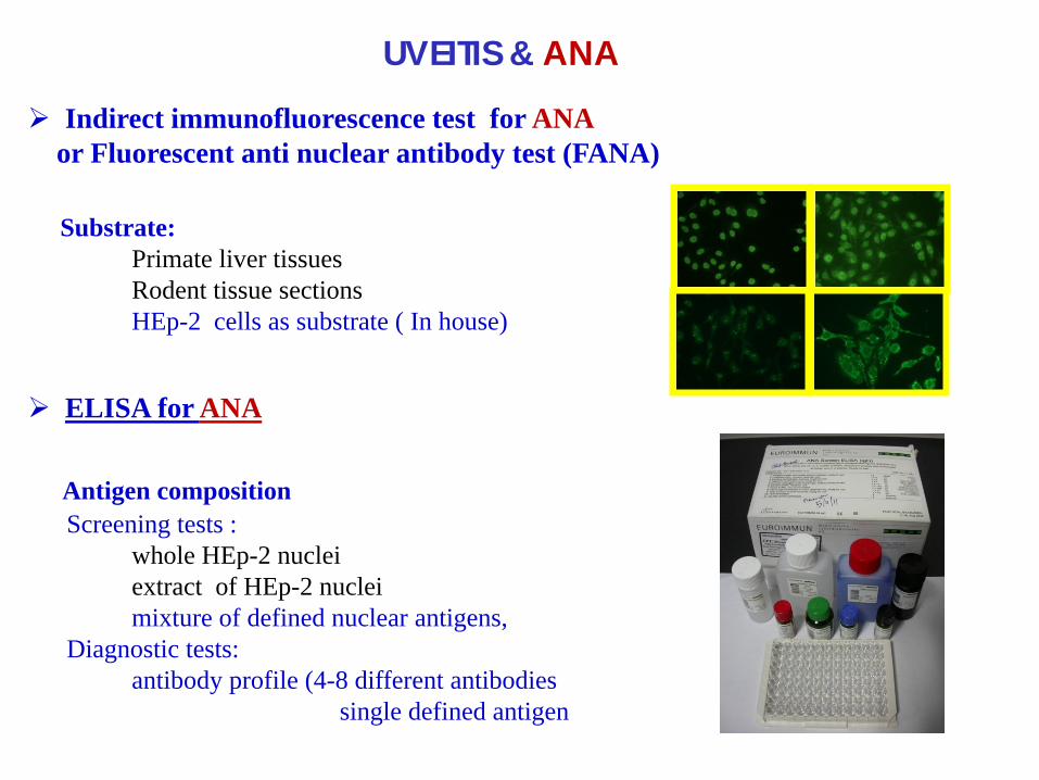
Indirect immunofluorescence test for ANA or Fluorescent anti nuclear antibody test (FANA)
Substrate: Primate liver tissuesRodent tissue sectionsHEp-2 cells as substrate ( In house)
ELISA for ANA
Antigen compositionScreening tests :
whole HEp-2 nucleiextract of HEp-2 nuclei mixture of defined nuclear antigens,
Diagnostic tests: antibody profile (4-8 different antibodies
single defined antigen
UVEITIS & ANA
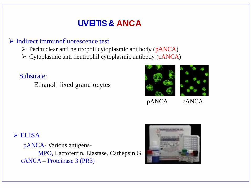
Indirect immunofluorescence test Perinuclear anti neutrophil cytoplasmic antibody (pANCA) Cytoplasmic anti neutrophil cytoplasmic antibody (cANCA)
Substrate: Ethanol fixed granulocytes
ELISA pANCA- Various antigens-
MPO, Lactoferrin, Elastase, Cathepsin GcANCA – Proteinase 3 (PR3)
pANCA cANCA
UVEITIS & ANCA
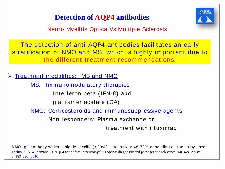
Detection of AQP4 antibodies
Jarius, S. & Wildemann, B. AQP4 antibodies in neuromyelitis optica: diagnostic and pathogenetic relevance Nat. Rev. Neurol. 6, 383–392 (2010)
NMO-IgG antibody which is highly specific (>99%) , sensitivity 48-72% depending on the assay used.
Neuro Myelitis Optica Vs Multiple Sclerosis
The detection of anti-AQP4 antibodies facilitates an early stratification of NMO and MS, which is highly important due to
the different treatment recommendations.
Treatment modalities: MS and NMOMS: Immunomodulatory therapies
Interferon beta (IFN-ß) and glatiramer acetate (GA)
NMO: Corticosteroids and immunosuppressive agents.Non responders: Plasma exchange or
treatment with rituximab
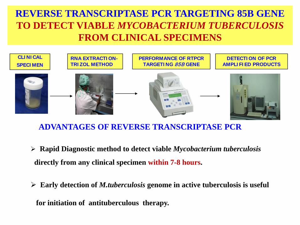
REVERSE TRANSCRIPTASE PCR TARGETING 85B GENE TO DETECT VIABLE MYCOBACTERIUM TUBERCULOSIS
FROM CLINICAL SPECIMENS
CLINICAL SPECIMEN
RNA EXTRACTION-TRIZOL METHOD
PERFORMANCE OF RTPCR TARGETING 85B GENE
DETECTION OF PCR AMPLIFIED PRODUCTS
ADVANTAGES OF REVERSE TRANSCRIPTASE PCR
Rapid Diagnostic method to detect viable Mycobacterium tuberculosis
directly from any clinical specimen within 7-8 hours.
Early detection of M.tuberculosis genome in active tuberculosis is useful
for initiation of antituberculous therapy.
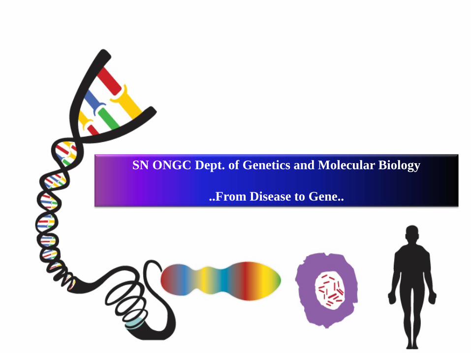
SN ONGC Dept. of Genetics and Molecular Biology
..From Disease to Gene..
Name of the test Condition for which the test is done Specimen
Rhodopsin (RHO) Gene Screening Retinitis pigmentosa & Congenital Stationary Night Blindness
RPE65 Gene Screening Retinitis Pigmentosa & Leber’sCongenital Amaurosis
CYP1B1 Gene ScreeningPeter’s anomaly, Congenital Glaucoma,Primary and Juvenile Open Angle Glaucoma
MYOC Gene Screening Primary and Juvenile Open Angle Glaucoma
PAX6 Gene Screening Peter’s anomaly & AniridiaRetinoschisis (RS1) Gene Screening RetinoschisisCytogenetic analysis for Retinoblastoma Retinoblastoma
Screening the three primary mitochondrial mutations for LebersHereditary Optic Neuropathy (LHON)
Lebers Hereditary Optic Neuropathy (LHON)
BBS Genes screening for Bardet –Biedl Syndrome Bardet – Biedl Syndrome
Chromosomal Study Chromosomal abnormalities
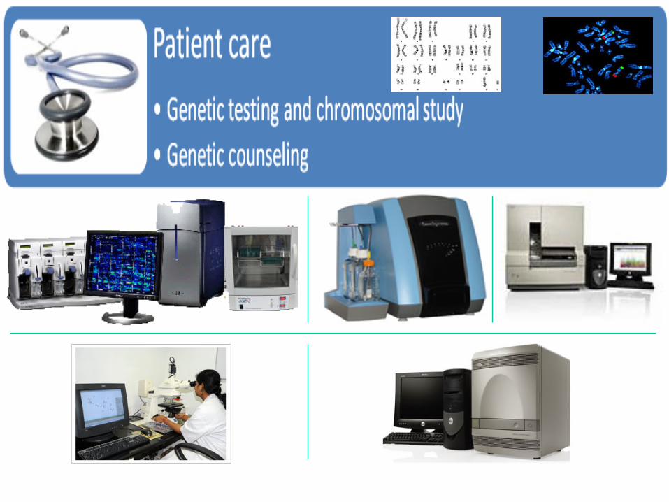
Genetic testing and Chromosomal study
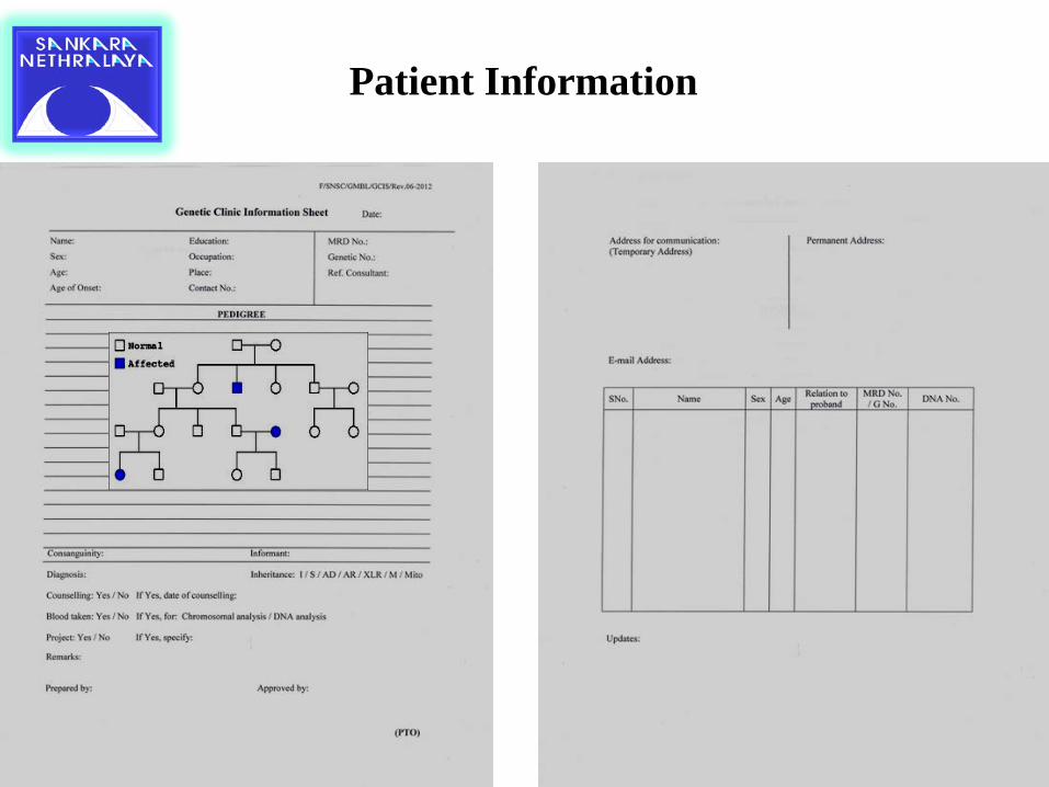
Patient Information
80
60
40
20
0
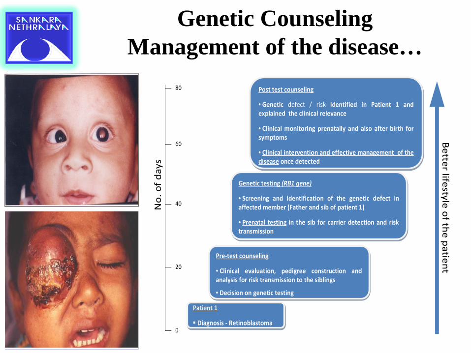
Patient 1
Diagnosis - Retinoblastoma
Patient 1
Diagnosis - Retinoblastoma
Pre-test counseling
• Clinical evaluation, pedigree construction and analysis for risk transmission to the siblings
• Decision on genetic testing
Pre-test counseling
• Clinical evaluation, pedigree construction and analysis for risk transmission to the siblings
• Decision on genetic testing
Genetic testing (RB1 gene)
• Screening and identification of the genetic defect in affected member (Father and sib of patient 1)
• Prenatal testing in the sib for carrier detection and risk transmission
Genetic testing (RB1 gene)
• Screening and identification of the genetic defect in affected member (Father and sib of patient 1)
• Prenatal testing in the sib for carrier detection and risk transmission
Post test counseling
• Genetic defect / risk identified in Patient 1 and explained the clinical relevance
• Clinical monitoring prenatally and also after birth for symptoms
• Clinical intervention and effective management of the disease once detected
Post test counseling
• Genetic defect / risk identified in Patient 1 and explained the clinical relevance
• Clinical monitoring prenatally and also after birth for symptoms
• Clinical intervention and effective management of the disease once detected
No.
of d
ays
Better lifestyle of the patientGenetic Counseling
Management of the disease…
Hematological Investigations
Dr.S.B.Vasanthi Dr.Doreen Gracias
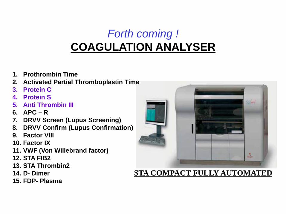
Forth coming !COAGULATION ANALYSER
1. Prothrombin Time2. Activated Partial Thromboplastin Time3. Protein C4. Protein S5. Anti Thrombin III6. APC – R 7. DRVV Screen (Lupus Screening) 8. DRVV Confirm (Lupus Confirmation)9. Factor VIII 10. Factor IX 11. VWF (Von Willebrand factor)12. STA FIB2 13. STA Thrombin2 14. D- Dimer15. FDP- Plasma
STA COMPACT FULLY AUTOMATED
Jaypee: publisherRs. 450/-
A Sankara Nethralaya publication
Thank you
SNSC clinical laboratory
21.7.13

![Review Article Role of Glutathione in Cancer Progression ...downloads.hindawi.com/journals/omcl/2013/972913.pdf · GCL and glutathione S-transferases [ ]. 2. GSH Biosynthesis Glutathione](https://cdn.vdocuments.site/doc/165x107/5edbd12aad6a402d666637cd/review-article-role-of-glutathione-in-cancer-progression-gcl-and-glutathione.jpg)


