Download - seminar on intercellular junctions

1

2
INTERCELLULAR JUNCTIONS
Presented By:Dr. Mamta SinghPGT - I
3
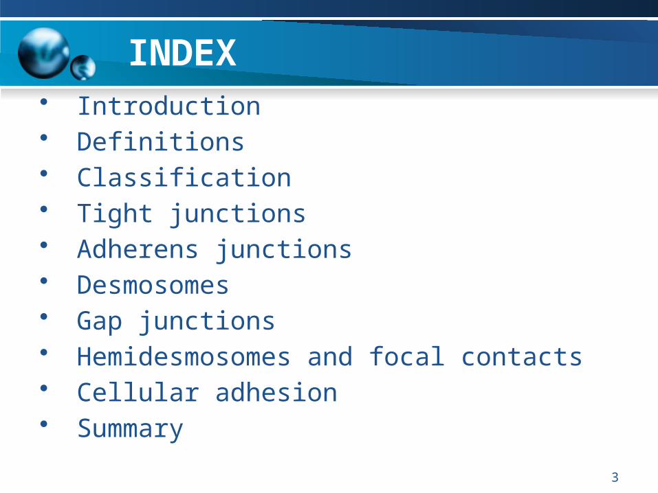
INDEX• Introduction• Definitions• Classification• Tight junctions• Adherens junctions• Desmosomes• Gap junctions• Hemidesmosomes and focal contacts• Cellular adhesion• Summary
4
INTERCELLULAR JUNCTIONS
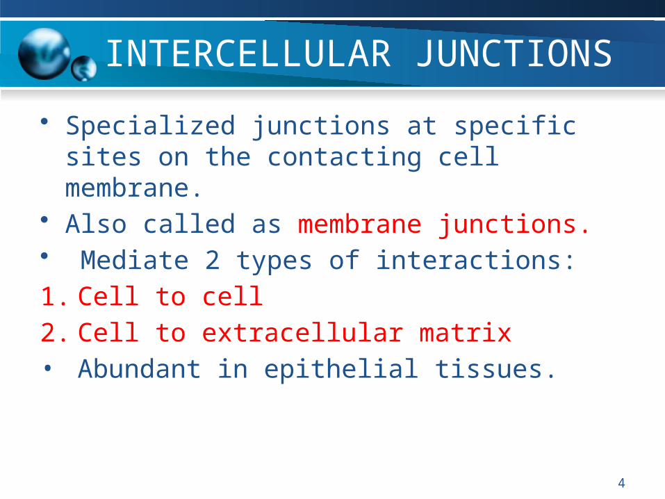
• Specialized junctions at specific sites on the contacting cell membrane.
• Also called as membrane junctions.• Mediate 2 types of interactions:
1. Cell to cell
2. Cell to extracellular matrix• Abundant in epithelial tissues.
5
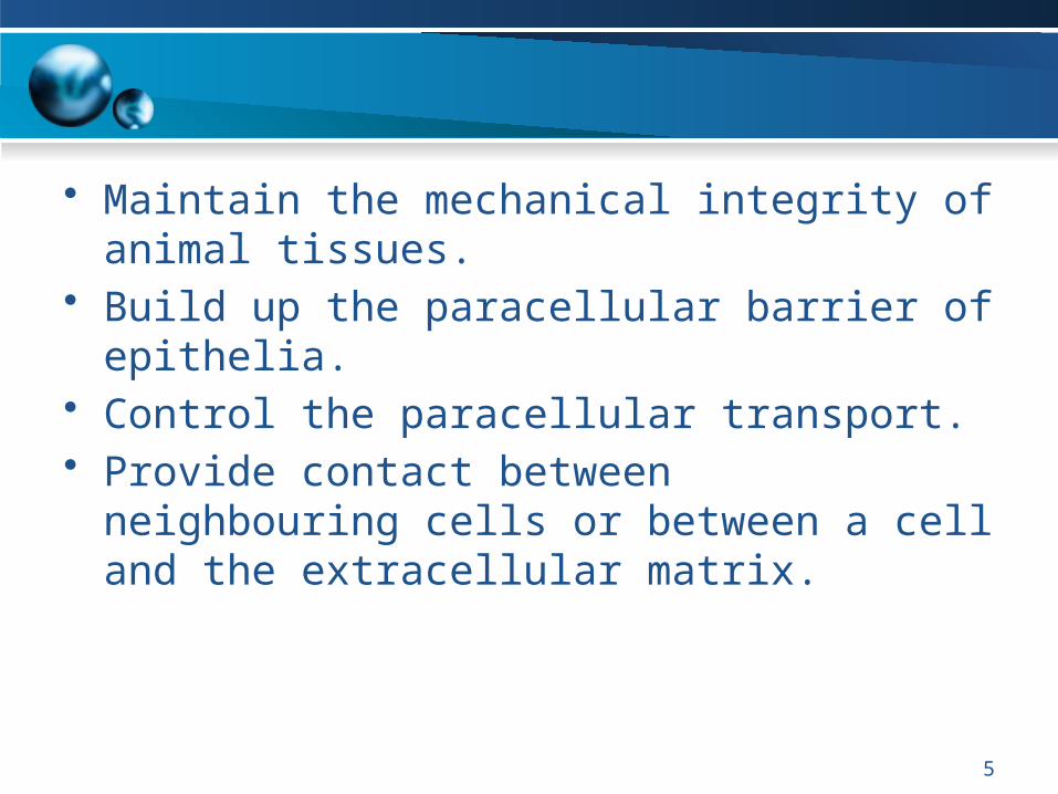
• Maintain the mechanical integrity of animal tissues.
• Build up the paracellular barrier of epithelia.• Control the paracellular transport.• Provide contact between neighbouring cells or
between a cell and the extracellular matrix.
6
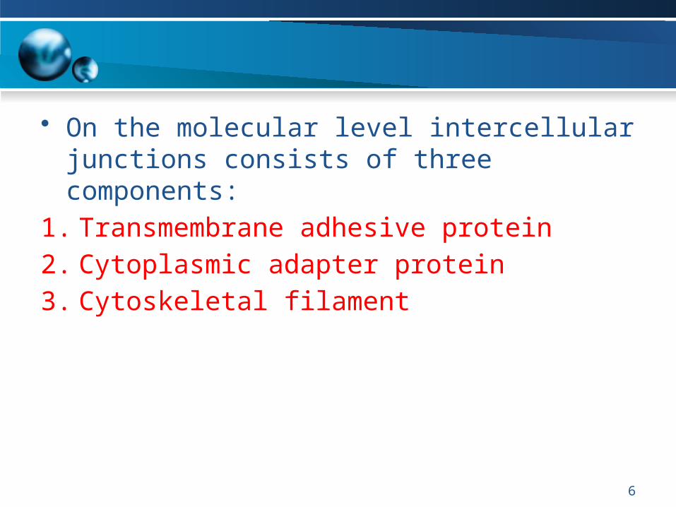
• On the molecular level intercellular junctions consists of three components:
1. Transmembrane adhesive protein
2. Cytoplasmic adapter protein
3. Cytoskeletal filament
7
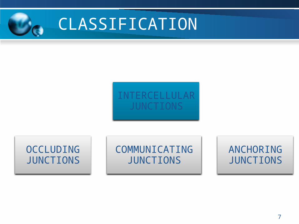
CLASSIFICATION
INTERCELLULAR JUNCTIONS
OCCLUDING JUNCTIONS
COMMUNICATING JUNCTIONS
ANCHORING JUNCTIONS
8
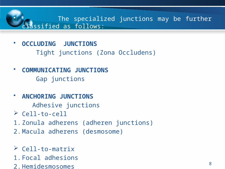
• The specialized junctions may be further classified as follows:
• OCCLUDING JUNCTIONS
Tight junctions (Zona Occludens)
• COMMUNICATING JUNCTIONS
Gap junctions
• ANCHORING JUNCTIONS
Adhesive junctions Cell-to-cell
1. Zonula adherens (adheren junctions)
2. Macula adherens (desmosome)
Cell-to-matrix
1. Focal adhesions
2. Hemidesmosomes

9
10
Definitions
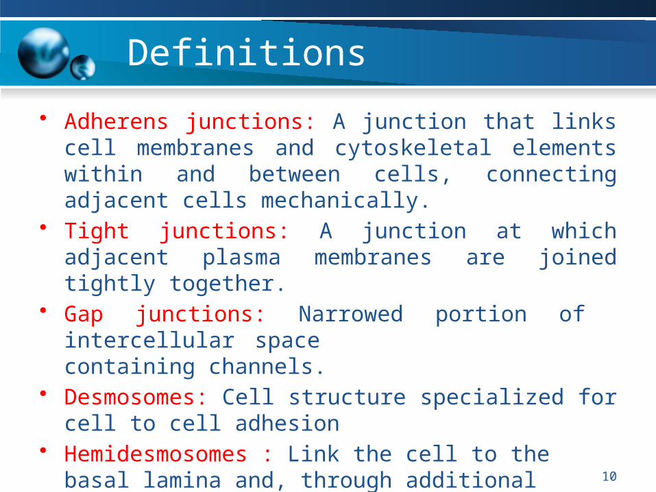
• Adherens junctions: A junction that links cell membranes and cytoskeletal elements within and between cells, connecting adjacent cells mechanically.
• Tight junctions: A junction at which adjacent plasma membranes are joined tightly together.
• Gap junctions: Narrowed portion of intercellular space containing channels.
• Desmosomes: Cell structure specialized for cell to cell adhesion
• Hemidesmosomes : Link the cell to the basal lamina and, through additional extracellular molecules, to ECM.
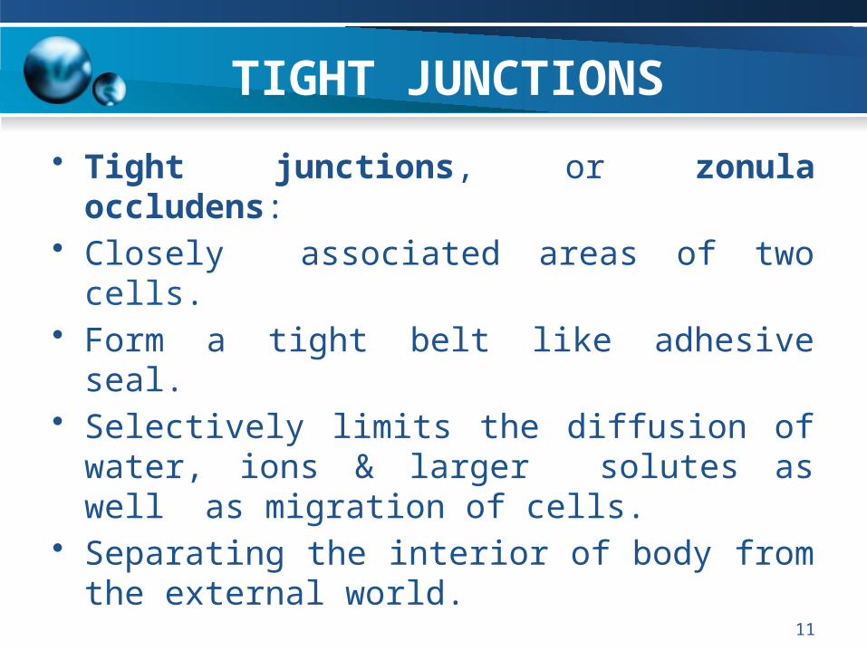
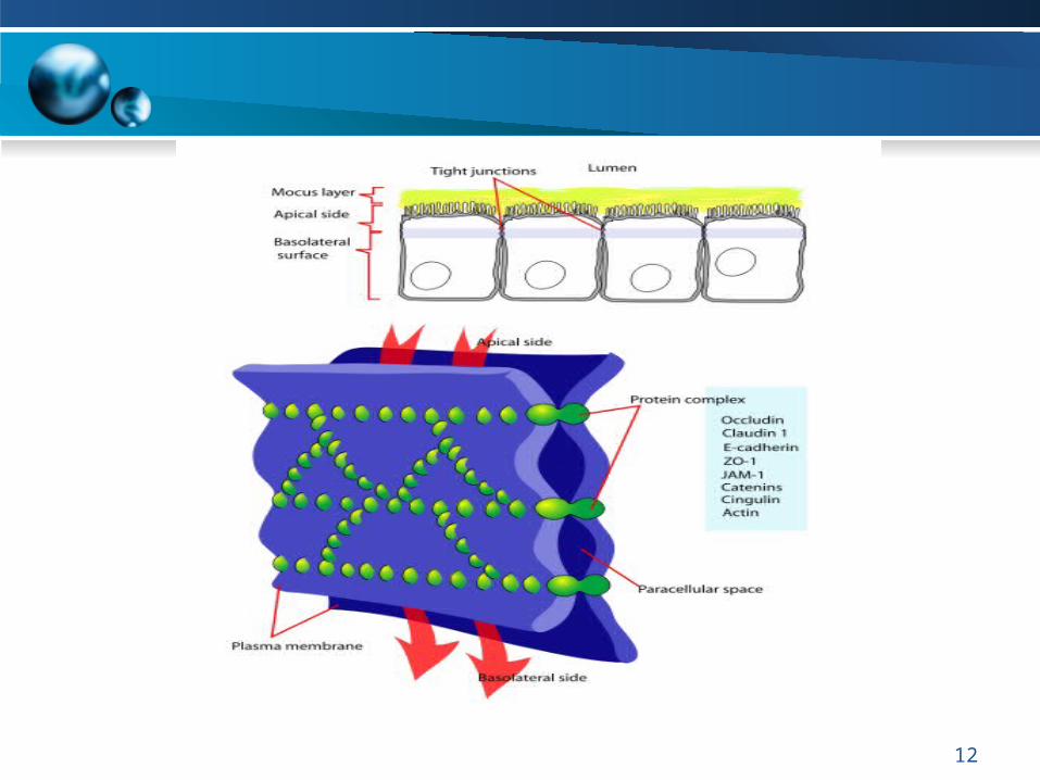
TIGHT JUNCTIONS
• Tight junctions, or zonula occludens: • Closely associated areas of two cells.• Form a tight belt like adhesive seal.• Selectively limits the diffusion of water, ions &
larger solutes as well as migration of cells.• Separating the interior of body from the external
world.
11
12
13
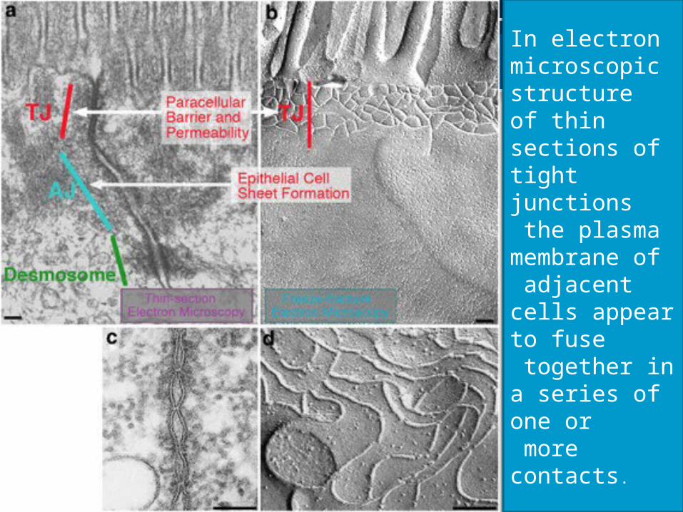
In electron microscopic structureof thin sections of tight junctions the plasma membrane of adjacent cells appear to fuse together in a series of one or more contacts.
• Two structural proteins have been identified in the structure of tight junctions namely:
• Occludins• Claudin
14
16
• A family of more than 20 proteins, called Claudins, constitutes the main structural proteins of tight junction strands.
• Claudins have four transmembrane sequences, but they are not related in sequence to occludin.
17
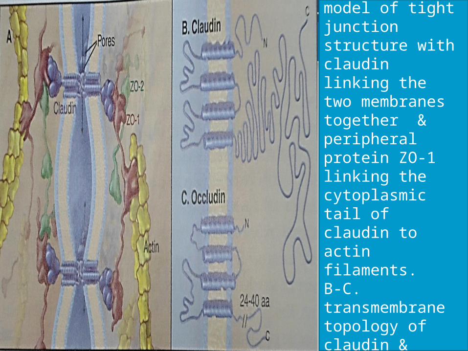
A.Preliminary model of tight junction structure with claudin linking the two membranes together & peripheral protein ZO-1 linking the cytoplasmic tail of claudin to actin filaments.B-C. transmembrane topology of claudin & occludin.
Tight junctions
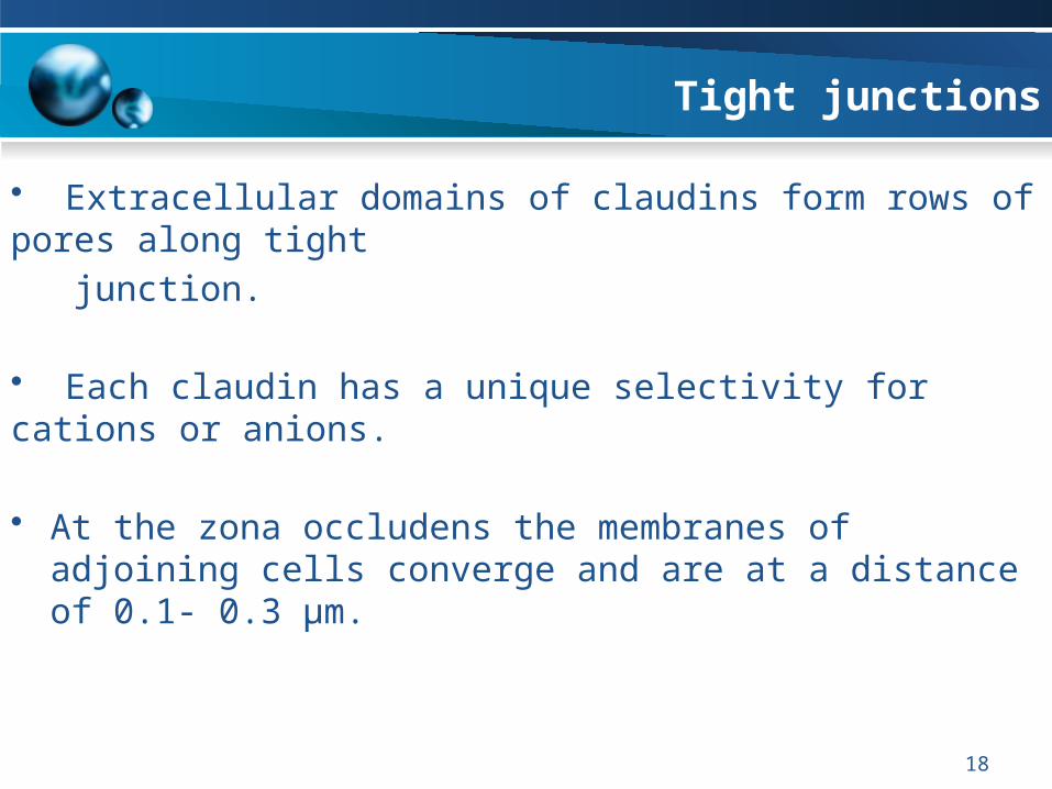
• Extracellular domains of claudins form rows of pores along tight
junction.
• Each claudin has a unique selectivity for cations or anions.
• At the zona occludens the membranes of adjoining cells
converge and are at a distance of 0.1- 0.3 µm.
18

19
• Human gingival keratinocytes (HGKs) were studied by means of freeze-fracture technique for the investigation of intercellular contacts.
• In vivo the tight junctions, which were of low complexity were co-distributed with desmosomes; in one case, the strands ran directly through desmosomal plaques.
• Where tight junctions and desmosomes occurred together, no gap junctions were seen.
• In contrast, where no tight junctions were present, gap junctions and desmosomes were co-localized.
Meyle J, Güttig K, Rascher G and Wolburg H: Transepithelial electrical resistance and tight junctions of human gingival keratinocvtes. J Periodont Res 1999; 34: 214–222
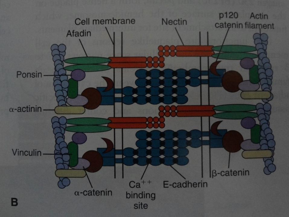
• Adherens junctions and Desmosomes are two types of adhesive junctions using homophilic interactions of cadherins to bind epithelial cells to adjacent cells.
20
ADHERENS JUNCTIONS
• The zonula adherens is a band like specialization of the membrane and cytoplasm that encircles the apex of adjoining cells and strongly bonds the cells together.
• In this junction the opposing membranes are 15 – 20 nm apart .
• It is a major site of epithelial cell cohesion.
Adherens Junction
• Cytoplasmic actin filaments bind adherens junctions.
• Homophilic interactions between densely clustered E-cadherens (the epithelial transmembranic adhesive protein) bind adjacent cells together at adherens junctions.
• β- catenin(cytoplasmic adapter protein) and Plakoglobin (desmosomal cytoplasmic adapter protein) bind the cytoplasmic domains of E-cadherin.
Adherens Junction
• An another cytoplasmic adapter protein, α-catenin, binds cadherins to actin filaments and β-catenin to actin filaments.
• Adherens junctions are first connections that are established between developing sheets of epithelial cells.
• The contact begins when cadherins on the tips of filopodia engage to the cadherins of another cell.
Adherens Junction
• Adherens junctions are a pre requisite for tight junctions that allow epithelial cells to establish polarity with proteins and lipids in plasma membranes.
• Zonula adherens is the major site for cell cohesion.
• It stabilizes the surface of epithelia.
• The junctions and polarity determine the orientation of mitotic spindle and the plane of division . This allows for asymmetrical division of stem cells (stratified epithelium).
Adherens junction
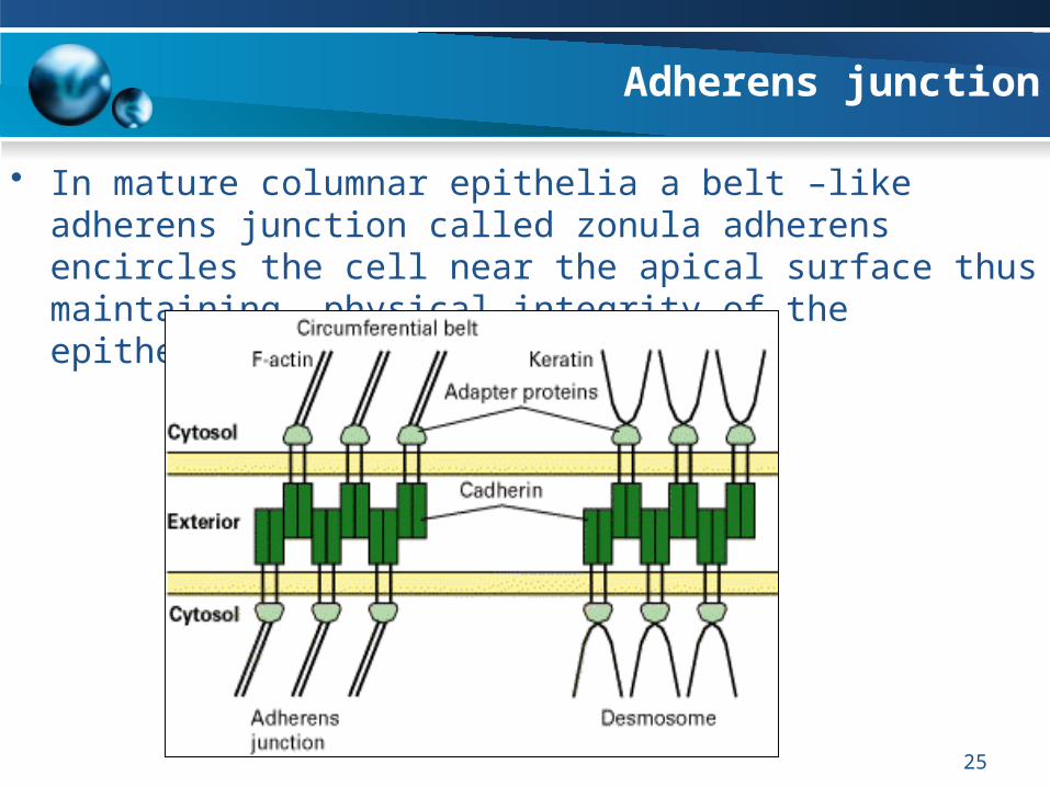
• In mature columnar epithelia a belt –like adherens junction called zonula adherens encircles the cell near the apical surface thus maintaining physical integrity of the epithelium.
25
26
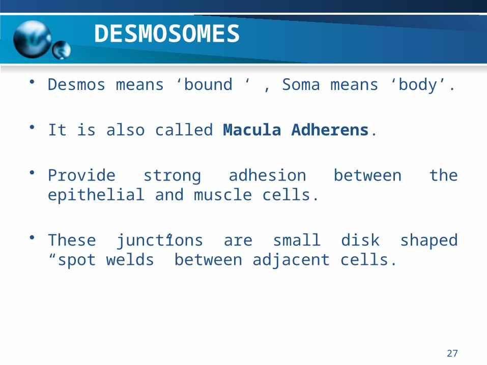
DESMOSOMES
• Desmos means ‘bound ‘ , Soma means ‘body’.
• It is also called Macula Adherens.
• Provide strong adhesion between the epithelial and muscle cells.
• These junctions are small disk shaped “spot welds” between adjacent cells.
27
28
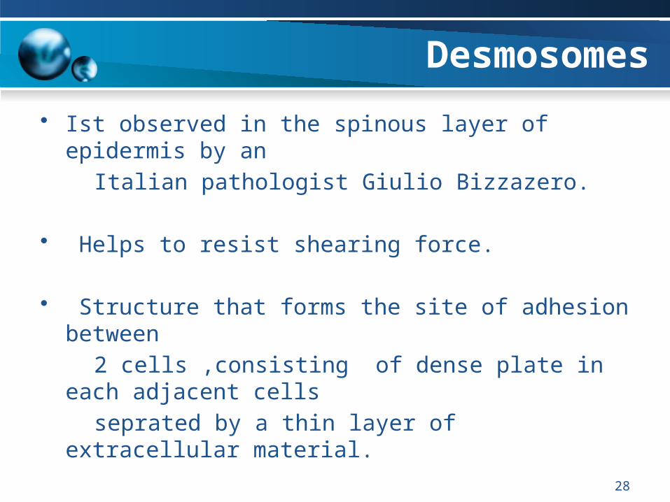
Desmosomes
• Ist observed in the spinous layer of epidermis by an
Italian pathologist Giulio Bizzazero.
• Helps to resist shearing force.
• Structure that forms the site of adhesion between
2 cells ,consisting of dense plate in each adjacent cells
seprated by a thin layer of extracellular material.
• Desmosomes link 2 cells together.
29
Desmosomes
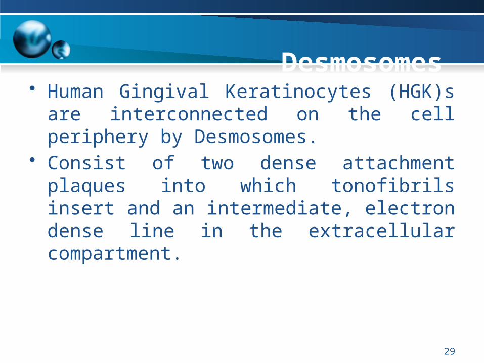
• Human Gingival Keratinocytes (HGK)s are interconnected on the cell periphery by Desmosomes.
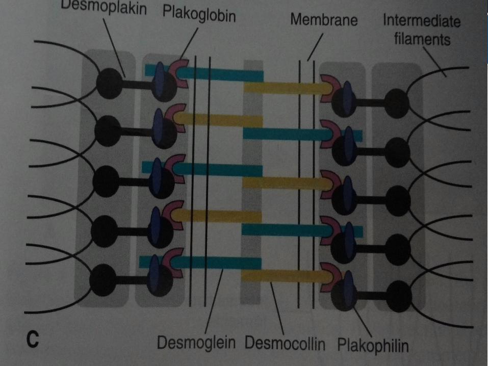
• Consist of two dense attachment plaques into which tonofibrils insert and an intermediate, electron dense line in the extracellular compartment.
Desmosomes
• Cellular adhesions at desmosomes are mediated by transmembrane proteins:
• Desmogleins • Desmocollins.
• Plakoglobin also called gamma- catenin.
• Molecular composition of desmosomes vary in particular tissues
30
Desmosomes
• Desmosomes are site for attachment, structural ability of epithelium linking cytoskeletal structures of two cells.
• Desmoglein -2 and desmocollin -2 are found in most of the desmosomes.
• The devlopment of animal tissues depends on desmosomes & their constituents proteins.
31
32
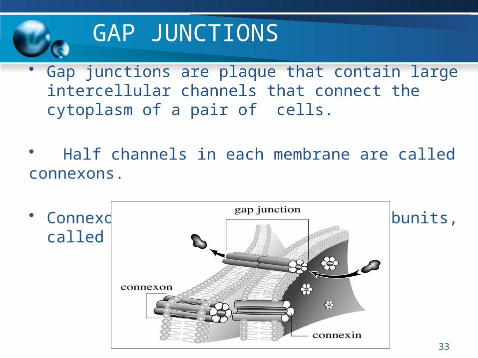
GAP JUNCTIONS• Gap junctions are plaque that contain large intercellular
channels that connect the cytoplasm of a pair of cells.
• Half channels in each membrane are called connexons.
• Connexons consists of six protein subunits, called
connexins.
33
Gap Junctions
• Connexin are named by their molecular weight.
• Found exclusively in chordates.
• Most connexons pair with identical connexons on the partner cell to form homotypic gap junctions.
• Gap junction communication is conditional• It depends on:• Number of channels• Fraction that are open or closed
34
Gap Junctions
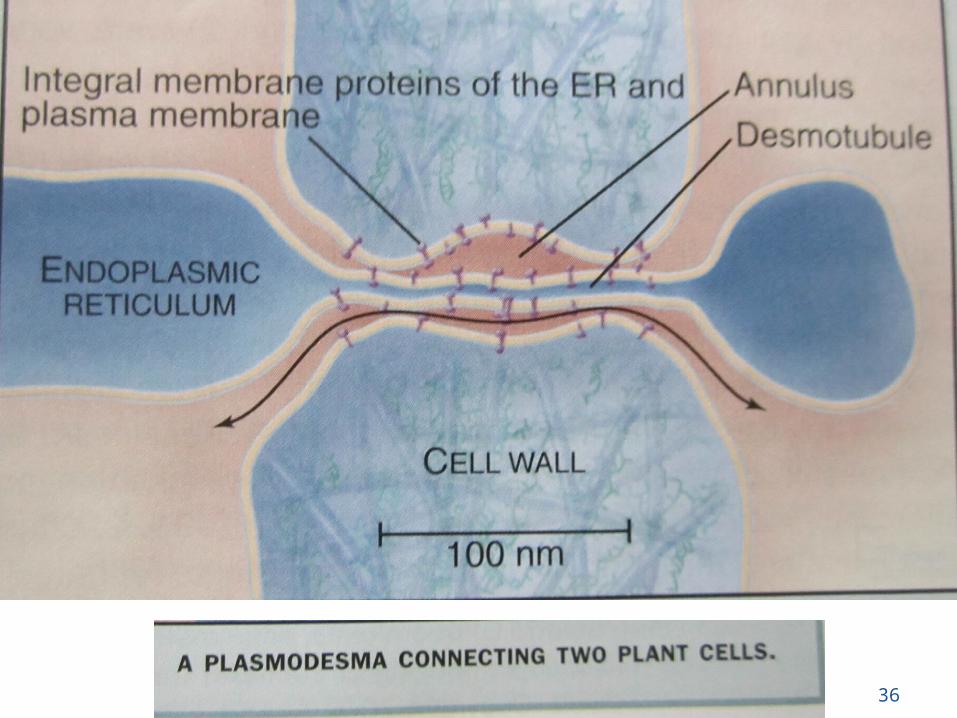
• Plants lacks gap junctions
• Cells in plant tissues maintain continuity through plasmodesmata.
• Molecules smaller than 1kd diffuse freely through plasmodesmata.
35
36
Gap Junctions
• Oleamide- fatty acid amide produced by the brain , blocks gap junction and induce sleep in animals.
• Gap junctions allow osteocytes to maintain cellular supply line to acquire nutrients from distant blood vessels.
• White blood cells may also form transient gap junctions with endothelial cells.
• Cells in most metazoans communicate by gap junctions.
37
Gap Junctions
• Mutations in connexins genes cause human disease.
• Recessive mutation in the connexin -26 gene are most common cause of human Deafness.
• Mutation in connexin-32 gene causes degeneration of myelin sheets around axons.
38
HEMIDESMOSOMES
39
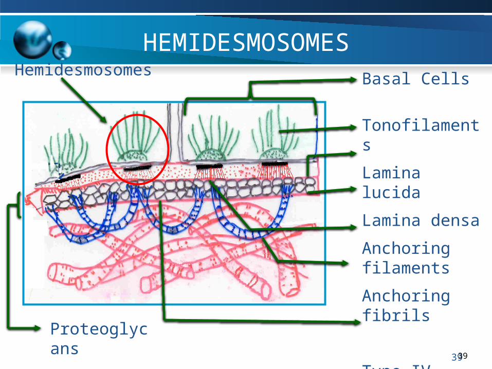
Basal Cells
Tonofilaments
Lamina lucida
Lamina densa
Anchoring filaments
Anchoring fibrils
Type IV collagenProteoglycans
39
Hemidesmosomes
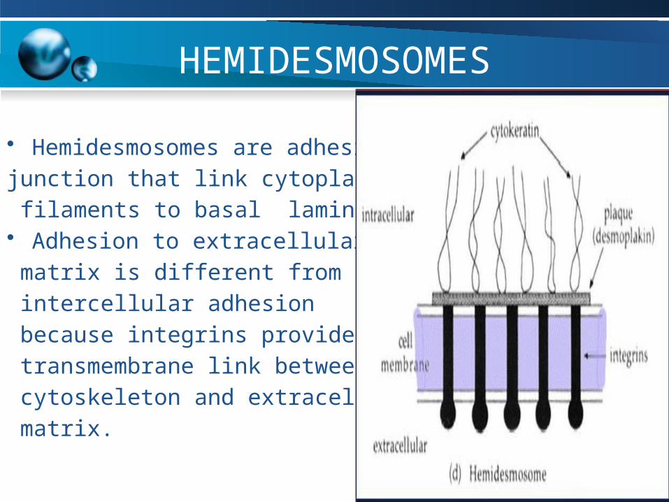
HEMIDESMOSOMES
• Hemidesmosomes are adhesive
junction that link cytoplasmic
filaments to basal lamina. • Adhesion to extracellular
matrix is different from
intercellular adhesion
because integrins provide
transmembrane link between
cytoskeleton and extracellular
matrix.
40
Contd…
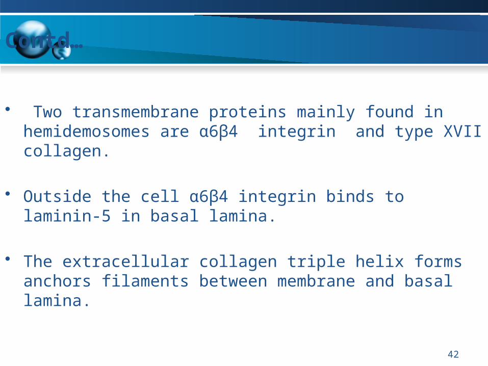
• Two transmembrane proteins mainly found in hemidemosomes are α6β4 integrin and type XVII collagen.
• Outside the cell α6β4 integrin binds to laminin-5 in basal lamina.
• The extracellular collagen triple helix forms anchors filaments between membrane and basal lamina.
42
Contd…
• STRUCTURE OF HEMIDESMOSOME:
– Adhesive protein – INTEGRIN– Cytoplasmic proteins – PECTIN, BP 180– Cytoskeletal element - INTERMEDIATE
FILAMENTS – Target molecule - LAMININ.
43
CELLULAR ADHESION
PRINCIPLES OF CELLULAR ADHESION
1. First principle of adhesion
• Cells define their capacity for adhesive interactions by selectively expressing plasma membrane receptors with limited ligand binding activity.• For example: Endothelial cells produce E – selectin only when stimulated by inflammatory hormones
44
Contd…
2. Second principle of adhesion• Many adhesion proteins bind one main ligand and many ligands bind a single type of receptor, for example Most cadherins bind to themselves, such homophilic interactions require Ca+2 ions.
3. Third principle of adhesion• It states that cell modulate adhesion by controlling the surface density, state of aggregation and state of activation of their adhesion receptors.
45
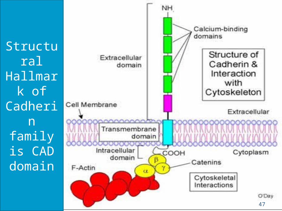
CADHERIN FAMILY OF ADHESION RECEPTORS
• Cadherins name is derived from calcium dependent adhesion
protein.
• Homophilic interactions of cadherins link epithelial and muscle cells to adjacent cells at specialized junctions called adherens junctions and desmosomes.
• The cytoplasmic domains of cadherin junctions interact with actin filaments to maintain physical integrity of tissues.
46
Contd…
47
Structural Hallmark
of Cadherin family is
CAD domain

Cadherins are named according to their location or cells to which they are attached. for eg:
•Epithelial tissue -E-cadherin •Nervous tissue - N-cadherin •Placenta- P-cadherin • osteoblasts- O-cadherin•Kidney-K cadherin•muscle -M-cadherin
49
• Integrins are the main cellular receptors for extra cellular matrix.
• Integrins tend to be more promiscous than most adhesion receptors as some bind to several protein ligands & many matrix molecule bind to 1 integrin.
50
51
SUMMARY
• Intercellular junctions are fundamental to the interactions between cells.
• Mucosal barrier integrity is maintained by the physical
interactions of intercellular junctional molecules on opposing epithelial cells.
• In the heart, cell junctions form the low-resistance pathways for rapid impulse conduction and propagation, enabling synchronous stimulation of myocyte contraction.
52
SUMMARY
• In kidney, cell junctions help to maintain concentrations of fluid creating a balance between osmotic gradients.
• By means of these junctions, the activities of the individual cells that make up tissues are co-ordinated, enabling each tissue system to function as an integrated whole.

53


