
SELF-ASSEMBLED MONOLAYERS OF PEPTIDE NUCLEIC ACIDS (PNA):
FROM THE MOLECULAR STRUCTURE TO BIOSENSOR APPLICATIONS.
C. Briones , E. Mateo-Martí , V. Parro, C. RogeroCentro de Astrobiología (CSIC-INTA). Madrid, Spain
J.A. Martín-Gago, C. Gómez-Navarro (UAM) , J. Mendez, E. Román, Instituto de Ciencia de Materiales de Madrid (CSIC). Madrid, Spain
Victor Fernandez, Marcos PitaInstituto de Catalisis (CSIC). Madrid, Spain
http://www.icmm.csic.es/esisna/
Interdisciplinary work: biology, chemistry, Physics

Motivation of the research project:
To investigate the interaction of nucleic acids with solid surfaces
Origin of life on earth could be a catalytic processMetallic oxides, pyrite, clay minerals, silicates
Surface science can model all these interactions
XPS,AFM, NEXAFS, IRSGold, pyrite, silicon, nanoparticles
1.- Nanoscience:
To build new biosensors from the bases
3SSpecificSensitiveSimple
2.-Nanotechnology:

ssDNA: (single stranded) single chain
dsDNA double stranded in double chain (helicoidal :Every nucleo-base recognaice by H-bridges a complementary one
Sugar(deoxyribose)
Phosphategroup
Nitrogeneous bases(nucleo-bases)
DNA (deoxyribonucleic acid): polynucleotide
nucleotide
Rapid bio-background for physicists
backbone

?? ??labelling
Two unknownnucleotidesWhich is the sequence
DNA- microaarrays, how does it work?
AG AT AC
GT GC TC
Robot imprinting
Inkjet or piezo actuation technology. Spots of 50-150 μm in diameter, and low-density microarrays with no more than 5,000 spots/cm2
Target
Probe

TC
?? ??labelling
Robot imprinting
?? = AG
Fluoresce Scanner
DNA- microaarrays, how does it work?Two unknownnucleotides

In every spot a sequence
DNA
Spacer
Surface
DNA- microaarrays, how does it work?
DNA-chip pattern

Drawbacks of the current technology:Nanotechnology comes to help…
1.- Fluorescence molecular label of the target
2.- Expensive equipments
3.- No quantitative information,lack of specificity, not very sensitive

-Alkanethiol layers have been extensively studied due to their important technological properties and their outstanding capability to form self-
assembled monolayers (SAMs).
AFM From E.Barrena and C.Ocal
Alkanethiols self-assembled monolayers
gold
S-head
Alkyl chain
Functional group

IDEA!!!
Let us add a S atom at the end of a DNA to recover the alkanethiol ideas and made a solid-state biosensor

Clean Au Immobilized DNA Hybridized DNA
More details:E. Casero, M. Darder, D. J. Díaz, F. Pariente, J. A.Martín-Gago,H. Abruña and E. Lorenzo
Lamgmuir 2003,19, 6230-6235
strong surface-molecule molecule-molecule interactions
-Thiolated DNA also immobilized on gold surfaces, but most of theresults have been disappointing: DNA fold into itself leading to a formless globular structure, with a very reduced bioactivity.

PNA is an achiral and uncharged DNA mimic
the sugar-phosphatebackbone has been replaced with a peptide-
like N-(2-aminoethyl)glycine polyamide structure. The nucleobases are connected by methylenecarbonyl linkages.
PEPTIDE NUCLEIC ACID (PNA)

Menchise et al., PNAS 2003
PNA recognizes complementary DNA with stronger affinity than DNA-DNA:
(5’)
(3’)
ON N
NH2
O
OO
OOO P
OO
OOO P
OO
OOO P
O
O
OOO P
N NH
O
O
NH2N
NN N
NH2
N
NN NH
O
(N)
O
N
NH
O
O
N
NH
O
O
N
NH
O
O
N
NH
O
(C)
NNH
O
O
N
NNN
H2N
NN
O
H2N
N
NH2N
HN
O
N
Can be used as probe for DNA biosensorsAbsence of P in the backbone:
Specific signature: no fluorescence

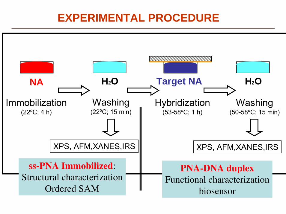
EXPERIMENTAL PROCEDURE
Cysteine
Spacer
ssPNA

Au
H2O H2OTarget NANA
Immobilization(22ºC; 4 h)
Washing(22ºC; 15 min)
Hybridization(53-58ºC; 1 h)
Washing(50-58ºC; 15 min)
XPS, AFM,XANES,IRS
ss-PNA Immobilized:Structural characterization
Ordered SAM
PNA-DNA duplexFunctional characterization
biosensor
EXPERIMENTAL PROCEDURE
XPS, AFM,XANES,IRS

H2O H2OTarget NANA
Immobilization(22ºC; 4 h)
Washing(22ºC; 15 min)
Hybridization(53-58ºC; 1 h)
Washing(50-58ºC; 15 min)
ss-PNA Immobilized:Structural characterization
Ordered SAM
PNA-DNA duplexFunctional characterization
biosensor
EXPERIMENTAL PROCEDURE
XPS, AFM,XANES,IRS XPS, AFM,XANES,IRS
H2O H2OTarget NANA
Immobilization(22ºC; 4 h)
Washing(22ºC; 15 min)
Hybridization(53-58ºC; 1 h)
Washing(50-58ºC; 15 min)
ss-PNA Immobilized:Structural characterization
Ordered SAM
PNA-DNA duplexFunctional characterization
biosensor
EXPERIMENTAL PROCEDURE
XPS, AFM,XANES,IRS XPS, AFM,XANES,IRS

H2O H2OTarget NANA
Immobilization(22ºC; 4 h)
Washing(22ºC; 15 min)
Hybridization(53-58ºC; 1 h)
Washing(50-58ºC; 15 min)
ss-PNA Immobilized:Structural characterization
Ordered SAM
PNA-DNA duplexFunctional characterization
biosensor
EXPERIMENTAL PROCEDURE
XPS, AFM,XANES,IRS XPS, AFM,XANES,IRS

49nm
50nm
RESULTS : Structure of the layers
AFM images: • ordered arrangement of the molecules, with reproducible aligned and meandering patterns.
• The ordered protrusions are 6 to 7 nm high from the bare surface.
• Width: 10 to 30 nm.
The ssPNA molecules stand-up on the surface with a small tilt.

RESULTS : Structure of the layers
- XANES spectra at the N-edge at grazing andnormal emission indicate a preferentialorientation of the molecule with the nucleo-bases nearly- paralel to the surface plane.
Inte
nsity
(arb
.uni
ts)
420415410405400Photon energy (eV)
Normal incidence
70º off normal incidence
Most of the π* orbitals lie along the backbone of the PNA and the σ* are parallel to the nucleobases plane,

49nm
Proposed Structural model
- Self-assembling of ssPNA molecules
promoted by non-complementary
H-bonding between nucleobases

RESULTS : Concentration dependence
41nm
[ssPNA] = 0.1μM
- Linear featuresfollowing crys-tallographic directions
- Height: ~ 1 nm- Length: ~ 10 nm- Individual mole-cules lying on the surface
200nm
[ssPNA] = 0.5μM
- Layer not com-plete
- Groups of mo-lecules standup as islands anchored to theupper part of the step edges
50nm
[ssPNA] ≥ 10μM
- Ordered featuresare lost
- Surface saturatedof amorphous groups of mole-cules
50nm
[ssPNA] = 1.0μM
- Aligned and meandering patterns
- Height: 6-7 nm- Width: 10-30 nm- Groups of mole-cules stand up on the whole surface
- Optimal coverage
With increasing PNA concentration

Advantage of ssPNA over ssDNAin the formation of bioSAMs
i) the lack of charged groups in the PNA backbone avoids electrostatic repulsions either among neighbouring molecules or among the solvent counterions.
ii) although relatively flexible, the PNA molecule is more rigid than DNA due to the planar amide
Now, let us see if they work as biosensors…

RESULTS : Concentration dependence
41nm
[ssPNA] = 0.1μM
200nm
[ssPNA] = 0.5μM
50nm
[ssPNA] ≥ 10μM[ssPNA] = 1.0μM
With increasing PNA concentration
PNA molecules standing up on the surface
PNAmolecules lying on
the surface
PNA molecules intermixed one with each other

RESULTS : Hybridization to DNA
The hybridization of complementary DNA is detected by XPS:- Increase of N1s/Au4f ratio in afactor of 2.8 to 3.
- Detection of aclear P2p signal.
130 132 134 136 138
396 398 400 402 404
1.4
1.2
1.0
0.8
0.6
0.4
0.2
0.0
Inte
nsity
(arb
. Uni
ts)
600 500 400 300 200 100 0Binding energy (eV)
O 2p N 1s
C 1s Au 4f
Au
Immob. PNA
PNA/DNA
XPS overview
P 2p

RESULTS : Characterization of the biosensor
0,001
0,01
0,1
1
10
0,1 1 10 100
ImmobilizedHybridized
N1s
/Au4
f rat
io
ssPNA Concentration (μM)
Evolution of the normalized XPS intensity with the concentration of immobilized PNA:
I
II
III 50nm
I
II
III
AFM imaging:

0,001
0,01
0,1
1
10
0,1 1 10 100
Immobilized
Hybridized
N1s
/Au4
f rat
io
ssPNA Concentration ( μM)
50nm
NA concentrations higher than 5 μM are not useful since:1. Surface is saturated of NA, and the possibility of hybridization with target NA is strongly reduced2. During the hybridization incubation non-specifically bound NA is removed from the surface, and therefore N1s/Au4f decreases

41nm
[ssPNA] = 0.1μM
200nm
[ssPNA] = 0.5μM
50nm
[ssPNA] ≥ 10μM[ssPNA] = 1.0μM
Is that a general rule for nucleic acids?
PNA molecules standing up on the surface
PNAmolecules lying on
the surface
PNA molecules intermixed one with each other
Just when the molecules stand-up, do not interact with the surface and are isolated, are biosensitive
Let us see on pyrite: a more reactive, metallic, natural surface

Evolution of the SAM structure with PNA concentration
Absence of thiol Group: covalent bonding
PM-RAIRS RESULTS on goldE. Briand, C. M. PradierLab. De reactivité des surfacesCNRS-Jussieu

PM-RAIRS RESULTS on pyrite
PNA adsorbs on pyrite surface, however…
Presence of thiol group:
unspecific bonding
Wide peaksAll transition allowed

XANES at N1s on gold
Inte
nsity
(arb
.uni
ts)
420415410405400Photon energy (eV)
XANES at N1s on pyrite
Orientation of PNA on pyrite

PNA-DNA interaction on pyrite surface
SugarPhosphate
phosphate
PM-RAIRS RESULTS on pyrite

On Pyrite, ssPNA adsorbs strongly interacting with the surface,Without forming covalent bonding through the S atom
DNA is always on the surface, independently of the sequence:Unspecific bonding

Immobilization and hybridization of single-stranded PNA on aldehyde terminated monolayers prepared at Si (111) surfaces
CH3
SiSiSi
Si
CH3 CH3
OH
CH3CH3OH OH
SiSiSi
SiH|
H| H
|
H|
1
100 % C10CHO 0 % C11
50 % C10CHO 50 % C11
10 % C10CHO 90 % C11
1 % C10CHO 99 % C11
0 % C10CHO 100 % C11
0 % C10CHO 0 % C11 (clean H-Si(111)
Si(111) substrate
Col. B HorrowckUniversity New castle

400nm 400nm
2 x 2 µmContact Tapping
C11-Si(111)

N
NH
OO
Base
N
NH
OO
Base
N
NH
OO
Base
Immobilization and hybridization of single-stranded PNA on aldehyde terminated monolayers prepared at Si (111) surfaces
CH3
SiSiSi
Si
CH3 CH3
OH
CH3CH3OH OH
SiSiSi
SiH|
H| H
|
H|
CH3
SiSiSi
Si
CH3 CH3
OH
1 2
N
NH
OO
Base
N
NH
OO
Base
N
NH
OO
Base
3
CH3
SiSiSi
Si
CH3
CH3
O
OO O
O
OP
O
OO O
O
OPBase
BaseO
OO O
O
OPBase
100 % C10CHO 0 % C11
50 % C10CHO 50 % C11
10 % C10CHO 90 % C11
1 % C10CHO 99 % C11
0 % C10CHO 100 % C11
0 % C10CHO 0 % C11 (clean H-Si(111)
Si(111) substrate

CONCLUSIONS
- PNA molecules, in spite of theirlength of up to 7 nm, can self-assemble on gold surfaces similarly to short alkanethyol molecules.
- Two main reasons for the clear advantage of PNA over DNA:• lack of charged groups and intermolecular electrostatic repulsions• higher rigidity and restrited conformational flexibility
- Mechanism for the formation of SAMs of ssPNA:• at low coverage densities, molecules condensate on the surface andare absorbed as lying molecules
• at a certain coverage (corresponding to [PNA] ≈ 1 μM) the layer undergoes a phase transition: realignment of the molecule backboneperpendicular to the surface
- XPS can be used for the monitorization of the hybridization of complementary DNA, the characterization of a PNA-based biosensor, and its optimization to discriminate point mutations in target DNA.

NUCLEIC ACIDS AND THEIR ANALOGUES AS NANOMATERIALS FOR BIOSENSOR DEVELOPMENTC. Briones, J.A. Martín-Gago . In press: current nanotechnology
E. Mateo-Martı´, C. Briones, E. Roma´n, E. Briand, C. M. Pradier, and J. A. Martín-GagoLangmuir 2005, 21, 9510-9517
Briones, C.; Mateo-Marti, E.; Gomez-Rodriguez, C.; Parro, V.Roman, E.; Martín-Gago, J. A.
Phys. Rev. Lett. 2004, 93, 208103
Understanding surface-molecule interactions could be of great help to design 3S biosensors
(specific, sensitive, simple)
More information:

No N1s signal is detected
108 106 104 102 100 98 96
s83 100% C10CHO s84 100% C10CHO PNA version 1
Si 2p
Binding energy (eV)108 106 104 102 100 98 96
s83 100% C10CHO
Si 2p
Binding energy (eV)
PNA immobilization:PG142- noE (AATCCCCGCAT).
• Version 1 (microarray-like): •drop of PNA solution (5 μM in milli-Q water) .•When the drop is dried,•30 μl of 5.0 M aqueous Sodium Cyanoborohydride (Sigma) is deposited on top. The solution is left to react for 18 h
• Version 2 : •drop of PNA solution (5 μM in milli-Q water) .•Immediately, a frop of 30 μl of 5.0 M aqueous Sodium Cyanoborohydride is added. The solution is left to react for 18 h
Not perfect. We damage the surface!!!!!
Surface completely oxidized
410 408 406 404 402 400 398 396 394 392 390
sample84 100% C10CHO PNA version 1 sample83 100% C10CHO
N 1s
Binding energy (eV)410 408 406 404 402 400 398 396 394 392 390
sample82 100% C10CHO PNA version 2 sample84 100% C10CHO PNA version 1 sample83 100% C10CHO
N 1s
Binding energy (eV)
108 106 104 102 100 98 96
s83 100% C10CHO
Si 2p
Binding energy (eV)

RESULTS : Identification of mutations in DNA
MISMATCH
SCOMP.
S
S
COMPLEMENTARYHYBRIDIZATION IS
MAINTAINED
S
WASHING
MISMATCHEDHYBRIDIZATION IS
REMOVED
Mutation screening experiment:160
140
120
100
80
60
40
20
0
Inte
nsity
(arb
. Uni
ts)
408406404402400398396
Binding Energy (eV)
Fully complementary target Mismatched target, after washing Immbilized NA
XPS result:
3,5
ssP
NA
N1s/Au4fP2p/Au4f
2,5
1,5
0,5
2,0
1,0
0ss
PN
A+
ssD
NA
(M)
ssP
NA
+ss
DN
A (M
M)
ssP
NA
ssP
NA
+ss
DN
A (M
)
ssP
NA
+ss
DN
A (M
M)
3, 0
Optimization of washing buffer:

DNART-PCR with
Cy5 -dUTP
STRAIN SELECTION
HYBRIDIZATION
MatchingProbe (M)
WASHING
M MM
SCANNING
M MM
RNA
Naturalsample
LabeledTarget DNA
MismatchingProbe (MM)

200nm
0 50 100 150 200 250 300
02
46
810
X[nm]
Z[nm]
100nm

EXPERIMENTAL PROCEDURE
Au
Target DNAH2O W.S.PNA
Immobilization(22ºC; 4 h)
Washing(22ºC; 15 min)
Hybridization(53-58ºC; 1 h)
Washing(50-62ºC; 30 min)
Immobilized PNA 1: HS-Cys-O-O-AATCCCCGCGTTarget- G142 (M): 5'-CCGCCAGTGCACGCGGGGATTTGGCTCACCT-3' Target- E142 (MM): 5'-CCGCCAGTGCACGCGAGGATTTGGCTCACCT-3'
Immobilized PNA 2: HS-Cys-O-O- GCCATCTCTTarget- M41 (M): 5'-GAAATTTGTACAGAGATGGAAAAGGAAGGGA-3' Target- L41 (MM): 5'-GAAATTTGTACAGAGTTGGAAAAGGAAGGGA-3'
We have Choosen sequencies of clinical relevanceAnd similar to the one used in microarray technoology

Table. Infrared assignment for the main frequencies of PNA on gold surface
PNA Concentration Assignment
2977
2931
2854
1250
1168
1086
933
2974
2928
2854
1240
1161
1084
2963
2931
2864
1736
1669
1613
1549
1250
1168
1100
933
2967
2935
2857
1680
1602
1235
1161
1083
933
0.01µM 1µM0.1µM 10µMνasym(CH3)
νasym(CH2)
νsym(CH2)
ν(C=O)
N-H
NH2 C=N
C=C, C=N
νasym(C-O-C)
ν(C-N,C-C),δ(C-H)
νsym(C-O-C)
ν(C-N)
δ(N-H)oop

Study of the N(1s) core level of PNA by means of XPS
N 1s Binding energy
Assignment % N Calculated % N Experimental
398.93 -N= 25 23.4
400.13 -NH- 59.4 62.2
401.10 NH2 15.6 14.4
Table. Assignment of N(1s) core level peak of PNA

XPS core-level peak of N (1s) for PNA adsorbed on Pyrite surface
XPS core-level peak of N (1s) for PNA-DNA adsorbed on Pyrite surface
Table . Experimental reported binding energy (eV) of of N(1s) core level peak for % of different chemical statesof nitrogen involved in the PNA chemical structure.
Specie 0.1μM PNA 1μM PNA 10μM PNA 1μM PNA-DNA
NH2 2.7 % 3.5% 11.3% 3.1%
-NH- 40.0% 30.1% 32.8% 21.1%
-N= 34.1% 44.1% 44.8% 42.6%
N-Fe 23.0% 22.3% 11.1% 33.2%

Specie 1μM PNA 1μM PNA-DNA
18.7% 20.2%
FeS2 (Cys) 55.1% 65.0%
Free-thiols SH 12.0% 8.3%
SO42- 14.2% 6.5%
Table . Experimental reported binding energy (eV) of of S(2p) core level peak for % of different chemical states of sulphur involved in the PNA-Pyrite interaction.
S (2p) Core Level of PNA S (2p) Core Level of PNA-DNA

XANES
XANES was used to corroborate the absence of any preferential orientation of PNA
adsorbed on pyrite surface.

AFM study
100nm 100nm100nm100nm
limpia 0,1μm 1μm 10 μm
120100806040200
1.6
1.4
1.2
1
0.8
0.6
0.4
0.2
0
X [nm ]Z[
nm]
150100500
2.5
2
1.5
1
0.5
0
X[nm]
Z[nm
]
80706050403020100
1.5
1
0.5
0
X[nm]
Z[nm
]
100806040200
3.5
3
2.5
2
1.5
1
0.5
0
X[nm]
Z[nm
]

200nm 200nm200nm200nm
limpia 0,1μm 1μm 10 μm

Experiments realised on this year:
Nov 2004- Nov 2005
Si modified surfaces:
C11 C10CHO C11S C11NH2
Mixed layers:PNA immobilizedDNA hybridized
Mixed layers:Au nanoparticlePNA immobilizedDNA hybridized
For future PNA synthesis
Surface science techniques:
AFM (contact and tapping)XPS (home lab and synchrotron)XANES (normal and grazing emission)FTIR

OBJECTIVES
- Study the immobilization of thiol-modified ssPNA molecules on gold surfaces.
- Characterize the structure and ordering of the layers.
- Monitor the hybridization of complementary ssDNA to layers of immobilized ssPNA.
- Characterize the PNA-based biosensor: concentration, temperature,time, buffer composition.
- Determine the ability of the biosensor to detect point mutations(SNPs) in target DNA.
-Perform this structural and functional characterization by meansof powerful label-free techniques for surface characterization: atomic force microscopy (AFM), X-ray photoemission spectroscopy(XPS) and X-ray absorption near-edge spectroscopy (XANES)

Biosensors based on DNA-chips
DNA-chip pattern
Detection of hybridization is performed by using fluorescence molecules

- XPS Experiments: performed ex situ in an Ultra High Vacuumchamber (UHV: Pressure 10-9 mbar). The sample was transported inair for a few seconds. The chamber is equipped with a double-passcylinder mirror analyser (CMA) . A Mg anode in the X-ray source wasused for experiments (1253.6 eV photons).
- Synchrotron radiation experiments (XPS and XANES): performed atELETTRA-superESCA beam line. The overall resolution was around 80 meV.
- AFM images: recorded in air using a NANOTEC-AFM working intapping mode.
- Substrates used:• XPS: polycristaline Au layers evaporated on glass (Arrandee, Germany)
and single-crystal Au(111)• AFM: Au films Annealed to 600ºC for few minutes.
EXPERIMENTAL PROCEDURE


![Splicing isoform-specific functional genomic in cancer cells · 2018. 12. 14. · morpholinos, peptide nucleic acid (PNA), locked nucleic acid (LNA)] confer resistance to various](https://cdn.vdocuments.site/doc/165x107/614a7e2a12c9616cbc697388/splicing-isoform-specific-functional-genomic-in-cancer-cells-2018-12-14-morpholinos.jpg)