Zita Krūmiņa
RARE INBORN ERRORS OF METABOLISM IN
CHILDREN IN LATVIA
Summary of doctoral theses
Speciality – medical genetics
Riga, 2011
2
Doctoral theses elaborated in Medical Genetics Clinic of Childrens’ University Hospital, Riga, Latvia
Scientific supervisors:
Dr. Med. assoc. Prof. Rita Lugovska - Medical Genetics Clinic of Childrens’ University Hospital,
Riga Stradins university, Department of Biology and microbiology
Dr. Med. Baiba Lāce - Medical Genetics Clinic of Childrens’ University Hospital, Latvian Biomedical
research and study center
Official reviewers:
1. Dr. Biol. Prof. N. Sjakste - University of Latvia, Medical Faculty, Riga, Latvia
2. Dr. Biol. Asoc. Prof. E. Miklašēvičs – Riga Stradins University, Department of Biology and
microbiology, Riga, Latvia
3. Dr. Med. Prof. Anna Tylki-Szymanska - Children’s Memorial Health Institute Departament of
Metabolic Diseases, Warsaw, Poland
Doctoral theses will be deffended on “_21.12._2011. _16.p.m.___” in open Fundamental science
doctoral council meeting at Hippocrates’ auditorium, Riga Stradins University, Dzirciema street 16,
Riga
Doctoral theses could be found at library of Riga Stradins University and at Riga Stradins university
website: www.rsu.lv
Financially study was supported by ESF project No 2009/0147/1DP/1.1.2.1.2/09/IPIA/VIAA/009
“Enhancement of competencies, qualification and skills of health care and health promotion
professionals” Sub-activity No 1.1.2.1.2 “Support to doctor’s studies”, (2011)
Secretary of doctoral council:
Zinātniskais grāds latīņu val., zinātniskais nosaukums Vārds Uzvārds
3
CONTENT
1. INTRODUCTION ................................................................................................................... 4
Hypothesis ............................................................................................................................... 5
The aim of the study ................................................................................................................ 5
Tasks of the study .................................................................................................................... 5
Scientific novelty ..................................................................................................................... 6
Actuality of subject .................................................................................................................. 6
2. REVIEW OF LITERATURE ................................................................................................. 7
2.1. Inborn errors of metabolism ......................................................................................... 7
2.2. Long-chain hydroxyacyl-CoA dehydrogenase deficiency............................................ 8
2.3. Urea Cycle disorders ..................................................................................................... 9
2.4. Lysosomal storage disorders ....................................................................................... 11
3. MATERIAL AND METHODS ........................................................................................ 13
4. RESULTS........................................................................................................................... 15
4.1. Long-chain 3-hydroxyacyl-CoA dehydrogenase deficiency ...................................... 15
4.2. Urea cycle disorders .................................................................................................... 18
4.3. Lysosomal storage disorders ....................................................................................... 20
5. DISCUSSION .................................................................................................................... 31
5.1. Long–chain 3–hydroxyacyl–CoA dehydrogenase (LCHAD) deficiency ................... 31
5.2. Urea cycle disorders .................................................................................................... 32
5.3. Lysosomal storage disorders ....................................................................................... 33
5.4. Diagnosed rare inborn errors of metabolism in Latvia ............................................... 34
6. CONCLUSIONS ................................................................................................................ 36
7. RECOMMENDATIONS HOW TO IMPROVE DIAGNOSTICS OF RARE INBORN
ERROR OF METABOLISM IN LATVIA ............................................................................... 37
APPROBATION ....................................................................................................................... 38
REFERENCES .......................................................................................................................... 43
THE STRUCTURE OF WORK AND VOLUME .................................................................... 47
4
1. INTRODUCTION
Inborn errors of metabolism (IEM) or inherited metabolic diseases comprise a large class of
genetic diseases. In most of the disorders, problems arise due to accumulation of substances
which are toxic and can cause acute or chronic intoxication, hypoglycaemia or other metabolic
disturbances.
First clinical manifestation can be seen already in antenatal period or later in newborn,
children, juvenile or even in adult period. Clinical presentation in most cases is unspecific and
in the neonatal period or infancy could be misdiagnosed as manifestation of sepsis, birth
trauma, encephalitis, sudden infant death syndrome or other disease. In childhood or juvenile
period IEM may manifest as schizophrenia, epilepsy, progressive mental retardation,
unspecific hepatitis, eye, kidney, cardiac and other organ pathology. IEM are rare individually
but collectively they are common, and numbers of them are rising as diagnostic techniques are
improved. The number of described IEM currently is close to 2000. Data from the literature
suggest that risk for a baby to be born with any of IEM is about 1:500. It means, that there
have to be about 40 newborns with IEM in Latvia every year (presuming that each year there
are about 20 000 newborns in Latvia).
Disorders that are not included in newborn screening are more difficult to diagnose due to
clinical variety. These disorders are diagnosed using selective screening (specialised genetic
analyses done only for individuals with clinical symptoms of IEM or positive family history).
Many countries have enlarged newborn screening with more treatable IEM and also cystic
fibrosis (CF), that still is one of lethal disorders (Sommerburg, 2010). CF is included in
newborn screening because early diagnosis and adequate therapy before clinical signs of
disease delays development of bronchectasis and other severe complications, that in longer
period gives possibility to elongate patient’s quality of life and lifetime (Farell, 2005, Grosse,
2006).
The precise frequency of two IEM – phenylketonuria and congenital hypothyroidism is known
in Latvia due to newborn screening. The newborn screening for phenylketonuria started since
year 1987 and for congenital hypothyroidism since 1996. Other IEM that are not included in
newborn screening very often are not diagnosed or diagnosed very late. A delayed diagnosis
can cause physical and mental retardation, invalidity and patient’s early death. The diagnosis
5
of untreatable IEM is also important, because it gives opportunity for a family to recieve a
qualitative genetic consultation, including a calculation of a recurrency risk for birth of an
affected individual in the family and allows to take preventive actions.
The decision of European Parliament and Council No 1295/1999/EC (it was accepted on 29
April 1999) was adopting a programme of Community action on rare diseases within the
framework for action in the field of public health (1999 to 2003). It was declared that rare IEM
are disorders that affect less than 5 in 10 000 people. According to this document a work
group was organised in Latvia in November of 2010 to develop the strategic plan in the field
of rare IEM (The decision of Health Ministry No 229 (15.11.2010). The accepted plan
contains 5 major directions: (1) to increase awareness of rare IEM, (2) prevention of rare IEM
and its early diagnostics, (3) treatment, (4) integration of social and health care, (5) training for
patients, their families and health care professionals.
In 2009 the government of the Republic of Latvia assigned one year budget of 700 000 LVL
for medication for children with rare IEM.
Hypothesis
Most of patients with rare IEM are not diagnosed or diagnostics is delayed in Latvia.
The aim of the study
To evaluate situation in the field of rare inborn errors of metabolism in Latvian children.
Tasks of the study
1) The selection of patients with diagnosed rare inborn errors of metabolism, excluding
phenylketonuria and congenital hypothyroidism;
2) The analysis of clinical symptoms and laboratory data of patients with rare inborn
errors of metabolism in Latvia and data comparison with data from other countries;
3) The determination of prevalence for rare inborn errors of metabolism in Latvia;
4) The efficiency evaluation of the diagnostics of inborn errors of metabolism in Latvia;
5) The development of indications for further investigations in case of possible inborn
error of metabolism.

6
6) The development of suggestions for the diagnostic improvement of rare inborn errors
of metabolism in children in Latvia.
Scientific novelty
1) The data about children with rare inborn errors of metabolism in Latvia will be
summarised for the first time. The data will include clinical and laboratory data of
patients with diagnosed rare inborn errors of metabolism, which is important for
further disease investigation and worldwide recognition.
2) The calculation of prevalence for rare inborn errors of metabolism in Latvia will be
done for the first time.
Actuality of subject
1) Increased attention payed to rare disorders in Latvia from 2010 (The decision of Health
Ministry No 229 /2010 of developing the strategic plan in the field of rare disorders in
Latvia).
2) The information about rare IEM in children in Latvia currently has not been
summarised.
3) Most of children with rare IEMs are not diagnosed and treated, which increases
children’s mortality and disability in Latvia.
4) There are no informative materials about rare IEM, no guidelines for investigations or
management of acute metabolic crisis in Latvian.

7
2. REVIEW OF LITERATURE
2.1. Inborn errors of metabolism
Inborn errors of metabolism (IEM) or inherited metabolic diseases comprise a large class of
genetic diseases involving disorders of metabolism caused by several enzyme defects. Most of
IEM are genetically determined monogenic disorders that are usually inherited in autosomal
recessive manner, but can show also other modes of inheritance. IEM are classified by main
disorder groups (Table 2.1. (Pons Ruiz, 2007)).
Table2.1.
Classification of inborn errors of metabolism (Pons Ruiz, 2007)
Disease group Specific diseases
IEM of amino acids Hyperphenylalaninemia or phyenylketonuria,
tyrosinaemia, non-ketotic hyperglycinemia,
homocystinuria, maple syrup urine disease etc.
IEM of organic acids 3-methylglutaconic aciduria, glutaric aciduria,
methylmalonic aciduria etc.
IEM of carbohydrates Glycogenosis, fructose intolerance, galactosaemia,
glucose transport defects, defects of glycerol metabolism
Fatty acid and ketone body metabolism Fatty acid oxidation and ketogenesis disorders
Lysosomal storage disorders Mucopolysaccharidoses, oligosaccharidoses,
sfingolipidoses, mucolipidoses, lipid storage disorders,
lysosomal transport defects, neural ceroid lypofuscinoses,
glycogenosis type II (Pompe disease)
Mitochondrial disorders Pyruvate dehydrogenase complex deficiency, Leigh
syndrome etc.
Protein glycosylation disorders Congenital disorders of glycosylation
Peroxisomal defects Adrenoleukodystrophy, Refsum disease etc.
Sterol metabolism defects Smith – Lemli – Opitz syndrome, Antley- Bixler
syndrome etc.
Lipoprotein metabolism defects Hypercholesterolaemias, hyperlipidaemias,
hypertriglyceridaemias
Purine and pyrimidine metabolism
defects
Lesch – Nyhan syndrome, podagra etc.
Urea cycle disorders Ornitine transcarbamylase deficiency, citrullinaemia,
argininaemia etc.
Neurotransmitter defects Tetrahydrobiopterin deficiency, tyrosine hydroxylase
deficiency etc.
Metal metabolism disorders Wilson disease, Menkes disease
Vitamin metabolism disorders IEM of folate, cobalamine, biotine, B 6, etc.
Membrane transport system defects Renal tubular acidosis, cystic fibrosis
8
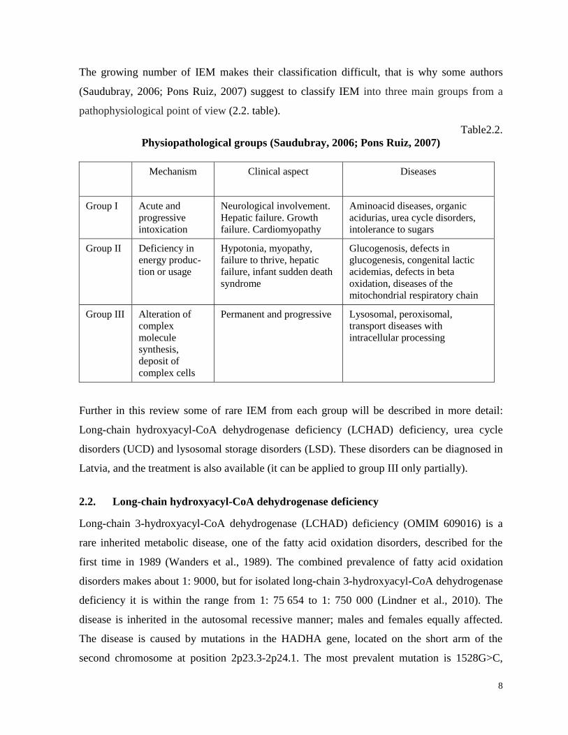
The growing number of IEM makes their classification difficult, that is why some authors
(Saudubray, 2006; Pons Ruiz, 2007) suggest to classify IEM into three main groups from a
pathophysiological point of view (2.2. table).
Table2.2.
Physiopathological groups (Saudubray, 2006; Pons Ruiz, 2007)
Mechanism Clinical aspect Diseases
Group I Acute and
progressive
intoxication
Neurological involvement.
Hepatic failure. Growth
failure. Cardiomyopathy
Aminoacid diseases, organic
acidurias, urea cycle disorders,
intolerance to sugars
Group II Deficiency in
energy produc-
tion or usage
Hypotonia, myopathy,
failure to thrive, hepatic
failure, infant sudden death
syndrome
Glucogenosis, defects in
glucogenesis, congenital lactic
acidemias, defects in beta
oxidation, diseases of the
mitochondrial respiratory chain
Group III Alteration of
complex
molecule
synthesis,
deposit of
complex cells
Permanent and progressive Lysosomal, peroxisomal,
transport diseases with
intracellular processing
Further in this review some of rare IEM from each group will be described in more detail:
Long-chain hydroxyacyl-CoA dehydrogenase deficiency (LCHAD) deficiency, urea cycle
disorders (UCD) and lysosomal storage disorders (LSD). These disorders can be diagnosed in
Latvia, and the treatment is also available (it can be applied to group III only partially).
2.2. Long-chain hydroxyacyl-CoA dehydrogenase deficiency
Long-chain 3-hydroxyacyl-CoA dehydrogenase (LCHAD) deficiency (OMIM 609016) is a
rare inherited metabolic disease, one of the fatty acid oxidation disorders, described for the
first time in 1989 (Wanders et al., 1989). The combined prevalence of fatty acid oxidation
disorders makes about 1: 9000, but for isolated long-chain 3-hydroxyacyl-CoA dehydrogenase
deficiency it is within the range from 1: 75 654 to 1: 750 000 (Lindner et al., 2010). The
disease is inherited in the autosomal recessive manner; males and females equally affected.
The disease is caused by mutations in the HADHA gene, located on the short arm of the
second chromosome at position 2p23.3-2p24.1. The most prevalent mutation is 1528G>C,
9
traced in homozygous form in up to 87% of the total number of patients (Kahler et al., 2010).
The HADHA gene’s encoded protein is long-chain 3-hydroxyacyl-CoA dehydrogenase
(LCHAD), an enzyme required for long-chain fatty acid oxidation for energy production. The
disease affects the Krebs cycle and adenosine triphosphate (ATP) synthesis, therefore the
metabolic crisis is accompanied by nonketotic hypoglycemia.
Hypoglycemia, elevated liver transaminases and elevated creatine kinase level in blood are
observed in the acute disease period. Moderately elevated levels of lactate and ammonia in
blood are observed quite often. Nonketotic hypoglycemic acidosis and absence of ketons in
the urine are observed during coma. Dicarboxylic and 3-hydro-dicarboxylic acid excretion in
the urine, as well as changes in the acylcarnitines profile in blood, both being specific
diagnostic criteria are found in metabolic crisis. The diagnosis is confirmed by DNA tests and
finding two mutant alleles of the HADHA gene or by lowered enzyme activity in skin
fibroblasts.
Early diagnostics and an adequate therapy might prevent a sudden death of patients; in most
cases normalization of clinical symptoms is possible, except in the cases of progressive
retinopathy and peripheral myopathy. Therefore, in many countries of the world a
comprehensive newborn screening has been started, including fatty acid oxidation disorders,
among them long-chain 3-hydroxyacyl-CoA dehydrogenase deficiency.
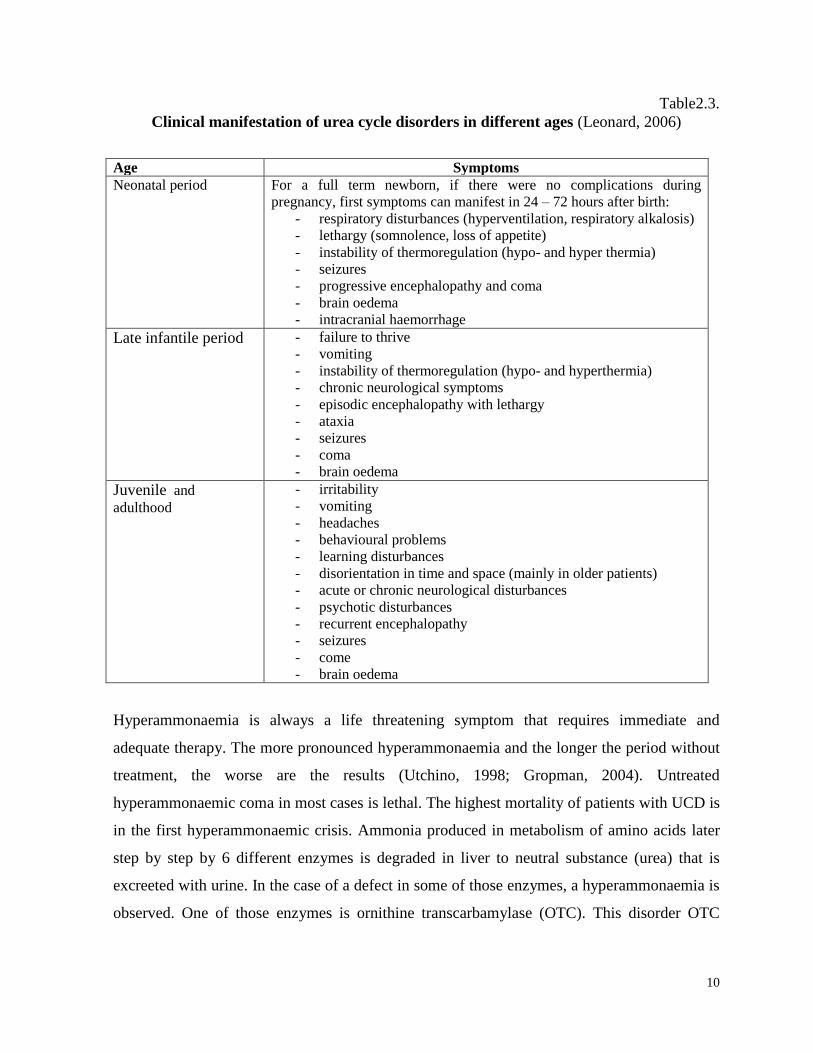
2.3. Urea Cycle disorders
Urea cycle disorders (UCD) are among the most common IEM with cumulative incidence
approx. 1:8000 (Zchocke, 2004). First symptoms can manifest at any age: starting from
neonatal age to adult period, but most frequently in newborns, todlers and during puberty
(Leonard, 2006). Although urea cycle disorders are considered easy to diagnose, the clinical
symptoms quite often are not recognised, which results in delayed diagnosis. Urea cycle
disorders have a characteristic symptom triad: encephalopathy, respiratory alkalosis,
hyperammonaemia (Brusilow, 1996). The manifestations of clinical symptoms in different
ages are shown in table 2.4. (Leonard, 2006). There is a wide variation of clinical symptoms
even in one family with the same mutation. Early diagnostics is very important, because
majority of patients with urea cycle disorders are treatable using diet with decreased protein
amount and medication normalising ammonia level.
10
Table2.3.
Clinical manifestation of urea cycle disorders in different ages (Leonard, 2006)
Age Symptoms
Neonatal period
For a full term newborn, if there were no complications during
pregnancy, first symptoms can manifest in 24 – 72 hours after birth:
- respiratory disturbances (hyperventilation, respiratory alkalosis)
- lethargy (somnolence, loss of appetite)
- instability of thermoregulation (hypo- and hyper thermia)
- seizures
- progressive encephalopathy and coma
- brain oedema
- intracranial haemorrhage
Late infantile period - failure to thrive
- vomiting
- instability of thermoregulation (hypo- and hyperthermia)
- chronic neurological symptoms
- episodic encephalopathy with lethargy
- ataxia
- seizures
- coma
- brain oedema
Juvenile and
adulthood
- irritability
- vomiting
- headaches
- behavioural problems
- learning disturbances
- disorientation in time and space (mainly in older patients)
- acute or chronic neurological disturbances
- psychotic disturbances
- recurrent encephalopathy
- seizures
- come
- brain oedema
Hyperammonaemia is always a life threatening symptom that requires immediate and
adequate therapy. The more pronounced hyperammonaemia and the longer the period without
treatment, the worse are the results (Utchino, 1998; Gropman, 2004). Untreated
hyperammonaemic coma in most cases is lethal. The highest mortality of patients with UCD is
in the first hyperammonaemic crisis. Ammonia produced in metabolism of amino acids later
step by step by 6 different enzymes is degraded in liver to neutral substance (urea) that is
excreeted with urine. In the case of a defect in some of those enzymes, a hyperammonaemia is
observed. One of those enzymes is ornithine transcarbamylase (OTC). This disorder OTC
11
deficiency will be described futher, because there are 6 patients diagnosed with this disorder in
Latvia.
Ornitine transcarbamylase deficiency (OMIM 311250) is the most frequent urea cycle
disorder in Europe and worldwide with frequency 1:14 000 newborns. Enzyme coding gene
OTC is located on the short arm of X chromosome (Xp21.1), that’s why clinical symptoms are
most severe in boys and girls may be clinically unaffected. Clinical symptoms are similar for
all urea cycle disorders, but there is an important fact that girls in the neonatal period usually
are clinically healthy, while in boys the clinical symptoms manifest already 24 – 72 hours after
birth. A characteristic symptom for OTC deficiency in the neonatal period is
hyperammonaemia with respiratory alkalosis. During a metabolic crisis there are elevated
glutamine, alanine and lysine concentrations in blood aminoacid spectrum, but citrulline and
in some cases arginine level is decreased. A diagnostic symptom is an elevated orotic acid
level in urine. If there are laboratory changes characteristic of OTC deficiency, there is a
necessity of DNA diagnostics for confirmation of the diagnosis. If the mutation is not found,
it’s possible to measure enzyme activity in liver tissue and decreased enzyme activity also
confirms the diagnosis (Zchocke, 2004).
2.4. Lysosomal storage disorders
There are 50 – 70 different rare genetic diseases (Staretz-Chacham, 2009; Aerts, 2011) in
lysosomal storage disorders (LSD) group, caused by absence of lysosomal enzymes, which are
involved in degradation of complex molecules. The prevalence of each LSD separately is in
the range from 1:20 000 to 1:100 00, but combined prevalence is about 1: 5 000 to 1: 10 000
(Poorthuis, 1999; Aerts, 2011). Due to an enzyme deficiency, undegraded products of
metabolism are stored in lysosomes, resulting in cells’ and organs’ enlargement and their
impaired function (Moog, 2010). In most cases undegraded substrate is stored in central
nervous system, which causes progressive mental retardation. Sphingolipidoses,
mucopolysacchariodoses, olygosaccharidoses, mucolipidoses, lipid storage disorders, lysosmal
transport defects, glycogen storage disorders type II or Pompe disease are the main LSD
groups (Zschocke, 2004).
LSD is a group of slowly progressive disorders without acute metabolic crises (Moog, 2010).
In most cases the neonatal period is without pathology; in some cases hydrops fetalis, face
dysmorphism, umbilical or inguinal hernia and cardiomegaly is observed. Quite often there are
12
early hypotonia and motor development delay, later a progressive mental retardation,
organomegaly, coarse facial features, skeletal changes (dysostosis multiplex) are evident.
There are no characteristic changes in routine biochemical analyses, that’s why for most of
LSD diagnostics are difficult. The clinical variability is the main reason why some of those
disorders are diagnosed late or misdiagnosed. As most of LSD are rare, there are no data about
definite incidence, but in many countries the prevalence of total and individual LSD is
calculated by using total diagnosed patient number in known period of time (Poupĕtová, 2010;
Pinto, 2004; Poorthuis, 1999). For most of LSD the therapy is still not discovered. Enzyme
replacement therapy is available for Gaucher disease, Fabry disease, Pompe disease and for
MPS types I, II and VI (Kakis, 2001, Muenzer 2002, Harmatz 2004).
Mucopolysaccharidoses (MPS) are the most frequenly diagnosed LSD pathologies in Latvia.
MPS are caused due to the absence of glycoasminoglycans (mucopolysacharides) degrading
enzymes. MPS are classified in several types (I, II, III, IV, VI, VII, IX) and subtypes. Usually
MPS is inherited in an autosomal recessive manner, except MPS type II, which is X- linked.
Clinical variability is significant between MPS types and even in one type depending from
enzyme activity. The diagnostics is started primarily by the quantative analysis of
glycosaminoglycans (GAG) in urine and GAG electrophoresis and confirmed by enzyme
analysis.
13
3. MATERIAL AND METHODS
Retrospective analysis of clinical and laboratory findings of diagnosed 108 children with rare
IEM. From 4600 children, that were sent to Medical Genetics Clinic of Children’s University
Hospital during 1997 to 2010 years with unclear possible genetic pathology, later after
examination, excluding chromosomal, neuromuscular and other syndromic cases, 2500
patients were sent to selective screening for IEM and in 108 of them a diagnosis of rare inborn
error of metabolism was confirmed.
The disease prevalence was calculated, using the methods, published by Poorthuis BJ (1999),
Pinto R (2004), Poupetova H (2010). The prevalence reflects the number of patients with IEM
per 100 000 live births. The total number of patients with particular disease is divided by the
total number of live births in the given period. The birth period is the time span between the
year of birth of the oldest diagnosed patient and the year of birth of the youngest patient. The
total live births within the years 1997-2010 were calculated by using the data of the Central
Statistical Bureau of the Republic of Latvia (http://www.csb.gov.lv/).
As IEM are rare diseases it was not possible to accumulate a sufficient information amount for
applying either the Fisher test or ANOVA for data comparison within the period of 10-20
years. Consequently, the analysis of clinical and laboratory findings was done by using
descriptive statistics.
For diagnostics of rare IEM selective screening was usually used, when patient with suspicion
of IEM (clinical symptoms, laboratory data or family anamnesis) was sent to specialised
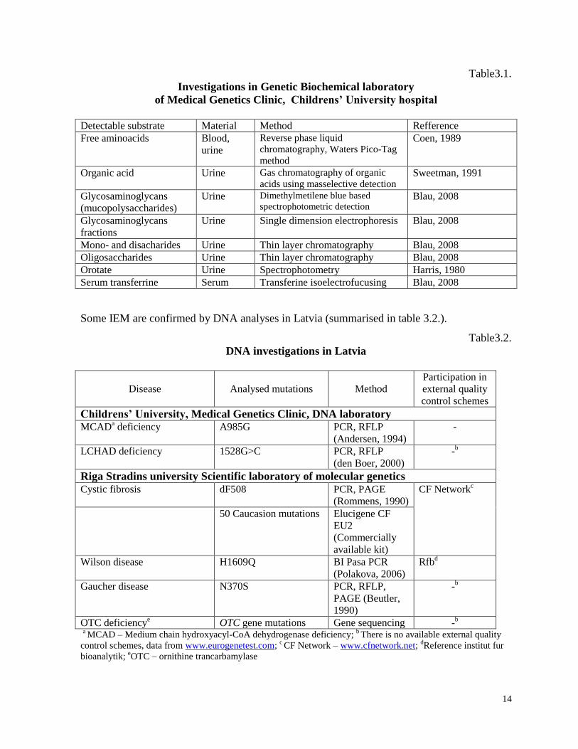
genetic analyses. Genetic biochemical analyses that are available in Medical Genetics Clinic
of Childrens’ University hospital in Latvia are summarised in table 3.1.
Research was done in Medical Genetics Clinic of Childrens’ University hospital. Study was
approved by Riga Stradins university Ethical committee.
Research was supported by ESF grant No.2009/0147/1DP/1.1.2.1.2/09/IPIA/VIAA/009.
14
Table3.1.
Investigations in Genetic Biochemical laboratory
of Medical Genetics Clinic, Childrens’ University hospital
Detectable substrate Material Method Refference
Free aminoacids Blood,
urine
Reverse phase liquid
chromatography, Waters Pico-Tag
method
Coen, 1989
Organic acid Urine Gas chromatography of organic
acids using masselective detection Sweetman, 1991
Glycosaminoglycans
(mucopolysaccharides)
Urine Dimethylmetilene blue based
spectrophotometric detection Blau, 2008
Glycosaminoglycans
fractions
Urine Single dimension electrophoresis Blau, 2008
Mono- and disacharides Urine Thin layer chromatography Blau, 2008
Oligosaccharides Urine Thin layer chromatography Blau, 2008
Orotate Urine Spectrophotometry Harris, 1980
Serum transferrine Serum Transferine isoelectrofucusing Blau, 2008
Some IEM are confirmed by DNA analyses in Latvia (summarised in table 3.2.).
Table3.2.
DNA investigations in Latvia
Disease Analysed mutations Method
Participation in
external quality
control schemes
Childrens’ University, Medical Genetics Clinic, DNA laboratory
MCADa deficiency A985G PCR, RFLP
(Andersen, 1994)
-
LCHAD deficiency 1528G>C PCR, RFLP
(den Boer, 2000)
-b
Riga Stradins university Scientific laboratory of molecular genetics Cystic fibrosis dF508 PCR, PAGE
(Rommens, 1990)
CF Networkc
50 Caucasion mutations Elucigene CF
EU2
(Commercially
available kit)
Wilson disease H1609Q BI Pasa PCR
(Polakova, 2006)
Rfbd
Gaucher disease N370S PCR, RFLP,
PAGE (Beutler,
1990)
-b
OTC deficiencye
OTC gene mutations Gene sequencing -b
a MCAD – Medium chain hydroxyacyl-CoA dehydrogenase deficiency;
b There is no available external quality
control schemes, data from www.eurogenetest.com; c CF Network – www.cfnetwork.net;
dReference institut fur
bioanalytik; eOTC – ornithine trancarbamylase
15
4. RESULTS
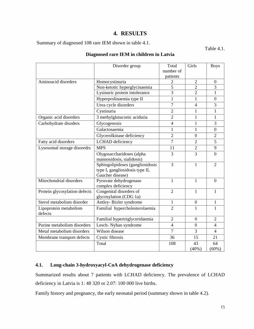
Summary of diagnosed 108 rare IEM shown in table 4.1.
Table 4.1.
Diagnosed rare IEM in children in Latvia
Disorder group Total
number of
patients
Girls Boys
Aminoacid disorders Homocystinuria 2 2 0
Non-ketotic hyperglycinaemia 5 2 3
Lysinuric protein intolerance 3 2 1
Hyperprolinaemia type II 1 1 0
Urea cycle disorders 7 4 3
Cystinuria 2 1 1
Organic acid disorders 3 methylglutaconic aciduria 2 1 1
Carbohydrate disoders Glycogenosis 4 1 3
Galactosaemia 1 1 0
Glycerolkinase deficiency 2 0 2
Fatty acid disorders LCHAD deficiency 7 2 5
Lysosomal storage disoredrs MPS 11 2 9
Olygosaccharidoses (alpha
mannosidosis, sialidosis)
3 3 0
Sphingolipidoses (gangliosidosis
type I, gangliosidosis type II,
Gaucher disease)
3 1 2
Mitochondrial disorders Pyruvate dehydrogenase
complex deficiency
1 1 0
Protein glycosylation defects Congenital disorders of
glycosylation (CDG 1a)
2 1 1
Sterol metabolism disorder Antley- Bixler syndrome 1 0 1
Lipoprotein metabolism
defects
Familial hypercholesterolaemia 2 1 1
Familial hypertriglyceridaemia 2 0 2
Purine metabolism disorders Lesch- Nyhan syndrome 4 0 4
Metal metabolism disorders Wilson disease 7 3 4
Membrane transport defects Cystic fibrosis 36 15 21
Total 108 43
(40%)
64
(60%)
4.1. Long-chain 3-hydroxyacyl-CoA dehydrogenase deficiency
Summarized results about 7 patients with LCHAD deficiency. The prevalence of LCHAD
deficiency in Latvia is 1: 48 320 or 2.07: 100 000 live births.
Family history and pregnancy, the early neonatal period (summary shown in table 4.2).
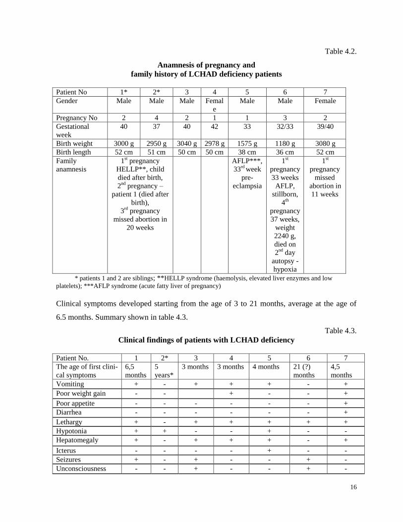
16
Table 4.2.
Anamnesis of pregnancy and
family history of LCHAD deficiency patients
Patient No 1* 2* 3 4 5 6 7
Gender Male Male Male Femal
e
Male Male Female
Pregnancy No 2 4 2 1 1 3 2
Gestational
week
40 37 40 42 33 32/33 39/40
Birth weight 3000 g 2950 g 3040 g 2978 g 1575 g 1180 g 3080 g
Birth length 52 cm 51 cm 50 cm 50 cm 38 cm 36 cm 52 cm
Family
anamnesis
1st pregnancy
HELLP**, child
died after birth,
2nd
pregnancy –
patient 1 (died after
birth),
3rd
pregnancy
missed abortion in
20 weeks
AFLP***,
33rd
week
pre-
eclampsia
1st
pregnancy
33 weeks
AFLP,
stillborn,
4th
pregnancy
37 weeks,
weight
2240 g,
died on
2nd
day
autopsy -
hypoxia
1st
pregnancy
missed
abortion in
11 weeks
* patients 1 and 2 are siblings; **HELLP syndrome (haemolysis, elevated liver enzymes and low
platelets); ***AFLP syndrome (acute fatty liver of pregnancy)
Clinical symptoms developed starting from the age of 3 to 21 months, average at the age of
6.5 months. Summary shown in table 4.3.
Table 4.3.
Clinical findings of patients with LCHAD deficiency
Patient No. 1 2* 3 4 5 6 7
The age of first clini-
cal symptoms
6,5
months
5
years*
3 months 3 months 4 months 21 (?)
months
4,5
months
Vomiting + - + + + - +
Poor weight gain - - + - - +
Poor appetite - - - - - - +
Diarrhea - - - - - - +
Lethargy + - + + + + +
Hypotonia + + - - + - -
Hepatomegaly + - + + + - +
Icterus - - - - + - -
Seizures + - + - - + -
Unconsciousness - - + - - + -
17
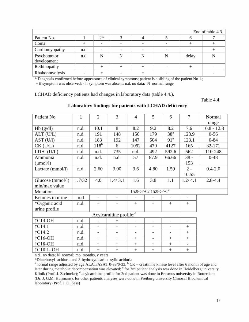
End of table 4.3.
Patient No. 1 2* 3 4 5 6 7
Coma + - + - - + +
Cardiomyopathy n.d. - - - - - +
Psychomotor
development
n.d. N N N N delay N
Rethinopathy - + + + - + -
Rhabdomyolysis - + - + - - -
* Diagnosis confirmed before appearance of clinical symptoms; patient is a sibling of the patient No 1.;
+ if symptom was observed; - if symptom was absent; n.d. no data; N normal range
LCHAD deficiency patients had changes in laboratory data (table 4.4.).
Table 4.4.
Laboratory findings for patients with LCHAD deficiency
Patient No 1 2 3 4 5 6 7 Normal
range
Hb (g/dl) n.d. 10.1 8 8.2 9.2 8.2 7.6 10.8 - 12.8
ALT (U/L) n.d. 191 148 156 179 38a
123.9 0-56
AST (U/l) n.d. 183 192 147 504 91a
123.1 0-84
CK (U/L) n.d. 118b
6 1092 470 4127 165 32-171
LDH (U/L) n.d. n.d. 735 n.d. 492 592.6 562 110-248
Ammonia
(µmol/l)
n.d. n.d. n.d. 57 87.9 66.66 38 -
153
0-48
Lactate (mmol/l) n.d. 2.60 3.00 3.6 4.80 1.59 2 -
10.55
0.4-2.0
Glucose (mmol/l)
min/max value
1.7/32
4.0 1.4/ 3.1 1.6 3.8 1.1 1.2/ 4.1 2.8-4.4
Mutation 1528G>C/ 1528G>Cc
Ketones in urine n.d - - - - - -
*Organic acid
urine profile
n.d. + + + + + +
Acylcarnitine profile:d
↑C14-OH n.d. - + - - - -
↑C14:1 n.d. - - - - - +
↑C14:2 n.d. - - - - - +
↑C16-OH n.d. + + + - + +
↑C18-OH n.d. + + + + + -
↑C18:1- OH n.d. + + + + + + n.d. no data; N normal; mo months, y years
*Dicarboxyl -aciduria and 3-hydroxydicarbo -xylic aciduria a normal range adjusted by age ALAT/ASAT 0-33/0-33,
b CK – creatinine kinase level after 6 month of age and
later during metabolic decompensation was elevated; c for 3rd patient analysis was done in Heidelberg university
Klinik (Prof. J. Zschocke); d acylcarnitine profile for 2nd patient was done in Erasmus university in Rotterdam
(Dr. J. G.M. Huijmans), for other patients analyses were done in Freiburg university Clinocal Biochemical
laboratory (Prof. J. O. Sass)

18
4.2. Urea cycle disorders
Here follow data summarised about seven patients with UCD; six of them were confirmed
with OTC deficiency. Patients were from two unconsanguineous families. All OTC deficiency
patients were from one family – the patients’ mothers were unaffected sisters (see figure 4.1.).
Fig.4.1. Pedigree tree of OTC deficiency patients
Used symbols: circle – female, square – male, white colour – healthy individual, black colour –
affected individual, crossed out symbol – individual that is dead, circle with spot – healthy female
mutation carrier
Calculated UCD prevalence in Latvia is 1,49 : 100 000. Calculation of prevalence of OTC
deficiency wouldn’t be correct, because patients are from one family.
The patients with OTC deficiency had positive family history – unexplained sudden deaths of
newborns, infant and child. A characteristic sign is that all boys died in early neonatal period
(2nd or 3rd day of life). The brothers of patients’ mothers died also on the first and second day
of life. All children were born as full term newborns in normal deliveries, one of newborns
was large for gestational age.
The manifestation of clinical symptoms for patients with OTC deficiency was from second
day of life till age of 3,5 years with distinct difference between boys and girls (Table 4.5.) The
manifestation of clinical symptoms in girls in average was 1 year and 10 months (from 9,5
months till 3,5 years). The manifestation of clinical symptoms in boys (patients No. 2 and 3)
started on the second day of life, when fast deterioration was observed with rapid superficial
breathing, lethargy, coma and death.
19
Table 4.5.
Clinical findings of UCD patients
Patients
(No.)
1a 2
a 3
a 4
b 5
b 6
b 7
c
Gender female male male female Female female male
In figure
4.1.
III-10 III-11 III-12 III-5 III-6 III-8
Beginning
of first
clinical
symptoms
9,5
months
2nd
day
of life
2nd
day of
life
4 years ? 15 months from
birth
Clinical
symptoms
Vomiting + - - + - + +
Lethargy + + - - - - +
Respiratory
disturbances
+ + + - - - -
Termoregul
ation
disturbances
+
- - - - - -
Hepatho-
megaly
+ - - + - + -
Behavioral
changes
- - - + - +
Coma + + - + - - -
Seizures - - + - - - -
Change in
appetite
+
- - - + - +
Other
symptoms
- - Focal and
generalised
seizures
- Sometimes
headaches
- Nystag-
mus
Situation at
the moment
Death at
age of 10
months
Death
at 3rd
day of
life
Death at 2nd
day of life
Death at
5 years
of age
20 years of
age, normal
psycho-
motor
development
4,5 years of age,
normal
psychomotor
develop-ment
Death at
age of
10,5
months
a siblings in one family; bsiblings in other family; c
UCD not precise
The ammonia level and aminoacid profile in blood were examined only in three patients (5, 6
and 7) and all three patients had elevated glutamine level, but patients 6 and 7 had
hyperammonaemia 210 and 304µmol/l (normal range till 48 µmol/l) during acute
manifestation of the disease. During metabolic crisis all girls had elevated liver enzymes, and
respiratory alkalosis was observed in boys.
20
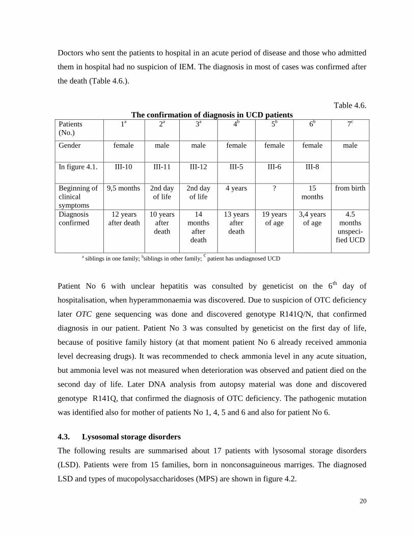
Doctors who sent the patients to hospital in an acute period of disease and those who admitted
them in hospital had no suspicion of IEM. The diagnosis in most of cases was confirmed after
the death (Table 4.6.).
Table 4.6.
The confirmation of diagnosis in UCD patients Patients
(No.)
1a 2
a 3
a 4
b 5
b 6
b 7
c
Gender female male male female female female male
In figure 4.1. III-10 III-11 III-12 III-5 III-6 III-8
Beginning of
clinical
symptoms
9,5 months 2nd day
of life
2nd day
of life
4 years ? 15
months
from birth
Diagnosis
confirmed
12 years
after death
10 years
after
death
14
months
after
death
13 years
after
death
19 years
of age
3,4 years
of age
4.5
months
unspeci-
fied UCD
a siblings in one family; bsiblings in other family; c
patient has undiagnosed UCD
Patient No 6 with unclear hepatitis was consulted by geneticist on the 6th
day of
hospitalisation, when hyperammonaemia was discovered. Due to suspicion of OTC deficiency
later OTC gene sequencing was done and discovered genotype R141Q/N, that confirmed
diagnosis in our patient. Patient No 3 was consulted by geneticist on the first day of life,
because of positive family history (at that moment patient No 6 already received ammonia
level decreasing drugs). It was recommended to check ammonia level in any acute situation,
but ammonia level was not measured when deterioration was observed and patient died on the
second day of life. Later DNA analysis from autopsy material was done and discovered
genotype R141Q, that confirmed the diagnosis of OTC deficiency. The pathogenic mutation
was identified also for mother of patients No 1, 4, 5 and 6 and also for patient No 6.
4.3. Lysosomal storage disorders
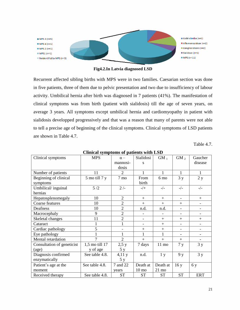
The following results are summarised about 17 patients with lysosomal storage disorders
(LSD). Patients were from 15 families, born in nonconsaguineous marriges. The diagnosed
LSD and types of mucopolysaccharidoses (MPS) are shown in figure 4.2.
21
Fig4.2.In Latvia diagnosed LSD
Recurrent affected sibling births with MPS were in two families. Caesarian section was done
in five patients, three of them due to pelvic presentation and two due to insufficiency of labour
activity. Umbilical hernia after birth was diagnosed in 7 patients (41%). The manifestation of
clinical symptoms was from birth (patient with sialidosis) till the age of seven years, on
average 3 years. All symptoms except umbilical hernia and cardiomyopathy in patient with
sialidosis developped progressively and that was a reason that many of parents were not able
to tell a precise age of beginning of the clinical symptoms. Clinical symptoms of LSD patients
are shown in Table 4.7.
Table 4.7.
Clinical symptoms of patients with LSD Clinical symptoms
MPS
α –
mannosi-
dosis
Sialidosi
s
GM 1
GM 2
Gaucher
disease
Number of patients 11 2 1 1 1 1
Beginning of clinical
symptoms
5 mo till 7 y 7 mo From
birth
6 mo 3 y 2 y
Umbilical/ inguinal
hernias
5 /2 2 /- -/+ -/- -/- -/-
Hepatosplenomegaly 10 2 + + - +
Coarse features 10 2 + + + -
Deafness 10 2 n.d. n.d. - -
Macrocephaly 9 2 - - - -
Skeletal changes 11 2 - + + +
Cataract 1 1 - + - -
Cardiac pathology 5 - + + - -
Eye pathology 1 1 1 1 - -
Mental retardation 7 2 + + + -
Consultation of geneticist
(age)
1,5 mo till 17
y of age
2,5 y
5 y
7 days 11 mo 7 y 3 y
Diagnosis confirmed
enzymatically
See table 4.8. 4,11 y
5 y
n.d. 1 y 9 y 3 y
Patient’s age at the
moment
See table 4.8. 7 and 22
years
Death at
10 mo
Death at
21 mo
16 y
6 y
Received therapy See table 4.8. ST ST ST ST ERT
22
mo month; y year, GM 1 gangliosidosis type I; GM 2 gangliosidosis type II; n.d. no data; ST – symtptomatic
therapy, ERT – ensyme replacement therapy
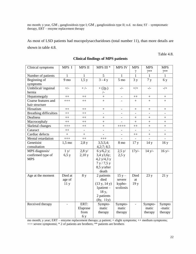
As most of LSD patients had mucopolysaccharidoses (total number 11), than more details are
shown in table 4.8.
Table 4.8.
Clinical findings of MPS patients
Clinical symptoms MPS I MPS II MPS III * MPS IV
MPS
?
MPS
?**
MPS
?**
Number of patients 1 1 5 1 1 1 1
Beginning of
symptoms
9 mo 1,5 y 3 - 4 y
5 mo 3 y 7 y 6 y
Umbilical/ inguinal
hernia
+/- + /- + (2p.)
/-
-/- +/+ -/- -/+
Hepatomegaly ++ ++ + - ++ + +
Coarse features and
hair structure
+++ ++ + - + + +
Hirsutism ++ ++ + - + + +
Breathing difficulties ++ ++ - - - - -
Deafness ++ ++ + - + + +
Macrocephaly ++ ++ + - + + +
Skeletal changes +++ ++ + ++++ ++ + +
Cataract ++ - - - - - -
Cardiac defects + + - - ++ + +
Mental retardation +++ ++ +++ - - - -
Genetisist
consultation
1,5 mo 2,8 y 3,5;3,4;
4,2;7; 8,5
8 mo 17 y 14 y 16 y
MPS diagnosis/
confirmed type of
MPS
1 y/
6,5 y
2,8 y/
2,10 y
6 y/6,2 y;
3,4 y3,6y;
4,2 y/4,3 y
7 y / 7,5 y
8,5 y/after
death
2,5 y/
2,5 y
17y/- 14 y/- 16 y/-
Age at the moment Died at
age of
11 y
8 y
2 patients
died
(13 y, 14 y)
1patient –
18 y,
2 patients
(8y, 11y)
15 y –
severe
kypho-
scoliosis
Died
at
19 y
23 y 21 y
Received therapy - ERT:
Elaprase
from
6 y
Sympto-
matic
therapy
Sympto-
matic
therapy
- Sympto-
matic
therapy
Sympto
-matic
therapy
mo month; y year; ERT – enzyme replacement therapy; p patient; + slight symptoms; ++ medium symptoms;
+++ severe symptomsi; * 2 of patients are brothers; ** patients are brothers
23
Patients didn’t have characteristic changes on rutine biochemical analyses, except anaemia and
thrombocytopenia observed for Gaucher patient. Gaucher patient had also eleveted
chitotriosidase activity in serum. GAG quantitative detection in urine was done in suspicion of
MPS and GAG electrophoresis if GAG level was elevated. Patients with alpha mannosidosis,
gangliosidosis type I and II and sialidosis had typical changes in olygosaccharides in urine.
The detection of enzyme activity was done in foreign laboratories (mainly in Poland), because
these investigations are not available in Latvia. X chromosome inactivation and deletion of
IDS gene in other X chromosome was found for girl with MPS II.
Prevalence of LSD in Latvia is 1,931: 100 000 live births (table 4.9.) and it’s lower compared
to other countries (table 4.10).
Table 4.9.
Prevalence of LSD in Latvia
Disorders No of
patientsa,b
1997-2010
Years of
birth
No of live
birthsc
Prevalence
per 100,000
CI 95%
LSD total 17 1980 - 2010 880 527 1,931 1,162-3,028
MPS total 11 (9) 1980 - 2003 728 315 1,510 0,794-2,625
MPS III 5 (4) 1989 - 2003 373 032 1,340 0,490 – 2,971
MPS I 1* 1997 - 2010 289 920 0,350 0,017 – 1,700
MPS IV 1* 1997 - 2010 289 920 0,350 0,017 – 1,700
α-manno-
sidosis
2 1989 - 2004 393 464 0,508 0,080 – 0,679
GM1 1* 1997 - 2010 289 920 0,350 0,017 – 1,700
GM2 1* 1997 - 2010 289 920 0,350 0,017 – 1,700
Sialidosis 1* 1997 - 2010 289 920 0,350 0,017 – 1,700 a number of patients who were diagnosed in time from 1997 – 2010;
b in brackets there is shown number of
families; c the data from Central Statistical Bureau of Latvia (http://www.csb.gov.lv/); CI confidence interval;
* If there was only one diagnosed patient than in calculation there were used number of live births in inevstigated
period.

24
Table 4.10.
Prevalence of LSD in Latvia comparing with other countries a,b
Diseases Prevalence
Latvia
Prevalence
Netherlands
Prevalence
Portugal
Prevalence
Australia
Prevalence
Czech
Republic
Prevalence
Germany
LSD total 1,93 14,00 25,00 12,90 12,25 n.d.
MPS total 1,51 4,50 4,80 4,44 3,72 3,53
MPS III 1,34 1,89 0,84 1,42 0,91 1,57
MPS I 0,35 1,19 1,33 1,14 0,72 0,69
MPS IV 0,35 0,36 0,60 0,59 0,73 0,38
α-manno-
sidosis
0,51 0,09 0,12 0,10 0,38 n.d.
GM1 0,35 0,41 0,62 0,26 0,26 n.d
GM2 0,35 0,34 1,49 0,26 0,19 n.d
Sialidosis 0,35 0,05 0 0,02 0,07 n.d n.d. no data;
a prevalence calculated on 100 000 live births;
b the data of other countries (Poupĕtová 2010);
The recognition of LSD among doctors is insufficient, because only two patients (with
suspicion of MPS and Gaucher disease) were sent to geneticist. One patient with alpha
mannosidosis came to us with already confirmed diagnosis abroad. Time period from first
clinical symptoms till first consultation of mediacl geneticist was in average 3 years (from 7
days to 17 years), and median time of consultation till confirmation the diagnosis was two
years.
4.4 Results of diagnosed rare IEM in children in Latvia.
Prevalence of IEM, except that of LSD are shown in table 4.11.
Table 4.11.
Prevalence of IEM in Latvia and Orphanet data
Diseases
Number
of
patients a,b
Years of
birth
No of live
birthsc
Prevalen-
ce per
100,000
CI 95% Orphanet
datad
LSD total 17 (15) 1980 - 2010 880527 1.931 1.162-3.028 -
LCHAD deficiency 7 (6) 1997 - 2010 289810 2.070 0.838-4.304 1
UCD 7 (2) 1990 - 2009 467103 1.499 0.655-2.964 1
Non-ketotic
hyperglycinaemia 5 ( 4) 1997 - 2010 289810 1.725 0.632-3.824 0.2
Homocystinuria 2 1991- 2003 296192 0.675 1.13-22.31e 0.4
Lysinuric protein
intolerance 3 1984 - 2005 620831 0.483 1.23-13.15
e -
Lesch – Nyhan
syndrome 4 (2) 1996 - 2007 244748 1.634 0.519-3.942 0.38

25
a number of patients diagnosed during 1997 – 2010;
b number of families shown in brackets;
c the data from
Central Statistical Bureau of Latvia (http://www.csb.gov.lv/; d Orhanet data summarise prevalence data in Europe,
if there are no available data, that is because of small diagnosed number of patients * If there was only one
diagnosed patient, number of live births in inevstigated period was used in calculation; e CI 95% calculated for
prevalence 1: 1 000 000.
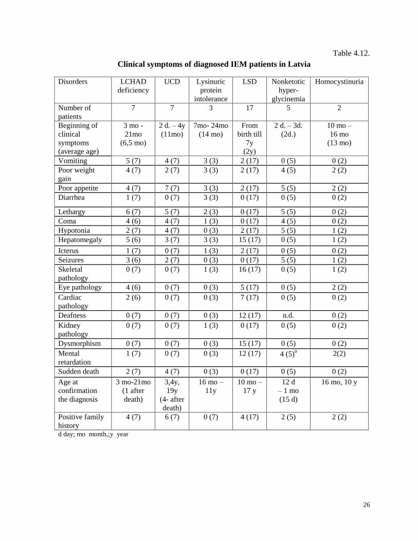
Clinical symptoms of diagnosed IEM patients is shown in table 4.12.
End of table 4.11.
Diseases
Number
of
patients a,b
Years of
birth
No of live
birthsc
Prevalen-
ce per
100,000
CI 95% Orphanet
datad
Glycerolkinase
deficiency 2 2001- 2009 193707 1.032 0.173-3.411 -
Cystinuria 2 1989 - 2010 525244 0.380 0.63-12.58e
14
Wilson disease 7 1990 - 2004 354444 1.975 0.863-3.907 5.84
3-methylglutaconic
aciduria 2 2001 - 2009 193707 1.032 0.173-3.411 -
Pyruvate
dehydrogenase
complex deficiency
1* 1997 - 2010 289810 0.344 0.17-17.01e -
Congenital
disorders of
glycosylation
(CDG 1a)
2 (1) 1997 - 2010 289810 0.689 1.15-22.79e -
Familial
hypertriglycerid-
aemia
2 2000 - 2007 148082 1.351 0.226-4.462 -
Familial
hypercholesterol-
aemia
2 1999 - 2007 187726 1.065 0.179-3.520 -
Glycogenosis 4 (3) 1997 - 2007 224966 1.778 0.565-4.289 -
Hyperprolinaemia
type II 1* 1997 - 2010 289810 0.345 0.17-17.01
e -
Antley- Bixler
syndrome 1* 1997 - 2010 289810 0.345 0.17-17.01
e -
26
Table 4.12.
Clinical symptoms of diagnosed IEM patients in Latvia
Disorders LCHAD
deficiency
UCD
Lysinuric
protein
intolerance
LSD
Nonketotic
hyper-
glycinemia
Homocystinuria
Number of
patients
7 7 3 17 5 2
Beginning of
clinical
symptoms
(average age)
3 mo -
21mo
(6,5 mo)
2 d. – 4y
(11mo)
7mo- 24mo
(14 mo)
From
birth till
7y
(2y)
2 d. – 3d.
(2d.)
10 mo –
16 mo
(13 mo)
Vomiting 5 (7) 4 (7) 3 (3) 2 (17) 0 (5) 0 (2)
Poor weight
gain
4 (7) 2 (7) 3 (3) 2 (17) 4 (5) 2 (2)
Poor appetite 4 (7) 7 (7) 3 (3) 2 (17) 5 (5) 2 (2)
Diarrhea 1 (7) 0 (7) 3 (3) 0 (17) 0 (5) 0 (2)
Lethargy 6 (7) 5 (7) 2 (3) 0 (17) 5 (5) 0 (2)
Coma 4 (6) 4 (7) 1 (3) 0 (17) 4 (5) 0 (2)
Hypotonia 2 (7) 4 (7) 0 (3) 2 (17) 5 (5) 1 (2)
Hepatomegaly 5 (6) 3 (7) 3 (3) 15 (17) 0 (5) 1 (2)
Icterus 1 (7) 0 (7) 1 (3) 2 (17) 0 (5) 0 (2)
Seizures 3 (6) 2 (7) 0 (3) 0 (17) 5 (5) 1 (2)
Skeletal
pathology
0 (7) 0 (7) 1 (3) 16 (17) 0 (5) 1 (2)
Eye pathology 4 (6) 0 (7) 0 (3) 5 (17) 0 (5) 2 (2)
Cardiac
pathology
2 (6) 0 (7) 0 (3) 7 (17) 0 (5) 0 (2)
Deafness 0 (7) 0 (7) 0 (3) 12 (17) n.d. 0 (2)
Kidney
pathology
0 (7) 0 (7) 1 (3) 0 (17) 0 (5) 0 (2)
Dysmorphism 0 (7) 0 (7) 0 (3) 15 (17) 0 (5) 0 (2)
Mental
retardation
1 (7) 0 (7) 0 (3) 12 (17) 4 (5)a 2(2)
Sudden death 2 (7) 4 (7) 0 (3) 0 (17) 0 (5) 0 (2)
Age at
confirmation
the diagnosis
3 mo-21mo
(1 after
death)
3,4y,
19y
(4- after
death)
16 mo –
11y
10 mo –
17 y
12 d
– 1 mo
(15 d)
16 mo, 10 y
Positive family
history
4 (7) 6 (7) 0 (7) 4 (17) 2 (5) 2 (2)
d day; mo month,;y year

27
Continuation of table 4.12. Disorders Lesch
Nyhan
syndro
-me
Wilson
disease
3 methyl-
glutaconic
aciduria
Pyruvate
dehydroge-
nase
complex
deficiency
Gala-
ctosaemia
Glycoge
nosis
Congenital
disorder of
glycosilation
type Ia
Number of
patients
4 7 2 1 1 4 2
Beginning of
clinical
symptoms
(average age)
6 mo –
9 mo
(7,5
mo)
3 y –
5 y
(4 y)
2 d. –
3 y
(15 mo)
From birth
6 d
6 mo –
3 y
(1,5 y)
1 y – 3 y
(2 y)
Vomiting 0 (4) 1 (7) 1 (2) + + 1(4) 0 (2)
Poor weight
gain
2 (4) 2 (7) 1 (2) + + 1 (4) 2 (2)
Poor appetite 1 (4) 2 (7) 1 (2) + + 1 (4) 0 (2)
Diarrhea 1 (4) 0 (7) 0 (2) - + 0 (4) 0 (2)
Lethargy 0 (4) 0 (7) 1 (2) + - 1 (4) 1 (2)
Coma 0 (4) 0 (7) 0 (2) - - 1 (4) 0 (2)
Hypotonia 4 (4) 0 (7) 2 (2) + + 0 (4) 2 (2)
Hepatomegaly 0 (4) 7(7) 1 (2) + + 4 (4) 0 (2)
Icterus 0 (4) 0 (7) 1 (2) + + 0 (4) 0 (2)
Seizures 0 (4) 0 (7) 1 (2) + - 1 (4) 0 (2)
Skeletal
pathology
3 (4) 0 (7) 1 (2) + - 1 (4) 2 (2)
Eye pathology 0 (4) 0 (7) 1 (2) + + 0 (4) 2 (2)
Cardiac
pathology
0 (4) 0 (7) 0 (2) - - 0 (4) 0 (2)
Deafness 0 (4) 0 (7) 1 (2) + - 0 (4) 0 (2)
Kidney
pathology
4 (4) 0 (7) 0 (2) - - 0 (4) 0 (2)
Dismorphism 0 (4) 0 (7) 1 (2) + - 1 (4) 2 (2)
Mental
retardation
4 (4)b
0 (7) 1 (2) + -
1 (4) 2 (2)
Sudden death 0 (4) 0 (7) 0 (2) - - 0 (4) 0 (2)
Age at
confirmation
the diagnosis
9 mo-
4 y
5 y –
16 y
8 mo –
7 y
10 mo 1,5 mo 10 mo
- 10 y
10 y –
12 y
Positive family
history
4 (4) 1 (7) 0 (2) - - 2 (4) 1 (2)
d day; mo month,;y year
28
End of table 4.12. Disorders Lipid
metabolism
disorder
Hyper-
prolinaemia
type I
Antley
Bixler
s.
Cystinuria Glycerol
kinase
complex
deficien
cy
CF
Number of
all patients
(%)
Number of
patients
4 1 1 2 2 36 108
Beginning of
clinical
symptoms
(average age)
no 1 y From
birth
3 y – 4 y
(3,5 y)
1 week-
3 week
(2 week)
From
birth –
4 y
(10 mo)
Vomiting 0 (4) - - 0 (2) 2(2) 10 (36) 29 (27%)
Poor weight
gain
0 (4) - - 0 (2) 2 (2) 30 (36) 59 (55%)
Poor appetite 0 (4) - - 0 (2) 2 (2) 0 (36) 30 (28%)
Diarrhea 0 (4) - - 0 (2) 0 (2) 24 (36) 29 (27%)
Lethargy 0 (4) - - 0 (2) 1 (2) 0 (36) 22 (20%)
Coma 0 (4) - - 0 (2) 0 (2) 0 (36) 14 (13%)
Hypotonia 0 (4) + - 0 (2) 2 (2) 0 (36) 27 (25%)
Hepatomegaly 0 (4) + - 0 (2) 0 (2) 12 (36) 56 (52%)
Icterus 0 (4) - - 0 (2) 1 (2) 0 (36) 8 (8%)
Seizures 0 (4) + - 0 (2) 0 (2) 0 (36) 15 (14%)
Skeletal
pathology
0 (4) - + 0 (2) 1 (2) 5 (36) 33 (31%)
Eye pathology 0 (4) - - 0 (2) 0 (2) 0 (36) 14 (13%)
Cardiac
pathology
0 (4) - - 0 (2) 0 (2) 1 (36) 10 (9%)
Deafness 0 (4) - - 0 (2) 0 (2) 0 (36) 14 of 103
(14%)
Kidney
pathology
0 (4) - - 2 (2) 0 (2) 2 (36) 9 (8%)
Dysmorphism 0 (4) + + 0 (2) 1 (2) 0 (36) 23 (21%)
Mental
retardation
0 (4) + - 2 (2) 2 (2) 0 (36) 33 (31%)
Sudden death 0 (4) - - 0 (2) 0 (2) 0 (36) 6 (5%)
Age at
confirmation
the diagnosis
1 y
– 8 y
8 y 1 mo 9 y and
17 y
2,5 mo
and
2 y
1 mo -
15 y
Positive family
history
4 (4) - - 0 (2) 1 (2) 2 (36) 33 (31%)
d day; mo month,;y year
Clinical symptoms in neonatal period started in 28 patients (24%) and in 31 patients (28%) –
before the age of one year. The most frequent symptoms in early infancy were vomiting,
diarrhea, poor weight gain, lethargy with coma and seizures. Progressive mental retardation
29
(mostly for LSD), skeletal changes and hepatomegaly were observed quite often in patients
aged 2 and 3 years.
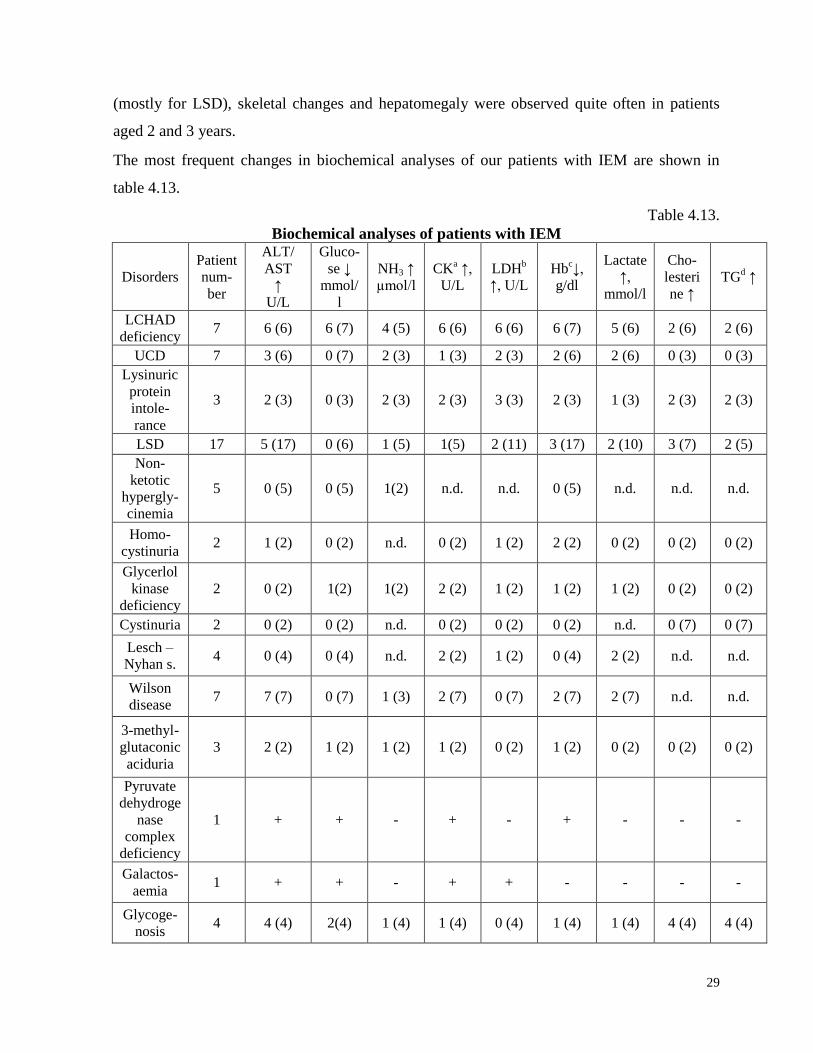
The most frequent changes in biochemical analyses of our patients with IEM are shown in
table 4.13.
Table 4.13.
Biochemical analyses of patients with IEM
Disorders
Patient
num-
ber
ALT/
AST
↑
U/L
Gluco-
se ↓
mmol/
l
NH3 ↑
µmol/l
CKa ↑,
U/L
LDHb
↑, U/L
Hbc↓,
g/dl
Lactate
↑,
mmol/l
Cho-
lesteri
ne ↑
TGd ↑
LCHAD
deficiency 7 6 (6) 6 (7) 4 (5) 6 (6) 6 (6) 6 (7) 5 (6) 2 (6) 2 (6)
UCD 7 3 (6) 0 (7) 2 (3) 1 (3) 2 (3) 2 (6) 2 (6) 0 (3) 0 (3)
Lysinuric
protein
intole-
rance
3 2 (3) 0 (3) 2 (3) 2 (3) 3 (3) 2 (3) 1 (3) 2 (3) 2 (3)
LSD 17 5 (17) 0 (6) 1 (5) 1(5) 2 (11) 3 (17) 2 (10) 3 (7) 2 (5)
Non-
ketotic
hypergly-
cinemia
5 0 (5) 0 (5) 1(2) n.d. n.d. 0 (5) n.d. n.d. n.d.
Homo-
cystinuria 2 1 (2) 0 (2) n.d. 0 (2) 1 (2) 2 (2) 0 (2) 0 (2) 0 (2)
Glycerlol
kinase
deficiency
2 0 (2) 1(2) 1(2) 2 (2) 1 (2) 1 (2) 1 (2) 0 (2) 0 (2)
Cystinuria 2 0 (2) 0 (2) n.d. 0 (2) 0 (2) 0 (2) n.d. 0 (7) 0 (7)
Lesch –
Nyhan s. 4 0 (4) 0 (4) n.d. 2 (2) 1 (2) 0 (4) 2 (2) n.d. n.d.
Wilson
disease 7 7 (7) 0 (7) 1 (3) 2 (7) 0 (7) 2 (7) 2 (7) n.d. n.d.
3-methyl-
glutaconic
aciduria
3 2 (2) 1 (2) 1 (2) 1 (2) 0 (2) 1 (2) 0 (2) 0 (2) 0 (2)
Pyruvate
dehydroge
nase
complex
deficiency
1 + + - + - + - - -
Galactos-
aemia 1 + + - + + - - - -
Glycoge-
nosis 4 4 (4) 2(4) 1 (4) 1 (4) 0 (4) 1 (4) 1 (4) 4 (4) 4 (4)
30
End of tabele 4.13
Disorders
Patient
num-
ber
ALT/
AST
↑
U/L
Gluco-
se ↓
mmol/
l
NH3 ↑
µmol/l
CKa ↑,
U/L
LDHb
↑, U/L
Hbc↓,
g/dl
Lactate
↑,
mmol/l
Cho-
lesteri
ne ↑
TGd ↑
Congeni-
tal diso-
rders of
glycosi-
lation
type Ia
2 0 (2) 0(2) 0 (2) 2 (2) 0 (2) 1 (2) 0 (2) n.d. n.d.
Lipid
metabo-
lism
defects
4 2 (4) 0 (4) 0 (4) 0 (4) 0 (4) 0 (4) 0 (4) 4 (4) 4 (4)
Hyper-
prolinae-
mia type
II
1 + - - - - - - + -
Antley
Bixler
syndrome
1 - - n.d. - - - - - -
Cystic
fibrosis 36 16 (36) 0 (36) n.d. n.d. n.d. 7 (36) 2 (36) n.d. n.d.
Total 108
51 out
of 106
(48%)
12 out
of 97
(12%)
16 out
of 38
(42%)
17 out
of 48
(35%)
17 out
of 54
(31%)
29 out
of 107
(27%)
18 out
of 90
(20%)
16 out
of 44
(36%)
14 out
of 42
(33%) aCK creatinine kinase,;
hLDH lactate dehydrogenase,;
iHb hemoglobine,;
jTG triglycerides
The most frequent biochemical change in IEM patients were elevated liver tranaminases (48%
of patients), that were detected in almost all patients.
31
5. DISCUSSION
5.1. Long–chain 3–hydroxyacyl–CoA dehydrogenase (LCHAD) deficiency
LCHAD deficiency prevalence in Latvia constitutes 2.07: 100 000, this being relatively higher
than average in Europe where it is 1: 100 000 (Orphanet Report Series, 2010). Differing from
Western Europe where the most frequent fatty acids oxidation disorders are medium chain
hydroxyacyl-CoA dehydrogenase deficiency, in Latvia, presumably, like in Poland, the Czech
Republic an Russia, patients with long-chain hydroxyacyl-CoA dehydrogenase deficiency
predominate.
The data analysis of our patients reveals that in the cases of identical genotype most of the
clinical and laboratory data are similar. Differences do appear, but they are less pronounced;
the fact being stressed also by a part of researchers (den Boer, 2002, Spiekerkoetter, 2010).
However, it should be noted that due to the small number of patients categorical statements
can’t be done. Hypoglycemia developed in five out of six patients or in 83% cases during the
first metabolic crisis. Hypoglycemia may not develop at the initial stage of metabolic crisis,
and the fact makes diagnosing difficult and might cause lethal consequences as it was in the
case with patient No 1, when stating moderate hyperglycemia, insulin therapy was prescribed
as a result of which within less than an hour developed hypoglycemic coma and seizures and
later death. It is likely that in the stress situations due to the hormones secreted by the adrenal
glands for a short period a normal or even elevated glucose level in the blood might be
observed, but the organism already experiences intracellular hypoglycemia. All available
sources refer to hypoglycaemia as a characteristic symptom of LCHAD deficiency, but the
possibility of a short-term hyperglycemia is overlooked. Though the literature data mention
creatine kinase as a sensitive marker in patients with LCHAD deficiency our data reveal that
up to the age of 6 months lactate dehydrogenase is a more sensitive indicator for the acute
period of the disease. A sudden worsening of general health, accompanied by vomiting,
lethargy, hypoglycemia with the absence of ketone substances in the urine are important
symptoms that might indicate of possible disorders of fatty acids metabolism. The Latvian
doctors have poor information and knowledge about the clinical and laboratory symptoms and
emergency therapy in the cases of acute decompensation period of LCHAD deficiency. As
acylcarnitines profile in the blood is not tested in Latvia and doing the test is possible only
32
abroad on the basis of individual agreement the given fact has led to extension of the diagnosis
confirmation. Thus, lately molecular diagnostics of the most frequent mutation in HADHA
gene 1528G>C is done to patients with clinical symptoms and changes in the organic acid
spectrum, as a result of which the diagnosis may be confirmed within a period of five to seven
days.
Early diagnostics of the disease before the manifestation of clinical symptoms is very
important for decreasing the patients’ mortality rate and the number of complications. It might
be possible if a comprehensive newborn screening included testing for disorders of fatty acids
oxidation, including LCHAD deficiency. It might also provide a possibility for measuring the
real disease frequency or incidence in Latvia. However, under the present conditions it is
important to improve the doctors’ knowledge about LCHAD deficiency, so as on the basis of
clinical and laboratory data they might identify the patients and timely do the required tests for
confirmation the diagnosis and adequate therapy.
5.2. Urea cycle disorders
The clinical data of our patients with UCD are similiar to those of other countries. The evident
difference of clinical symptoms between girls and boys was noticed in children with OTC
deficiency. The first characteristic clinical symptom in boys was respiratory failure, and later,
if hyperammonaemia persisted, lethargy and coma appeared. The repeated episodes of
vomiting before acute decompensation was characteristic sign in girls. There are no precisely
data what is the proportion between symptomatic and asymptomatic females with OTC
deficiency in literature. The time of clinical presentation in our girls was earlier compared to
data of M. Summar (Summar, 2008). The letality among our girls with OTC deficiency was
higher (50%), compared to median letality in Europe (11%). The data were collected about
110 female patients from 19 metabolic centers in 11 Eurpean countries (Häberle, 2010). The
peculiarity of clinical signs and severity among patients in one family, more likely is due to
difference in X chromosome inactivation. Our four patients with OTC deficiency died during
first undiagnosed hyperammonaemic coma. The reason of diagnostic failure was impossibility
to detect ammonia level in Latvia in patients 1, 2 and 4. Respiratory alkalosis was diagnosed
in three patients, elevated liver enzymes were observed in all girls during metabolic
decompensation. It’s difficult to summarise laboratory results because ammonia level,
aminoacids and orotic acid haven’t been done in most of patients. The family pedigree with

33
sudden death in boys during neonatal period showed the connection with X chromosome,
that’s why DNA analysis of OTC gene was done. The mortality of our patients with UCT is
71.4%, including OTC deficiency - 66.6%, but in UK – 14,4% and 15% (Chakrapani, 2010).
The highest mortality is reported during neonatal period: from 32% to 36% (Summar, 2008;
Chakrapani, 2010), but in Latvia it runs to 66,6 %. Latvian data show a serious problem which
is necessary to start solving. We could affirm that most of patients with UCD are still not
diagnosed in Latvia. It’s confirmed by increasing number of patients in countries where
diagnostics of UCD is included in newborn screening. The majority of doctors have no
knowledge about clinical symptoms of hyperammonaemia and it’s risks. For instance in year
2007, when patient Nr.7 was in Intensive Care Unit of Children’s University Hospital for three
weeks, the ammonia level was checked only after geneticist consultation. The geneticist was
informed about the result of ammonia level - 214 µmol/l (normal range below 48 µmol/l) only
on the fifth day and to that day no therapy to decrease ammonia level had been prescribed. The
tactic was wrong also in case with patient Nr.3, when his health condition became worse, the
ammonia control and appropriate therapy wasn’t done and boy died on the second day of life.
Sudden health aggravation as respiratory failure, lethargy, vomiting episodes, acute
neurological disturbances, coma could indicate the possibility of UCD independently of age
and ammonia level must be checked immediately or child transported to other hospital where
it’s available. The parameters of mortality with UCD in Latvia are 50% – 70% higher
compared to data of other countries.
The lack of knowledge about UCD among doctors in Latvia leads to late diagnosis and high
mortality in many cases. Presently it’s important to improve the awareness about UCD among
doctors in order to recognise patients and take analyses and start adequate therapy on time.
The protocol about activities in case of hyperammonaemia is necessary.
5.3. Lysosomal storage disorders
Clinical symptoms of patients with LSD in Latvia are similiar to those in other countries. In
spite of fact that clinical symptoms are persistent and slowly progressive, the diagnostic
process is often extended and difficult, because some symptoms appear in later period of life.
Comparatively high amount of revealed MPS is due to possibility to do GAG quantitation and
electrophoretic separation of different GAGs and relatively pronounced progressive clinical
symptoms. The pathological findings of heart in all cases developed secondary as a
34
complication of LSD. However comparatively small number of all patients with LSD (20%
compared to Chech Republic, Portugal, Netherlands and others) definetely shows, that many
patients with LSD are still not diagnosed. Partially it’s because the only method of diagnostics
for many LSD is enzyme studies in leukocytes or fibroblasts, which is not done in Latvia. The
clinical variability and lack of knowledge about LSD among doctors causes diagnostic
difficulties. The proper diagnosis is important for family planning and prenatal diagnosis, but
at the moment the most important thing is to find patients with treatable LSD. Patients with
hepatosplenomegaly, coarse facial features, and skeletal changes must be examined to exclude
LSD. The symptoms of LSD could be also behavioural problems, regress of psychomotor
development and hearing impairment. Gaucher disease must be excluded in patients with
unknown etiology of spleno- or hepatosplenomegaly and trombocytopenia.
It will be advisable to start checking chitotriosidase activity in Latvia, because it’s a good
marker for several LSD.
5.4. Diagnosed rare IEM in Latvia
The results of studies showed that incidence of CF in Latvia is similiar to incidence in Europe
1:3300 (Krumina, 2001). It means that in Latvia about six patients are born every year, but
only two patients or one third are diagnosed in a year. It was confirmed also by pilotproject
done in Latvia (Lace, 2009) when from 7000 screened newborns, two new patients with CF
were found. During last fifteen years only one patient was found with classical galactosaemia
in Latvia and this also indicates diagnostic problems in our country. In Estonia during time
from 1986 through 2008 nine patients with galactosaemia were diagnosed (Ounap, 2010).
Diagnostic difficulties are also with mitochondrial disorders, because immunohistochemistry
or enzyme histochemistry of muscle biopsy is not done in Latvia. Diagnostic problems are
connected also with quickly increasing number of new IEM during last years. For example,
the first defect of congenital disorders of glycosylation (CDG) was discovered in 1980, but
now the number is over 50 (Lefeber, 2011).
There are two main preconditions in diagnostics of IEM: examination facilities in Latvia to
confirm the disease and awareness of doctors about IEM. Many investigations are not
available, also the knowledge of doctors about IEM is poor in Latvia. Only with improving
these both things, we could hope for better results. First of all it’s important to improve
diagnostics of treatable IEM, accordingly decrease mortality and disability. For improving the
35
knowledge of practitioners of medicine about IEM it’s necessary to provide more information
in Latvian and Medical Genetics Association of Doctors must organise courses and workshops
about IEM. As half of our patients had symptoms already during first year of life, training of
neonatologists and pediatricians is very important.

36
6. CONCLUSIONS
1. Clinical symptoms in children with rare IEM are similiar with data from other countries.
2. Patients with LCHAD deficiency at the beginning of metabolic crises for short period may
have normoglycemia or hyperglycemia.
3. LDH is a good marker for metabolic decompensation in LCHAD deficiency for infants
before six months of age.
4. The prevalence of IEM in most cases is lower compared to average data of other European
countries.
5. Rare IEM in most cases are not recognised or diagnosed too late, which leads to early death
and severe complications.
6. The knowledge about clinical symptoms, diagnostics and therapy of rare IEM among
Latvian doctors are insufficient.

37
7. RECOMMENDATIONS HOW TO IMPROVE DIAGNOSTICS OF
RARE INBORN ERROR OF METABOLISM IN LATVIA
1. To extend a comprehensive newborn screening embraced testing for relatively more
frequent and treatable disorders, including disorders of fatty acids oxidation, UCD,
galactosaemia, cystic fibrosis.
2. The ammonia level in blood must be checked immediately to all patients with sudden
health aggravation, lethargy, coma, seizures (an informative letter about hyperammonaemia
has been created).
3. A possibility to check ammonia level in Riga’s maternity home and in all prenatal centers
of our country must be organised.
4. Common guidelines about measures in case of hyperammonaemia must be developped.
5. Newborns with sepsis and hepatopathy must be examined to sugars reducing substances
and in case of positive result selective screening for galactosaemia must be done rapidly.
6. For infants with LCHAD deficiency before six months of age it is recommended to
examine lactatdehydrogenase, which presents evidence of metabolic decompensation.
7. Sweat test must be done for children with unknown etiology of malnutrition, recurrent
obstructive bronchitis for excluding cystic fibrosis.
8. Medical Genetics Association must facilitate the knowledge about IEM organising lectures
and workshops among neonatologists, paediatricians and others practitioners of medicine.

38
APPROBATION
Publications:
1. Krūmiņa Z., Daneberga Z., Piekuse L, Kreile M., Valeiņe S., Lace B., Lugovska R. Long-
chain 3-Hydroxyacyl-CoA Dehydrogenase Deficiency in Latvia. Proceedings of the
Latvian Academy of Sciences, 2011, (submitted).
2. Krumina Z. Lizosomālās uzkrāšanās slimības. Latvijas Ārsts, 2011, 7-8: 54-56.
3. Wortmann S.B., Vaz F.M., Vissers L.E.M., Gardeitchik T., Schuurs-Hoeijmakers J.H.M.,
Kulik W., Lammens M, Christin C., Kluijtmans L.A.J., Rodenburg R., van Hasselt P.M.,
Kloosterman W., Baric I., Pronicka E., Kalkan S., Naess K., Singhal K., Krumina Z., van
Bokhoven H., Veltman J.A., Smeitink J.A.M., Lefeber D.J., Wevers R.A., Morava E., de
Brouwer A.P.. SERAC1 mutations cause MEGDEL syndrome, a phospholipid remodeling
disorder with mitochondrial dysfunction and impaired intracellular cholesterol trafficking.
SERAC1 mutations cause MEGDEL syndrome, a phospholipid remodeling disorder with
mitochondrial dysfunction and impaired intracellular cholesterol trafficking. Journal „
Nature Genetics”, 2011, (submitted).
4. Jurecka A., Krumina Z., Zuber Z., Rodzynski-Swiakowska A., Kloska A., Tylki-
Szymanska A. MPS Type II in Females and Response to Enzyme Replacement Therapy.
American Journal of Medical Genetics, 2011 (accepted).
5. Pētersons A., Ābola Z., Villeruša., Pilmane M., Lugovska R., Proņina N., Daneberga Z.,
Krūmiņa Z., Šterna O., Kreicberga I., Rezebega D., Lubaua I. Uz modernām
tehnoloģijām balstītu iedzimtu patoloģiju diagnostikas un ārstēšanas algoritmu izstrāde
bērniem.- Latvijas iedzīvotāju dzīvildzi un dzīves kvalitāti apdraudošās slimības,
Zinātniskā analīze un galvenās rekomendācijas. V. Pīrāga redakcijā. Rīga, 2009, pp.77-94.
6. Jurecka A., Popowska E., Tylki-Szymanska A., Kubalska J.,Ciara E., Krumina Z., Sykut-
Cegielska J., Pronicka E. Hypoxanthine-guanine phosphoribosylotransferase deficiency –
clinical, biochemical and molecular characteristics of patients. Przeglad Pediatryczny
2008, vol.38, Nr.3, 227-236
7. Jurecka A., Popowska E., Tylki-Szymanska A., Kubalska J.,Ciara E., Krumina Z., Sykut-
Cegielska J., Pronicka E. Hypoxanthine-guanine phosphoribosylotransferase deficiency-
The spectrum of Polish mutations. JIMD Short Report # 136 (2008) Online.

39
8. Krūmiņa A., Keiss J., Sondore V., Chernusenko A., Zarina A., Micule I., Piekuse L.,
Kreile M., Lace B., Krumina Z., Rozentāle B. From clinical and biochemical to molecular
genetic diagnosis of Wilson disease in Latvia. – Genetika, 2008, 44(10),pp.1379-1384.
9. Krumina A, Kroshkina V, Krumina L, Svabe V, Krumina Z, Tamane I, Baumanis V
(2001) Cystic fibrosis mutation dF508 in the Latvian population. RSU/AML Scientific
Proceedings, 161-166.
10. Krumina Z., Lugovska R., Vevere P. Long chain 3 hydroxyacyl- Co A dehydrogenase
deficiency – case report. Balcan Journal of Medical Genetics, International J.of Medical
Genetics, 1999,Vol.2(1),pp.37-3
The oral presentations in International conferences:
09.2011. LCHAD deficiency in Latvia.
Baltic Paediatric Ophthalmology conference, Riga, Latvia
06.2011. Patient with Alexander disease – case report. 11th International conference
of the Baltic child neurology association, Riga, Latvia
05.2009.
The infantile form of GM1 gangliosidosis: case report. 10th International
conference of the Baltic child neurology association, Tartu, Estonia
10.2008.
.
Situation with rare diseases in Latvia. MPS and rare diseases conference.
Warsaw, Poland
09.2008.
Rare inborn errors of metabolism in Latvia. 9th
Baltic Congress of
Laboratory Medicine, Jurmala, Latvia
03.2007.
Experience with vitamin B6 non-responsive homocystinuria patients in
Latvia. Meeting of Baltic Metabolic Specialists, Talinn, Estonia
03.2005.
09.2003.
Lysinuric protein intolerance.10. AEWIEM European-Asian conference of
metabolic diseases, Cairo, (Egypt)
Difficulties in diagnosis of CF in Latvia. 7th
International Symposium for
Cystic Fibrosis, Bratislava, Slovakia
Theses:
1. Krumina Z., Kreile M., Daneberga Z., Piekuse L., Vevere P, Krumina A, Lugovska R.
Large pedigree of ornithine transcarbamylase (OTC) deficiency. Case report. – J Inherit Metab
Dis (2011) 34 (suppl3): S93
40
2. Krumina Z, Daneberga Z, Kreile M, Lugovska R. Long-chain-3-hydroxyacil-CoA
dehydrogenase deficiency - the most frequent fatty acid oxidation disorder in Latvia: 7 cases. -
European Society of Human Genetics Conference, 2011, Abstract book, pp. 124-125.
3. Krūmiņa Z, Daneberga Z, Lugovska R. Garo ķēžu taukskābju hidroksiacilkoenzīma A
dehidrogenāzes nepietiekamība – biežākais diagnosticētais taukskābju defekts bērniem
Latvijā. - Rīgas Stradiņa universitātes zinātniskā konference, 2011., Rīga, Latvija, tēžu
grāmata 231 lpp.
4. Krūmiņa Z, Miklasevics E, Berzina D, Lugovska R. Patient with Alexander disease – case
report. - 11th International conference of the Baltic child neurology association, Riga, Latvia,
2011, Abstract book, p 15.
5. Krumina Z. Enzyme replacement therapy in a girl with Hunter syndrome.- Starptautiskā
konferencē par lizosomālajām slimībām, 2010, Prāga, Čehija. Abstract book.
6. Kreile M, Dzivite- Krisane I, Krumina Z, Daneberga Z, Piekuse L. Long chain 3-
hydroxyacyl-coenzyme A dehydrogenase deficiency with unusual presentation of extremely
low vitam D level. J Inherit Metab Dis, 2010, 33 (Suppl 1): S161.
7. Osipova O, Pronina N, Piekuse L, Krumina Z, Strautmanis J, Krumina A, Baumanis V.
Quantitative analysis of mitochondrial DNA in patients with suspected mitochondrial
depletion syndrome. 5th Baltic Sea reagion conference in medical sciences, 2010, Abstracts,
pp125-126.
8. Krumina Z, Daneberga Z, Czartoryska B, Kornejeva A, Lugovska R. The infantile form of
GM1 gangliosidosis: case report. 11th International conference of the Baltic child neurology
association, 2009, Abstract book, p
9. Krūmiņa A, Laganovska G, Baumane K, Pliss L, Brakmanis A, Lāce B, Krūmiņa Z,
Baumanis V. Mitohondriālās DNS analīze cilvēka patoloģijas diagnostikā. RSU 2008.gada
zinātniskā konference. Tēzes, Rīga, 102 lpp
10. Krumina Z, Lace B, Lugovska R, Vevere P, Daneberga Z, Grauduma I, Kornejeva A,
Pronina N, Micule I, Kornejeva L, Locmele Dz, Zarina A, Jasko J. Rare inborn errors of
metabolism in Latvia. Laboratorine Medicina (ISSN 1392-6470), 2008, Vol. 10, Special
Suppl, p. 14.
11. Krūmiņa Z, Vēvere P, Korņejeva L, Lugovska R. Lizinūriskā proteīnu nepanesība.
Klīniskais gadījums. RSU 2008.gada zinātniskā konference. Tēzes, 97 lpp.

41
12. Lugovska R, Krumina Z, Lace B, Vevere P, Pronina N, Daneberga Z, Kornejeva A.
Newborn screening and diagnosis of inborn errors of metabolism in Latvian children.
Laboratorine Medicina, 2008, Vol. 10, Special Suppl, p. 15.
13. Krūmiņa Z, Czartoryska B, Grauduma I, Lugovska R. Hunter syndrome in a girl: case
report. – Journal of Inherited Metabolic Disease, Volume 30, Suppl. 1, August 2007, p.98
14. Krumina Z, Muceniece Z, Lugovska R. Two patients with LCHAD deficiency in Latvia.-
Abstract book 6th International Congress on Fatty Acid Oxidation Clinical, Biochemical and
Molecular Aspects, 2005, Abstract book, p.39
15. Muceniece Z, Krūmiņa Z, Vēvere P, Pronina N, Lugovska R. Long-chain 3-hydroxyacyl-
CoA dehydrogenase (LCHAD) deficiency: Biochemical and mutation analysis in Latvia.
European Journal of Human Genetics Vol. 13 Suppl. 1, 2005, pp.269-270
16. Pronina N, Lugovska R, Muceniece Z, Olhovaya O, Krumina Z, Lace B, Balode.
Experience of molecular diagnostics for genetic disorders in Latvia. European Journal of
Human Genetics Vol. 13 Suppl. 1, 2005, p.286
17. Lugovska R, Krumina Z, Vevere P, Lace B, Pronina N, Muceniece Z, Grauduma I,
Detection of inborn errors of metabolism in Latvia. Abstract “International workshop on
Advances in Pediatrics Metabolomics”, 2005, Tartu, p.27
18. Krumina Z, Vevere P, Lugovska R. Two patients with B6 non-responsive homocystinuria
– case report. “International workshop on Advances in Pediatrics Metabolomics”, 2005, Tartu,
Abstract book, p.57
19. Lugovska R., Krumina Z., Vevere P., Pronina N., Muceniece Z., Purina G., Lace B.,
Grauduma I. Inborn errors of metabolism in Latvia. – Abstr. Bratislava Medical Journal
105(9) , 2004. p. 332
20. Muceniece Z., Krumina Z., Sass J. O., Vevere P., Grauduma I., Lugovska R. Laboratory
findings of Long-chain 3-hydroxyacyl-CoA dehydrogenase (LCHAD) deficiency – case
report. Abstr. The 7th Baltic Congress in Laboratory Medicine. Parnu, Estonia, 2004. p.127
21. Lugovska R., Krumina Z., Vevere P., Muceniece Z., Purina G., Lace B., Grauduma
I.Screening for Inherited Metabolic Diseases in Latvia. – Dietary Management of Inborn
Errors of Metabolic Disease. Inborn Errors Review Series No. 14. London, 2004, p. 36

42
22. Krumina Z., Grauduma I., Muceniece Z., Lugovska R.. Difficulties in diagnosis of MPS in
Latvia. Conference of MPS, Mainz, Germany, 2004. (stenda referāts)
23. Muceniece Z., Krumina Z., Sass J.O., Vevere P., Grauduma I., Lugovska R. Laboratory
findings of Long-chain 3-hydroxyacyl-CoA dehydrogenase (LCHAD) deficiency- case report.
Abstr. The 7th
Baltic Congress in Laboratory Medicine, Parnu, Estonia, 2004. (stenda referāts)
24. Krumina Z., Sass JO, Muceniece Z., Lugovska R. Long-Chain Hydroxyacyl-CoA
Dehydrogenase (LCHAD) Deficiency- case report. - Dietary Management of Inborn Errors of
Metabolic Disease. Inborn Errors Review Series No. 14. London, 2004, p.33
25. Krumina Z., Krumina A., Krumina L., Svabe V. Difficulties in diagnosis of CF in Latvia.
Abstr. 7th
International Symposium for Cystic Fibrosis, Bratislava, Slovakia, 2003, p.7
26. Maliseva – Lace B., Krūmiņa Z., Tamane I., Krūmiņa A, Baumanis V. Alfa-1-antitripsīna
nepietiekamības klīniskā gadījuma apraksts bērnam. – RSU Zinātniskā conference, 2002,
Riga, tēzes, p 75
27. Muceniece Z., Lugovska R., Vevere P., Krumina Z., Purina G., Lace B., Grauduma I.
Biochemical screening methods for inborn errors of metabolism in Latvia. - Laboratorine
Medicina (ISSN 1392-0), 2002, Special supplement , pp. 52-53; 63
28. Krumina Z., Vevere P., Lace B., Lugovska R., Late diagnosis of homocystinuria (case
report) Laboratorine Medicina (ISSN 1392-0), 2002, Special supplement , p. 49
29. Krumina A., Krumina L., Lazdins M., Svabe V., Krumina Z., Teibe U., Baumanis V.. CA
repeats in the first intron of the CFTR gene in cystic fibrosis patients and healthy Latvians.
European Journal of Human Genetics Vol. 10 Suppl. 1, May 2002, p.213.
30. Krumina A., Kroshkina V., Svabe V., Krumina Z., Krumina L., Tamane I.,The major
cystic fibrosis mutation in Latvia; frequency, origin and age of the dF508 mutation, Journal of
the European Society of Human Genetics, 2001, Vol.9.,Suppl.,1.,p. 276
43
REFERENCES
1. Aerts J, Kallemeijn W, Wegdam W, Ferraz M, Breemen M, Dekker N, Kramer G,
Poorthuis B, Groener J, Brinkman J, Rombach S, Hollak C, Linthorst G, Witte M, Gold
H, Marel G, Overkleeft S, Boot R (2011) Biomarkers in the diagnosis of lysosomal
storage disorders: proteins, lipids, and inhibodies. J Inherit Metab Dis 34: 605-619
2. Andersen BS, Jensen TG, Bross P, Knudsen I, Winter V, Kolvraa S, Bolund L, Ding
JH, Chen YT, Van Hove J, Curtis D, Yokota I, Tanaka K, Kim JP, Gregersen N (1994)
Disease-causing Mutations in Exon 11 of the Medium-Chain Acyl-CoA Dehydrogenase
Gene. Am J Hum Genet 54: 975-988
3. Bachmann C (2003) Outcome and survival of 88 patients with urea cycle disorders: a
retrospective evaluation. Eur. J Pediatr. 162:410-416
4. Beutler E, Gelbart T, West C (1990) The facile detection of the NT 1226 mutation of
glucocerebrosidase by “mismatched” PCR. Clin Chim Acta 194:161–166
5. Blau N, Duran M, Gibson KM (2008 a) Laboratory guide to
the methods in biochemical genetics. Springer-Verlag, pp 287-324, pp 325 – 333
6. Brusilow S, Maestry N (1996) Urea cycle disorders: diagnosis, pathophysiology and
therapy. Advances in Pediatrics, 43: 127-139
7. Chakrapani A, Champion B, Grunevald S, Lachmann R, Shortland G, Williams M,
Morris A (2010), Experience of urea cycle disorders in the UK. Poster V6: Layout 1
8. Coen SA, Meys M, Tarvin TD (1989) The Pico-Tag Method. A Manual of Advanced
Techniques for Amino Acid Analysis. Waters, Millipore Corporation.
9. den Boer ME, Ijlst L, Wijburg FA, Oostheim W, van Werkhoven MA, van Pampus
MG, Heymans HS, Wanders RA (2000) Heterozygosity for the common LCHAD
mutation (1528G>C) is not a major cause of HELLP syndrome and the prevalence of
the mutation in the Dutch population is low. Pediatric Research, Vol 48, No 2: 151-154
10. den Boer ME, Wanders RJA, Morris AA, IJlst L, Heymans HS, Wijburg FA (2002)
Long-chain 3–hydroxyacyl–CoA dehydrogenase deficiency: clinical presentation and
follow up of 50 patients. Pediatrics 109: 99-104
44
11. Farrell PM, Lai HJ, Li Z, Kosorok MR, Laxova A, Green CG, Collins J, Hoffman G,
Laessig R, Rock MJ, Splaingard ML (2005) Evidence on improved outcomes with early
diagnosis of cystic fibrosis through neonatal screening: enough is enough! J Pediatr
147: S30 – S36
12. Gropman A, Batshaw M (2004) Cognitive outcome in urea cycle disorders. Molecular
Genetics and Metabolism 81(1): 58-62
13. Grosse SD, Rosenfeld M, Devine OJ, Lai HJ, Farell PM (2006) Potential impact of
newborn screening for cystic fibrosis on child survival: a systematic review and
analysis. J Pediatr 149: 362-366
14. Häberle J, Lachmann R (2010) Diagnosis and treatment of OTC in females. European
Metabolic Group Workshop proceedings 2010., Milupa, Metabolics, pp 49-59
15. Harmatz P, Whitley CB, Waber L et al (2004) Enzyme replacement therapy in
mucopolysacharidosis VI (Maroteaux-Lamy syndrome). J Pediatr 144:574-580
16. Kahler S (2010) LCHAD (Including Trifunctional Protein) Deficiency. In: Hoffmann
G, Zschocke J, Nyhan L (eds) Inherited Metabolic Diseases. A clinical approach.
Springer-Verlag Berlin Heidelberg pp 171-172
17. Kakkis ED, Muenzer J, Tiller GE, Waber L, Belmont J, Passage M, Izykoxski B,
Phillips J, Doroshow R, Walot I, Hoft R, Neufeld EF (2001) Enzyme-replacement
therapy in mucopolysaccharidosis. N Engl J Med 344:182–188
18. Krumina A, Kroshkina V, Krumina L, Svabe V, Krumina Z, Tamane I, Baumanis V
(2001) Cystic fibrosis mutation dF508 in the Latvian population. RSU/AML Scientific
Proceedings, 161-166.
19. Lace B, Grīnblate S, Korņejeva L, Svabe V, Grauduma I,Vevere P, Lugovska R,
Martinsons A (2009) Neonatal Cystic Fibrosis screening in Latvia: a pilot project.
Proceedings of the Latvian Academy of Sciences, Section B. Natural, Exact, and
Applied Sciences, 63 (4/5): p 24
20. Lazovskis I (2001) Klīniskie simptomi un sindromi. Clinical signs, symptoms and
syndromes. Nacionālais medicīnas apgāds, Rīga, Latvija

45
21. Leonard J (2006) Disorders of the Urea Cycle and and Related Enzymes. In: Fernandes
J, Saudubray JM, van den Berghe G, Walter JH (eds) Inborn Metabolic Diseases 4th
edition, Springer Medizin Verlag Heidelberg pp 264-272
22. Lindner M, Hoffmann G, Matern D (2010) Newborn screening for disorders of fatty-
acid oxidation: experience and recommendations from an expert meeting. J Inherit
Metab Dis 33: 521-526.
23. Moog U, Zchocke I, Grünewald S (2010) Lysosomal Storage Disorders. Disturbed
degradation of macromolecules in lysosomes. In: Hoffmann GF, Zschocke J, Nyhan
WL (edit) Inherited metabolic diseases. A clinical approach, Springer-Verlag Berlin
Heidelberg, pp 220-225
24. Muenzer J, Lamsa JC, Garcia A, Dacosta J, Garcia J, Treco DA (2002) Enzyme
replacement therapy in mucopolysaccharidosis type II (Hunter syndrome): a
preliminary report. Acta Paediatr Suppl 91:98–99
25. Munnich A (2006) Defects of the Respiratory Chain. In: Fernandes J, Saudubray JM,
van den Berghe G, Walter JH (eds) Inborn Metabolic Diseases 4th
edition, Springer
Medizin Verlag Heidelberg pp 198-208
26. Orphanet Report Series – Prevalence of rare diseases: Bibliographic data – May 2010 –
Number1;http://www.orpha.net/orphacom/cahiers/docs/GB/ Prevalence of rare diseases
by alphabetical list.pdf.
27. Ounap K, Joost K, Temberg T, Krabbi L, Tonisson N (2010) Classical galactosemia in
Estonia: selective neonatal screening, incidence, and genotype/ phenotype data of
diagnosed patients. J Inherit Metab Dis 33: 175-176
28. Pinto R, Caseiro C, Lemos M et al (2004) Prevalence of lysosomal storage diseases in
Portugal. Eur J Hum Genet 12(2): 87-92
29. Polakova H, Minarik G, Ferekova E, Ficek A, Baldovic M, Kadasi L (2007) Detection
of His1069Gln mutation in Wilson disease by bidirectional PCR amplification of
specific alleles (BI-PASA) test. Gen Physiol Biophys 26(2): 91-6
30. Pons Ruiz M, Sanchez-Valverde V, Dalmau Serra J (2007) Classification. Nutritional
treatment of inborn errors of metabolism. Nutricia, Madrid pp 7-11
46
31. Poorthuis BJ, Wevers RA, Kleijer Wj et al (1999) The frequency of lysosomal storage
diseases in The Netherlands. Hum Genet 105(1-2): 151-156
32. Poupĕtová H, Ledvinová J, Berná L, Dvoráková L, Kozich V, Elleder M (2010) The
birth prevalence of lysosomal storage disorders in the Czech Republic: comparison with
data in different populations. J Inherit Metab Dis 33: 387-396
33. Rommens JM, Kerem BS, Greer W, Chang P, Tsui LC, Ray P (1990) Rapid
nonradioactive detection of the major CF mutation. Am J Hum Genet 46:395-396
34. Saudubray J, Desguerre I, Sedel F, Charpentier C (2006) Classification of inborn errors
of metabolism. In: Fernandes J, Saudubray J, van den Berghe G, Walter J (eds) Inborn
Metabolic Diseases 4th
edition, Springer Medizin Verlag Heidelberg pp 5-6
35. Sommerburg O, Lindner M, Muckenthaler M, Kohlmueller D, Leible S, Feneberg R,
Kulozik A, Mall M, Hoffmann G (2010) Initial evaluation of a biochemical cystic
fibrosis newborn screening by sequential analysis of immunoreactive trypsinogen and
pancreatitis-associated protein (IRT/PAP) as a strategy that does not involve DNA
testing in a Northen European population. J Inherit Metab Dis 33 (Suppl 2):S263-S271
36. Staretz-Chacham O, Lang TC, LaMarca ME, Krasnewitch D, Sidransky E (2009)
Lysosomal storage disorders in the newborn. Pediatrics 123(4):1191-1207
37. Summar M, Dobbelaere D, Brusilow S, Lee B (2008) Diagnosis, symptoms, frequency
and mortality of 260 patients with urea cycle disorders from a 21-year multicentre study
of acute hyperammonaemia episodes. Acta Paediatrica, 97: 1420-1425
38. Sweetman L (1991) Techniques in Diagnostic Human Biochemical Genetics: A
laboratory Manual. Wiley-Lics, Inc.pp 143-176
39. Tuchman M (2001) Proceedings of a Consensus Conference for the Managment of
patients with Urea Cycle disorders. J Pediatr 138 (Suppl 1):S1-S80
40. Utchino T, Endo F, Matsuda I (1998) Neurodevelopmental outcome of long-term
therapy of urea cycle disorders in Japan. J Inherit Metab Dis 21:151-159
41. Wanders RJ, Duran M, Ijlst L, de Jager JP, van Gennip AH, Jakobs C, Dorland L, van
Sprang FJ (1989) Sudden infant death and long chain 3-hydroxyacyl-CoA
dehydrogenase. Lancet 2:52-53
47
42. Zschocke J, Hoffmann G (2004) Disorders of ammonia detoxification – urea cycle
defects. Allopurinol test. Vademecum Metabolicum. Manual of Metabolic Paediatrics,
Milupa, Heidelberg, p 51, pp 61-63, p.96, pp. 111-123
43. Lefeber DJ, Morava E, Jaeken J (2011) How to find and diagnose a CDG due to
defective N-glycosylation. J Inherit Metab Dis 34:849-852
ACKNOWLEDGEMENTS
I am very grateful for my supervisors Dr.med. asoc. Prof. Rita Lugosvka and Dr.med. Baiba
Lāce and for colleagues from Medical Genetics Clinic (Childrens’ University Hospital) for
support in my work.
Research was supported by ESF grant No.2009/0147/1DP/1.1.2.1.2/09/IPIA/VIAA/009.
THE STRUCTURE OF WORK AND VOLUME
The doctoral thesis are written in Latvian. There are introduction, rewiev of literature,
materials and methods, results, discussion, conclusions, practical recommendations, and
refferences (101). The volume of thesis are 104 pages, apart from appendix, 32 tables and 15
figures.


