
Qeeg-guidedneurofeedBAck:
new BrAin-BAsed individuAlized evAluAtionAnd treAtment for Autism
By JaMES NEuBRaNdER, Md,1 MIChaEl lINdEN, Phd,2
Jay guNkElMaN, QEEgd,3,4 aNd CyNThIa kERSON, Phd 3,5,6
Affiliations: 1 Road to Recovery Clinic, Iselin, NJ. 2 Attention Learning Center, San Juan Capistrano, CA. 3 Brain Science International, Pleasanton, CA. 4 Behavioral Medicine Research and Training Foundation, Port Angeles, WA.5 Marin Biofeedback, San Rafael, CA. 6 ISNR Research Foundation, San Rafael, CA.
QEEG-guided neurofeedback is based on normalizing dysregulated brain regions that relate to specific clinical presentation. With ASD, this means that the approach is specific to each individual’s QEEG subtype patterns and presentation. The goal of neurofeedback with ASD is to correct amplitude abnormalities and balance brain functioning, while coherence neurofeedback aims to improve the connectivity and plasticity between brain regions. This tailored approach has implications that should not be underestimated. . . . Clinicians, including the authors, have had amazing results with ASD, including significant speech and communication improvements, calmer and less aggressive behavior, increased attention, better eye contact, and improved socialization. Many of our patients have been able to reduce or eliminate their medications aftercompletion of QEEG-guided neurofeedback.
prEfAcE Parents of children with autism know me ( JN) as a physician who uses various biomedical treatments to help children move toward recovery. Several years ago, I was introduced to the powerful modality of QEEg-guided neurofeedback. This treatment uses EEg biofeedback, also known as neurofeedback, guided by the QEEg, or quantitative electroencephalogram. Neurofeedback has since become an important addition to my practice because it offers therapeutic options that are not possible through biomedical treatments alone.
To date, I have obtained QEEgs on hundreds
of children with autism and have watched the neurofeedback process help them take one or more steps forward on their roads to recovery. That is why it pleases me to have been asked by Autism Science Digest to write this article to introduce QEEg and QEEg-guided neurofeedback for children with autism as one more important treatment option for parents to consider.
although I have prescribed many neurofeedback sessions for my clients, I cannot claim to be an expert in QEEg interpretation. In that regard, I defer to those who evaluate my patients’ EEg tracings and subsequently recommend appropriate
neurofeedback protocols that my neurofeedback technicians then implement. My coauthors (Ml, Jg, and Ck), whose biographies speak for themselves, are some of the most respected names in the field of QEEg and QEEg-guided neurofeedback. In this paper, they provide an overview of the science behind the process, a theoretical platform, and an outline of the benefits this treatment can offer to the many children who have attention-deficit or attention-deficit/hyperactivity disorder (add/adhd), asperger’s syndrome, pervasive developmental disorder-not otherwise specified (Pdd-NOS), or autism spectrum disorder (aSd).
I have obtained QEEgs on hundreds of children with autism and have watched the neurofeedback process help them take one or more steps forward on their roads to recovery. www.autismone.org REPRINTED WITH PERMISSION AUTISM SCIENCE DIGEST: THE JOURNAL OF AUTISMONE ISSUE 03 91
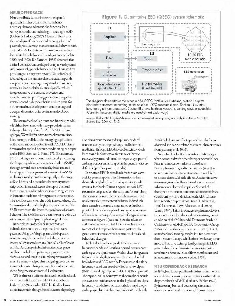
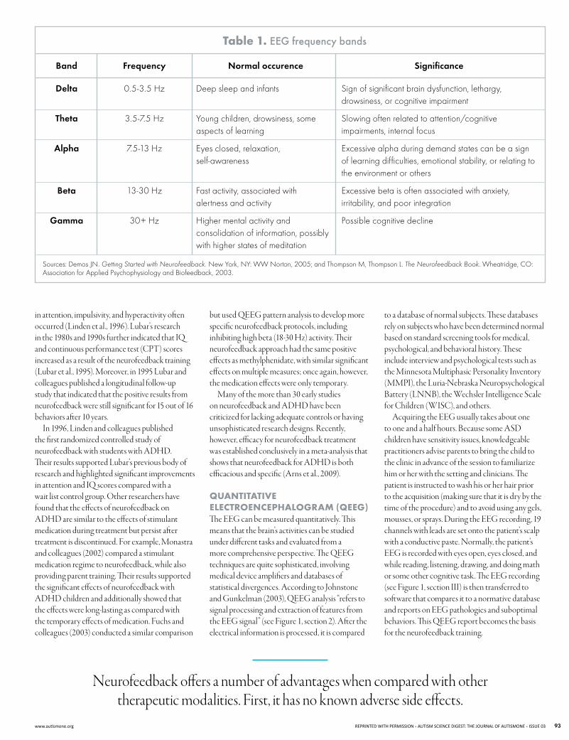
table 1. eeG frequency bands
Band Frequency Normal occurence Significance
Delta 0.5-3.5 Hz Deep sleep and infants Sign of significant brain dysfunction, lethargy, drowsiness, or cognitive impairment
Theta 3.5-7.5 Hz Young children, drowsiness, someaspects of learning
Slowing often related to attention/cognitive impairments, internal focus
Alpha 7.5-13 Hz Eyes closed, relaxation, self-awareness
Excessive alpha during demand states can be a sign of learning difficulties, emotional stability, or relating to the environment or others
Beta 13-30 Hz Fast activity, associated with alertness and activity
Excessive beta is often associated with anxiety, irritability, and poor integration
Gamma 30+ Hz Higher mental activity and consolidation of information, possibly with higher states of meditation
Possible cognitive decline
Sources: Demos JN. Getting Started with Neurofeedback. New York, NY: WW Norton, 2005; and Thompson M, Thompson L. The Neurofeedback Book. Wheatridge, CO: Association for Applied Psychophysiology and Biofeedback, 2003.
in attention, impulsivity, and hyperactivity often occurred (linden et al., 1996). lubar’s research in the 1980s and 1990s further indicated that IQ and continuous performance test (CPT) scores increased as a result of the neurofeedback training (lubar et al., 1995). Moreover, in 1995 lubar and colleagues published a longitudinal follow-up study that indicated that the positive results from neurofeedback were still significant for 15 out of 16 behaviors after 10 years.
In 1996, linden and colleagues published the first randomized controlled study of neurofeedback with students with adhd. Their results supported lubar’s previous body of research and highlighted significant improvements in attention and IQ scores compared with a wait list control group. Other researchers have found that the effects of neurofeedback on adhd are similar to the effects of stimulant medication during treatment but persist after treatment is discontinued. for example, Monastra and colleagues (2002) compared a stimulant medication regime to neurofeedback, while also providing parent training. Their results supported the significant effects of neurofeedback with adhd children and additionally showed that the effects were long-lasting as compared with the temporary effects of medication. fuchs and colleagues (2003) conducted a similar comparison
but used QEEg pattern analysis to develop more specific neurofeedback protocols, including inhibiting high beta (18-30 hz) activity. Their neurofeedback approach had the same positive effects as methylphenidate, with similar significant effects on multiple measures; once again, however, the medication effects were only temporary.
Many of the more than 30 early studies on neurofeedback and adhd have been criticized for lacking adequate controls or having unsophisticated research designs. Recently, however, efficacy for neurofeedback treatment was established conclusively in a meta-analysis that shows that neurofeedback for adhd is both efficacious and specific (arns et al., 2009).
quAntItAtIvE ElEctroEncEphAlogrAm (qEEg) The EEg can be measured quantitatively. This means that the brain’s activities can be studied under different tasks and evaluated from a more comprehensive perspective. The QEEg techniques are quite sophisticated, involving medical device amplifiers and databases of statistical divergences. according to Johnstone and gunkelman (2003), QEEg analysis “refers to signal processing and extraction of features from the EEg signal” (see figure 1, section 2). after the electrical information is processed, it is compared
to a database of normal subjects. These databases rely on subjects who have been determined normal based on standard screening tools for medical, psychological, and behavioral history. These include interview and psychological tests such as the Minnesota Multiphasic Personality Inventory (MMPI), the luria-Nebraska Neuropsychological Battery (lNNB), the Wechsler Intelligence Scale for Children (WISC), and others.
acquiring the EEg usually takes about one to one and a half hours. Because some aSd children have sensitivity issues, knowledgeable practitioners advise parents to bring the child to the clinic in advance of the session to familiarize him or her with the setting and clinicians. The patient is instructed to wash his or her hair prior to the acquisition (making sure that it is dry by the time of the procedure) and to avoid using any gels, mousses, or sprays. during the EEg recording, 19 channels with leads are set onto the patient’s scalp with a conductive paste. Normally, the patient’s EEg is recorded with eyes open, eyes closed, and while reading, listening, drawing, and doing math or some other cognitive task. The EEg recording (see figure 1, section III) is then transferred to software that compares it to a normative database and reports on EEg pathologies and suboptimal behaviors. This QEEg report becomes the basis for the neurofeedback training.
Neurofeedback offers a number of advantages when compared with other therapeutic modalities. first, it has no known adverse side effects.
www.autismone.org REPRINTED WITH PERMISSION AUTISM SCIENCE DIGEST: THE JOURNAL OF AUTISMONE ISSUE 03 93
The QEEg report interprets the following three metrics:
1. Absolute power measures the amplitude of the signal, measured in hz (or cycles per second).
2. Relative power looks at the percentage that each frequency encompasses on the overall profile.
3. Multivariate connectivity measures the similarity of the electrical waveforms to determine their level of communication. Brain areas associated with specific tasks communicate best when their electrical profiles are coherent or similar.
Many current studies support the use of QEEg in a variety of domains. for example, QEEg was found to be highly sensitive (96%) in identifying post-concussive syndrome (duff, 2004). a recent meta-analysis that recounts developments in the field observes that the QEEg has acquisition properties not achievable by other imaging technologies (such as MRI, PET, and CT scanning) because QEEg allows for the nonlinear and temporal aspects of brain activity (Thakor & Tong, 2004). Studies have used the QEEg for analysis of responses to the following:
Psychopharmacology (fingelkurts et al., 2005; hunter et al., 2005)
dementia (Chapman, 2004; yener et al., 1996)
delirium ( Jacobson et al., 1993)
Epilepsy (Clemens, 2004; van Cott, 2002)
alzheimer’s disease (Bennys et al., 2001; Jeong, 2002)
Concussion (duff, 2004)
Child and adolescent psychiatric disorders (studies reviewed by Chabot et al., 2005).
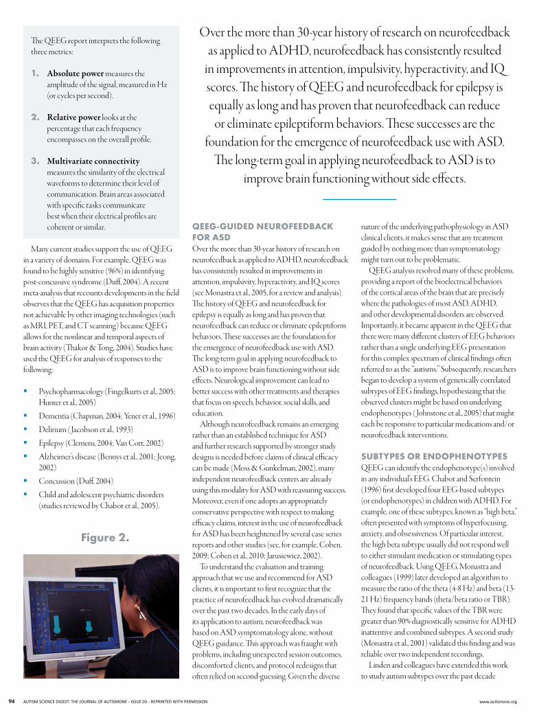
figure 2.
Over the more than 30-year history of research on neurofeedback as applied to adhd, neurofeedback has consistently resulted
in improvements in attention, impulsivity, hyperactivity, and IQ scores. The history of QEEg and neurofeedback for epilepsy is equally as long and has proven that neurofeedback can reduce
or eliminate epileptiform behaviors. These successes are the foundation for the emergence of neurofeedback use with aSd.
The long-term goal in applying neurofeedback to aSd is to improve brain functioning without side effects.
qEEg-guIdEd nEurofEEdbAck for Asd Over the more than 30-year history of research on neurofeedback as applied to adhd, neurofeedback has consistently resulted in improvements in attention, impulsivity, hyperactivity, and IQ scores (see Monastra et al., 2005, for a review and analysis). The history of QEEg and neurofeedback for epilepsy is equally as long and has proven that neurofeedback can reduce or eliminate epileptiform behaviors. These successes are the foundation for the emergence of neurofeedback use with aSd. The long-term goal in applying neurofeedback to aSd is to improve brain functioning without side effects. Neurological improvement can lead to better success with other treatments and therapies that focus on speech, behavior, social skills, and education.
although neurofeedback remains an emerging rather than an established technique for aSd and further research supported by stronger study designs is needed before claims of clinical efficacy can be made (Moss & gunkelman, 2002), many independent neurofeedback centers are already using this modality for aSd with reassuring success. Moreover, even if one adopts an appropriately conservative perspective with respect to making efficacy claims, interest in the use of neurofeedback for aSd has been heightened by several case series reports and other studies (see, for example, Coben, 2009; Coben et al., 2010; Jarusiewicz, 2002).
To understand the evaluation and training approach that we use and recommend for aSd clients, it is important to first recognize that the practice of neurofeedback has evolved dramatically over the past two decades. In the early days of its application to autism, neurofeedback was based on aSd symptomatology alone, without QEEg guidance. This approach was fraught with problems, including unexpected session outcomes, discomforted clients, and protocol redesigns that often relied on second-guessing. given the diverse
nature of the underlying pathophysiology in aSd clinical clients, it makes sense that any treatment guided by nothing more than symptomatology might turn out to be problematic.
QEEg analysis resolved many of these problems, providing a report of the bioelectrical behaviors of the cortical areas of the brain that are precisely where the pathologies of most aSd, adhd, and other developmental disorders are observed. Importantly, it became apparent in the QEEg that there were many different clusters of EEg behaviors rather than a single underlying EEg presentation for this complex spectrum of clinical findings often referred to as the “autisms.” Subsequently, researchers began to develop a system of genetically correlated subtypes of EEg findings, hypothesizing that the observed clusters might be based on underlying endophenotypes ( Johnstone et al., 2005) that might each be responsive to particular medications and/or neurofeedback interventions.
subtypEs or EndophEnotypEs QEEg can identify the endophenotype(s) involved in any individual’s EEg. Chabot and Serfontein (1996) first developed four EEg-based subtypes (or endophenotypes) in children with adhd. for example, one of these subtypes, known as “high beta,” often presented with symptoms of hyperfocusing, anxiety, and obsessiveness. Of particular interest, the high beta subtype usually did not respond well to either stimulant medication or stimulating types of neurofeedback. using QEEg, Monastra and colleagues (1999) later developed an algorithm to measure the ratio of the theta (4-8 hz) and beta (13-21 hz) frequency bands (theta/beta ratio or TBR). They found that specific values of the TBR were greater than 90% diagnostically sensitive for adhd inattentive and combined subtypes. a second study (Monastra et al., 2001) validated this finding and was reliable over two independent recordings.
linden and colleagues have extended this work to study autism subtypes over the past decade
94 AUTISM SCIENCE DIGEST: THE JOURNAL OF AUTISMONE ISSUE 03 REPRINTED WITH PERMISSION www.autismone.org
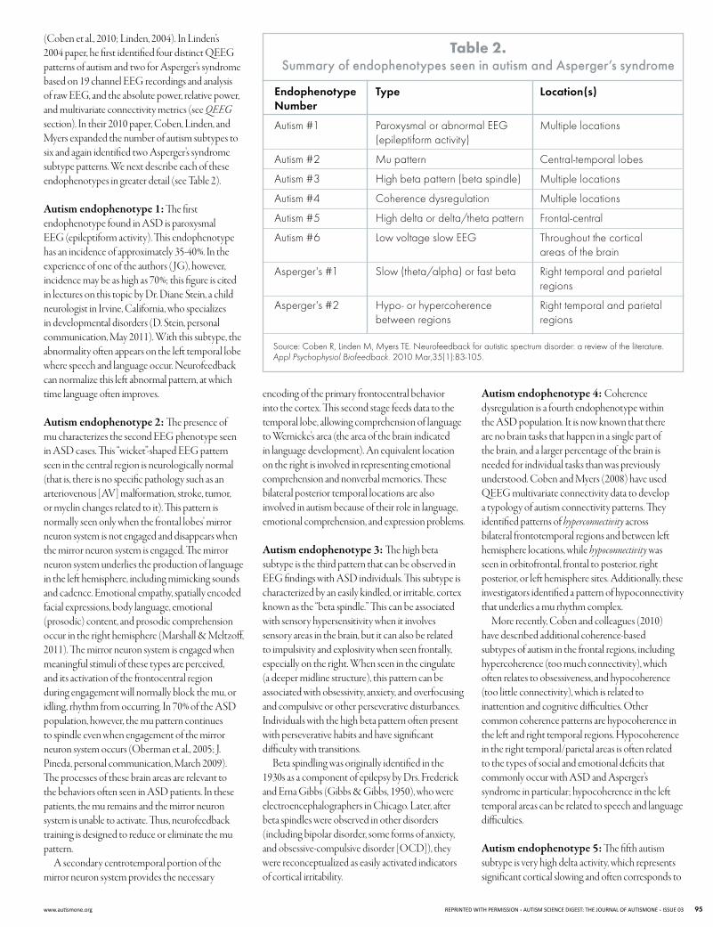
(Coben et al., 2010; linden, 2004). In linden’s 2004 paper, he first identified four distinct QEEg patterns of autism and two for asperger’s syndrome based on 19 channel EEg recordings and analysis of raw EEg, and the absolute power, relative power, and multivariate connectivity metrics (see QEEG section). In their 2010 paper, Coben, linden, and Myers expanded the number of autism subtypes to six and again identified two asperger’s syndrome subtype patterns. We next describe each of these endophenotypes in greater detail (see Table 2).
Autism endophenotype 1: The first endophenotype found in aSd is paroxysmal EEg (epileptiform activity). This endophenotype has an incidence of approximately 35-40%. In the experience of one of the authors ( Jg), however, incidence may be as high as 70%; this figure is cited in lectures on this topic by dr. diane Stein, a child neurologist in Irvine, California, who specializes in developmental disorders (d. Stein, personal communication, May 2011). With this subtype, the abnormality often appears on the left temporal lobe where speech and language occur. Neurofeedback can normalize this left abnormal pattern, at which time language often improves.
Autism endophenotype 2: The presence of mu characterizes the second EEg phenotype seen in aSd cases. This “wicket”-shaped EEg pattern seen in the central region is neurologically normal (that is, there is no specific pathology such as an arteriovenous [av] malformation, stroke, tumor, or myelin changes related to it). This pattern is normally seen only when the frontal lobes’ mirror neuron system is not engaged and disappears when the mirror neuron system is engaged. The mirror neuron system underlies the production of language in the left hemisphere, including mimicking sounds and cadence. Emotional empathy, spatially encoded facial expressions, body language, emotional (prosodic) content, and prosodic comprehension occur in the right hemisphere (Marshall & Meltzoff, 2011). The mirror neuron system is engaged when meaningful stimuli of these types are perceived, and its activation of the frontocentral region during engagement will normally block the mu, or idling, rhythm from occurring. In 70% of the aSd population, however, the mu pattern continues to spindle even when engagement of the mirror neuron system occurs (Oberman et al., 2005; J. Pineda, personal communication, March 2009). The processes of these brain areas are relevant to the behaviors often seen in aSd patients. In these patients, the mu remains and the mirror neuron system is unable to activate. Thus, neurofeedback training is designed to reduce or eliminate the mu pattern.
a secondary centrotemporal portion of the mirror neuron system provides the necessary
table 2. Summary of endophenotypes seen in autism and asperger’s syndrome
Endophenotype Number
Type Location(s)
Autism #1 Paroxysmal or abnormal EEG (epileptiform activity)
Multiple locations
Autism #2 Mu pattern Central-temporal lobes
Autism #3 High beta pattern (beta spindle) Multiple locations
Autism #4 Coherence dysregulation Multiple locations
Autism #5 High delta or delta/theta pattern Frontal-central
Autism #6 Low voltage slow EEG Throughout the cortical areas of the brain
Asperger's #1 Slow (theta/alpha) or fast beta Right temporal and parietal regions
Asperger's #2 Hypo- or hypercoherence between regions
Right temporal and parietal regions
Source: Coben R, Linden M, Myers TE. Neurofeedback for autistic spectrum disorder: a review of the literature.Appl Psychophysiol Biofeedback. 2010 Mar,35(1):83-105.
encoding of the primary frontocentral behavior into the cortex. This second stage feeds data to the temporal lobe, allowing comprehension of language to Wernicke’s area (the area of the brain indicated in language development). an equivalent location on the right is involved in representing emotional comprehension and nonverbal memories. These bilateral posterior temporal locations are also involved in autism because of their role in language, emotional comprehension, and expression problems.
Autism endophenotype 3: The high beta subtype is the third pattern that can be observed in EEg findings with aSd individuals. This subtype is characterized by an easily kindled, or irritable, cortex known as the “beta spindle.” This can be associated with sensory hypersensitivity when it involves sensory areas in the brain, but it can also be related to impulsivity and explosivity when seen frontally, especially on the right. When seen in the cingulate (a deeper midline structure), this pattern can be associated with obsessivity, anxiety, and overfocusing and compulsive or other perseverative disturbances. Individuals with the high beta pattern often present with perseverative habits and have significant difficulty with transitions.
Beta spindling was originally identified in the 1930s as a component of epilepsy by drs. frederick and Erna gibbs (gibbs & gibbs, 1950), who were electroencephalographers in Chicago. later, after beta spindles were observed in other disorders (including bipolar disorder, some forms of anxiety, and obsessive-compulsive disorder [OCd]), they were reconceptualized as easily activated indicators of cortical irritability.
Autism endophenotype 4: Coherence dysregulation is a fourth endophenotype within the aSd population. It is now known that there are no brain tasks that happen in a single part of the brain, and a larger percentage of the brain is needed for individual tasks than was previously understood. Coben and Myers (2008) have used QEEg multivariate connectivity data to develop a typology of autism connectivity patterns. They identified patterns of hyperconnectivity across bilateral frontotemporal regions and between left hemisphere locations, while hypoconnectivity was seen in orbitofrontal, frontal to posterior, right posterior, or left hemisphere sites. additionally, these investigators identified a pattern of hypoconnectivity that underlies a mu rhythm complex.
More recently, Coben and colleagues (2010) have described additional coherence-based subtypes of autism in the frontal regions, including hypercoherence (too much connectivity), which often relates to obsessiveness, and hypocoherence (too little connectivity), which is related to inattention and cognitive difficulties. Other common coherence patterns are hypocoherence in the left and right temporal regions. hypocoherence in the right temporal/parietal areas is often related to the types of social and emotional deficits that commonly occur with aSd and asperger’s syndrome in particular; hypocoherence in the left temporal areas can be related to speech and language difficulties.
Autism endophenotype 5: The fifth autism subtype is very high delta activity, which represents significant cortical slowing and often corresponds to
www.autismone.org REPRINTED WITH PERMISSION AUTISM SCIENCE DIGEST: THE JOURNAL OF AUTISMONE ISSUE 03 95
extreme activity (hyperactivity), impulsive behaviors, and inattention. Sometimes high delta activity overlaps or occurs in combination with theta activity (which also presents as inattention, impulsivity, and hyperactivity). high frontal-central slow findings are also often related to adhd.
Autism endophenotype 6: In some aSd cases a sixth pattern is seen, characterized by very low voltage EEg and dominated by slower wave activity. This low voltage slow EEg is classically identified in diffuse encephalopathies and specifically suggests that toxic or metabolic etiologies be ruled out. Some researchers believe that this low voltage pattern may be related to environmental influences (such as mercury in vaccines, pollution, pesticides, and so forth) or to metabolic issues such as mitochondrial or hormonal changes.
Asperger’s syndrome endophenotypes: Two EEg/QEEg patterns have been found to be present in most individuals with asperger’s syndrome (Coben et al., 2010; linden, 2004; Thompson et al., 2010). The first is either slow (theta/alpha) or fast beta EEg activity in the right temporal and parietal regions. These sites are involved in social skills and emotional recognition mechanisms as well as emotional expression and emotional control. The second is either too low (hypo) or too high (hyper) coherence between the right temporal/parietal brain regions and other regions. for example, hypocoherence between the right parietal and frontal regions (related to attention) may present as difficulty paying attention to emotional and social cues.
prEvAlEncE of Asd subtypEs As dEtEctEd by qEEg In our clinical work over the past 11 years and in our recent research, we have used QEEg to estimate the prevalence of the subtypes just discussed. In our experience, the high beta subtype and coherence abnormalities are the most common. We estimate the prevalence of the subtypes in children with aSd as follows:
high beta subtype (70%) Coherence abnormalities (70%) abnormal EEg subtype (33%) delta/theta subtype (30%) Metabolic/toxic (low voltage/low frequency)
subtype (10%).
Coben and colleagues (in press) recently published data that used QEEg analysis to reveal five subtypes in relative power for 91 individuals with autism and 310 normal controls. In contrast to our clinical and research estimates, these researchers observed pure excesses of beta and alpha in about one-fourth of the aSd sample (26.5% and 25.3%,
respectively) and excess theta in approximately 4.1%. Specific frontal dysfunction, including excesses of theta and alpha, was evident in 10.9% of the aSd group. Overall, more than four-fifths (83%) of the individuals with autism exhibited connectivity anomalies when compared with normal controls.
In our experience, many types of dysfunction overlap in people with autism, and most reveal a combination of QEEg findings. Our current work strongly suggests that all people with aSd display multiple brain wave pattern subtypes. In addition, individuals with autism can exhibit asperger’s patterns (and vice versa), and individuals with asperger’s may also have add/adhd QEEg patterns (for example, the high theta/beta ratio that is related to impulsivity, hyperactivity, and inattentive behaviors and symptoms). Thus, multiple diagnoses are possible and can be illuminated by EEg and QEEg subtype patterns.
thE ImportAncE of pErsonAlIzEd mEdIcInE as we have seen, EEg patterns are not simplistic or linear, and more than one pattern is usually evident. On a case-by-case basis, however, the EEg subtypes seem to correlate well with individuals’ clinical presentation. Thus, although the EEg/ QEEg subtypes (which cut across the dSM-Iv-TR categories) are not generally considered diagnostically specific, the phenotype framework can be used to guide a personalized approach to medicine through its ability to predict a given subgroup’s treatment response (gunkelman, 2007). for example, when the phenotype model was tested with adhd, it was predictive of effective response to stimulant medication (see arns et al., 2008).
In using QEEg-guided neurofeedback to treat a person with a condition as complex and heterogeneous as aSd, it seems obvious that the baseline EEg measurements would be both relevant and necessary for designing a personalized neurofeedback treatment plan. By using the QEEg report to identify a person’s phenotype patterns and then using those patterns to guide subsequent EEg training, it becomes possible to develop a customized protocol that seeks to normalize and optimize each individual’s EEg.
QEEg-guided neurofeedback is based on normalizing dysregulated brain regions that relate to specific clinical presentation. With aSd, this means that the approach is specific to each individual’s QEEg subtype patterns and presentation. The goal of neurofeedback with aSd is to correct amplitude abnormalities and balance brain functioning, while coherence neurofeedback aims to improve the connectivity and plasticity between brain regions. This tailored approach has implications that should not be underestimated. for example, correcting left temporal lobe abnormalities will affect speech and communication symptoms; working with right
parietal or temporal-sided abnormalities will affect social and emotional functions; a shift in frontal abnormalities will influence attention; addressing central abnormalities will affect impulsivity; and attention to posterior abnormalities can influence sensory functions. Clinicians, including the authors, have had amazing results with aSd, including significant speech and communication improvements, calmer and less aggressive behavior, increased attention, better eye contact, and improved socialization. Many of our patients have been able to reduce or eliminate their medications after completion of QEEg-guided neurofeedback.
not All stAtIstIcAl outlIErs ArE AbnormAl When using the QEEg, the EEg results are compared with a normative reference population to assess which average values differ between the two groups. Because it is highly likely that divergences from the mean will be seen in many domains, such as absolute and relative power and multivariate connectivity, it is most important to focus on the meaningfulness of a given divergence, which allows the neurofeedback training protocol to be further personalized. It should be recognized that while a statistical divergence may be associated with an actual abnormal finding, there are three other possibilities. Specifically, a divergence also may be due to one of the following:
1. a compensatory mechanism that helps the individual cope with the real abnormality (Barry et al., 2011)
2. an uniquely outlying measure that presents as a special skill or performance (such as very fast alpha and declarative memory performance) but not compensatory for any other finding
3. a central nervous system arousal “tuning” issue, with multiple divergent statistics seen due to frequency drifting outside normally expected ranges
an extremely important task of the clinician is to continuously monitor both clinical and behavioral changes to be assured that one of these three mechanisms is not being affected negatively. for example, in the case of example number two, if memory issues present and the training was in the alpha frequency (specifically, in the temporal areas), the training should be changed and the patient carefully monitored.
Asd And nEurofEEdbAck rEsEArch fIndIngs Notwithstanding the fact that the use of neurofeedback with aSd is still relatively recent, a
96 AUTISM SCIENCE DIGEST: THE JOURNAL OF AUTISMONE ISSUE 03 REPRINTED WITH PERMISSION www.autismone.org
number of studies have now been conducted that point to this modality’s potential. These include two pilot studies not guided by QEEg, and a small number of somewhat larger experimental studies, some of which were QEEg-guided (Coben, 2007; Coben & hudspeth, 2006; Coben & Padolsky, 2007).
PIlOT STudIES Two pilot group studies of the effects of neurofeedback on aSd symptoms have been conducted. In the first ( Jarusiewicz, 2002), 12 children each were assigned to an experimental or a control group. The experimental group received a mean of 36 neurofeedback training sessions (range = 20-69). Treatment protocols were based on Susan Othmer’s Protocol Guide for Neurofeedback Clinicians (Othmer, 2008) to determine over-, under-, and unstable arousal. The study used the autism Treatment Evaluation Checklist (aTEC) (Rimland & Edelson, 2000) to assess outcomes. Children who completed neurofeedback training attained an average 26% reduction in total aTEC-rated autism symptoms in contrast to 3% for the control group. Parents reported improvement in socialization, vocalization, anxiety, schoolwork, tantrum behaviors, and sleep habits; the control group had minimal changes in these domains. however, the outcome measures used were based solely on parent report with no other objective outcome measures.
The second pilot study (kouijzer et al., 2009a) included 14 children with aSd. Seven were in the treatment group and 7 in the wait list (no treatment) control group; controls were matched for age, gender, and IQ scores but were not randomly assigned. The treatment group received 40 sessions of neurofeedback on the right sensory motor strip. Theta activity (4-7 hz) was inhibited while SMR activity (12-15 hz) was rewarded. Pre- and post-assessment consisted of EEg learning curves, QEEg analyses, tests of executive functioning, and behavior rating scales. The neurofeedback-trained group demonstrated significant improvement in attentional control, cognitive flexibility, and goal-setting compared with the control group. Results of parent rating scales also showed improvements in social interaction and communication skills. These changes were associated with improvements in EEg learning curves. Interestingly, this same research group performed a 12-month follow-up of the treated patients with aSd (kouijzer et al., 2009b). Changes in executive functioning and behavior were both maintained, suggesting that neurofeedback may have long-lasting effects for children with autism.
although these two pilot studies showed positive results, caution should be exercised due to their very small sample sizes. Nevertheless, optimism regarding their findings led to more controlled research with larger sample sizes.
CONTROllEd STudIES WIThOuT QEEg guIdaNCE Two neurofeedback studies have focused on abnormal mu rhythms (Oberman et al., 2005). In a series of two experiments, Pineda and colleagues (2008) studied 27 children with high-functioning autism. In study 1, eight high-functioning males were randomly assigned to an experimental (n = 5) or placebo (n = 3) group. One subject dropped out of the experimental group midway through the training. Neurofeedback training included thirty 30-minute sessions with rewards for mu-like activity (8-13 hz) and inhibits for EMg (30-60 hz) at C4 (right central location). Parent rating scale data using the aTEC showed small changes (9-13%) in two of the four experimental participants. These pilot data should be considered preliminary due to the very small sample size.
In the second study (Pineda et al., 2008), 19 children with high-functioning aSd were randomly assigned to an experimental (n = 9) or placebo (n = 10) group. One very positive addition to this study was the verification of participants’ diagnoses through the autism diagnostic Observation Schedule (adOS) (lord et al., 1999) and the autism diagnostic Interview-Revised (adI-R) (Rutter et al., 2003). The neurofeedback training was similar to that provided in study 1, except that the reward band in study 2 was 10–13 hz. again, parent ratings showed a small but significant reduction in symptoms (aTEC total score). however, of concern was an increase in ratings of sensory/cognitive awareness in excess of 40% that did not occur in the placebo control group. This suggests that, according to their parents, participants improved in some areas but worsened in others. The areas of improvement may have been based on the frequencies and locations trained.
CONTROllEd STudIES WITh QEEg guIdaNCE In the largest published, controlled study to date of neurofeedback for autistic disorders, Coben and Padolsky (2007) studied 49 aSd children. The experimental group included 37 children who received QEEg-guided connectivity neurofeedback (20 sessions performed twice per week); the wait list control group included 12 children matched for age, gender, race, handedness, other treatments, and severity of aSd. The study used a broad range of assessments, including parental judgment of outcome, neuropsychological tests, behavior rating scales, QEEg analyses, and infrared imaging. Treatment protocols were assessment-based (including QEEg power and coherence) and individualized for each child. Children received neurofeedback training with a specific focus on the remediation of connectivity anomalies. Based on parental judgment of outcome, there was an 89% success rate for neurofeedback
and an average 40% reduction in core aSd symptomatology. There were also significant improvements, as compared with the control group, on neuropsychological measures of attention, executive functioning, visual perceptual processes, and language functions. Reduced cerebral hyperconnectivity was associated with positive clinical outcomes in this population. In all cases of reported improvement in aSd symptomatology, positive outcomes were confirmed by neuropsychological and neurophysiological assessment.
In another study related to mu rhythms, Coben and hudspeth (2006) studied 14 children with aSd who were identified as having significantly high levels of mu rhythm activity and a failure to suppress mu during observational activity. all 14 children received assessment-guided neurofeedback, with a strong focus on aspects of mu power and connectivity. The participants were non-randomly assigned to an interhemispheric bipolar training group (n = 7) or a coherence training (n = 7) group designed to increase connectivity between central regions and the peripheral frontal cortex. all patients were given neurobehavioral and neuropsychological testing and QEEg assessment. Both groups of patients improved significantly on neurobehavioral and neuropsychological measures. however, only in the coherence training treatment group was mu activity significantly reduced. Increased coherence was associated with diminished mu and improved levels of social functioning.
lastly, Coben (2007) conducted a controlled neurofeedback study focused on intervention for prominent social skills deficits based on a facial/ emotional-processing model. fifty individuals with autism were included, and all had previously had some neurofeedback. all patients underwent pre- and post-neuropsychological, QEEg, and parent rating scale assessments. The 50 individuals were non-randomly assigned to active neurofeedback (n = 25) and wait list control (n = 25) groups. The two groups were matched for age, gender, race, handedness, medication usage, autistic symptom severity, social skill ratings, and visual-perceptual impairment levels. Neurofeedback training was QEEg-connectivity-guided and included coherence training (along with amplitude inhibits) between maximal sites of hypocoherence over the right posterior hemisphere. The group that received the coherence training showed significant improvements in autism symptoms, social skills, and visual perceptual abilities. In addition, regression analyses showed that changes in visual-perceptual abilities significantly predicted improvements in social skills. QEEg analyses were also significant, showing improvements in connectivity and source localization of brain regions (fusiform gyrus, superior temporal sulcus) associated with enhanced visual/facial/emotional processing.
www.autismone.org REPRINTED WITH PERMISSION AUTISM SCIENCE DIGEST: THE JOURNAL OF AUTISMONE ISSUE 03 97
ImplIcAtIons And lImItAtIons In the five controlled studies that have examined neurofeedback and aSd, three of which were QEEg-guided, a total of 180 individuals with autism have been studied with positive results reported in each study. These findings have included positive changes as evidenced by parental report, neuropsychological findings, and changes in the EEg (Coben, 2007). Based on the guidelines of Coben and Padolsky (2007) and yucha and Montgomery (2008), neurofeedback for autism is considered “possibly efficacious.” added to these initial findings of efficacy is preliminary evidence that the effects of neurofeedback on the symptoms of autism are long-lasting (1–2 years) (Coben & Wagner, 2010; kouijzer et al., 2009b).
We are currently working on structured research that incorporates the emerging clinical application of neurofeedback for aSd cases with the phenotype approach, correlating EEg/QEEg patterns with brain structure using functional magnetic resonance imaging (f MRI) and diffusion tensor imaging (dTI). for example, the National Institutes of health (NIh) recently funded a study at the university of California, San diego (uCSd), that is evaluating the impact of neurofeedback on aSd in which one of the authors (Ml) is involved. Specifically, this study is investigating QEEg, f MRI, and dTI results of both QEEg-guided and mu neurofeedback in both aSd and typical students. These imaging tools utilize an MRI scanner to look at blood flow and water density, respectively.
another important use of the EEg/QEEg for the aSd population involves measuring brain wave activity to guide treatment with other commonly used therapeutic modalities, such as medication, hyperbaric oxygen therapy (hBOT), and biomedical treatments. Three of the authors ( JN, Jg, and Ml) are currently beginning preliminary research in these areas of application.
There are five limitations that prevent firm conclusions from being drawn from the studies conducted to date. Some of these limitations are being addressed by our current research.
1. first, the studies have largely included non-randomized samples, meaning that an unknown selection bias could have existed that could have influenced the findings.
2. Second, none of the completed studies (with the exception of the uCSd study in progress) have included participants or therapists/experimenters who were blind to the treatment condition. knowledge of group placement could have affected the findings to the extent that those in treatment (and their parents) may have been more prone to report significant changes.
3. Third, none of the studies attempted to control for placebo effects, attention from a caring professional, or expectations of treatment benefit. however, in the current uCSd study, we (Ml) are also having typical students complete neurofeedback. a randomized, double-blinded, placebo-controlled study, although complicated and difficult to do, would be optimal to further demonstrate efficacy.
4. a fourth limitation is that very youngchildren (under four years of age)and adults have not been representedin these studies, so generalization tothese groups is not possible. Thesepopulations should be the focus offuture research investigations.
5. lastly, aSd individuals who are lower functioning or who have more severe symptoms associated with autism have not been included in research to date, although clinicians, including the authors, have had successful treatment outcomes.
Overall, the use of QEEg to assess subtype patterns of aSd is important in both analysis of brain bioelectrical pathologies and for treatment selection and success. The use of neurofeedback with aSd is becoming a highly personalized and successful treatment option and continues to be very promising.
onE fInAl thought as I ( JN) mentioned at the beginning of this article, QEEgs and QEEg-guided neurofeedback have significantly increased the benefits I can offer my patients on the autism spectrum. Though the clinical outcomes I observe from biomedically
oriented treatments have been significant, at times leading to full recovery, the addition of QEEg-directed neurofeedback has given a high percentage of my patients the ability to get “unstuck” and begin moving again on the road to recovery. Once unstuck, many of them have gone much farther than they would have ever gone with the other biomedical, behavioral, and educational treatments I use or recommend.
One of the subtypes described above, aberrant EEg or short intermittent episodes of epileptiform behaviors (a term coined by some as subclinical seizures), has guided me to suggest a clinical trial of anticonvulsant therapy even when children do not have true seizure activity. In the past, only children with documented seizure activity were prescribed anticonvulsant medications. Research studies vary as to the incidence of true seizure activity in the autism population; 33% would be a close average. Now, however, it is becoming more accepted for children on the autism spectrum who do not have documented seizures but who have atypical, aberrant EEg brain wave activity (approximately 66%-75%) to at some point be given a clinical trial of anticonvulsant therapy, especially when other treatments are not producing the expected results. It is not uncommon for parents to report that the addition of an anticonvulsant medication to their child’s treatment regimen resulted in increased language, focus, attention, cognition, and positive behavioral changes. With the QEEg subtype analysis and QEEg-guided neurofeedback protocols developed by my coauthors ( Jg and Ml), I have become more successful in choosing appropriate treatments, whether medications or natural agents. By knowing this important information, I have been able to target specific medications or natural agents rather than “blindly prescribing” neuropsychological or neuropsychiatric medications as is commonly done by psychiatrists and neurologists who do not believe in or obtain QEEgs to help guide their choice of medications.
Though the clinical outcomes I observe from biomedically oriented treatments have been significant, at times leading to full recovery, the addition of QEEg-directed neurofeedback
has given a high percentage of my patients the ability to get “unstuck” and begin moving again on the road to recovery. Once unstuck, many of them have gone much farther than
they would have ever gone with the other biomedical, behavioral, and educational treatments I use or recommend.
98 AUTISM SCIENCE DIGEST: THE JOURNAL OF AUTISMONE ISSUE 03 REPRINTED WITH PERMISSION www.autismone.org
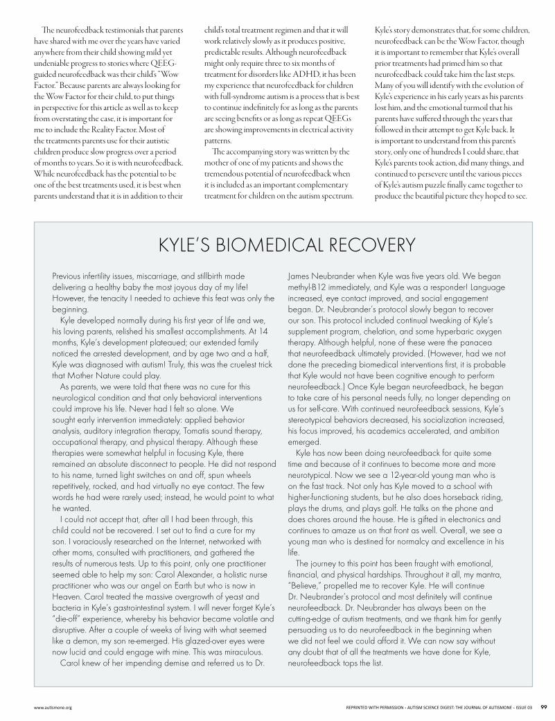
The neurofeedback testimonials that parents have shared with me over the years have varied anywhere from their child showing mild yet undeniable progress to stories where QEEg-guided neurofeedback was their child’s “Wow factor.” Because parents are always looking for the Wow factor for their child, to put things in perspective for this article as well as to keep from overstating the case, it is important for me to include the Reality factor. Most of the treatments parents use for their autistic children produce slow progress over a period of months to years. So it is with neurofeedback. While neurofeedback has the potential to be one of the best treatments used, it is best when parents understand that it is in addition to their
child’s total treatment regimen and that it will work relatively slowly as it produces positive, predictable results. although neurofeedback might only require three to six months of treatment for disorders like adhd, it has been my experience that neurofeedback for children with full-syndrome autism is a process that is best to continue indefinitely for as long as the parents are seeing benefits or as long as repeat QEEgs are showing improvements in electrical activity patterns.
The accompanying story was written by the mother of one of my patients and shows the tremendous potential of neurofeedback when it is included as an important complementary treatment for children on the autism spectrum.
kyle’s story demonstrates that, for some children, neurofeedback can be the Wow factor, though it is important to remember that kyle’s overall prior treatments had primed him so that neurofeedback could take him the last steps. Many of you will identify with the evolution of kyle’s experience in his early years as his parents lost him, and the emotional turmoil that his parents have suffered through the years that followed in their attempt to get kyle back. It is important to understand from this parent’s story, only one of hundreds I could share, that kyle’s parents took action, did many things, and continued to persevere until the various pieces of kyle’s autism puzzle finally came together to produce the beautiful picture they hoped to see.
KYLE’SBIOMEDICALRECOVERYPrevious infertility issues, miscarriage, and stillbirth made delivering a healthy baby the most joyous day of my life!However, the tenacity I needed to achieve this feat was only the beginning.
Kyle developed normally during his first year of life and we, his loving parents, relished his smallest accomplishments. At 14 months, Kyle’s development plateaued; our extended familynoticed the arrested development, and by age two and a half, Kyle was diagnosed with autism! Truly, this was the cruelest trick that Mother Nature could play.
As parents, we were told that there was no cure for this neurological condition and that only behavioral interventions could improve his life. Never had I felt so alone. Wesought early intervention immediately: applied behavior analysis, auditory integration therapy, Tomatis sound therapy, occupational therapy, and physical therapy. Although these therapies were somewhat helpful in focusing Kyle, there remained an absolute disconnect to people. He did not respond to his name, turned light switches on and off, spun wheels repetitively, rocked, and had virtually no eye contact. The few words he had were rarely used; instead, he would point to whathe wanted.
I could not accept that, after all I had been through, this child could not be recovered. I set out to find a cure for my son. I voraciously researched on the Internet, networked with other moms, consulted with practitioners, and gathered the results of numerous tests. Up to this point, only one practitioner seemed able to help my son: Carol Alexander, a holistic nurse practitioner who was our angel on Earth but who is now in Heaven. Carol treated the massive overgrowth of yeast and bacteria in Kyle’s gastrointestinal system. I will never forget Kyle’s “die-off” experience, whereby his behavior became volatile anddisruptive. After a couple of weeks of living with what seemed like a demon, my son re-emerged. His glazed-over eyes were now lucid and could engage with mine. This was miraculous.
Carol knew of her impending demise and referred us to Dr.
James Neubrander when Kyle was five years old. We beganmethyl-B12 immediately, and Kyle was a responder! Language increased, eye contact improved, and social engagement began. Dr. Neubrander’s protocol slowly began to recoverour son. This protocol included continual tweaking of Kyle’s supplement program, chelation, and some hyperbaric oxygen therapy. Although helpful, none of these were the panacea that neurofeedback ultimately provided. (However, had we not done the preceding biomedical interventions first, it is probable that Kyle would not have been cognitive enough to perform neurofeedback.) Once Kyle began neurofeedback, he began to take care of his personal needs fully, no longer depending on us for self-care. With continued neurofeedback sessions, Kyle’s stereotypical behaviors decreased, his socialization increased, his focus improved, his academics accelerated, and ambition emerged.
Kyle has now been doing neurofeedback for quite some time and because of it continues to become more and more neurotypical. Now we see a 12-year-old young man who ison the fast track. Not only has Kyle moved to a school withhigher-functioning students, but he also does horseback riding, plays the drums, and plays golf. He talks on the phone and does chores around the house. He is gifted in electronics and continues to amaze us on that front as well. Overall, we see a young man who is destined for normalcy and excellence in his life. The journey to this point has been fraught with emotional,
financial, and physical hardships. Throughout it all, my mantra, “Believe,” propelled me to recover Kyle. He will continueDr. Neubrander’s protocol and most definitely will continueneurofeedback. Dr. Neubrander has always been on thecutting-edge of autism treatments, and we thank him for gently persuading us to do neurofeedback in the beginning when we did not feel we could afford it. We can now say without any doubt that of all the treatments we have done for Kyle, neurofeedback tops the list.
www.autismone.org REPRINTED WITH PERMISSION AUTISM SCIENCE DIGEST: THE JOURNAL OF AUTISMONE ISSUE 03 99
rEfErENCES
Arns M, de Ridder S, Strehl U, Breteler M, Coenen A.Efficacy of neurofeedback treatment in ADHD: the effects on inattention, impulsivity and hyperactivity: a meta-analysis. Clin EEG Neurosci. 2009 Jul;40(3):180-9.
Arns M, Gunkelman J, Breteler M, Spronk D. EEGphenotypes predict treatment outcome to stimulants in children with ADHD. J Integr Neurosci. 2008 Sep;7(3):421-38.
Barry RJ, Clarke AR, Hajos M, Dupuy FE, McCarthy R,Selikowitz M. EEG coherence and symptom profiles ofchildren with Attention-Deficit/Hyperactivity Disorder. Clin Neurophysiol. 2011 Jul;122(7):1327-32.
Bennys K, Rondouin G, Vergnes C, Touchon J. Diagnosticvalue of quantitative EEG in Alzheimer’s disease. Neurophysiol Clin. 2001 Jun;31(3):153-60.
Chabot RJ, Serfontein G. Quantitative electroencephalographic profiles of children with attention deficit disorder. Biol Psychiatry. 1996 Nov;40(10:951-63.
Chabot RJ, di Michele F, Prichep L. The role of quantitativeelectroencephalography in child and adolescent psychiatric disorders. Child Adolesc Psychiatr Clin N Am. 2005 Jan;14(1):21-53, v-vi.
Chapman H. qEEG and dementia. Arq Neuropsiquiatr. 2004 Sep; 62(3A):749.
Clemens B. Abnormal quantitative EEG scores identify patients with complicated idiopathic generalised epilepsy. Seizure. 2004 Sep;13(6):366-74.
Coben R. Autistic spectrum disorder: outcome of EEG coherence training targeting social skills deficits. J Neurother. 2007;12(1):60.
Coben R. Efficacy of connectivity guided neurofeedback for autistic spectrum disorder: controlled analysis of 75 cases with a 1 to 2 year follow-up. J Neurother. 2009;13(1):81.
Coben R, Hirshberg L, Chabot R. EEG discriminant power and subtypes in autistic spectrum disorder. Int J Psychophysiol. (In press).
Cohen R, Hudspeth W. Mu-like rhythms in autistic spectrumdisorder: EEG analyses and neurofeedback. Presented at the 14th Annual Conference of the International Society for Neuronal Regulation. Atlanta, GA: September, 2006.
Coben R, Linden M, Myers TE. Neurofeedback forautistic spectrum disorder: a review of the literature. Appl Psychophysiol Biofeedback. 2010 Mar;35(1):83-105.
Coben R, Myers TE. Connectivity theory of autism: use ofconnectivity measures in assessing and treating autistic disorders. J Neurother. 2008;12(2-3):161-79.
Coben R, Myers TE. The relative efficacy of connectivityguided and symptom based EEG biofeedback for autistic disorders. Appl Psychophysiol Biofeedback. 2010 Mar;35(1):13-23.
Coben R, Padolsky I. Assessment-guided neurofeedback for autistic spectrum disorder. J Neurother. 2007;11(1):5-23.
Coben R, Wagner L. Emerging empirical evidence supporting connectivity-guided neurofeedback for autistic disorders. Pp. 153-82 in Neurofeedback and Neuromodulation Techniques and Applications, Chapter 6, R Coben & JR Evans (eds.). New York, NY: Academic Press,2010.
Demos JN. Getting Started with Neurofeedback. New York,NY: WW Norton, 2005.
Duff J. The usefulness of quantitative EEG (QEEG) and neurotherapy in the assessment and treatment of post-concussion syndrome. Clin EEG Neurosci. 2004 Oct;35(4):198-209.
Fingelkurts AA, Fingelkurts AA, Kähkönen S. Newperspectives in pharmaco-electroencephalography. Prog Neuropsychopharmacol Biol Psychiatry. 2005 Feb;29(2):193-9.
Fuchs T, Birbaumer N, Lutzenberger W, Gruzelier JH,Kaiser J. Neurofeedback treatment for attention-deficit/hyperactivity disorder in children: a comparison with methylphenidate. Appl Psychophysiol Biofeedback. 2003 Mar;28(1):1-12.
Gibbs FA, Gibbs EL Atlas of Electroencephalography, Volume 1. Cambridge, MA: Addison-Wesley, 1950.
Gunkelman J. Transcend the DSM using phenotypes.Biofeedback. 2006 Fall;34(3):95-8.
Hunter AM, Leuchter AF, Morgan ML, Cook IA, AbramsM, Siegman B, DeBrota DJ, Potter WZ. Neurophysiologiccorrelates of side effects in normal subjects randomized tovenlafaxine or placebo. Neuropsychopharmacology. 2005 Apr;30(4):792-9.
Jacobson SA, Leuchter AF, Walter DO. Conventional and quantitative EEG in the diagnosis of delirium among the elderly. J Neurol Neurosurg Psychiatry. 1993 Feb;56(2):153-8.
Jarusiewicz B. Efficacy of neurofeedback for children in the autistic spectrum: a pilot study. J Neurother. 2002 Win;6(4):39-49.
Jeong J. Nonlinear dynamics of EEG in Alzheimer’s disease.Drug Dev Res. 2002 Jun;56(2):57-66.
Johnstone J, Gunkelman J. Use of databases in QEEG evaluation. J Neurother. 2003;7(3/4):31-52.
Johnstone J, Gunkelman J, Lunt J. Clinical database development: characterization of EEG phenotypes. Clin EEG Neurosci. 2005 Apr;36(2):99-107.
Kouijzer MEJ, de Moor JMH, Gerrits BJL, Congedo M, vanSchie HT. Neurofeedback improves executive functioning inchildren with autism spectrum disorders. Res Autism Spectr Disord. 2009a Jan;3(1):145-62.
Kouijzer MEJ, de Moor JMH, Gerrits BJL, Buitelaar JK, vanSchie HT. Long-term effects of neurofeedback treatment in autism. Res Autism Spectr Disord. 2009b Apr;3(2):496–501.
Laibow RE. Medical applications of neurobiofeedback.Pp. 83-102 in Introduction to Quantitative EEG and Neurofeedback, Evans JR and Abarbanel A, eds. San Diego, CA: Academic Press, 1999.
Linden, M. Case studies of QEEG mapping andneurofeedback with autism. Presented at the 12th Annual Conference of the International Society for NeuronalRegulation, Fort Lauderdale, FL, August 2004.
Linden M, Habib T, Radojevic V. A controlled study of theeffects of EEG biofeedback on cognition and behavior of children with attention deficit disorder and learning disabilities. Biofeedback Self Regul. 1996 Mar;21(1):35-49.
Lord C, Rutter M, DiLavore PC, Risi S. Autism Diagnostic Observation Schedule-WPS (ADOS-WPS). Los Angeles, CA: Western Psychological Services, 1999.
Lubar JF. Neocortical dynamics: implications forunderstanding the role of neurofeedback and related techniques for the enhancement of attention. Appl Psychophysiol Biofeedback. 1997 Jun;22(2):111-26.
Lubar JF, Bahler WW. Behavioral management of epileptic seizures following EEG biofeedback training of the sensorimotor rhythm. Biofeedback Self Regul. 1976 Mar;1(1):77-104.
Lubar JF, Swartwood MO, Swartwood JN, O’DonnellPH. Evaluation of the effectiveness of EEG neurofeedback training for ADHD in a clinical setting as measured by changes in T.O.V.A. scores, behavioral ratings, and WISE-Rperformance. Biofeedback Self Regul. 1995 Mar;20(1):83-99.
Marshall PJ, Meltzoff AN. Neural mirroring systems:Exploring the EEG mu rhythm in human infancy. Dev Cogn Neurosci. 2011 Apr;1(2):110–23.
Monastra VJ, Lubar JF, Linden M. The development of aquantitative electroencephalographic scanning process for attention-deficit/hyperactivity disorder: reliability and validity studies. Neuropsychology. 2001;15(1):136-44.
Monastra VJ, Lubar JF, Linden M, VanDeusen P,Green G, Wing W, Phillips A, Fenger TN. Assessingattention deficit hyperactivity disorder via quantitative electroencephalography: an initial validation study. Neuropsychology. 1999;13(3):424-33.
Monastra VJ, Lynn S, Linden M, Lubar JF, Gruzelier J,LaVaque TJ. Electroencephalographic biofeedback in thetreatment of attention-deficit/hyperactivity disorder. Appl Psychophysiol Biofeedback. 2005 Jun;30(2):95-114.
Monastra VJ, Monastra DM, George S. The effects ofstimulant therapy, EEG biofeedback, and parenting style on the primary symptoms of attention-deficit/hyperactivity disorder. Appl Psychophysiol Biofeedback. 2002 Dec;27(4):231-49.
Moss D, Gunkelman J. Task force report on methodologyand empirically supported treatments: introduction. Appl Psychophysiol Biofeedback. 2002;27(4):271-2.
MTA Cooperative Group. National Institute of Mental Healthmultimodal treatment study of ADHD follow-up: 24-month outcomes of treatment strategies for attention-deficit/ hyperactivity disorder. Pediatrics. 2004;113(4):754-61.
Oberman LM, Hubbard EM, McCleery JP, Altschuler EL,Ramachandran VS, Pineda JA. EEG evidence for mirrorneuron dysfunction in autism spectrum disorders. Brain Res Cogn Brain Res. 2005 Jul;24(2):190-8.
Othmer S. Protocol Guide for Neurofeedback Clinicians, 2nd edition. Woodland Hills, CA: EEG Info, 2008.
Pineda JA, Brang D, Hecht E, Edwards L, Carey S, Bacon M, Futagaki C, Suk D, Tom J, Birnbaum C, Rork A. Positivebehavioral and electrophysiological changes following neurofeedback training in children with autism. Res Autism Spectr Disord. 2008 Jul-Sep;2(3):557-81.
Rangaswamy M, Porjesz B, Chorlian DB, Wang K, Jones KA,Bauer LO, Rohrbaugh J, O’Connor SJ, Kuperman S, Reich T, Begleiter H. Beta power in the EEG of alcoholics. Biol Psychiatry. 2002 Oct;52(8):831-42.
Rimland B, Edelson SM. Autism Treatment Evaluation Checklist. San Diego, CA: Autism Research Institute, 2000.
Rutter M, LeCouteur A, Lord C.Manual for the ADI–WPS version. Los Angeles, CA: Western Psychological Services, 2003.
Sherlin LH, Arns M, Lubar J, Heinrich H, Kerson C, Strehl U,Sterman MB. Neurofeedback and basic learning theory:implications for research and practice. J Neurother. (In press.)
Skinner BF. Reinforcement today. American Psychologist. 1958 Mar;13(3):94-99.
Sterman MB, Friar L. Suppression of seizures in anepileptic following sensorimotor EEG feedback training. Electroencephalogr Clin Neurophysiol. 1972 Jul;33(1):89-95.
Sterman MB, LoPresti RW, Fairchild MD.Electroencephalographic and behavioral studies of monomethl hydrazine toxicity in the cat. J Neurother. 2010;14(4):293-300.
Tansey MA. Ten-year stability of EEG biofeedback resultsfor a hyperactive boy who failed fourth grade perceptually impaired class. Biofeedback Self Regul. 1993 Mar;18(1):33-44.
Thakor NV, Tong S. Advances in quantitativeelectroencephalogram analysis methods. Annu Rev Biomed Eng. 2004;6:453-5.
Thompson M, Thompson L. The Neurofeedback Book. Wheatridge, CO: Association for Applied Psychophysiology and Biofeedback, 2003.
Thompson L, Thompson M, Reid A. Functional neuroanatomyand the rationale for using EEG biofeedback for clients with Asperger’s syndrome. Appl Psychophysiol Biofeedback. 2010 Mar;35(1):39-61.
Van Cott AC. Epilepsy and EEG in the elderly. Epilepsia. 2002 Mar;43(suppl 3):94-102.
Yener GG, Leuchter AF, Jenden D, Read SL, Cummings JL,Miller BL. Quantitative EEG in frontotemporal dementia. Clin Electroencephalogr. 1996 Apr;27(2):61-8.
Yucha C, Montgomery D. Evidence-Based Practice in Biofeedback and Neurofeedback. Wheat Ridge, CO: Association for Applied Psychophysiology and Biofeedback, 2008.
100 AUTISM SCIENCE DIGEST: THE JOURNAL OF AUTISMONE ISSUE 03 REPRINTED WITH PERMISSION www.autismone.org


