Outline - Membranes
1. Fluid Mosaic Model of Membrane Structure
2. Membrane Proteins
1. Kinds of membrane proteins
2. Membrane protein structure
Single pass
Multi-pass: Channels, Pores & Carriers
3. Transport MechanismsPassive: Diffusion & Facilitated Diffusion
Active: Molecular & Bulk
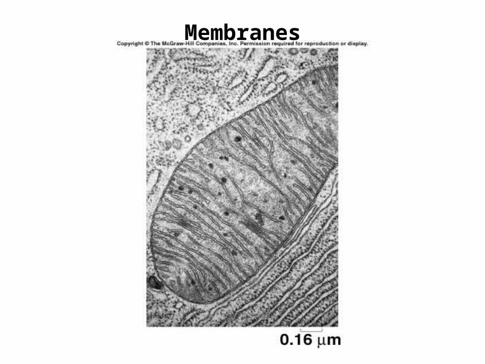
Membranes
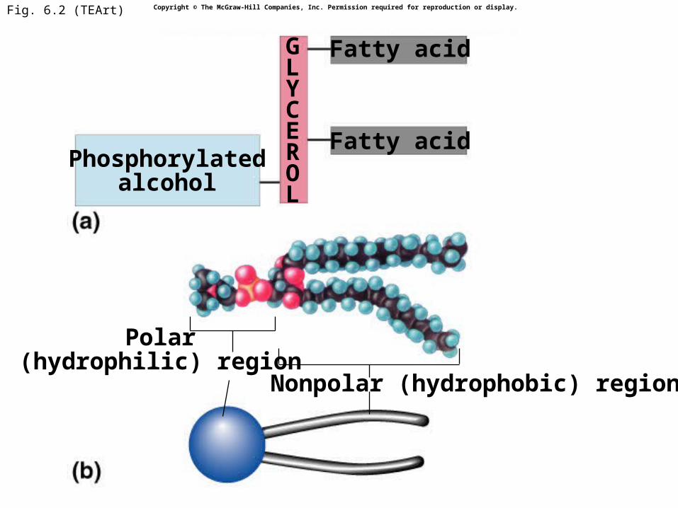
Fig. 6.2 (TEArt)
Fatty acidPhosphorylated
alcohol
Polar(hydrophilic) region
Nonpolar (hydrophobic) region
Fatty acidGLYCEROL
Copyright © The McGraw-Hill Companies, Inc. Permission required for reproduction or display.
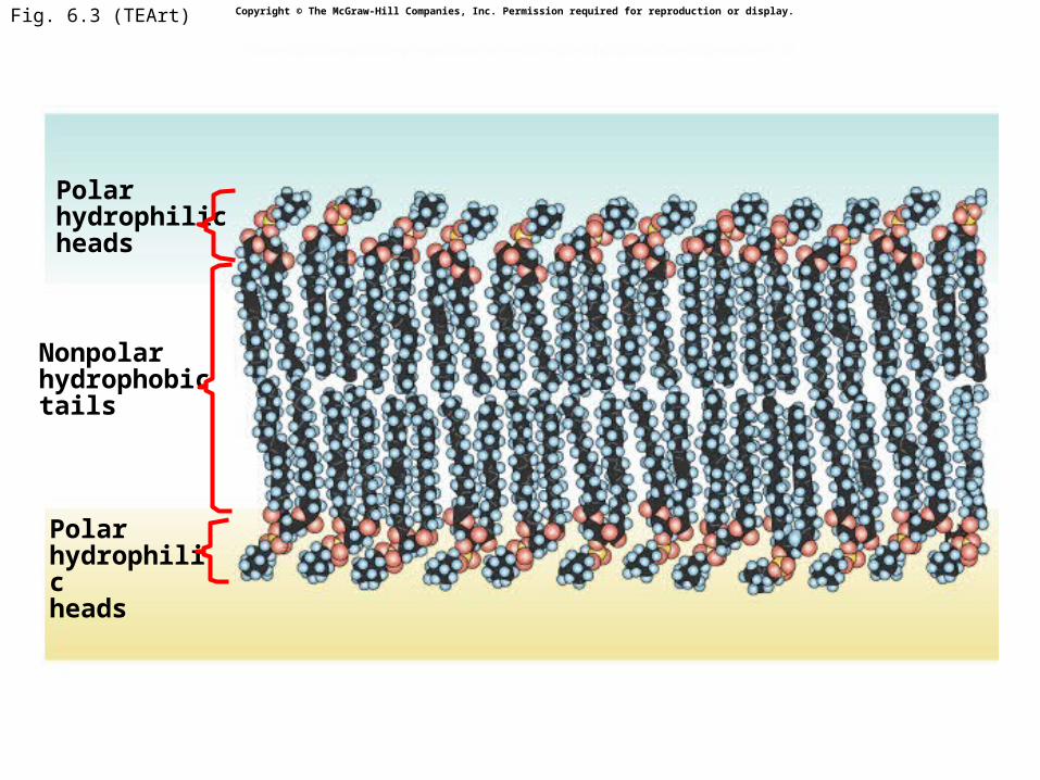
Fig. 6.3 (TEArt)
Polarhydrophilicheads
Nonpolarhydrophobictails
Polarhydrophilicheads
Copyright © The McGraw-Hill Companies, Inc. Permission required for reproduction or display.
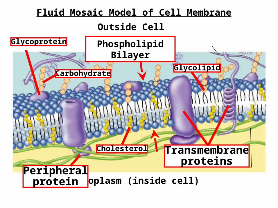
Outside Cell
Cytoplasm (inside cell)
Cholesterol Transmembraneproteins
Peripheralprotein
Glycoprotein
CarbohydrateGlycolipid
Phospholipid Bilayer
Fluid Mosaic Model of Cell Membrane
6
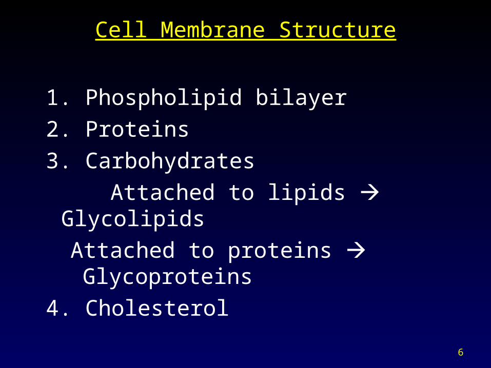
Cell Membrane Structure
1. Phospholipid bilayer
2. Proteins
3. Carbohydrates
Attached to lipids Glycolipids
Attached to proteins Glycoproteins
4. Cholesterol
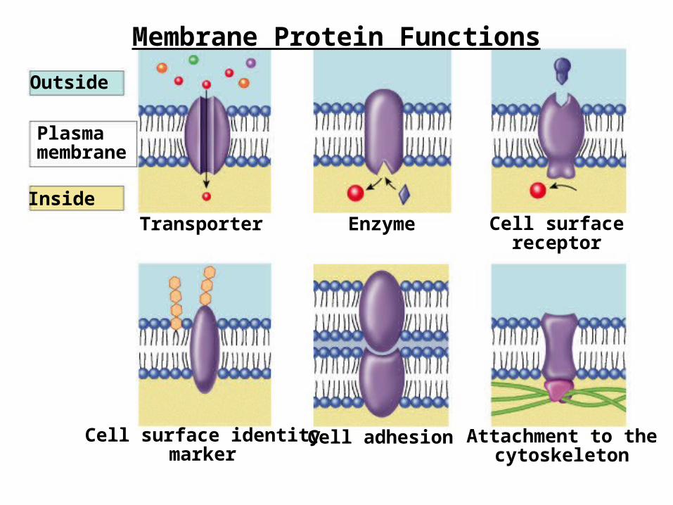
Outside
Plasmamembrane
InsideTransporter Cell surface
receptorEnzyme
Cell surface identitymarker
Attachment to thecytoskeleton
Cell adhesion
Membrane Protein Functions
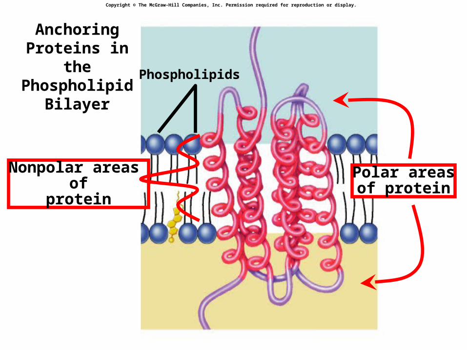
Phospholipids
Polar areasof protein
Nonpolar areas of
protein
Copyright © The McGraw-Hill Companies, Inc. Permission required for reproduction or display.
Anchoring Proteins in the Phospholipid
Bilayer
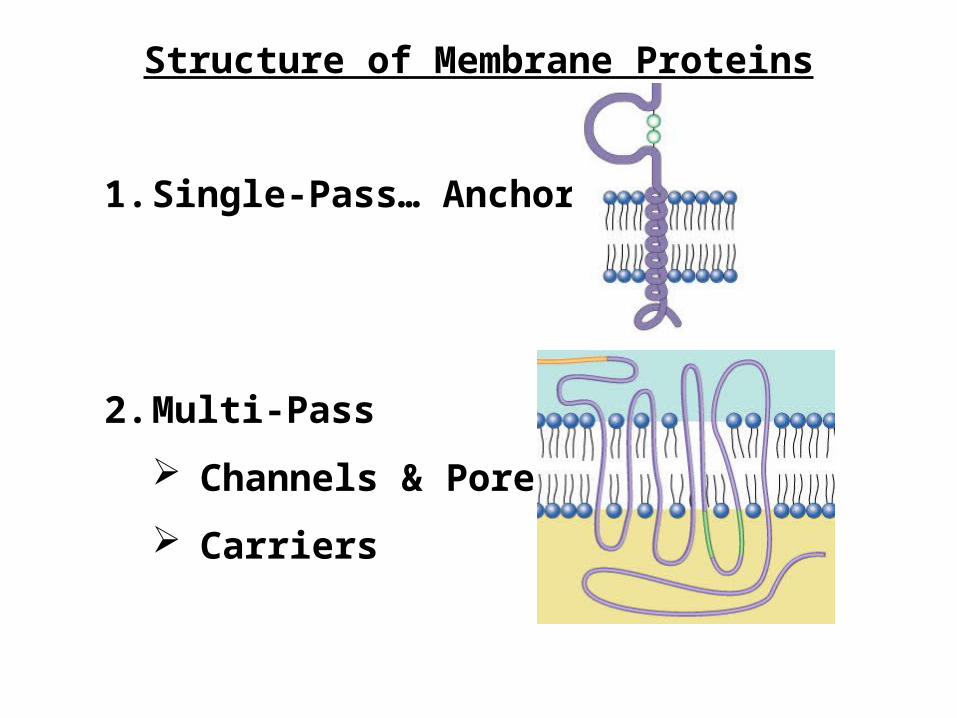
1. Single-Pass… Anchors
2. Multi-Pass
Channels & Pores
Carriers
Structure of Membrane Proteins
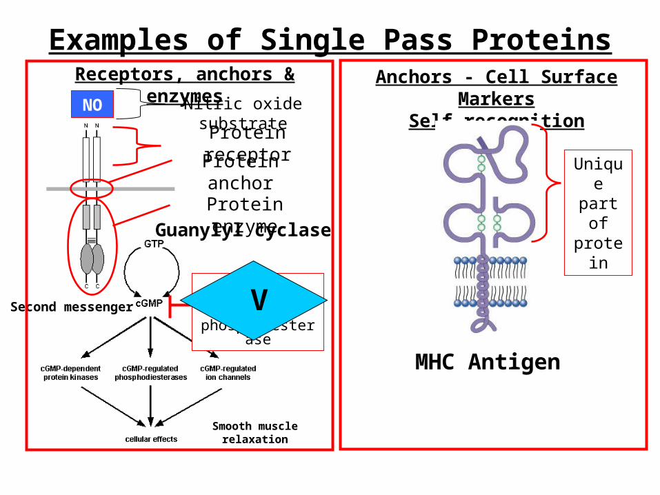
Examples of Single Pass ProteinsReceptors, anchors & enzymes Anchors - Cell Surface Markers
Self-recognition
MHC Antigen
Guanylyl cyclase
Second messenger
Protein anchor
Protein receptor
Protein enzyme
Smooth muscle relaxation
NO Nitric oxide substrate
Unique part of protein
cGMP degradation by
phosphdiesteraseV
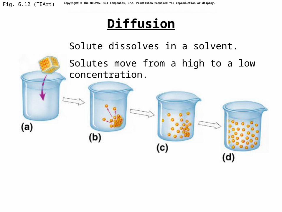
Fig. 6.12 (TEArt)
Lumpof sugar
Sugarmolecule
Copyright © The McGraw-Hill Companies, Inc. Permission required for reproduction or display.
Solute dissolves in a solvent.
Solutes move from a high to a low concentration.
Diffusion
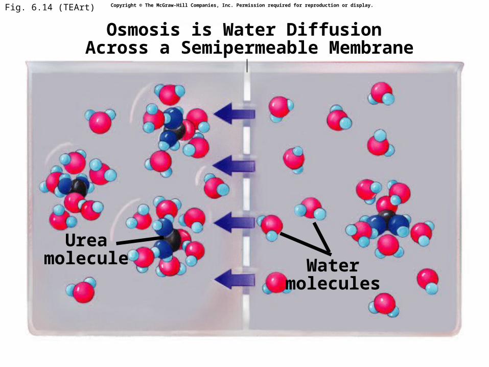
Fig. 6.14 (TEArt)
Ureamolecule Water
molecules
Osmosis is Water Diffusion Across a Semipermeable Membrane
Copyright © The McGraw-Hill Companies, Inc. Permission required for reproduction or display.
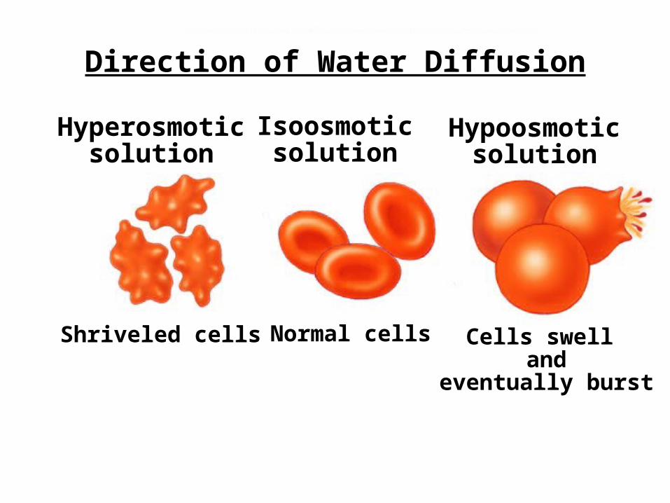
Shriveled cells Normal cells Cells swell and
eventually burst
Hyperosmoticsolution
Isoosmoticsolution
Hypoosmoticsolution
Direction of Water Diffusion

Fig. 6.15c (TEArt)
Plasmolysis Cell body shrinks
from cell wall
Normal turgid cellTurgor Pressure
Copyright © The McGraw-Hill Companies, Inc. Permission required for reproduction or display.
HyperosmoticExternal Solution
Water Diffusion in Plant Cells
IsoosmoticExternal Solution
HypoosmoticExternal Solution
15
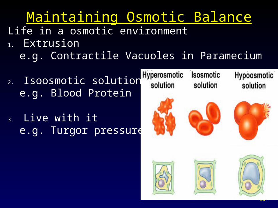
Maintaining Osmotic BalanceLife in a osmotic environment1. Extrusion
e.g. Contractile Vacuoles in Paramecium
2. Isoosmotic solutionse.g. Blood Protein
3. Live with ite.g. Turgor pressure
Moving Molecules into or out of Cells- Overview of Passive & Active Transport
I. Passive Transport1. Always “down” a concentration gradient
2. Always involves proteins calledA. ChannelsB. CarriersC. Pores… “porins”
II. Active Transport1. Always “down” a concentration gradient2. Small molecules transported through
A. Protein Pumps3. Large molecules transported by vesicles
A. EndocytosisB. Exocytosis
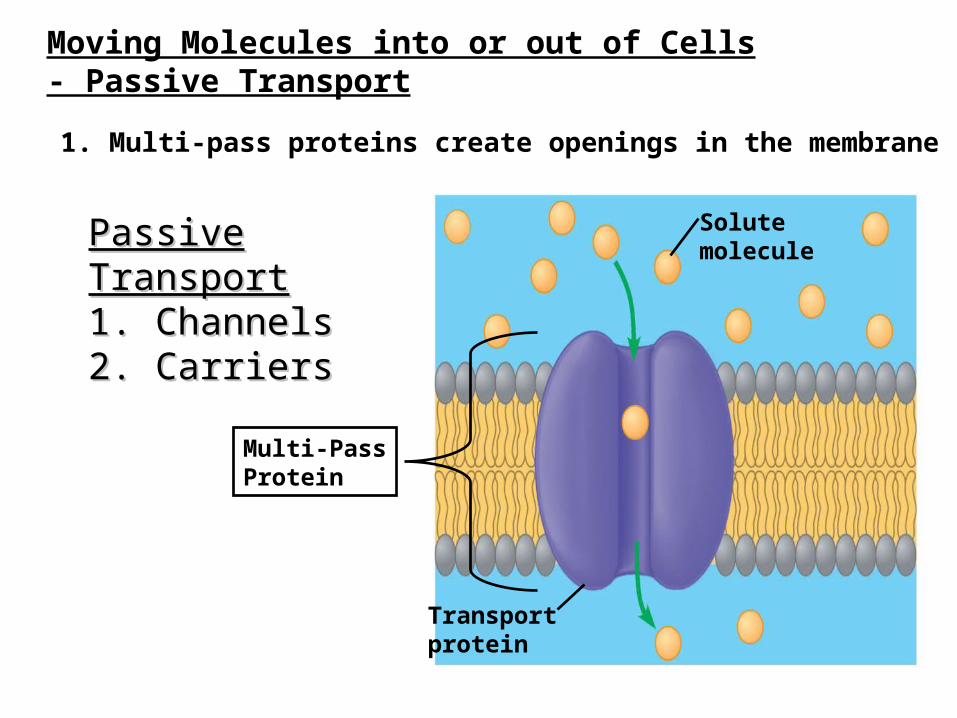
1. Multi-pass proteins create openings in the membrane
Moving Molecules into or out of Cells- Passive Transport
Passive TransportPassive Transport1. Channels1. Channels2. Carriers2. Carriers
Solutemolecule
Transportprotein
Multi-PassProtein
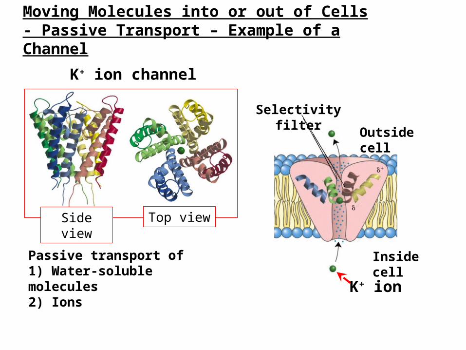
Moving Molecules into or out of Cells- Passive Transport – Example of a Channel
Passive transport of1) Water-soluble molecules2) Ions
Selectivity filter
Inside cell
Outside cell
K+ ion
K+ ion channel
Side view Top view
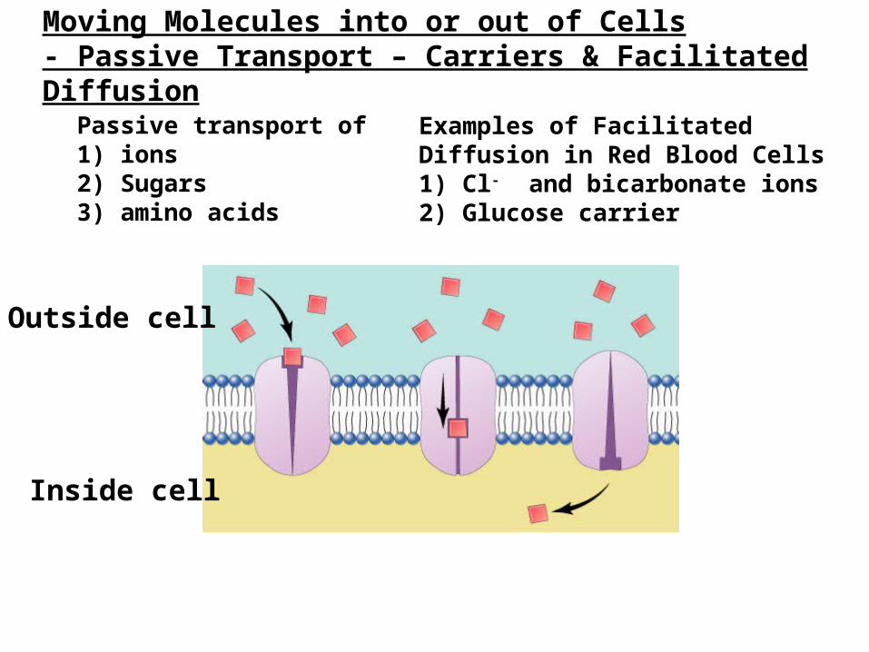
Moving Molecules into or out of Cells- Passive Transport – Carriers & Facilitated Diffusion
Passive transport of1) ions2) Sugars3) amino acids
Outside cell
Inside cell
Examples of Facilitated Diffusion in Red Blood Cells1) Cl- and bicarbonate ions2) Glucose carrier
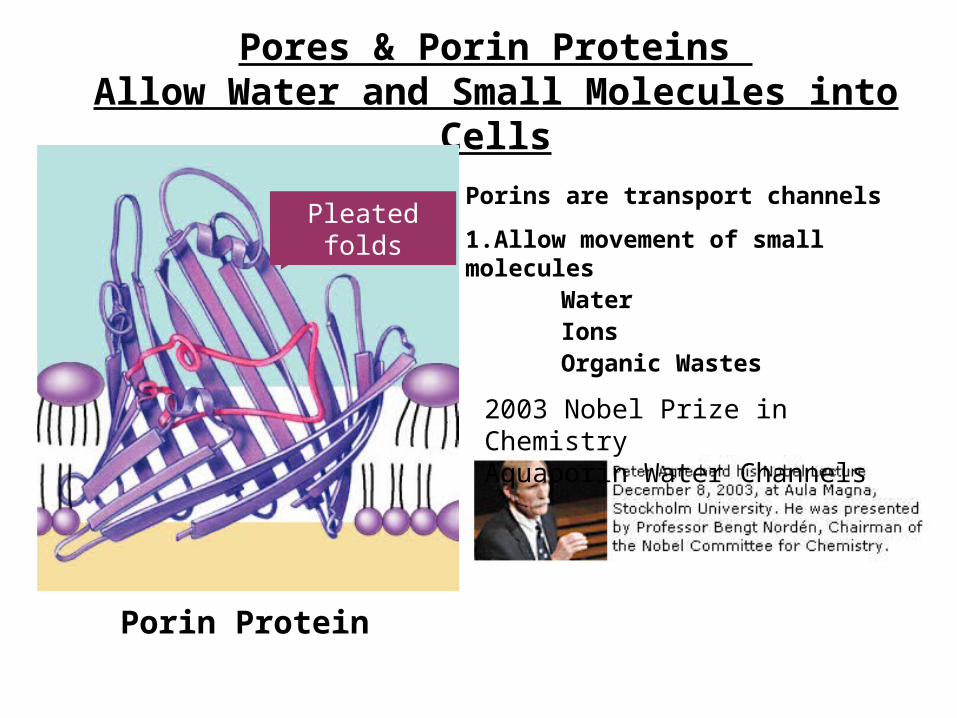
Pores & Porin Proteins Allow Water and Small Molecules into Cells
Porins are transport channels
1.Allow movement of small moleculesWaterIonsOrganic Wastes
Porin Protein
Pleated folds
2003 Nobel Prize in ChemistryAquaporin Water Channels
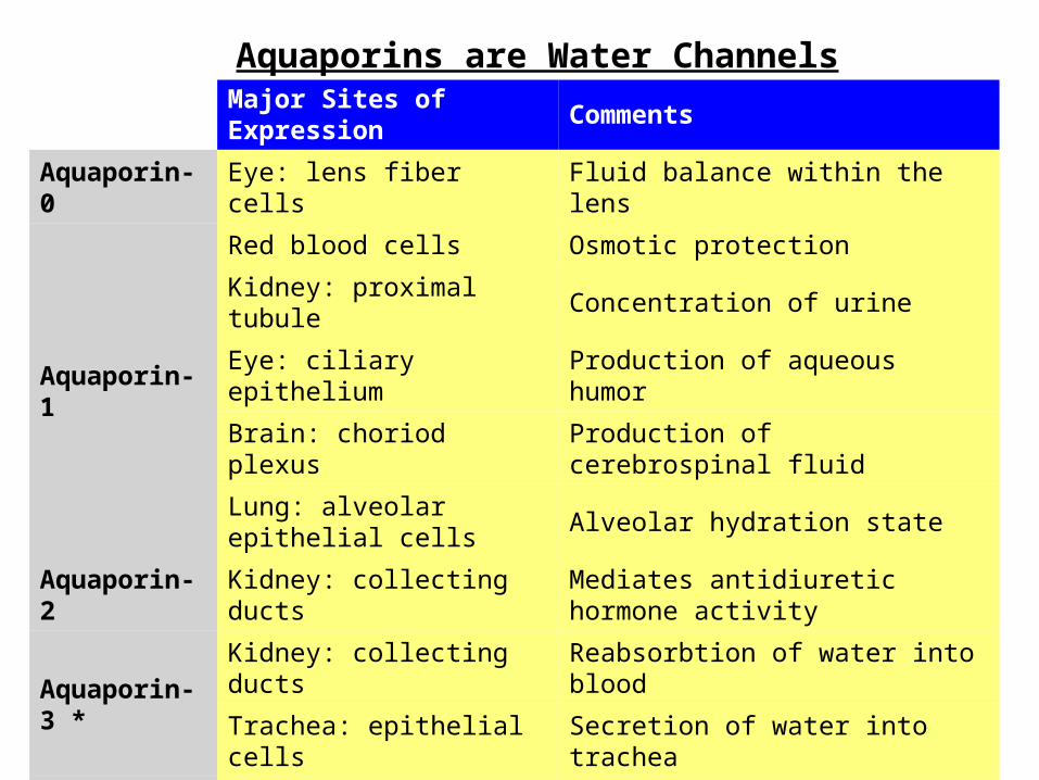
Major Sites of Expression Comments
Aquaporin-0 Eye: lens fiber cells Fluid balance within the lens
Aquaporin-1
Red blood cells Osmotic protection
Kidney: proximal tubule Concentration of urine
Eye: ciliary epithelium Production of aqueous humor
Brain: choriod plexus Production of cerebrospinal fluid
Lung: alveolar epithelial cells Alveolar hydration state
Aquaporin-2 Kidney: collecting ducts Mediates antidiuretic hormone activity
Aquaporin-3 *Kidney: collecting ducts Reabsorbtion of water into blood
Trachea: epithelial cells Secretion of water into trachea
Aquaporin-4
Kidney: collecting ducts Reabsorbtion of water
Brain: ependymal cells CSF fluid balance
Brain: hypothalamus Osmosensing function?
Lung: bronchial epithelium Bronchial fluid secretion
Aquaporin-5Salivary glands Production of saliva
Lacrimal glands Production of tears
Aquaporins are Water Channels
1. Proteins allow transport2. Mechanisms of movement through proteins
1. Passive Transport Channels, carriers & pores Simple Diffusion Facilitated Diffusion
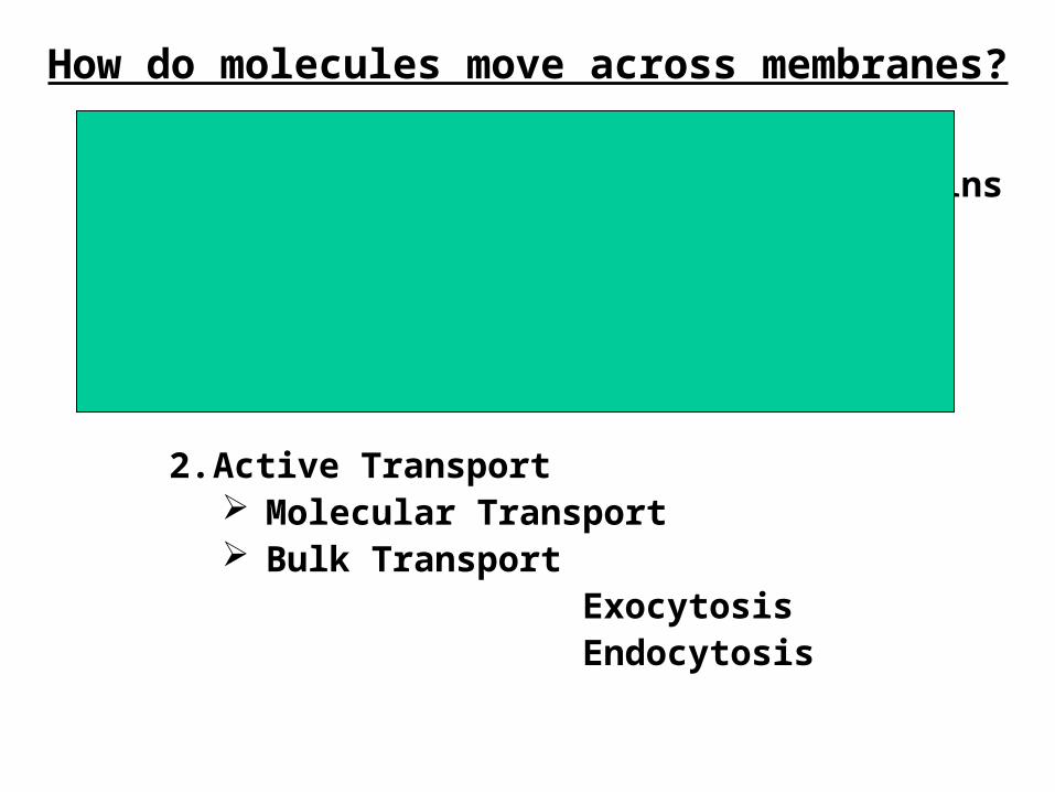
2. Active Transport Molecular Transport Bulk Transport
ExocytosisEndocytosis
How do molecules move across membranes?
PPPA
PPPA
Na+
Extracellular
Intracellular
ATP ATP
PPPA
ATP
PPA
P
ADP
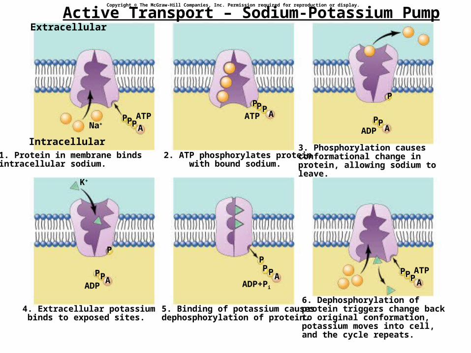
1. Protein in membrane bindsintracellular sodium.
2. ATP phosphorylates proteinwith bound sodium.
3. Phosphorylation causesconformational change inprotein, allowing sodium to leave.
PPA
P
ADP
4. Extracellular potassiumbinds to exposed sites.
K+
PPA
P
ADP+Pi
5. Binding of potassium causesdephosphorylation of protein.
6. Dephosphorylation ofprotein triggers change backto original conformation,potassium moves into cell,and the cycle repeats.
Copyright © The McGraw-Hill Companies, Inc. Permission required for reproduction or display.
Active Transport – Sodium-Potassium Pump
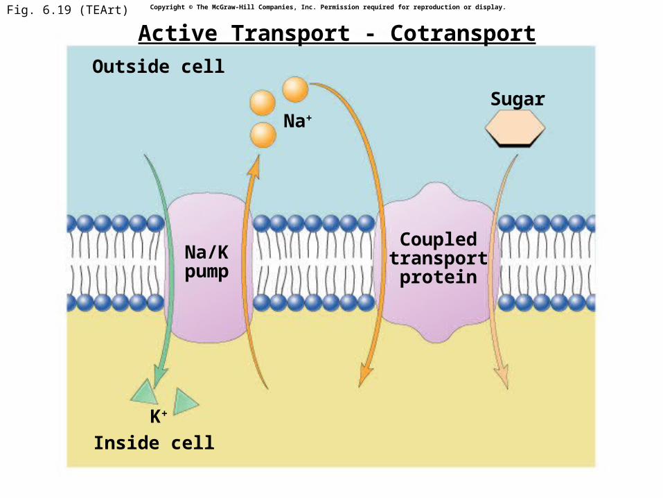
Fig. 6.19 (TEArt)
Outside cell
Inside cell
Na+
Coupledtransportprotein
Sugar
K+
Na/Kpump
Copyright © The McGraw-Hill Companies, Inc. Permission required for reproduction or display.
Active Transport - Cotransport
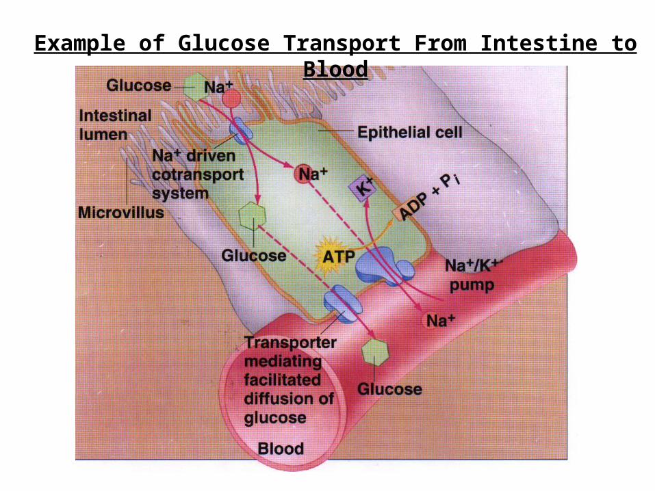
Example of Glucose Transport From Intestine to Blood
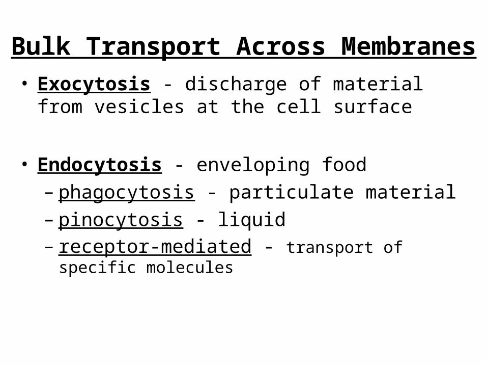
Bulk Transport Across Membranes• Exocytosis - discharge of material from vesicles at
the cell surface
• Endocytosis - enveloping food– phagocytosis - particulate material– pinocytosis - liquid– receptor-mediated - transport of specific molecules
27
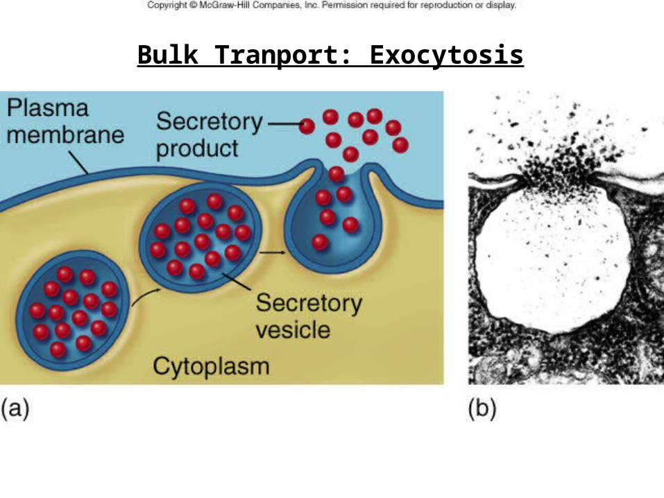
Bulk Tranport: Exocytosis
Copyright © The McGraw-Hill Companies, Inc. Permission required for reproduction or display.
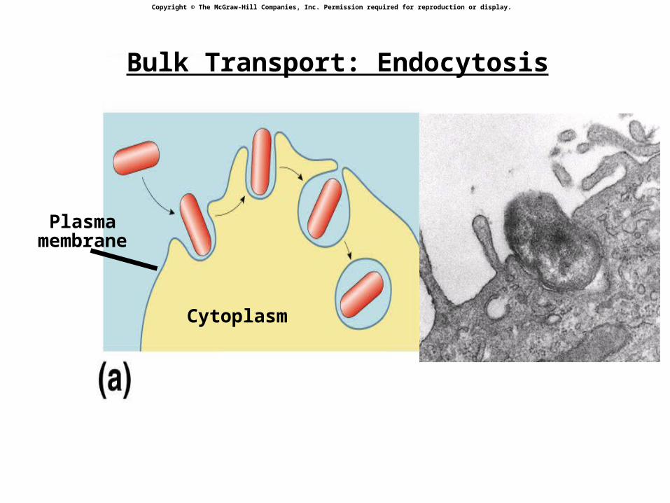
Cytoplasm
Plasmamembrane
Bulk Transport: Endocytosis
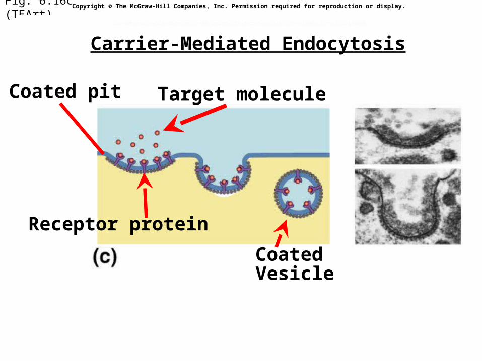
Fig. 6.16c (TEArt)
Coated pit Target molecule
Receptor protein
Coated Vesicle
Copyright © The McGraw-Hill Companies, Inc. Permission required for reproduction or display.
Carrier-Mediated Endocytosis
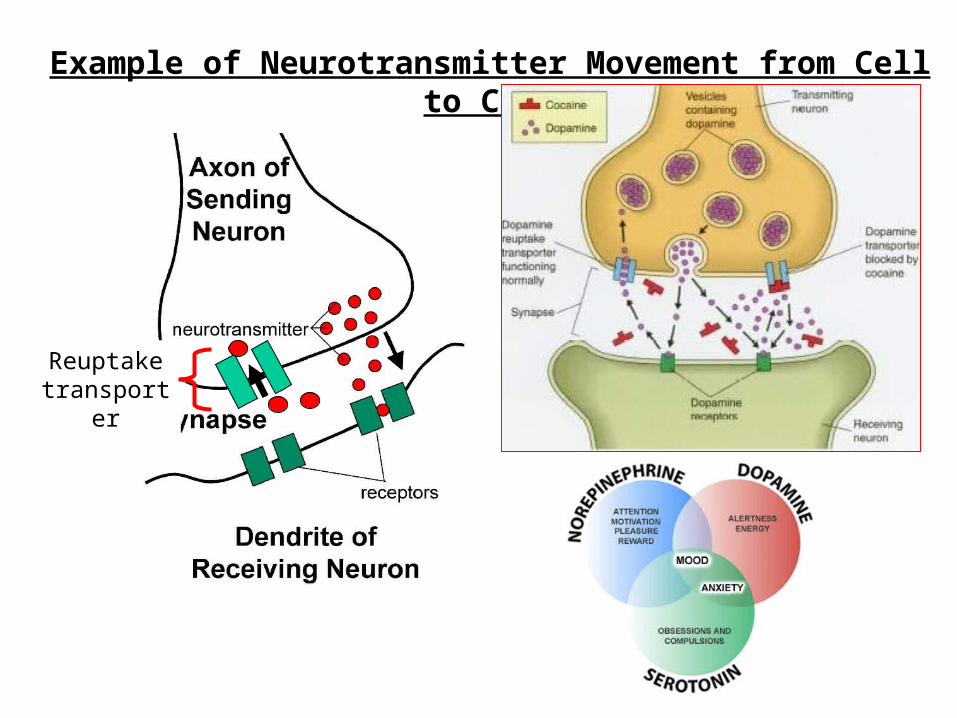
Example of Neurotransmitter Movement from Cell to Cell
Reuptake transporter
END
Membranes & Transport


