12
Organophosphorus Pesticides - Mechanisms Of Their Toxicity
Tina Eleršek and Metka Filipič National Institute of Biology
Slovenia
1. Introduction
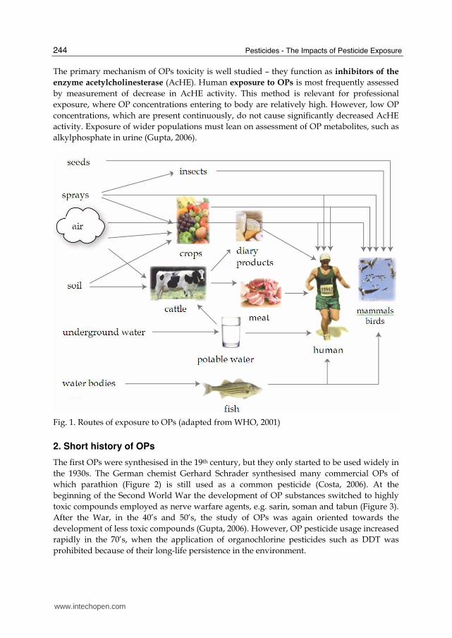
In this rapidly developing, capitalist world, people are continually exposed to numerous environmental pollutants such as industrial waste, polluted air and pesticides. These invariably comprise complex mixtures of chemicals. The effects of the mixtures and their mode of action in humans are insufficiently well studied. The majority of pollutants are potentially toxic for organisms, some being connected to disease development. In this context, the increase of chronic degenerative disease including cancer in humans, is of considerable concern (Gupta, 2006). Pesticides are a very important group of environmental pollutants used in intensive agriculture for protection against diseases and pests. The estimated annual application is more than 4 million tons, but only 1% of this reaches the target pests (Gavrilescu, 2005). Functionwise they are divided into herbicides (protection against weeds), insecticides (against insects), fungicides (against fungi), and others. While their use improves the quantity of agricultural products it potentially affects their quality, as pesticides may enter human diet (Grlić, 1988). This is a matter of major current concern. Organophosphorus compounds or organophosphates (OPs) form a large group of chemicals used over the past 60 years for protecting crops, livestock, human health and as warfare agents. On the basis of structural characteristics they are divided into at least 13 types, including phosphates, phosphonates, phosphinates, phosphorothioates (S=), phosphonothioates (S=), phosphorothioates (S substituted), phosphonothioates (S substituted), phosphorodithioates, phosphorotrithioates, phosphoramidothioates (Gupta, 2006). OPs are the most widely used pesticides worldwide and their metabolites are widespread across different populations (Aprea, 2000; Barr, 2004; Curl, 2003). The adverse short-term effects of exposure to these chemicals have been studied mostly in the nervous system, which is their primary target (Gupta at al., 2001), but there is a growing concern about their possible toxic effects in non-target tissues and (long-term) chronic effects that have not been studied in such detail. The majority of people are continually exposed to low OP concentrations, and long-term epidemiologic studies reveal linkage to higher risk of cancer development (Brown et al., 1990; Waddell at al., 2001). The World Health Organization estimates that every year 3 million people experience acute poisoning by OPs, 200.000 people terminally (WHO, 1990). The main routes of OP exposure are shown in Figure 1. Humans are exposed to OPs via ingested food and drink and by breathing polluted air (WHO, 2001). The exposure of workers in closed areas and of agricultural workers or people living near farms, is also very important (Gupta, 2006).
www.intechopen.com
Pesticides - The Impacts of Pesticide Exposure
244
The primary mechanism of OPs toxicity is well studied – they function as inhibitors of the
enzyme acetylcholinesterase (AcHE). Human exposure to OPs is most frequently assessed by measurement of decrease in AcHE activity. This method is relevant for professional exposure, where OP concentrations entering to body are relatively high. However, low OP concentrations, which are present continuously, do not cause significantly decreased AcHE activity. Exposure of wider populations must lean on assessment of OP metabolites, such as alkylphosphate in urine (Gupta, 2006).
Fig. 1. Routes of exposure to OPs (adapted from WHO, 2001)
2. Short history of OPs
The first OPs were synthesised in the 19th century, but they only started to be used widely in the 1930s. The German chemist Gerhard Schrader synthesised many commercial OPs of which parathion (Figure 2) is still used as a common pesticide (Costa, 2006). At the beginning of the Second World War the development of OP substances switched to highly toxic compounds employed as nerve warfare agents, e.g. sarin, soman and tabun (Figure 3). After the War, in the 40’s and 50’s, the study of OPs was again oriented towards the development of less toxic compounds (Gupta, 2006). However, OP pesticide usage increased rapidly in the 70’s, when the application of organochlorine pesticides such as DDT was prohibited because of their long-life persistence in the environment.
www.intechopen.com
Organophosphorus Pesticides - Mechanisms Of Their Toxicity
245
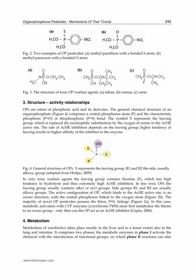
Fig. 2. Two examples of OP pesticides: (a) methyl-parathion with a bonded S atom; (b) methyl-paraoxon with a bonded O atom.
Fig. 3. The structure of toxic OP warfare agents: (a) tabun, (b) soman, (c) sarin
3. Structure – activity relationships
OPs are esters of phosphoric acid and its derivates. The general chemical structure of an organophosphate (Figure 4) comprises a central phosphorus atom (P) and the characteristic phosphoric (P=O) or thiophosphoric (P=S) bond. The symbol X represents the leaving group, which is replaced (by nucleophilic substitution) by the oxygen of serine in the AcHE active site. The rate of AcHE inhibition depends on the leaving group; higher tendency of leaving results in higher affinity of the inhibitor to the enzyme.
Fig. 4. General structure of OPs. X represents the leaving group, R1 and R2 the side, usually alkoxy, group (adapted from Hreljac, 2009)
In very toxic warfare agents the leaving group contains fluorine (F), which has high tendency to hydrolysis and thus extremely high AcHE inhibition. In less toxic OPs the leaving group usually contains alkyl or aryl groups. Side groups R1 and R2 are usually alkoxy groups. The active configuration of OP, which binds to the AcHE active site, is an oxono structure, with the central phosphorus linked to the oxygen atom (Figure 2b). The majority of novel OP pesticides possess the thion, P=S, linkage (Figure 2a). In this case, metabolic activation with CYP enzymes (cytochrome P450) must first metabolize the thiono to an oxono group – only then can the OP act as an AcHE inhibitor (Gupta, 2006).
4. Metabolism
Metabolism of xenobiotics takes place mostly in the liver and to a lesser extent also in the lung and intestine. It comprises two phases; the metabolic enzymes in phase I activate the chemical with the introduction of functional groups, on which phase II reactions can take
www.intechopen.com
Pesticides - The Impacts of Pesticide Exposure
246
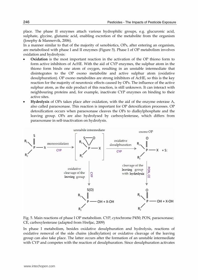
place. The phase II enzymes attach various hydrophilic groups, e.g. glucuronic acid, sulphate, glycine, glutamic acid, enabling excretion of the metabolite from the organism (Josephy & Mannervik, 2006). In a manner similar to that of the majority of xenobiotics, OPs, after entering an organism, are metabolised with phase I and II enzymes (Figure 5). Phase I of OP metabolism involves oxidation and hydrolysis: • Oxidation is the most important reaction in the activation of the OP thiono form to
form active inhibitors of AcHE. With the aid of CYP enzymes, the sulphur atom in the thiono form binds one atom of oxygen, resulting in an unstable intermediate that disintegrates to the OP oxono metabolite and active sulphur atom (oxidative desulphuration). OP oxono metabolites are strong inhibitors of AcHE, so this is the key reaction for the majority of neurotoxic effects caused by OPs. The influence of the active sulphur atom, as the side product of this reaction, is still unknown. It can interact with neighbouring proteins and, for example, inactivate CYP enzymes on binding to their active sites.
• Hydrolysis of OPs takes place after oxidation, with the aid of the enzyme esterase A, also called paraoxonase. This reaction is important for OP detoxification processes. OP detoxification occurs when paraoxonase cleaves the OPs to dialkylphosphate and the leaving group. OPs are also hydrolysed by carboxylesterase, which differs from paraoxonase in self-inactivation on hydrolysis.
Fig. 5. Main reactions of phase I OP metabolism. CYP, cytochrome P450; PON, paraoxonase; CE, carboxylesterase (adapted from Hreljac, 2009)
In phase I metabolism, besides oxidative desulphuration and hydrolysis, reactions of oxidative removal of the side chains (dealkylation) or oxidative cleavage of the leaving group can also take place. The latter occurs after the formation of an unstable intermediate with CYP and competes with the reaction of desulphuration. Since desulphuration activates
www.intechopen.com
Organophosphorus Pesticides - Mechanisms Of Their Toxicity
247
OPs, while oxidative cleavage of the leaving group detoxifies them, the equilibrium between these two reactions is very important for the final OP toxicity outcome. The outcome of oxidation is often a more hydrophilic compound, which can be more easily conjugated in phase II metabolism, thus enabling faster excretion from the organism (Gupta, 2006). The OP metabolites resulting from phase I metabolism are conjugated with hydrophilic groups under catalysis by enzymes of phase II, and excreted in urine. In phase II metabolism, detoxification reactions take place exclusively. Many studies of the metabolism of model OPs have shown that the most important enzymes are cytochromes CYP1A1, CYP3A4, CYP2B6 and CYP2C19. The first three have the highest affinity for desulphuration and activation to oxon, but for oxidative cleavage of the leaving group and detoxification CYP2C19 is the most effective (Mutch & Williams, 2006). The high degree of polymorphism in the various human CYPs means that the susceptibility of individuals to toxic effects of OPs depends on the expression level of specific CYP isoforms (e.g. Buratti et al., 2007).
5. Mechanisms of toxicity
The toxicity of OPs depends on their chemical structure, metabolism in target organism, concentration (i.e. dose), mode of application, degree of decomposition, mode of entering organisms, etc. (Grlić, 1988). The best described OP toxic effects are the neurological symptoms following acute poisoning as a consequence of the primary target (AcHE). Potential secondary targets and toxic effects outside the nerve system have not been well studied, but are nevertheless very important for risk assessment. Unlike other man-made chemicals, OP pesticides can affect a large proportion of the human population, as a result of exposure through domestic use, proximity to agricultural activities and consumption of contaminated food and water (Maroni at al., 2000).
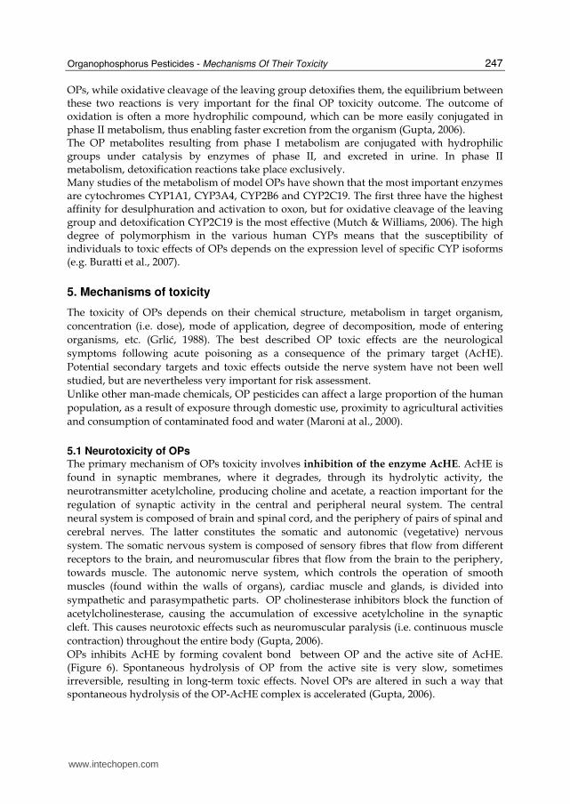
5.1 Neurotoxicity of OPs The primary mechanism of OPs toxicity involves inhibition of the enzyme AcHE. AcHE is found in synaptic membranes, where it degrades, through its hydrolytic activity, the neurotransmitter acetylcholine, producing choline and acetate, a reaction important for the regulation of synaptic activity in the central and peripheral neural system. The central neural system is composed of brain and spinal cord, and the periphery of pairs of spinal and cerebral nerves. The latter constitutes the somatic and autonomic (vegetative) nervous system. The somatic nervous system is composed of sensory fibres that flow from different receptors to the brain, and neuromuscular fibres that flow from the brain to the periphery, towards muscle. The autonomic nerve system, which controls the operation of smooth muscles (found within the walls of organs), cardiac muscle and glands, is divided into sympathetic and parasympathetic parts. OP cholinesterase inhibitors block the function of acetylcholinesterase, causing the accumulation of excessive acetylcholine in the synaptic cleft. This causes neurotoxic effects such as neuromuscular paralysis (i.e. continuous muscle contraction) throughout the entire body (Gupta, 2006). OPs inhibits AcHE by forming covalent bond between OP and the active site of AcHE. (Figure 6). Spontaneous hydrolysis of OP from the active site is very slow, sometimes irreversible, resulting in long-term toxic effects. Novel OPs are altered in such a way that spontaneous hydrolysis of the OP-AcHE complex is accelerated (Gupta, 2006).
www.intechopen.com
Pesticides - The Impacts of Pesticide Exposure
248
Fig. 6. The binding of OP to the active site of AcHE (adapted from Hreljac, 2009)
Neurotransmitters of the autonomic nerve system act on target organs by binding to specific receptors. Adrenergic (┙, ┚) and cholinergic (nicotine, muscarine) receptors are affected by different types of neurotransmitter. The excess of acetylcholine in the peripheral nerve synapse causes the activation of muscarine and nicotine receptors and increases the activation of the sympathetic and parasympathetic parts. Since the synapses in the central nerve system are quite inaccessible for experimental work, compared with neuromuscular junctions in the peripheral nerve system, the mechanism of poisoning by OPs in the central nerve system has been less studied. Symptoms of acute OP poisoning can be divided according to the site of acetylcholine accumulation in the organism (Figure 7). In addition to acute symptoms, some OPs can cause other symptoms that arise a few days after exposure or poisoning with OP. Weakness in muscles and breathing difficulties usually appear 1 – 4 days after poisoning while, after 7- 21 days, weakness in peripheral muscles also occurs. The cause of these delayed symptoms is inhibition of the neuropathy target esterase (NTE) located in the neural system, rather than AcHE inhibition. NTE belongs to the same group of serine esterases as AcHE, however its primary role in the organism is not well known (Kamanyire & Karalliedde, 2004). Several other neurotoxic symptoms that cannot be ascribed to AcHE inhibition, but act on different secondary targets inside the neural system, have been also proposed.
www.intechopen.com

Organophosphorus Pesticides - Mechanisms Of Their Toxicity
249
ACETYLCHOLINE ACCUMULATION SITE
SYMPTOMS
central neural system
headache, insomnia, vertigo, spasms, confusion, sleeplessness, speaking disorders, coma and respiratory paralysis
peripheral autonomic nerve system
muscarine receptors
muscarine symptoms; increased elimination (salivation, perspiration, lachrymation), indigestion (spasms, vomiting, diarrhoea), lowered heart beat, visual disorders
nicotine receptors nicotine symptoms; uncontrolled muscle contractions and paralysis
Fig. 7. Symptoms of acute OP poisoning that differ according to the site of acetylcholine accumulation.
5.2 Non-neuronal molecular targets of OPs Although the neurotoxicity of OPs is well described, little is known about their secondary mechanisms of activity and the consequences of chronic exposure to OPs on non-target (non-neuronal) tissues and organs in humans. Recent studies have revealed several secondary targets for OPs that possibly disturb a variety of biological processes. Among the enzymes that are inhibited by OPs are carboxylases, which take part in xenobiotic metabolism. Their inhibition with OPs can block metabolic transformation of various substances (Hodgson & Rose, 2005). OPs can also influence xenobiotic metabolism via the active sulphur atom that arises from desulphuration in phase I of metabolism and strongly inhibits CYP enzymes (Hodgson & Rose, 2006). OPs also inhibit lipases, which play an important role in cell signalling (Quistad et al., 2006). Bomser et al. (2002) showed that OP chlorpyrifos-oxon, by inhibiting DAG-lipase indirectly, activates ERK kinases that are members of the group of mitogen-activated protein kinases (MAPK) that regulate cell proliferation and differentiation. OPs can also affect signalling pathways by activation of protein kinase (PKC) (Bagchi et al., 1997). However, this activation is probably indirect through OP mediated formation of reactive oxygen species (ROS) that activate PKC. OP induced ROS formation and oxidative stress have also been shown to be associated with apoptosis in different tissues (Oral et al., 2006; Yu et al., 2008). Kojima et al. (2004) showed that OPs can inhibit steroid androgen (AR) receptor, which can cause steroid hormone disturbances in the organism. Further targets of OPs have been revealed, by in vitro and in vivo studies, five of which were particularly sensitive (Casida & Quistad, 2004): • malathion and malaoxon (IC50 = 1-9 nM) inhibit lysyl oxidase in homogenates of
Xenopus embryos, suggesting that they alter posttranslational modification of collagen, with resulting morphological defects in connective tissue (Snawder & Chambers, 1993);
• chlorpyrifos and chlorpyrifos oxone (at below 1 nM) are reported to activate Ca2+/cAMP response element binding protein in cultured rat neurons, as a possible mechanism for neurotoxicity (Schuh et al., 2003);
www.intechopen.com

Pesticides - The Impacts of Pesticide Exposure
250
• paraoxon (1-10 nM) causes apoptotic cell death in a leukaemia cell line by disruption of mitochondria, leading to activation of caspase-9 (Saleh et al., 2003);
• ethyl arachidonyl fluorophosphonate (at < 1 µM) and diisopropylfluorophosphate (at 100 µM) inhibit platelet activating factor acetylhydrolase (Kell et al., 2003);
• fenitrothion (at 22 nM) acts as an androgen receptor antagonist in vitro (Tamura et al., 2003) and inhibits the development of androgen-dependent tissues in vivo (Tamura et al., 2001).
5.3 Immunotoxicity of OPs Toxic effects of OPs on the immune system can be direct or indirect and are reflected in different immune organ pathologies and lowered humoral and/or cell immunity. Direct immunotoxic effects of OPs can be due to: • inhibition of serine hydrolases (complement system) or esterases (lymphocyte and
monocyte membranes) in the immune system; • oxidative damage of immune system organs; • changes in signal transduction pathways that control proliferation and immune cell
differentiation. Indirect immunotoxicity of OPs is expressed as changes in the nervous system or chronic effects of altered metabolism on the immune system. Selecting appropriate biomarkers and biological methods is very difficult since the physiological diversity of immune systems among organisms is very high (Galloway & Handy, 2003).
5.4 Genotoxicity and carcinogenicity of OPs Because of the chronic exposure of human populations to low OP concentrations, it is very important to study the influence of OPs on cancer development and progress, and to elucidate the underlying mechanisms. The most informative data regarding potential human carcinogenicity come from epidemiological studies of exposed populations. Chronic occupational exposure to OPs has been linked to increased risk for cancer development such as non-Hodgkin lymphoma (Waddell et al., 2001) and some types of leukaemia (Brown et al., 1990). However, the major limitation of these studies is the fact that exposure has been assessed based on questionnaires and that workers were exposed not only to OPs but also to other pesticides. Cancer development is a multi-stage process that involves initiation, promotion and progression. Many carcinogens are genotoxic and initiate cancer development by causing DNA damage and mutation, while non-genotoxic carcinogens mostly induce neoplastic cell transformation and promote cell proliferation by different mechanisms, such as avoiding apoptosis, stimulation of growth factors, and avoiding growth suppression signals. Experimental in vitro and in vivo studies have shown that several OPs exert genotoxic activity (Bolognesi, 2003), and there are also reports showing that OPs can induce neoplastic transformation of cells (Cabello et al., 2001; Isoda et al., 2005). OPs have been reported • to be weakly mutagenic in bacteria, but mutagenic in yeast (IARC, 1987); • to induce DNA damage in peripheral lymphocytes in vitro (Blasiak and Kowalik, 1999;
Ündeger and Basaran, 2005) and in vivo in occupationally exposed workers (Garaj-Vrhovac et al., 2000);
• to induce chromosomal aberrations and sister chromatid exchange (Galloway et al., 1987);
www.intechopen.com
Organophosphorus Pesticides - Mechanisms Of Their Toxicity
251
• to induce micronuclei formation in bone marrow (Mathew et al., 1990); • to cause sperm abnormalities in OP exposed mice (Mathew et al., 1992). Hreljac et al. (2008) have recently studied the mechanisms of genotoxicity and potential carcinogenicity of selected model OPs (parathion (PT), paraoxon (PO) and dimefox (DF)) in the in vitro experimental model with human hepatoma (HepG2) cells. They demonstrated that OPs act on several targets: • low concentrations of parathion and paraoxon were genotoxic, while dimefox acted as a
mitogen; • the three model OPs induced numerous variations of gene expression, particularly of
genes involved in stress response, inseparably connected to basic cell processes important in cancer development (cell cycle, apoptosis, xenobiotic metabolism, DNA repair);
• in addition, changes in phosphorylation of kinases, which are connected to stress response, were observed.
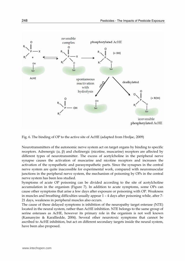
The methodologies used in these studies have involved a series of different genotoxicity assays (Salmonella typhimurium reverse mutation assay, and comet and micronucleus assay in HepG2 cells) and methods for measuring cell proliferation (MTT assay, cell-cycle analysis by flow cytometry and Ki-67 immunostaining), that were associated with molecular methods for gene expression analysis (quantitative real time PCR) and biochemical methods for enzyme activity measurements. Here we describe some of these methods in more detail. The comet assay (also known as Single Cell Gel Electrophoresis assay) is a relatively simple and sensitive technique for the detection of DNA damage at the level of the individual cell (Figure 8). HepG2 cells are exposed to graded doses of model OPs and DNA damage is determined with the comet assay after 4- and 24-hr exposures to OPs. (Hreljac & Filipič, 2008). The comet assay was performed as described by Singh et al. (1988). The slides were stained with ethidium bromide and analyzed using a fluorescence microscope and image analysis software.
Fig. 8. The principle of the comet assay (adapted from Hreljac, 2009)
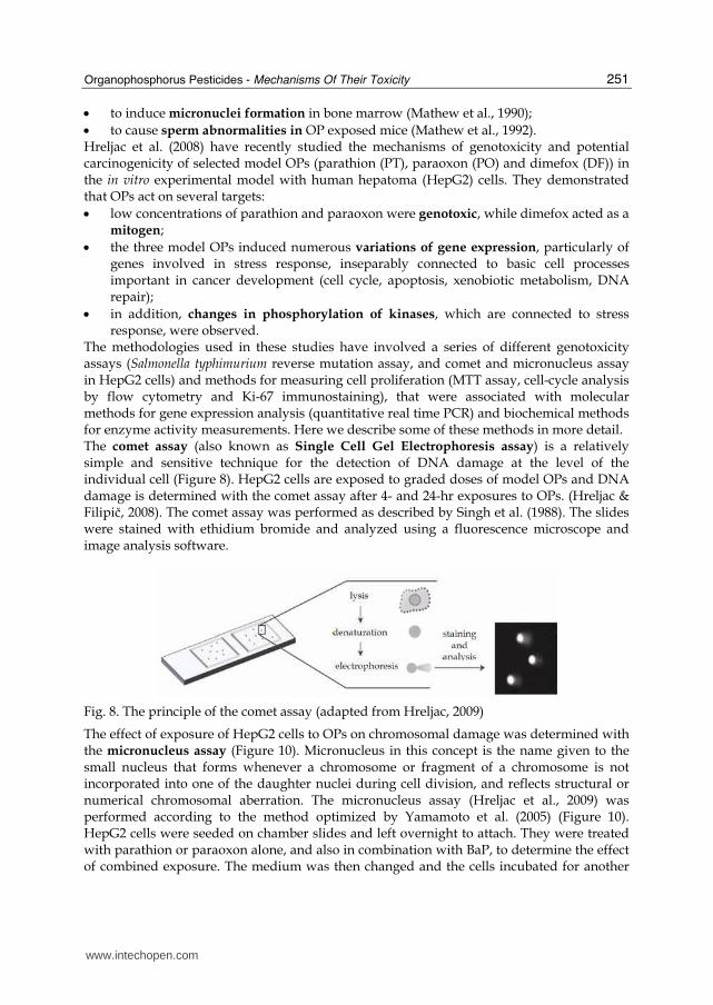
The effect of exposure of HepG2 cells to OPs on chromosomal damage was determined with the micronucleus assay (Figure 10). Micronucleus in this concept is the name given to the small nucleus that forms whenever a chromosome or fragment of a chromosome is not incorporated into one of the daughter nuclei during cell division, and reflects structural or numerical chromosomal aberration. The micronucleus assay (Hreljac et al., 2009) was performed according to the method optimized by Yamamoto et al. (2005) (Figure 10). HepG2 cells were seeded on chamber slides and left overnight to attach. They were treated with parathion or paraoxon alone, and also in combination with BaP, to determine the effect of combined exposure. The medium was then changed and the cells incubated for another
www.intechopen.com
Pesticides - The Impacts of Pesticide Exposure
252
68 h to allow cell division to take place. The cells were then washed, incubated in hypotonic KCl solution, and fixed. The slides were dried and stained with acridine orange, and examined under the fluorescence microscope. Micronuclei were scored according to established criteria (Fenech, 2000). To determine cytotoxicity, cells were seeded in parallel, with the same treatment and recovery protocol as for micronuclei, and assayed for cell viability using the MTS assay according to the manufacturer’s protocol.
Fig. 9. The principle of the micronucleus test (adapted from Hreljac, 2009)
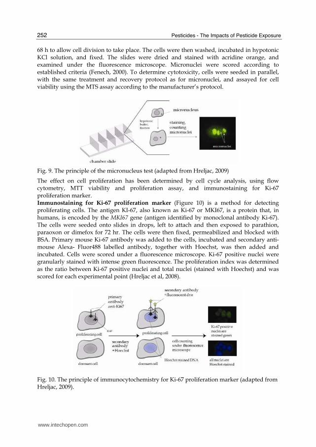
The effect on cell proliferation has been determined by cell cycle analysis, using flow cytometry, MTT viability and proliferation assay, and immunostaining for Ki-67 proliferation marker. Immunostaining for Ki-67 proliferation marker (Figure 10) is a method for detecting proliferating cells. The antigen KI-67, also known as Ki-67 or MKI67, is a protein that, in humans, is encoded by the MKI67 gene (antigen identified by monoclonal antibody Ki-67). The cells were seeded onto slides in drops, left to attach and then exposed to parathion, paraoxon or dimefox for 72 hr. The cells were then fixed, permeabilized and blocked with BSA. Primary mouse Ki-67 antibody was added to the cells, incubated and secondary anti-mouse Alexa- Fluor488 labelled antibody, together with Hoechst, was then added and incubated. Cells were scored under a fluorescence microscope. Ki-67 positive nuclei were granularly stained with intense green fluorescence. The proliferation index was determined as the ratio between Ki-67 positive nuclei and total nuclei (stained with Hoechst) and was scored for each experimental point (Hreljac et al, 2008).
Fig. 10. The principle of immunocytochemistry for Ki-67 proliferation marker (adapted from Hreljac, 2009).
www.intechopen.com
Organophosphorus Pesticides - Mechanisms Of Their Toxicity
253
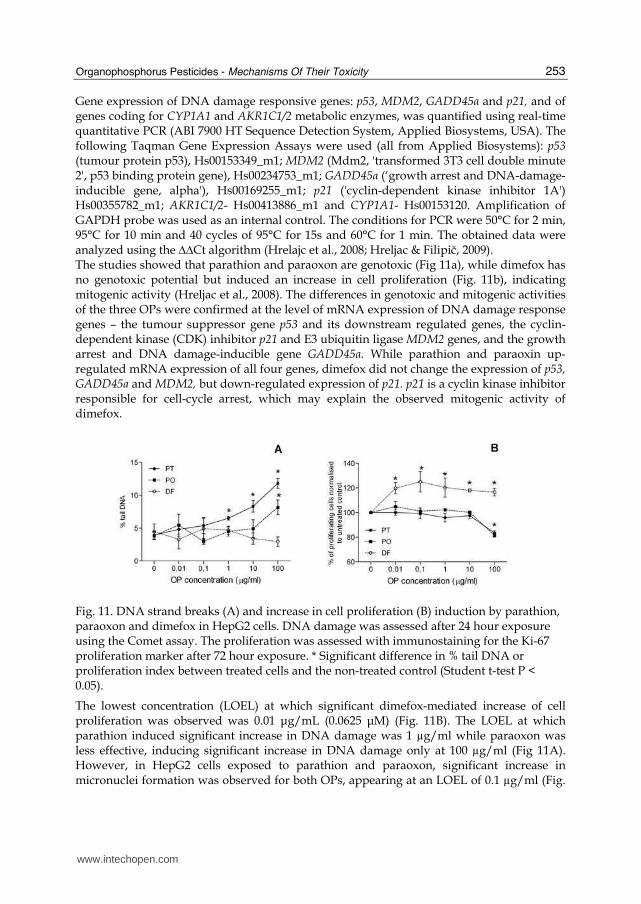
Gene expression of DNA damage responsive genes: p53, MDM2, GADD45α and p21, and of genes coding for CYP1A1 and AKR1C1/2 metabolic enzymes, was quantified using real-time quantitative PCR (ABI 7900 HT Sequence Detection System, Applied Biosystems, USA). The following Taqman Gene Expression Assays were used (all from Applied Biosystems): p53 (tumour protein p53), Hs00153349_m1; MDM2 (Mdm2, 'transformed 3T3 cell double minute 2', p53 binding protein gene), Hs00234753_m1; GADD45α (‘growth arrest and DNA-damage-inducible gene, alpha'), Hs00169255_m1; p21 ('cyclin-dependent kinase inhibitor 1A') Hs00355782_m1; AKR1C1/2- Hs00413886_m1 and CYP1A1- Hs00153120. Amplification of GAPDH probe was used as an internal control. The conditions for PCR were 50°C for 2 min, 95°C for 10 min and 40 cycles of 95°C for 15s and 60°C for 1 min. The obtained data were analyzed using the ∆∆Ct algorithm (Hrelajc et al., 2008; Hreljac & Filipič, 2009). The studies showed that parathion and paraoxon are genotoxic (Fig 11a), while dimefox has no genotoxic potential but induced an increase in cell proliferation (Fig. 11b), indicating mitogenic activity (Hreljac et al., 2008). The differences in genotoxic and mitogenic activities of the three OPs were confirmed at the level of mRNA expression of DNA damage response genes – the tumour suppressor gene p53 and its downstream regulated genes, the cyclin-dependent kinase (CDK) inhibitor p21 and E3 ubiquitin ligase MDM2 genes, and the growth arrest and DNA damage-inducible gene GADD45α. While parathion and paraoxin up-regulated mRNA expression of all four genes, dimefox did not change the expression of p53, GADD45a and MDM2, but down-regulated expression of p21. p21 is a cyclin kinase inhibitor responsible for cell-cycle arrest, which may explain the observed mitogenic activity of dimefox.
Fig. 11. DNA strand breaks (A) and increase in cell proliferation (B) induction by parathion, paraoxon and dimefox in HepG2 cells. DNA damage was assessed after 24 hour exposure using the Comet assay. The proliferation was assessed with immunostaining for the Ki-67 proliferation marker after 72 hour exposure. * Significant difference in % tail DNA or proliferation index between treated cells and the non-treated control (Student t-test P < 0.05).
The lowest concentration (LOEL) at which significant dimefox-mediated increase of cell proliferation was observed was 0.01 μg/mL (0.0625 μM) (Fig. 11B). The LOEL at which parathion induced significant increase in DNA damage was 1 µg/ml while paraoxon was less effective, inducing significant increase in DNA damage only at 100 µg/ml (Fig 11A). However, in HepG2 cells exposed to parathion and paraoxon, significant increase in micronuclei formation was observed for both OPs, appearing at an LOEL of 0.1 µg/ml (Fig.
www.intechopen.com
Pesticides - The Impacts of Pesticide Exposure
254
12) (Hreljac & Filipič, 2009). On the basis of pharmacokinetic models and biomonitoring data, Buratti et al. (2007) recently proposed that OP concentrations lower than 10 μM (corresponding to 2.6, 2.5 and 1.6 μg/mL of methyl parathion, methyl paraoxon and dimefox, respectively) in the in vitro studies reflect conditions in human during environmental exposure, and that concentrations higher than 100 μM reflect acute accidental intoxication. These studies thus showed that parathion, paraoxon and dimefox can cause adverse effects at concentrations that are relevant, not only for occupational, but also general human exposure. Thiono forms of OPs have been considered safer for use as pesticides because of their lower acute toxicity mediated by direct AChE inhibition, however this study indicated that, at chronic exposure, the thiono form of OPs may pose a similar or even greater health risk than the oxono form.
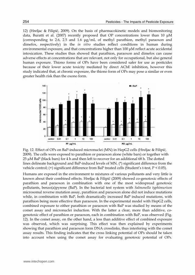
Fig. 12. Effect of OPs on BaP-induced micronuclei (MN) in HepG2 cells (Hreljac & Filipič, 2009). The cells were exposed to parathion or paraoxon alone (white bars) or together with 25 µM BaP (black bars) for 4 h and then left to recover for an additional 68 h. The dotted lines delineate background and BaP-induced levels of MN. (*) significant difference from the vehicle control; (+) significant difference from BaP treated cells (Student’s t-test, P < 0.05).
Humans are exposed in the environment to mixtures of various pollutants and very little is known about their combined effects. Hreljac & Filipič (2009) showed co-genotoxic effects of parathion and paraoxon in combination with one of the most widespread genotoxic pollutants, benzo(a)pyrene (BaP). In the bacterial test system with Salmonella typhimurium microsomal reverse mutation assay, parathion and paraoxon alone did not induce mutations while, in combination with BaP, both dramatically increased BaP induced mutations, with parathion being more effective than paraoxon. In the experimental model with HepG2 cells, combined exposure to either parathion or paraoxon with BaP was studied by means of the comet assay and micronuclei induction. With the latter a clear, more than additive, co-genotoxic effect of parathion or paraoxon, each in combination with BaP, was observed (Fig. 12). In the comet assay, on the other hand, a less than additive effect of combined exposure was observed, which was surprising. This effect was then explained by experiments showing that parathion and paraoxon form DNA crosslinks, thus interfering with the comet assay results. This finding indicates that the cross linking potential of OPs should be taken into account when using the comet assay for evaluating genotoxic potential of OPs.
www.intechopen.com
Organophosphorus Pesticides - Mechanisms Of Their Toxicity
255
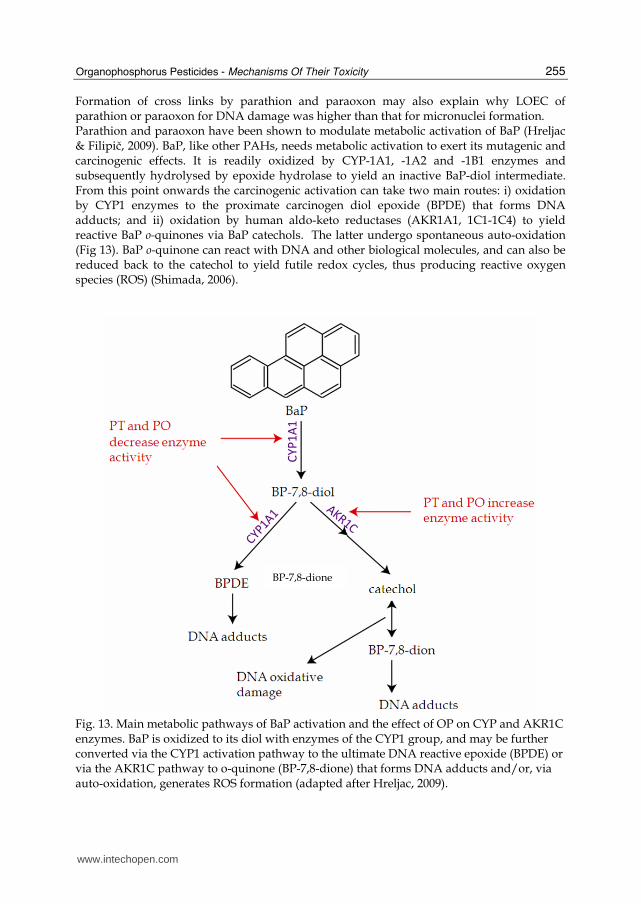
Formation of cross links by parathion and paraoxon may also explain why LOEC of parathion or paraoxon for DNA damage was higher than that for micronuclei formation. Parathion and paraoxon have been shown to modulate metabolic activation of BaP (Hreljac & Filipič, 2009). BaP, like other PAHs, needs metabolic activation to exert its mutagenic and carcinogenic effects. It is readily oxidized by CYP-1A1, -1A2 and -1B1 enzymes and subsequently hydrolysed by epoxide hydrolase to yield an inactive BaP-diol intermediate. From this point onwards the carcinogenic activation can take two main routes: i) oxidation by CYP1 enzymes to the proximate carcinogen diol epoxide (BPDE) that forms DNA adducts; and ii) oxidation by human aldo-keto reductases (AKR1A1, 1C1-1C4) to yield reactive BaP o-quinones via BaP catechols. The latter undergo spontaneous auto-oxidation (Fig 13). BaP o-quinone can react with DNA and other biological molecules, and can also be reduced back to the catechol to yield futile redox cycles, thus producing reactive oxygen species (ROS) (Shimada, 2006).
Fig. 13. Main metabolic pathways of BaP activation and the effect of OP on CYP and AKR1C enzymes. BaP is oxidized to its diol with enzymes of the CYP1 group, and may be further converted via the CYP1 activation pathway to the ultimate DNA reactive epoxide (BPDE) or via the AKR1C pathway to o-quinone (BP-7,8-dione) that forms DNA adducts and/or, via auto-oxidation, generates ROS formation (adapted after Hreljac, 2009).
BP-7,8-dione
www.intechopen.com
Pesticides - The Impacts of Pesticide Exposure
256
Treatment of HepG2 cells with BaP in the presence of parathion or paraoxon decreased mRNA expression and enzyme activity of CYP1A, while AKR1C1/2 levels were elevated. Based on these results it was proposed that the co-genotoxicity results from OP mediated modulation of BaP metabolism. This favours the induction of AKR1C enzymes known to catalyse the formation of DNA reactive BaP o-quinones and the production of reactive oxygen species (Fig. 13).
6. Conclusions
Organophosphorus pesticides are the most widely used pesticides worldwide and their metabolites are widespread across different populations. The primary mechanism of OP toxicity is the inhibition of acetylcholine esterase (AChE) in the central and peripheral nervous system, leading to a variety of short-term and chronic effects such as nausea, headache, confusion, depression, memory loss and chronic fatigue syndrome. Accordingly, safety evaluation of OP pesticides is generally based on the premise that AChE inhibition is the principal cause for their acute and chronic toxicity. Recent studies, however, have revealed a number of OP secondary targets that are not associated with the cholinergic system and may lead to immunotoxic, endocrine disrupting, genotoxic and potential carcinogenic effects. The genotoxicity and potential carcinogenicity of OPs are of particular concern. In commercial pesticide formulations, thiono forms of OPs are usually used because their acute neurotoxicity is lower than that of oxono forms. However, a recent comparative study of genotoxic potential of parathion and paraoxon in human hepatoma HepG2 cells revealed that parathion may have even higher genotoxic and co-genotoxic potential than paraoxon. In addition, genotoxic and co-genotoxic effects of parathion and paraoxon have been observed at concentrations that are relevant for human environmental exposure. These are important points that should be further investigated and considered in evaluations of the hazards and risk of exposure to these chemicals. Taken together, the scientific evidence supports the assertion that chronic exposure to OPs is associated with higher risk of developing diseases, including cancer, and that world usage of OPs should be reduced and better controlled. Further investigations are needed, particularly in regard to mechanisms of toxicity toward non cholinergic systems. Safety of the continuous use of OPs in agriculture and its expanding use in medicine depends on understanding the relevance of not only AChE inhibition but also of secondary targets in the effects of acute and long term exposure on health.
7. References
Aprea, C.; Strambi, M.; Novelli, M.T.; Lunghini, L. & Bozzi, N. (2000) Biologic monitoring of exposure to organophosphorus pesticides in 195 Italian children. Environ Health
Perspect 108 (6): 521-5 Bagchi, D.; Bagchi, M.; Tang, L. & Stohs, S.J. (1997) Comparative in vitro and in vivo protein
kinase C activation by selected pesticides and transition metal salts. Toxicology
Letters 91 (1): 31-37
www.intechopen.com
Organophosphorus Pesticides - Mechanisms Of Their Toxicity
257
Barr, D.B.; Bravo, R.; Weerasekera, G.; Caltabiano, L.M.; Whitehead, R.D. Jr.; Olsson, A.O.; Caudill, S.P.; Schober, S.E.; Pirkle, J.L. & Sampson, E.J. (2004) Concentrations of dialkyl phosphate metabolites of organophosphorus pesticides in the U.S. population. Environ Health Perspect 112 (2):186-200
Blasiak, J. & Kowalik, J. (1999) Effect of paraoxon-methyl and parathion-methyl on DNA in human lymphocytes and protective action of vitamin C. Pestic Sci 55: 1182-86
Bolognesi, C. (2003) Genotoxicity of pesticides: a review of human biomonitoring studies. Mutat Res 543 (3): 251-72
Bomser, J.A.; Quistad, G.B. & Casida, J.E. (2002) Chlorpyrifos oxon potentiates diacylglycerol-induced extracellular signal-regulated kinase (ERK 44/42) activation, possibly by diacylglycerol lipase inhibition. Toxicology and Applied
Pharmacology 178 (1):29-36. Brown, L.M.; Blair, A.; Gibson, R.; Everett, G.D.; Cantor, K.P.; Schuman, L.M.; Burmeister,
L.F.; Van Lier, S.F. & Dick, F. (1990) Pesticide exposures and other agricultural risk factors for leukemia among men in Iowa and Minnesota. Cancer Res 50 (20): 6585-91
Buratti, F.M.; Leoni, C. & Testai, E. (2007) The Human Metabolism of Organophosphorothionate Pesticides: Consequences for Toxicological Risk Assesment. J Verbr Lebensm 2: 37-44
Cabello, G; Valenzuela, M; Vilax, A; Duran, V; Rudolph, I; Hrepic, N & Calaf, G. (2001) A rat mammary tumor model induced by the organophosphorous pesticides parathion and malathion, possibly through acetylcholinesterase inhibition. Environ Health
Perspect 109: 471-9 Casida, J.E. & Qistad, G.B. (2004) Organophosphate Toxicology: Safety Aspects of
Nonacetylcholinesterase Secondary Targets: Chemical Research in Toxicology 17 (8): 983-998
Costa, L.G. (2006) Current issues in organophosphate toxicology. Clinica Chimica Acta 366: 1-13
Curl, C.L.; Fenske, R.A. & Elgethun, K. (2003) Organophosphorus pesticide exposure of urban and suburban preschool children with organic and conventional diets. Environ Health Perspect 111 (3): 377-82
Fenech, M. (2000) The in vitro micronucleus technique. Mutat. Res. 455: 81–95 Galloway, T. & Handy, R. (2003) Immunotoxicity of organophosphorus pesticides.
Ecotoxicology 12: 345-363 Galloway, S.M.; Armstrong, M.J.; Reuben, A.; Colman, S.; Brown, B.; Cannon, C.; Bloom,
A.D.; Nakamura, F.; Ahmed, M.; Duk, S.; Rimpo, J.; Margolin, B.H.; Resnick, M.A.; Anderson, B. & Zeiger, E. (1987) Chromosome aberrations and sister chromatid exchanges in Chinese hamster ovary cells: evaluations of 108 chemicals. Environ
Mol Mutagen 10 (10): 1-175 Garaj-Vrhovac, V. & Zeljezic D. (2000). Evaluation of DNA damage in workers
occupationally exposed to pesticides using single-cell gel electrophoresis (SCGE) assay: pesticide genotoxicity revealed by comet assay. Mutat Res 469: 279–285
www.intechopen.com
Pesticides - The Impacts of Pesticide Exposure
258
Gavrilescu, M. (2005) Fate of pesticides in the environment and its bioremediation. Eng Life
Sci 5 (6): 497-526 Grlić, L. (1988) Mali kemijski leksikon Napried, Zagreb Gupta, R.C. (2006) Toxicology of Organophosphate & Carbamate Compound. Elsevier Academic
Press Gupta, S.; Stravitz, R.T.; Dent, P. & Hylemon, P.B. (2001) Down-regulation of cholesterol
7alpha-hydroxylase (CYP7A1) gene expression by bile acids in primary rat hepatocytes is mediated by the c-Jun N-terminal kinase pathway. J Biol Chem 276 (19): 15816-22
Hodgson, E. & Rose, R.L. (2005) Human metabolism and metabolic interactions of deployment-related chemicals. Drug Metabolism Reviews 37 (1): 1-39
Hodgson, E. & Rose, R.L. (2006) Organophosphorus chemicals: Potent inhibitors of the human metabolism of steroid hormones and xenobiotics. Drug Metabolism Reviews 38 (1-2): 149-162
Hreljac, I. & Filipic, M. (2009) Organophosphorous pesticides enhance the genotoxicity of benzo(a)pyrene by modulating its metabolism. Mut Res 671: 84-92
Hreljac, I.; Zajc, I.; Lah, T. & Filipic, M. (2008) Effects of model organophosphorous pesticides on DNA damage and proliferation of HepG2 cells. Environ Mol Mutagen 49 (5): 360-7
Hrejac, I. (2009) Genotoxic, cogenotoxic and potential carcinogenic activity of model organophosphorous pesticiedes. Doctoral thesis, University of Ljubljana, Ljubljana, Slovenia 2009.
IARC. (1987) Evaluation of the carcinogenic risk of chemicals to humans: overall evaluations of carcinogenicity. IARC Monograph Supp 7., pp. 392. , Lyon: International Agency for Research on Cancer.
Isoda, H.; Talorete, T.P.; Han, J.; Oka, S.; Abe, Y. & Inamori, Y. (2005) Effects of organophosphorous pesticides used in china on various mammalian cells. Environ
Sci 12: 9-19 Josephy, P.D. & Mannervik, B. (2006) Molecular Toxicology. New York: Oxford University
Press Kamanyire, R. & Karalliedde, L. (2004) Organophosphate toxicity and occupational
exposure. Occupational Medicine-Oxford 54 (2): 69-75 Kell, P.J.; Creer, M.H.; Crown, K.N.; Wirsig, K. & Mchowat, J. (2003) Inhibition of platelate-
activating factor (PAF) acetylhydrolase by methyl arachidonyl fluorophosphonate potentiates PAF synthesis in thrombin-stimulated human coronary artery endothelial cells. J. Pharmacol. Exp. Ther. 307: 1163-1170
Kojima, H.; Katsura, E.; Takeuchi, S.; Niiyama, K. & Kobayashi, K. (2004) Screening for estrogen and androgen receptor activities in 200 pesticides by in vitro reporter gene assays using Chinese hamster ovary cells. Environmental Health Perspectives 112 (5): 524-531
Maroni, M.; Colosio, C.; Ferioli, A. & Fait, A. (2000) Chapter 1 – Organophosphorous pesticides, In: Toxicology 143: 5-37
Mathew, G.; Rahiman, M.A. & Vijayalaxmi, K.K. (1990) In vivo genotoxic effects in mice of Metacid 50, an organophosphorus insecticide. Mutagenesis 5: 147-9
www.intechopen.com
Organophosphorus Pesticides - Mechanisms Of Their Toxicity
259
Mathew, G.; Vijayalaxmi, K.K. & Abdul-Rahiman, M. (1992) Methyl parathion-induced sperm shape abnormalities in mouse. Mutat Res 280 (3): 169-73
Mutch, E. & Williams, F.M. (2006) Diazinon, chlorpyrifos and parathion are metabolised by multiple cytochromes P450 in human liver. Toxicology 224 (1-2): 22-32
Oral B.; Guney, M.; Demirin, H.; Ozguner, M.; Giray, S.G.; Take, G.; Mungan, T. & Altuntas, I. (2006) Endometrial damage and apoptosis in rats induced by dichlorvos and ameliorating effect of antioxidant vitamins E and C. Reprod Toxicol 22(4): 783-90
Quistad, G.B.; Liang, S.N.; Fisher, K.J.; Nomura, D.K. & Casida, J.E. (2006) Each lipase has a unique sensitivity profile for organophosphorus inhibitors. Toxicological Sciences 91 (1): 166-172
Saleh, A.M.; Vijayasarathy, C.; Masound, L.; Kumar, L.; Shahin, A. & Kambal, A. (2003) Paraoxon induces apoptosis in EL4 cells via activation of mitochondrial pathways. Toxicol Appl Pharmacol 190: 47-57
Schuh, R.A.; Lein, P.J.; Beckles, R.A. & Jett, D.A. (2002) Noncholinesterase mechanisms of chlorpyrifos neurotoxicity: altered phosphorylation of Ca2+/cAMP response element binding protein in cultured neurons. Toxicol Appl Pharmacol 182: 176-185
Shimada, T. (2006) Xenobiotic-metabolizing enzymes involved in activation and detoxification of carcinogenic polycyclic aromatic hydrocarbons. Drug Metab
Pharmacokinet 21: 257-276 Singh, N.P.; McCoy, M.T.; Tice, R.R. & Schneider, E.L. 1988. A simple technique for
quantitation of low levels of DNA damage in individual cells. Exp Cell Res 175 (1): 184-91
Snawder, J.E. & Chambers, J.E. (1993) Osteolathyrogenic effects of malthion in Xenopus embryos. Toxicol Appl Pharmacol 121: 210-216
Tamura, H.; Maness, S.C.; Reischmann, K.; Dorman, D.C.; Gray, L.E. & Gaido, K.W. (2001) Androgen receptor antagonism by the organophosphate insecticide fenitrothion. Toxicol Sci 60, 56-62
Tamura, H.; Yoshikawa, H.; Gaido, K.W.; Ross, S.M.; DeLisle, R.K.; Welsh, W.J. & Richard, A.M. (2003) Interaction of organophophate pesticides and related compounds with the androgen receptor. Environ Health Perspect 111: 545-552
Ündeger, U. & Basaran N. 2005. Effects of pesticides on human peripheral lymphocytes in vitro: induction of DNA damage. Arch Toxicol 79: 169-76
Waddell, B.L.; Zahm, S.H.; Baris, D.; Weisenburger, D.D.; Holmes, F.; Burmeister, L.F.; Cantor, K.P. & Blair, A. (2001) Agricultural use of organophosphate pesticides and the risk of non-Hodgkin's lymphoma among male farmers (United States). Cancer
Causes and Control 12 (6): 509-517 WHO (1990) Public health impact of pesticides used in agriculture. Geneva: WHO WHO (2001) Organophosphorous pesticides in the environment- Integrated Risk Assessment,
Geneva: WHO Yamamoto, M.; Motegi, A.; Seki, J. & Miyamae, Y. (2005) The optimized conditions for the in
vitro micronucleus (MN) test procedures using chamber slides, Environ Mut Res 27: 145–151
www.intechopen.com

Pesticides - The Impacts of Pesticide Exposure
260
Yu, F.; Wang, Z.; Ju, B.; Wang, Y.; Wang, J. & Bai, D. (2008) Apoptotic effect of organophosphorus insecticide chlorpyrifos on mouse retina in vivo via oxidative stress and protection of combination of vitamins C and E. Exp Toxicol Pathol 59 (6): 15-23
www.intechopen.com
Pesticides - The Impacts of Pesticides ExposureEdited by Prof. Margarita Stoytcheva
ISBN 978-953-307-531-0Hard cover, 446 pagesPublisher InTechPublished online 21, January, 2011Published in print edition January, 2011
InTech EuropeUniversity Campus STeP Ri Slavka Krautzeka 83/A 51000 Rijeka, Croatia Phone: +385 (51) 770 447 Fax: +385 (51) 686 166www.intechopen.com
InTech ChinaUnit 405, Office Block, Hotel Equatorial Shanghai No.65, Yan An Road (West), Shanghai, 200040, China
Phone: +86-21-62489820 Fax: +86-21-62489821
Pesticides are supposed to complete their intended function without “any unreasonable risk to man or theenvironment†. Pesticides approval and registration are performed “taking into account the economic,social and environmental costs and benefits of the use of any pesticide†. The present book documents thevarious adverse impacts of pesticides usage: pollution, dietary intake and health effects such as birth defects,neurological disorders, cancer and hormone disruption. Risk assessment methods and the involvement ofmolecular modeling to the knowledge of pesticides are highlighted, too. The volume summarizes the expertiseof leading specialists from all over the world.
How to referenceIn order to correctly reference this scholarly work, feel free to copy and paste the following:
Tina Elersek and Metka Filipic (2011). Organophosphorous Pesticides - Mechanisms of Their Toxicity,Pesticides - The Impacts of Pesticides Exposure, Prof. Margarita Stoytcheva (Ed.), ISBN: 978-953-307-531-0,InTech, Available from: http://www.intechopen.com/books/pesticides-the-impacts-of-pesticides-exposure/organophosphorous-pesticides-mechanisms-of-their-toxicity

© 2011 The Author(s). Licensee IntechOpen. This chapter is distributedunder the terms of the Creative Commons Attribution-NonCommercial-ShareAlike-3.0 License, which permits use, distribution and reproduction fornon-commercial purposes, provided the original is properly cited andderivative works building on this content are distributed under the samelicense.


