
Ocular Examination:FRONT TO BACK
Cornea Pathology
Lecture 4

Examination Tools
• Visual Acuity
• Gross evaluation
• Slit Lamp examination– Various illuminations
– Various magnifications
– Dyes• Na FL
• Rose bengal
• Lissamine green
• Visual Acuity
• Gross evaluation
• Slit Lamp examination– Various illuminations
– Various magnifications
– Dyes• Na FL
• Rose bengal
• Lissamine green

Diffuse Illumination /Parallelepiped
• Find problem areas(low mag)
• Then can increasemag andillumination
• Find problem areas(low mag)
• Then can increasemag andillumination

Optic Section Thru Cornea
• Depth

Retroillumination• Bounce light off retina and allow it to come
back thru cornea

Examination Tools• Keratometer– Mire quality
– Very small area
• About 3 mm/11-12mm
• Rigid gas perm lens– Gives smooth front
refractive surface
• Scrapings & cultures
• Visante
• Keratometer– Mire quality
– Very small area
• About 3 mm/11-12mm
• Rigid gas perm lens– Gives smooth front
refractive surface
• Scrapings & cultures
• Visante

• Anesthesiometer– Measures corneal sensitivity
– Teased cotton swab applicator
• Pachymetry– Ultrasound
– Measures corneal thickness
• Topography
• Specular microscopy– Endothelium
• Confocal microscopy– See living cells within cornea
– See hyphae of fungus
– Close up of acanthomoeba
• Photography
• Videography

Cornea• Transparent• Avascular• Refractive surface• 11.5 mm approximate diameter• 0.5-0.6 mm center thickness• 0.6-0.8 peripheral thickness• Borders the sclera at the limbus
• Transparent• Avascular• Refractive surface• 11.5 mm approximate diameter• 0.5-0.6 mm center thickness• 0.6-0.8 peripheral thickness• Borders the sclera at the limbus

What can go wrong?
• Foreign body• Abrasion• Superficial Punctate Keratitis
(SPK)• Edema• Neovascularization• Infiltrates• Ulcers• Opacities• Keratic Precipitate (KP)• Guttata• Endothelial folds• Ectasia
• Nerve• Shagreen• Vogt’s Limbal Girdle• Arcus• Posterior Embryotoxin• Depositions– Iron– Calcium– Drugs
• Dystrophies– Inherited
What can be seen?
Degenerations
• Foreign body• Abrasion• Superficial Punctate Keratitis
(SPK)• Edema• Neovascularization• Infiltrates• Ulcers• Opacities• Keratic Precipitate (KP)• Guttata• Endothelial folds• Ectasia
• Nerve• Shagreen• Vogt’s Limbal Girdle• Arcus• Posterior Embryotoxin• Depositions– Iron– Calcium– Drugs
• Dystrophies– Inherited

Corneal Pathology Symptoms• None– Find on “wellness”
examination
• Pain– Superficial– Proparacaine relieves
• Photophobia• Tearing• Reduced acuity• Fluctuating vision• Unstable refraction
• Blurry vision upon wakening• Eye pain upon waking• Irritation• Red eye(s)• See “white” spot on eye• Contact lens intolerance• Rub eyes• Blink too much
• None– Find on “wellness”
examination
• Pain– Superficial– Proparacaine relieves
• Photophobia• Tearing• Reduced acuity• Fluctuating vision• Unstable refraction
• Blurry vision upon wakening• Eye pain upon waking• Irritation• Red eye(s)• See “white” spot on eye• Contact lens intolerance• Rub eyes• Blink too much

• Nerve
• Shagreen– Corneal degeneration
• Nerve
• Shagreen– Corneal degeneration

• Vogt’s Limbal Girdle– Corneal degeneration

• Arcus– Corneal degeneration

• Posterior Embryotoxin– Prominent Schwalbe’s line
– Anterior segment dysgenesis• Axenfeld’s anomaly
• Reiger’s syndrome
• Peter’s anomaly– Glaucoma concern- get in glaucoma course
• Posterior Embryotoxin– Prominent Schwalbe’s line
– Anterior segment dysgenesis• Axenfeld’s anomaly
• Reiger’s syndrome
• Peter’s anomaly– Glaucoma concern- get in glaucoma course

• Hudson-Stahli line– Deep epithelium– Lower 1/3 of cornea– Iron deposition• Use cobalt blue filter
– Aging
• Stocker’s line– Iron deposition– Leading edge of pterygium
• Hudson-Stahli line– Deep epithelium– Lower 1/3 of cornea– Iron deposition• Use cobalt blue filter
– Aging
• Stocker’s line– Iron deposition– Leading edge of pterygium

• Fleischer’s line (ring)– Iron deposition
– Base of cone in keratoconus
• Ferry’s line– Iron deposit associated with filtering bleb
• Fleischer’s line (ring)– Iron deposition
– Base of cone in keratoconus
• Ferry’s line– Iron deposit associated with filtering bleb

• Band keratopathy– Subepithelial whitish depositions (calcium) in the
interpalpebral zone
• Medication toxicity
• Band keratopathy– Subepithelial whitish depositions (calcium) in the
interpalpebral zone
• Medication toxicity
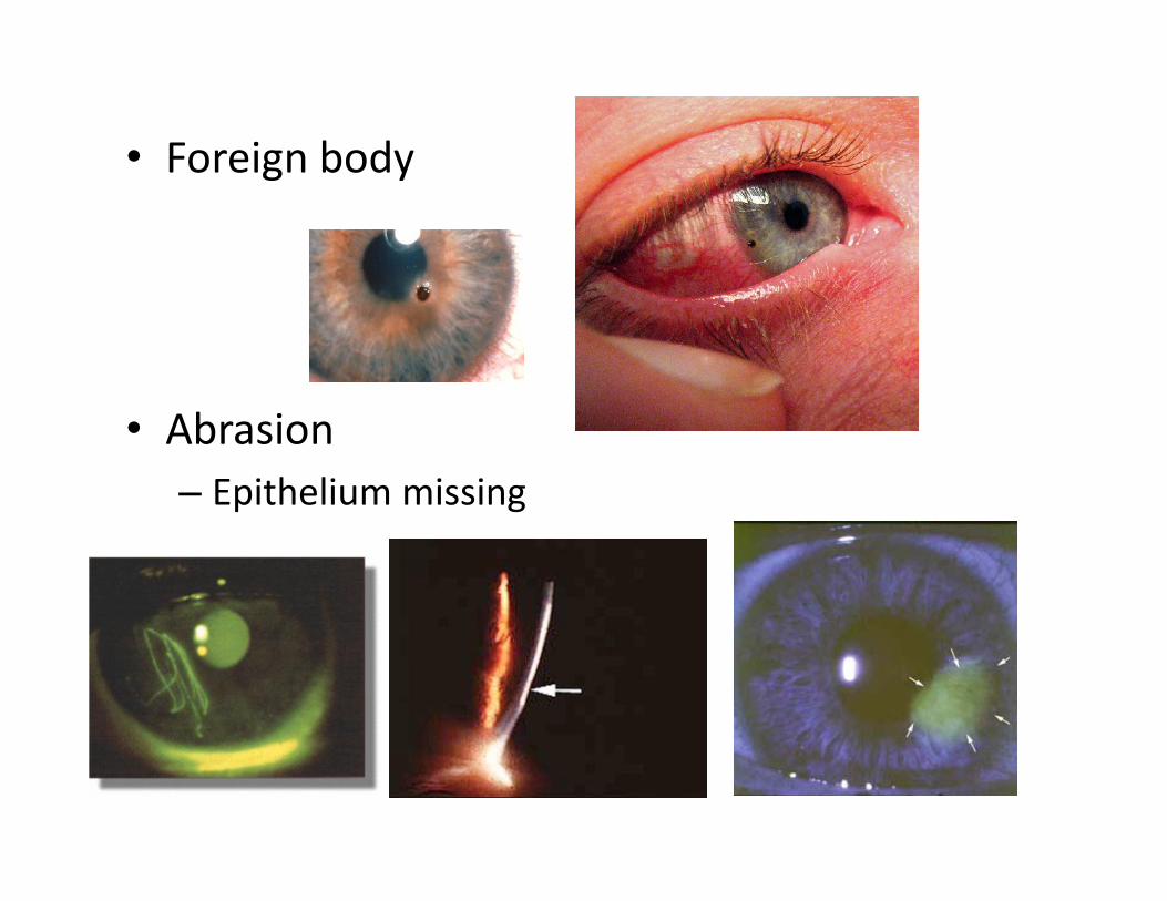
• Foreign body
• Abrasion– Epithelium missing
• Foreign body
• Abrasion– Epithelium missing

Recurrent Erosions• Unilateral• Wakes patient up• Sandy, irritated eye upon wakening• Look for cause– Rosacea, old trauma (fingernail in eye)
• Unilateral• Wakes patient up• Sandy, irritated eye upon wakening• Look for cause– Rosacea, old trauma (fingernail in eye)

Superficial Punctate Keratitis (SPK)• Sign, not diagnosis
• Disruption ofepithelial layer
CCLRU Punctate staining types

Location, Location, Location
• Diffuse– Toxic– Contact lens over wear
• Central– Adenovirus
• Upper 1/3– SLK– Vernal– Inclusive conjunctivitis– Trachoma
• Lower– Staph blepharitis– Lagophthalmos– Entropion– Acne rosacea
• Linear streak– FB
• Negative staining– Dystrophy/degeneration– Keratoconus
• Diffuse– Toxic– Contact lens over wear
• Central– Adenovirus
• Upper 1/3– SLK– Vernal– Inclusive conjunctivitis– Trachoma
• Lower– Staph blepharitis– Lagophthalmos– Entropion– Acne rosacea
• Linear streak– FB
• Negative staining– Dystrophy/degeneration– Keratoconus

• Negative Staining

• Edema– Epithelial/subepithelial– Stromal– Bullous keratopathy– Hydrops
– Striae– Microcystic

• Neovascularization– Response to lack of oxygen• Encroachment– Does not cross limbal band
• Superficial
• Deep
• Ghost
• Pannus
– Big ???• Etiology?
• Neovascularization– Response to lack of oxygen• Encroachment– Does not cross limbal band
• Superficial
• Deep
• Ghost
• Pannus
– Big ???• Etiology?

• Epithelial Infiltrates– Accumulation of WBC within superficial
cornea– Response to inflammation and/or infection
Viral- superficial, will stain with NaFl.
CL over wear
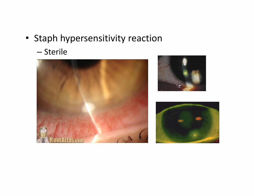
• Staph hypersensitivity reaction– Sterile

• Infected ulcerBacterial

• UlcerFungal
Confocal microscopy hyphae

• Acanthamoeba

Summary of corneal ulcers
Superficial staining of infiltrate
NaFl leaks into stroma

Dentritic Ulcer
• Herpes simplex keratitis– Epithelial disease
– Desensitized cornea

• Geographic lesion

• Interstitial disease– Stromal involvement

Neurotrophic keratopathy
• Lots of nerves within cornea
• Fed by CN 5 (sensory)
• Without them, cornea very sick and dies

• Dellen– Thinning of cornea due to dehydration
• Pterygium– Conjunctival overgrowth
• Dellen– Thinning of cornea due to dehydration
• Pterygium– Conjunctival overgrowth

• Limbal follicles
• Herbert’s pits
– Late stage trachoma• Earlier limbal follicles
• Limbal follicles
• Herbert’s pits
– Late stage trachoma• Earlier limbal follicles

• Endothelial pigmentKrukenberg’s spindle
• Endothelial pigment
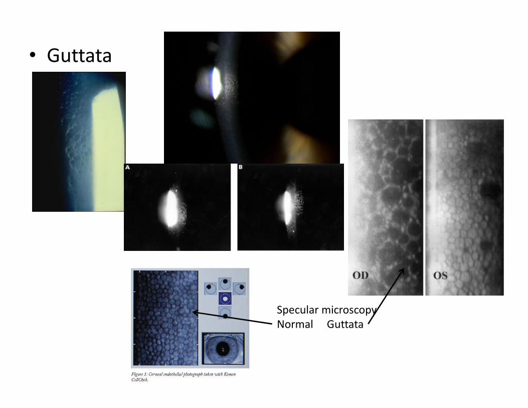
• Guttata
Specular microscopyNormal Guttata

• Endothelial Folds
• Vogt’s striae
• Endothelial Folds
• Vogt’s striae

• Keratic Precipitates (KP)– Inflammatory deposition on back
of endothelium
Mutton Fat
Retro Illumination
Fine
Stellate

– Corneal Ectasia– Bulging
– Keratoconus– Collagen disorder
• Opacities– Anything that makes cornea “not clear” or opaque–Why (etiology)• Developmental• Trauma– Scars» “War Wounds”
• Infection• Degenerations/dystrophies
– Types (from least to most dense)• Nebula• Macula• Leukoma
• Opacities– Anything that makes cornea “not clear” or opaque–Why (etiology)• Developmental• Trauma– Scars» “War Wounds”
• Infection• Degenerations/dystrophies
– Types (from least to most dense)• Nebula• Macula• Leukoma

• Nebular
• Macular
• Leukoma
• Nebular
• Macular

Surgical Scarring• Radial Keratotomy (RK)
• LASIK– The Flap NEVER heals
• Radial Keratotomy (RK)
• LASIK– The Flap NEVER heals

SummaryTABLE 1 Differentiating Between Dystrophy and Degeneration
CHARACTERISTIC DYSTROPHY DEGENERATION
Corneal location Central Peripheral
Laterality Bilateral Often unilateral
Symmetry Symmetric AsymmetricSymmetry Symmetric Asymmetric
VascularizationNone (Group of non-inflammatory disease)
Common
Family History Inherited Aging process
Onset age Often 10 or less Often 40 or more
Approx. 20 specificallynamed corneal dystrophies
Involutional = seen with age

• What does it look like?
• Where is it located?
it = opacity
• What does it look like?
• Where is it located?

Corneal Dystrophy VS Corneal Degeneration
• Corneal dystrophy– Over 20• Fuchs' dystrophy
• Keratoconus
• Lattice dystrophy
• Macular dystrophy
• Map-dot-fingerprint dystrophy (AKA Cogan’s, EpithelialBasement Membrane, or Microcystic CornealDystrophy)
– Inherited non-inflammatory diseases that affectboth eyes
Endothelial
• Corneal dystrophy– Over 20• Fuchs' dystrophy
• Keratoconus
• Lattice dystrophy
• Macular dystrophy
• Map-dot-fingerprint dystrophy (AKA Cogan’s, EpithelialBasement Membrane, or Microcystic CornealDystrophy)
– Inherited non-inflammatory diseases that affectboth eyes
Epithelial

• Fuch’s dystrophy– later-onset dystrophy that affects women more
than men
– Corneal guttata• Endothelial dystrophy
– Corneal haze
• Fuch’s dystrophy– later-onset dystrophy that affects women more
than men
– Corneal guttata• Endothelial dystrophy
– Corneal haze

• Keratoconus– Corneal Ectasis
– Inherited• Late teens to early 20’s
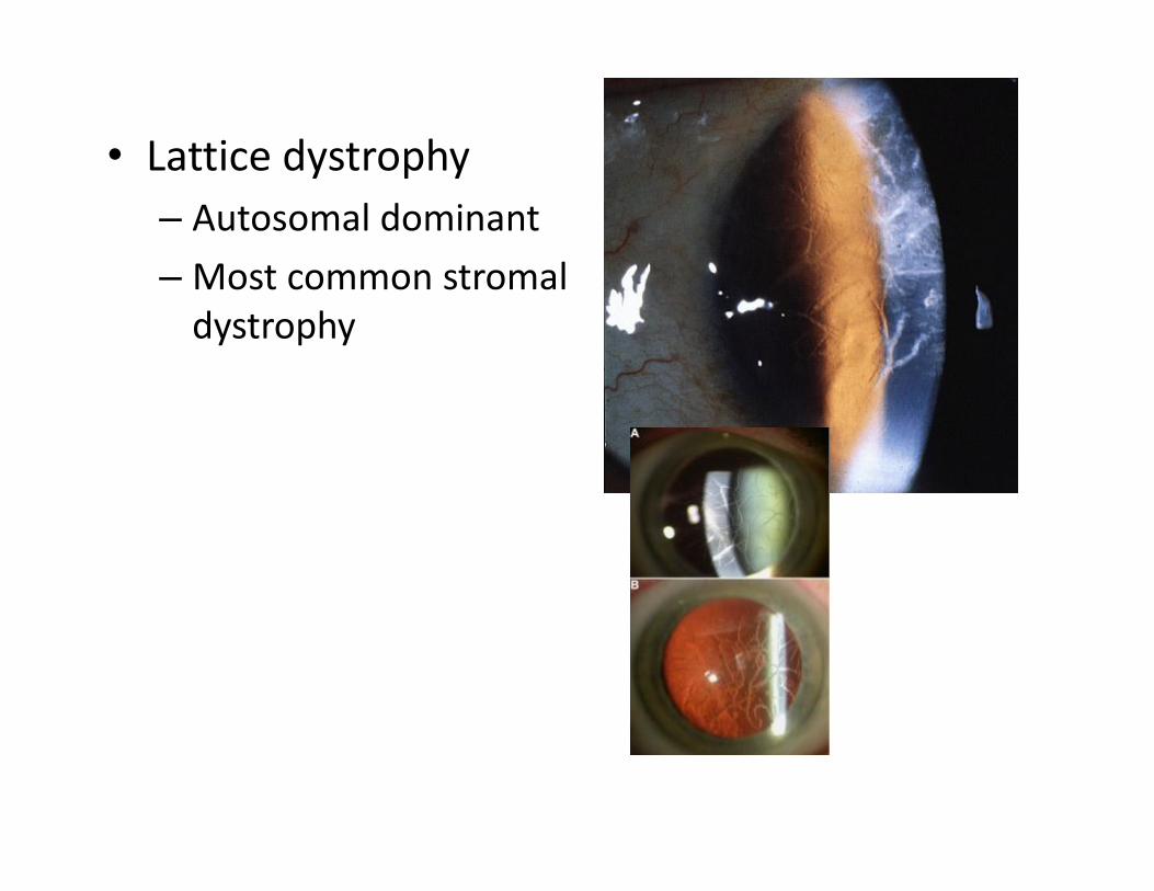
• Lattice dystrophy– Autosomal dominant
–Most common stromaldystrophy
• Lattice dystrophy– Autosomal dominant
–Most common stromaldystrophy
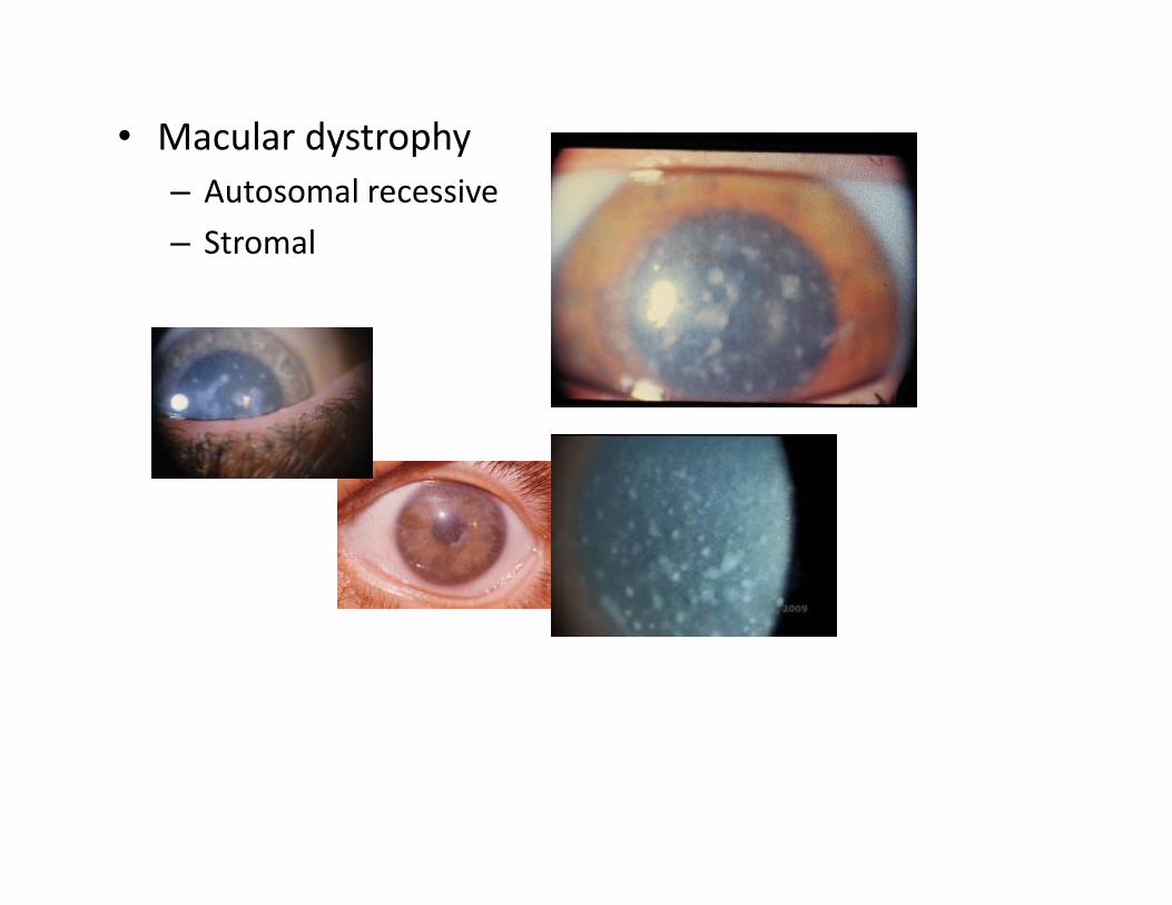
• Macular dystrophy– Autosomal recessive
– Stromal

• Map-Dot-Fingerprint– Epithelial basement membrane


