
Proc. Natl. Acad. Sci. USAVol. 82, pp. 3445-3449, May 1985Medical Sciences
Nucleotide sequence and structure of the human apolipoproteinE gene,
(Alu sequences/apo-E4 genotype/5' flanking region/Sl nuclease mapping/intron)
YOUNG-KI PAIK*, DAVID J. CHANG*, CATHERINE A. REARDON*, GLENN E. DAVIES*t,ROBERT W. MAHLEY*t§, AND JOHN M. TAYLOR*t¶II*Gladstone Foundation Laboratories for Cardiovascular Disease, tCardiovascular Research Institute, 1Department of Physiology and §Departments ofPathology and Medicine, University of California, San Francisco, CA 94140
Communicated by Stanley Cohen, January 7, 198S
ABSTRACT The gene for human apolipoprotein E (apo-E)was selected from a library of cloned genomic DNA byscreening with a specific cDNA hybridization probe, and itsstructure was characterized. The complete nucleotide sequenceof the gene as well as 856 nucleotides of the 5' flanking regionand 629 nucleotides of the 3' flanking region were determined.Analysis of the sequence showed that the mRNA-encodingregion of the apo-E gene consists of four exons separated bythree introns. In comparison to the structure of the mRNA, theintrons are located in the 5' noncoding region, in the codon forglycine at position -4 of the signal peptide region, and in thecodon for arginine at position +61 of the mature protein. Theoverall lengths of the apo-E gene and its corresponding mRNAare 3597 and 1163 nucleotides, respectively; a mature plasmaprotein of 299 amino acids is produced by this gene. Examina-tion of the 5' terminus of the gene by S1 nuclease mappingshows apparent multiple transcription initiation sites. Theproximal 5' flanking region contains a "TATA box" elementas well as two nearby inverted repeat elements. In addition,there are four Alu family sequences associated with the apo-Egene: an Alu sequence located near each end of the gene and twoAlu sequences located in the second intron. This knowledge ofthe structure permits a molecular approach to characterizingthe regulation of the apo-E gene.
Apolipoprotein E (apo-E) is a component of various classesof plasma lipoproteins in all mammals that have been studied(for review, see refs. 1 and 2). It is a single chain polypeptide(Mr, 34,000) of 299 amino acids (3) that is synthesized initiallywith an 18-residue signal peptide that is removed cotrans-lationally (4, 5). The amino acid sequence as well as themRNA nucleotide sequence are known for both the human(3, 6) and rat (7) species. The major site of synthesis is theliver, but relatively abundant levels of apo-E mRNA havebeen detected in many extrahepatic tissues, including thebrain and the adrenals (8). In addition, apo-E is produced bymouse peritoneal macrophages, as well as human monocyte-derived macrophages (9).A major function of apo-E is its mediation of the cellular
uptake of specific lipoproteins through an interaction withapo-B,E(LDL) receptors on extrahepatic and hepatic cellsurfaces and with distinct hepatic apo-E receptors (forreview, see ref. 10). The receptor binding domain of humanapo-E has been determined to be an arginine- and lysine-richregion in the vicinity of residues 140 and 160 (11, 12). Variantforms of apo-E with single amino acid substitutions in thisregion show decreased receptor binding activity (13-15) andare associated with type III hyperlipoproteinemia and ac-
celerated cardiovascular disease (for review, see refs. 16 and17). Apolipoprotein E with normal receptor binding activityis found in two common isoforms, the E3 and E4 phenotypes,with either cysteine or arginine, respectively, at residueposition 112 (13).Because of the central role that apo-E plays in the me-
tabolism of cholesterol and other lipids, knowledge of theregulation ofthe apo-E gene is important in understanding thealterations in lipid metabolism that occur in normal andpathological processes. Therefore, to provide a molecularbasis for examining its regulation, we have determined andanalyzed the nucleotide sequence of the human apo-E geneand its proximal flanking regions.
EXPERIMENTAL PROCEDURESDNA Library Screen. A human genome library of random,
partially Hae III/Alu I-digested fragments of fetal liver DNAcontained in the Charon 4A X bacteriophage (18) was pro-vided through the generosity of T. Maniatis (Harvard Uni-versity). The phage was grown in Escherichia coli LE392 andscreened essentially as described (19). About two millionphage plaques were screened with a 32P-labeled (20) restric-tion endonuclease fragment that was purified from a previ-ously characterized (6) full-length cloned cDNA to humanapo-E mRNA. A single recombinant bacteriophage wasidentified, and the DNA was prepared from plaque-purifiedmaterial (19). All experiments were done in accordance withthe National Institutes of Health Guidelines.DNA Mapping, Subcloning, and Sequencing. Bacteriophage
recombinant DNA was digested with various restrictionendonucleases (Boehringer Mannheim and New EnglandBiolabs) according to the suppliers' directions and wasexamined by electrophoresis in 0.8% agarose gels. The DNAwas transferred to nitrocellulose filters by blotting (21), thenhybridized to the 32P-labeled apo-E cDNA probe, and exam-ined by autoradiography to identify apo-E gene fragments.Based on these studies, EcoRI- and BamHI-digested DNAfragments were subcloned into plasmid pUC9 (22), and apo-Egene-containing recombinants were selected as describedabove. The apo-E gene DNA inserts in the subclones weresequenced by the method of Maxam and Gilbert (23).S1 Nuclease Mapping. A 67-base-pair BstNI/HindIII re-
striction endonuclease fragment from an apo-E genesubclone was prepared (23) that contained a portion of thefirst exon, the transcription initiation site, and a portion ofthe5'-terminal flanking region. The fragment was end-labeled atthe 5' ends by [y_32P]ATP and T4 polynucleotide kinase, and
Abbreviations: apo, apolipoprotein; kb, kilobases.tPresent address: Boehringer Mannheim, P.O. Box 50816, Indian-apolis, IN 46250.To whom reprint requests should be addressed.
3445
The publication costs of this article were defrayed in part by page chargepayment. This article must therefore be hereby marked "advertisement"in accordance with 18 U.S.C. §1734 solely to indicate this fact.
3446 Medical Sciences: Paik et al.
x
0c? Ot' W9'
23.1 -
0
xCla)
041)
z
0)-j
9.4 -
6.6 -
4.4 -
2.3 -
2.0 -
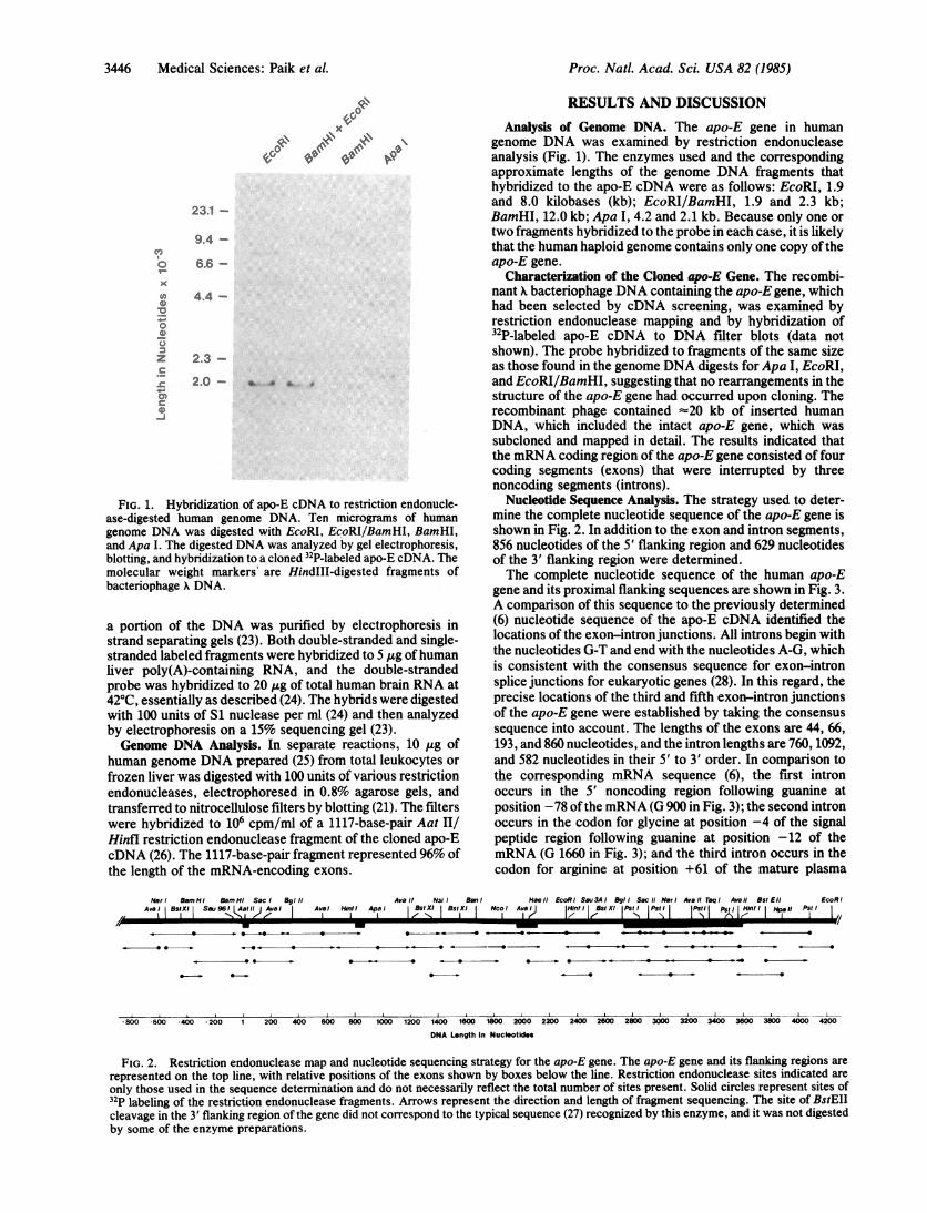
FIG. 1. Hybridization of apo-E cDNA to restriction endonucle-ase-digested human genome DNA. Ten micrograms of humangenome DNA was digested with EcoRI, EcoRI/BamHI, BamHI,and Apa I. The digested DNA was analyzed by gel electrophoresis,blotting, and hybridization to a cloned 32P-labeled apo-E cDNA. Themolecular weight markers' are HindIII-digested fragments ofbacteriophage X DNA.
a portion of the DNA was purified by electrophoresis instrand separating gels (23). Both double-stranded and single-stranded labeled fragments were hybridized to 5 ug ofhumanliver poly(A)-containing RNA, and the double-strandedprobe was hybridized to 20 ,ug of total human brain RNA at420C, essentially as described (24). The hybrids were digestedwith 100 units of S1 nuclease per ml (24) and then analyzedby electrophoresis on a 15% sequencing gel (23).Genome DNA Analysis. In separate reactions, 10 jg of
human genome DNA prepared (25) from total leukocytes orfrozen liver was digested with 100 units of various restrictionendonucleases, electrophoresed in 0.8% agarose gels, andtransferred to nitrocellulose filters by blotting (21). The filterswere hybridized to 106 cpm/ml of a 1117-base-pair Aat II/Hinfl restriction endonuclease fragment of the cloned apo-EcDNA (26). The 1117-base-pair fragment represented 96% ofthe length of the mRNA-encoding exons.
RESULTS AND DISCUSSIONAnalysis of Genome DNA. The apo-E gene in human
genome DNA was examined by restriction endonucleaseanalysis (Fig. 1). The enzymes used and the correspondingapproximate lengths of the genome DNA fragments thathybridized to the apo-E cDNA were as follows: EcoRI, 1.9and 8.0 kilobases (kb); EcoRI/BamHI, 1.9 and 2.3 kb;BamHI, 12.0 kb; Apa I, 4.2 and 2.1 kb. Because only one ortwo fragments hybridized to the probe in each case, it is likelythat the human haploid genome contains only one copy oftheapo-E gene.
Characterization of the Cloned apo-E Gene. The recombi-nant X bacteriophage DNA containing the apo-E gene, whichhad been selected by cDNA screening, was examined byrestriction endonuclease mapping and by hybridization of32P-labeled apo-E cDNA to DNA filter blots (data notshown). The probe hybridized to fragments of the same sizeas those found in the genome DNA digests for Apa I, EcoRI,and EcoRI/BamHI, suggesting that no rearrangements in thestructure of the apo-E gene had occurred upon cloning. Therecombinant phage contained -20 kb of inserted humanDNA, which included the intact apo-E gene, which wassubcloned and mapped in detail. The results indicated thatthe mRNA coding region of the apo-E gene consisted of fourcoding segments (exons) that were interrupted by threenoncoding segments (introns).
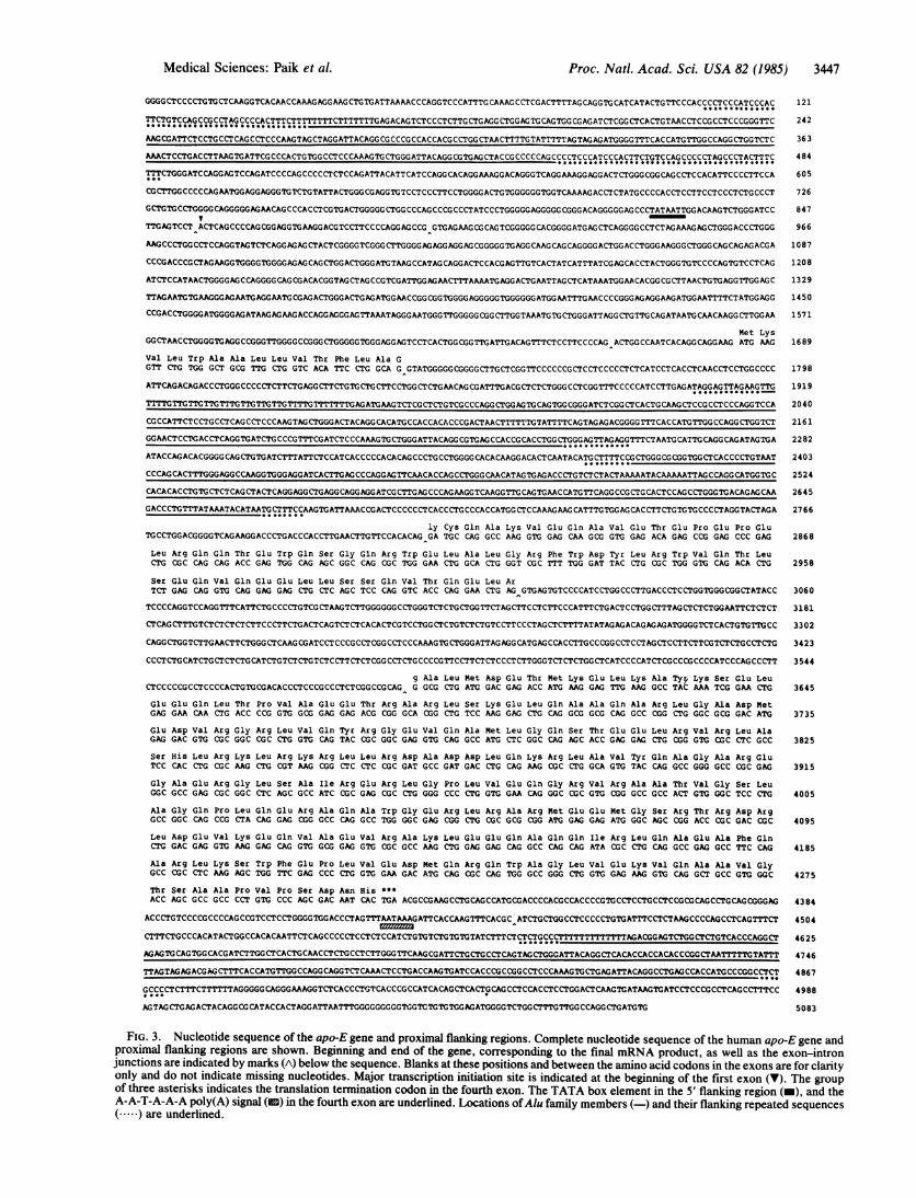
Nucleotide Sequence Analysis. The strategy used to deter-mine the complete nucleotide sequence of the apo-E gene isshown in Fig. 2. In addition to the exon and intron segments,856 nucleotides of the 5' flanking region and 629 nucleotidesof the 3' flanking region were determined.The complete nucleotide sequence of the human apo-E
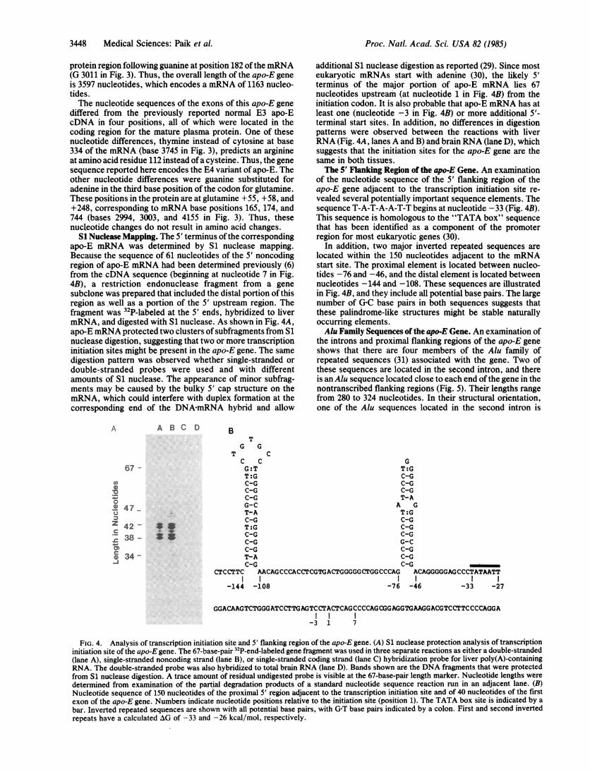
gene and its proximal flanking sequences are shown in Fig. 3.A comparison of this sequence to the previously determined(6) nucleotide sequence of the apo-E cDNA identified thelocations of the exon-intron junctions. All introns begin withthe nucleotides G-T and end with the nucleotides A-G, whichis consistent with the consensus sequence for exon-intronsplice junctions for eukaryotic genes (28). In this regard, theprecise locations of the third and fifth exon-intron junctionsof the apo-E gene were established by taking the consensussequence into account. The lengths of the exons are 44, 66,193, and 860 nucleotides, and the intron lengths are 760, 1092,and 582 nucleotides in their 5' to 3' order. In comparison tothe corresponding mRNA sequence (6), the first intronoccurs in the 5' noncoding region following guanine atposition -78 ofthe mRNA (G 900 in Fig. 3); the second intronoccurs in the codon for glycine at position -4 of the signalpeptide region following guanine at position -12 of themRNA (G 1660 in Fig. 3); and the third intron occurs in thecodon for arginine at position +61 of the mature plasma
Naor BamHI BamHi Sac BgIll Avail Nsil Ben Hoel/ EcoRl Sau3AI BgII SacIt NarI Avail TaqI Avail BstEI! EcoRIAval BstXI Sau 96! L|at! It AveI Aval Hint! Apoe BstXI BstXI Ncol Aval HinfI BstXI I Ps!jPstl I Ptt II fII lHPII Psta'I ,I >I/lll/lIrK E1XtII1X
-800 -600 -400 -200 1 200 400 60 80 00 10 1400 1600 1800 2000 2200 2400 2600 2800 300 320 40 60 30 400 20
DNA Length in Nucleotldes
FIG. 2. Restriction endonuclease map and nucleotide sequencing strategy for the apo-E gene. The apo-E gene and its flanking regions are
represented on the top line, with relative positions of the exons shown by boxes below the line. Restriction endonuclease sites indicated are
only those used in the sequence determination and do not necessarily reflect the total number of sites present. Solid circles represent sites of32P labeling of the restriction endonuclease fragments. Arrows represent the direction and length of fragment sequencing. The site of BstEIIcleavage in the 3' flanking region of the gene did not correspond to the typical sequence (27) recognized by this enzyme, and it was not digestedby some of the enzyme preparations.
Proc. Natl. Acad. Sci. USA 82 (1985)
Medical Sciences: Paik et al. Proc. Natl. Acad. Sci. USA 82 (1985) 3447
GGGGCTCCCCTGTGCTCAAGGTCACAACCAAAGAGGAAGCTGTGATTAAAACCCAGGTCCCATTTG CAAAGCCTCGACTTTTAGCAGGTGCATCATACTGTTCCCACCCCTCCCATCCCAC 121
TTCTGTCCAGCCGCCTAGCCCCACTTTCTTTTTTTTCTTTTTTTGAGACAGTCTCCCTCTTGCTGAGG CTGGAGTGCAGTGGCGAGATCTCGGCTCACTGTAACCTCCGCCTCCCGGGTTC 242
AAGCGATTCTCCTGCCTCAGCCTCCCAAGTAGCTAGGATTACAGG CGCCCGCCACCACGCCTGG CTAACTTTTG TATTTTTAGTAG AGATGGGGTTTCACCATGTTGGCCAGGCTGGTCTC 36 3
AAACTCCTGACCTTAAG TGATTCGCCCACTGTGGCCTCCCAAAG TGCTGGGATTACAGG CGTGAGCTACCGCCCCCAGCCCCTCCCATCCCACTTCTGTCCAG CCCCCTAGCCCTACTTTC 484
TTTCTGGGATCCAGGAGTCCAGATCCCCAGCCCCCTCTCCAGATTACATTCATCCAGG CACAGGAMAGGACAGGGTCAGGAAAGGAGGACTCTGGGCGGCAGCCTCCACATTCCCCTTCCA 60 5
CGCTTGGCCCCCAGAATGGAGGAGGGTGTCTGTATTACTGGG CGAGGTGTCCTCCCTTCCTGGGG ACTGTGGGGGGTGGTCAAAAG ACCTCTATGCCCCACCTCCTTCCTCCCTCTGCCCT 726
GCTGTGCCTGGGGCAGGGGGAGAACAGCCCACCTCGTGACTGGGGG CTGGCCCAGCCCGCCCTATCCCTGGGGGAGGGGGCGGGACAGGGGGAGCCCTATAATTGGACAAGTCTGGGATCC 84 7
TTGAGTCCT ACTCAGCCCCAGCGGAGGTGAAGGACGTCCTTCCCCAGGAG CCG GTGAGAAGCGCAGTCGGGGG CACGGGG ATGAGCTCAGGGG CCTCTAGAAAGAGCTGGGACCCTGGG 96 6
AAGCCCTGGCCTCCAGGTAGTCTCAGGAG AGCTACTCGGGGTCGGGCTTGGGG AGAGGAGGAGCGGGGGTGAGG CAAGCAGCAGGGGACTGGACCTGGGAAGGGCTGGGCAGCAGAGACGA 108 7
CCCGACCCGCTAGAAGGTGGGGTGGGGAGAGCAGCTGGACTGGGATGTAAGCCATAG CAGGACTCCACGAGTTGTCACTATCATTTATCG AGCACCTACTGGGTGTCCCCAGTGTCCTCAG 1 20 8
ATCTCCATAACTGGGGAG CCAGGGGCAGCGACACGGTAGCTAGCCGTCGATTGGAGAACTTTAAAATG AGGACTGAATTAGCTCATAAATGGAACACGG CGCTTAACTGTGAGGTTGG AG C 132 9
TTAGAATGTGAAGGGAGAATGAGGAATGCGAGACTGGGACTGAGATGGAACCGG CGGTGGGGAGGGGGTGGGGGGATGGAATTTGAACCCCGGGAGAGGAAGATGGAATTTTCTATGGAGG 145 0
CCGACCTGGGGATGGGGAGATAAGAGAAGACCAGGAGGGAGTTAAATAGGGAATGGGTTGGGGGCGG CTTGGTAAATGTGCTGGGATTAGGCTGTTGCAGATAATGCAACAAGGCTTGGAA 1 571
Met LysGGCTAACCTGGGGTGAGGCCGGGTTGGGGCCGGGCTGGGGGTGGGAGGAGTCCTCACTGGCGGTTGATTGACAGTTTCTCCTTCCCCAG ACTGGCCAATCACAGGCAGGAAG ATG AAG 168 9
Val Leu Trp Ala Ala Leu Leu Val Thr Phe Leu Ala GGTT CTG TGG GCT GCG TTG CTG GTC ACA TTC CTG GCA G GTATGGGGGOGGGGCTTGCTCGGTTCCCCCCGCTCCTCCCCCTCTCATCCTCACCTCAACCTCCTGGCCCC 17 98
ATTCAGACAGACCCTGGGCCCCCTCTTCTGAGGCTTCTGTGCTGCTTCCTGGCTCTGAACAG CGATTTGACGCTCTCTGGGCCTCGGTmcCCCCATCCTTGAGATAGGAGTTAGAAGTTG 191 9
TTTGTTGTTGTTGTTTGTTGTTGTTGTTTTGTTTTTTTGAGATGAAGTCTCGCTCTGTCGCCCAGG CTGGAGTGCAGTGGCGGGATCTCGGCTCACTGCAAGCTCCGCCTCCCAGGTCCA 204 0
CGCCATTCTCCTGCCTCAGCCTCCCAAGTAGCTGGGACTACAGGCACATGCCACCACACCCGACTAACTTTTTTGTATTTTCAGTAGAGACGGGGTTTCACCATGTTGG CCAGG CTGGTCT 2161
GGAACTCCTGACCTCAGG TGATCTGCCCGTTTCGATCTCCCAAAGTGCTGGGATTACAGG CGTGAGCCACCGCACCTGG CTGGGAGTTAGAGGTTTCTAATG CATTG CAGGCAGATAGTGA 2282
ATACCAGACACGGGG CAGCTGTGATCTTTATTCTCCATCACCCCCACACAG CCCTGCCTGGGGCACACAAGGACACTCAATACATG CTTTTCCGCTGGGCGCGGTGGCTCACCCCTGTAAT 2 40 3
CCCAGCACTTTGGGAGGCCAAGGTGGGAGGATCACTTGAG CCCAGGAGTTCAACACCAGCCTGGGCAACATAGTGAGACCCTGTCTCTACTAAAAATACAAAAATTAG CCAGGCATGGTG C 2 52 4
CACACACCTG TGCTCTCAGCTACTCAGGAGGCTGAGGCAGGAGGATCGCTTGAGCCCAGAAGGTCAAGGTTGCAGTGAACCATGTTCAGGCCGCTGCACTCCAGCCTGGGTGACAGAGCAA 264 5
GACCCTGTTTATAAATACATAATGCTTTCCAAGTGATTAAACCGACTCCCCCCTCACCCTGCCCACCATGG CTCCAAAGAAG CATTTGTGGAGCACCTTCTGTGTGCCCCTAGGTACTAGA 276 6
ly Cys Gln Ala Lys Val Glu Gln Ala Val Glu Thr Glu Pro Glu Pro GluTGCCTGGACGGGGTCAGAAGGACCCTGACCCACCTTGAACTTGTTCCACACAG GA TGC CAG GCC AAG GTG GAG CAA GCG GTG GAG ACA GAG CCG GAG CCC GAG 286 8
Leu Arg Gln Gln Thr Glu Trp Gln Ser Gly Gln Arg Trp Glu Leu Ala Leu Gly Arg Phe Trp Asp Tyr Leu Arg Trp Val Gln Thr LeuCTG CGC CAG CAG ACC GAG TGG CAG AGC GGC CAG CGC TGG GAA CTG GCA CTG GGT CGC TTT TGG GAT TAC CTG CGC TGG GTG CAG ACA CTG, 2958
Ser Glu Gln Val Gln Glu Glu Leu Leu Ser Ser Gln Val Thr Gln Glu Leu ArTCT GAG CAG GTG CAG GAG GAG CTG CTC AGC TCC CAG GTC ACC CAG GAA CTG AG GTGAGTGTCCCCATCCTGGCCCTTGACCCTCCTGGTGGGCGGCTATACC 3060
TCCCCAGGTCCAGGTTTCATTCTGCCCCTGTCGCTAAGTCTTGGGGGGCCTGGGTCTCTGCTGGTTCTAGCTTCCTCTTCCCATTTCTGACTCCTGGCTTTAGCTCTCTGGAATTCTCTCT 3181
CTCAGCTTTGTCTCTCTCTCTTCCCTTCTG ACTCAGTCTCTCACACTCGTCCTGG CTCTGTCTCTGTCCTTCCCTAGCTCTTTTATATAGAGACAGAGAGATGGGGTCTCACTGTGTTGCC 330 2
CAGGCTGGTCTTGAACTTCTGGGCTCAAG CGATCCTCCCGCCTCGGCCTCCCAAAGTGCTGGGATTAGAGGCATGAGCCACCTTGCCCGGCCTCCTAGCTCCTTCTTCGTCTCTG CCTCTG 3 42 3
CCCTCTGCATCTGCTCTCTGCATCTGTCTCTGTCTCCTTCTCTCGG CCTCTGCCCCGTTCCTTCTCTCCCTCTTGGGTCTCTCTGG CTCATCCCCATCTCG CCCGCCCCATCCCAGCCCTT 3 544
g Ala Leu Met Asp Glu Thr Met Lys Glu Leu Lys Ala Tyrp Lys 8er Glu LeuCTCCCCCGCCTCCCCACTGTGCGACACCCTCCCGCCCTCTCGGCCGCAG G GCG CTG ATG GAC GAG ACC ATG AAG GAG TTG AAG GCC TAC AAA TCG GAA CTG 3645
Glu Glu Gln Leu Thr Pro Val Ala Glu Glu Thr Arg Ala Arg Leu Ser Lys Glu Leu Gln Ala Ala Gln Ala Arg Leu Gly Ala Asp MetGAG GAA CAA CTG ACC CCG GTG GCG GAG GAG ACG CGG GCA CGG CTG TCC AAG, GAG CTG CAG GCG GCG CAG GCC CGG CTG GGC GCG GAC ATG 3735S
Glu Asp Val Arg Gly Arg Leu Val Gln Tyr Arg Gly Glu Val Gln Ala Met Leu Gly Gln Ser Thr Glu Glu Leu Arg Val Arg Leu AlaGAG GAC GTG CGC GGC CGC CTG GTG CAG TAC CGC GGC GAG GTG CAG GCC ATG CTC GGC CAG AGC ACC GAG GAG CTG CGG GTG CGC CTC GCC 3825
Ser His Leu Arg Lys Leu Arg Lys Arg Leu Leu Arg Asp Ala Asp Asp Leu Gln Lys Arg Leu Ala Val Tyr Gln Ala Gly Ala Arg GluTCC CAC CTG CGC AAG CTG CGT AAG CGG CTC CTC CGC GAT GCC GAT GAC CTG CAG AAG CGC CTG GCA GTG TAC CAG GCC GGG GCC CGC GAG 3915
Gly Ala Glu Arg Gly Leu Ser Ala Ile Arg Glu Arg Leu Gly Pro Leu Val Glu Gln Gly Arg Val Arg Ala Ala Thr Val Gly Ser LeuGGC GCC GAG CGC GGC CTC AGC GCC ATC CGC GAG CGC CTG GGG CCC CTG GTG GAA CAG GGC CGC GTG CGG GCC GCC ACT GTG GGC TCC CTG 4005
Ala Gly Gln Pro Leu Gln Glu Arg Ala Gln Ala Trp Gly Glu Arg Leu Arg Ala Arg Met Glu Glu Met Gly Ser Arg Thr Arg AsP ArgGCC GGC CAG CCG CTA CAG GAG CGG GCC CAG GCC TGG GGC GAG CGG CTG, CGC GCG CGG ATG GAG GAG ATG GGC AGC CGG ACC CGC GAC CGC 4095
Leu Asp Glu Val Lys Glu Gln Val Ala Glu Val Arg Ala Lys Leu Glu Glu Gln Ala Gln Gln Ile Arg Leu Gln Ala Glu Ala Phe GlnCTG GAC GAG GTG AAG GAG CAG GTG GCG GAG GTG CGC GCC AAG CTG GAG GAG CAG GCC CAG CAG ATA CGC CTG CAG GCC GAG GCC TTC CAG 4185
Ala Arg Leu Lys Ser Trp Phe Glu Pro Leu Val Glu Asp Met Gln Arg Gln Trp Ala Gly Leu Val Glu Lys Val Gln Ala Ala Val GlyGCC CGC CTC AAG AGC TGG TTC GAG CCC CTG GTG GMA GAC ATG CAG CGC CAG TGG GCC GGG CTG GTG GAG AAG GTG CAG GCT GCC GTG GGC 4275
Thr Ser Ala Ala Pro Val Pro Ser Asp Asn His ***ACC AGC GCC GCC CCT GTG CCC AGC GAC AAT CAC TGA ACGCCGAAGCCTGCAGCCATGCGACCCCACGCCACCCCGTGCCTCCTGCCTCCGCGCAGCCTGCAGCGGGAG 4384
ACCCTGTCCCCGCCCCAGCCGTCCTCCTGGGGTGGACCCTAGTTAATAAAGATTCACCAAGTTCACGC ATCTGCTGGCCTCCCCCTGTGATTTCCTCTAAGCCCCAG CCTCAGTTCT 450 4
CTTTCTGCCCACATACTGGCCACACAATTCTCAGCCCCCTCCTCTCCATCTGTGTCTGTGTGTATCTTTCTCT.CTGCCCTTTTTTTTTTTTAG ACGGAGTCTGGCTCTGTCACCCAGGCT 4625S
AGAGTGCAGTGGCACGATCTTGGCTCACTGCAACCTCTGCCTCTTGGG TTCAAGCGATTCTGCTGCCTCAGTAGCTGGGATTACAGGCTCACACCACCACACCCGGCTAATTTTGTATTT 4746
TTAGTAGAGACGAGCTTTCACCATGTTGGCCAGGCAGGTCTCAAACTCCTGACCAAG TGATCCACCCGCCGGCCTCCCAAAGTG CTGAGATTACAGGCCTGAGCCACCATGCCCGGCCTCT 4 86 7
GCCCCTCTTTCTTTTTTAGGGGG CAGGGAAAGGTCTCACCCT5GTCACCCG CCATCACAGCTCACTGCAGCCTCCACCTCCTGGACTCAAGTGATAAGTGATCCTCCCGCCTCAGCCTTTCC 4 98 8
AGTAGCTGAGACTACAGG CG CATACCACTAGGATTAATTTGGGGGGGGGIGGTGTGTGTGGAGATGGGGTCTGGCTTTGTTGGCCAGGCTGATGTG 5083
FIG. 3. Nucleotide sequence of the apo-E gene and proximal flanking regions. Complete nucleotide sequence of the human apo-E gene andproximal flanking regions are shown. Beginning and end of the gene, corresponding to the final mRNA product, as well as the exon-intronjunctions are indicated by marks (A) below the sequence. Blanks at these positions and between the amino acid codons in the exons are for clarityonly and do not indicate missing nucleotides. Major transcription initiation site is indicated at the beginning of the first exon (v). The groupof three asterisks indicates the translation termination codon in the fourth exon. The TATA box element in the 5' flanking region (-), and theA-A-T-A-A-A poly(A) signal (E) in the fourth exon are underlined. Locations ofAlu family members (-) and their flanking repeated sequences(.....)are underlined.
3448 Medical Sciences: Paik et al.
protein region following guanine at position 182 ofthe mRNA(G 3011 in Fig. 3). Thus, the overall length of the apo-E geneis 3597 nucleotides, which encodes a mRNA of 1163 nucleo-tides.The nucleotide sequences of the exons of this apo-E gene
differed from the previously reported normal E3 apo-EcDNA in four positions, all of which were located in thecoding region for the mature plasma protein. One of thesenucleotide differences, thymiine instead of cytosine at base334 of the mRNA (base 3745 in Fig. 3), predicts an arginineat amino acid residue 112 instead of a cysteine. Thus, the genesequence reported here encodes the E4 variant of apo-E. Theother nucleotide differences were guanine substituted foradenine in the third base position of the codon for glutamine.These positions in the protein are at glutamine +55, +58, and+248, corresponding to mRNA base positions 165, 174, and744 (bases 2994, 3003, and 4155 in Fig. 3). Thus, thesenucleotide changes do not result in amino acid changes.
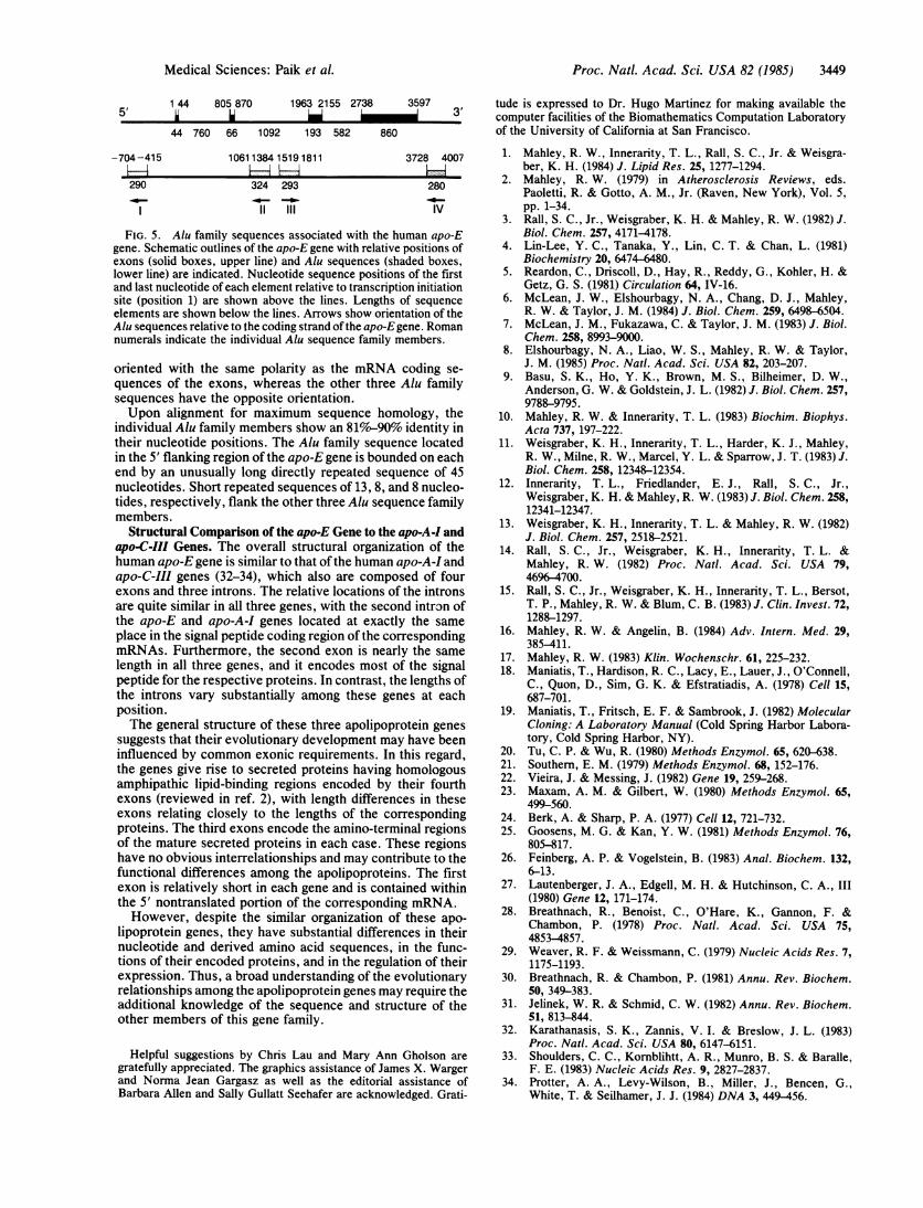
S1 Nuclease Mapping. The 5' terminus of the correspondingapo-E mRNA was determined by S1 nuclease mapping.Because the sequence of 61 nucleotides of the 5' noncodingregion of apo-E mRNA had been determined previously (6)from the cDNA sequence (beginning at nucleotide 7 in Fig.4B), a restriction endonuclease fragment from a genesubclone was prepared that included the distal portion of thisregion asmwell as a portion of the 5' upstream region. Thefragment was 32P-labeled at the 5' ends, hybridized to livermRNA, and digested with S1 nuclease. As shown in Fig. 4A,apo-E mRNA protected two clusters of subfragments from S1nuclease digestion, suggesting that two or more transcriptioninitiation sites might be present in the apo-E gene. The samedigestion pattern was observed whether single-stranded ordouble-stranded probes were used and with differentamounts of S1 nuclease. The appearance of minor subfrag-ments may be caused by the bulky 5' cap structure on themRNA, which could interfere with duplex formation at thecorresponding end of the DNA-mRNA hybrid and allow
A A B C D
67 -
v,
0
0._.@ 47
Z 42 -
s 38 -0)cr0 34--j
*.
additional S1 nuclease digestion as reported (29). Since mosteukaryotic mRNAs start with adenine (30), the likely 5'terminus of the major portion of apo-E mRNA lies 67nucleotides upstream (at nucleotide 1 in Fig. 4B) from theinitiation codon. It is also probable that apo-E mRNA has atleast one (nucleotide -3 in Fig. 4B) or more additional 5'-terminal start sites. In addition, no differences in digestionpatterns were observed between the reactions with liverRNA (Fig. 4A, lanes A and B) and brain RNA (lane D), whichsuggests that the initiation sites for the apo-E gene are thesame in both tissues.The 5' Flanking Region of the apo-E Gene. An examination
of the nucleotide sequence of the 5' flanking region of theapo-E gene adjacent to the transcription initiation site re-vealed several potentially important sequence elements. Thesequence T-A-T-A-A-T-T begins at nucleotide -33 (Fig. 4B).This sequence is homologous to the "TATA box" sequencethat has been identified as a component of the promoterregion for most eukaryotic genes (30).
In addition, two major inverted repeated sequences arelocated within the 150 nucleotides adjacent to the mRNAstart site. The proximal element is located between nucleo-tides -76 and -46, and the distal element is located betweennucleotides -144 and -108. These sequences are illustratedin Fig. 4B, and they include all potential base pairs. The largenumber of G-C base pairs in both sequences suggests thatthese palindrome-like structures might be stable naturallyoccurring elements.Alu Family Sequences of the apo-E Gene. An examination of
the introns and proximal flanking regions of the apo-E geneshows that there are four members of the Alu family ofrepeated sequences (31) associated with the gene. Two ofthese sequences are located in the second intron, and thereis an Alu sequence located close to each end ofthe gene in thenontranscribed flanking regions (Fig. 5). Their lengths rangefrom 280 to 324 nucleotides. In their structural orientation,one of the Alu sequences located in the second intron is
BT
G GT C
C C GG:T T:GT:G C-GC-G C-GC-G C-GC-G T-AG-C A GT-A T:GC-G C-GT:G C-GC-G C-GC-G G-CC-G C-GT-A C-GC-G C-G
CTCCTTC AACAGCCCACCTCGTGACTGGGGGCTGGCCCAG ACAGGGGGAGCCCTATAATTI
-144 -108 -76 -46 -33 -27
GGACAAGTCTGGGATCCTTGAGTCCTACTCAGCCCCAGCGGAGGTGAAGGACGTCCTTCCCCAGGA
-3 1 7
FIG. 4. Analysis of transcription initiation site and 5' flanking region of the apo-E gene. (A) Si nuclease protection analysis of transcriptioninitiation site of the apo-E gene. The 67-base-pair 32P-end-labeled gene fragment was used in three separate reactions as either a double-stranded(lane A), single-stranded noncoding strand (lane B), or single-stranded coding strand (lane C) hybridization probe for liver poly(A)-containingRNA. The double-stranded probe was also hybridized to total brain RNA (lane D). Bands shown are the DNA fragments that were protectedfrom S1 nuclease digestion. A trace amount of residual undigested probe is visible at the 67-base-pair length marker. Nucleotide lengths weredetermined from examination of the partial degradation products of a standard nucleotide sequence reaction run in an adjacent lane. (B)Nucleotide sequence of 150 nucleotides of the proximal 5' region adjacent to the transcription initiation site and of 40 nucleotides of the firstexon of the apo-E gene. Numbers indicate nucleotide positions relative to the initiation site (position 1). The TATA box site is indicated by abar. Inverted repeated sequences are shown with all potential base pairs, with G'T base pairs indicated by a colon. First and second invertedrepeats have a calculated AG of -33 and -26 kcal/mol, respectively.
Proc. Natl. Acad. Sci. USA 82 (1985)
Proc. Natl. Acad. Sci. USA 82 (1985) 3449
1 44 805 870 1963 2155 2738 35975' I I_ 3'
44 760 66 1092 193 582 860
-704-415 10611384 15191811 3728 4007
290 324 293 28011- -1
III IV
FIG. 5. Alu family sequences associated with the human apo-Egene. Schematic outlines of the apo-E gene with relative positions ofexons (solid boxes, upper line) and Alu sequences (shaded boxes,lower line) are indicated. Nucleotide sequence positions of the firstand last nucleotide of each element relative to transcription initiationsite (position 1) are shown above the lines. Lengths of sequenceelements are shown below the lines. Arrows show orientation of theAlu sequences relative to the coding strand of the apo-E gene. Romannumerals indicate the individual Alu sequence family members.
oriented with the same polarity as the mRNA coding se-quences of the exons, whereas the other three Alu familysequences have the opposite orientation.Upon alignment for maximum sequence homology, the
individual Alu family members show an 81%-90% identity intheir nucleotide positions. The Alu family sequence locatedin the 5' flanking region of the apo-E gene is bounded on eachend by an unusually long directly repeated sequence of 45nucleotides. Short repeated sequences of 13, 8, and 8 nucleo-tides, respectively, flank- the other three Alu sequence familymembers.
Structural Comparison of the apo-E Gene to the apo-A-I andapo-C-III Genes. The overall structural organization of thehuman apo-E gene is similar to that of the human apo-A-I andapo-C-III genes (32-34), which also are composed of fourexons and three introns. The relative locations of the intronsare quite similar in all three genes, with the second intron ofthe apo-E and apo-A-I genes located at exactly the sameplace in the signal peptide coding region of the correspondingmRNAs. Furthermore, the second exon is nearly the samelength in all three genes, and it encodes most of the signalpeptide for the respective proteins. In contrast, the lengths ofthe introns vary substantially among these genes at eachposition.The general structure of these three apolipoprotein genes
suggests that their evolutionary development may have beeninfluenced by common exonic requirements. In this regard,the genes give rise to secreted proteins having homologousamphipathic lipid-binding regions encoded by their fourthexons (reviewed in ref. 2), with length differences in theseexons relating closely to the lengths of the correspondingproteins. The third exons encode the amino-terminal regionsof the mature secreted proteins in each case. These regionshave no obvious interrelationships and may contribute to thefunctional differences among the apolipoproteins. The firstexon is relatively short in each gene and is contained withinthe 5' nontranslated portion of the corresponding mRNA.However, despite the similar organization of these apo-
lipoprotein genes, they have substantial differences in theirnucleotide and derived amino acid sequences, in the func-tions of their encoded proteins, and in the regulation of theirexpression. Thus, a broad understanding of the evolutionaryrelationships among the apolipoprotein genes may require theadditional knowledge of the sequence and structure of theother members of this gene family.
Helpful suggestions by Chris Lau and Mary Ann Gholson are
gratefully appreciated. The graphics assistance of James X. Wargerand Norma Jean Gargasz as well as the editorial assistance ofBarbara Allen and Sally Gullatt Seehafer are acknowledged. Grati-
tude is expressed to Dr. Hugo Martinez for making available thecomputer facilities of the Biomathematics Computation Laboratoryof the University of California at San Francisco.
1. Mahley, R. W., Innerarity, T. L., Rall, S. C., Jr. & Weisgra-ber, K. H. (1984) J. Lipid Res. 25, 1277-1294.
2. Mahley, R. W. (1979) in Atherosclerosis Reviews, eds.Paoletti, R. & Gotto, A. M., Jr. (Raven, New York), Vol. 5,pp. 1-34.
3. Rall, S. C., Jr., Weisgraber, K. H. & Mahley, R. W. (1982) J.Biol. Chem. 257, 4171-4178.
4. Lin-Lee, Y. C., Tanaka, Y., Lin, C. T. & Chan, L. (1981)Biochemistry 20, 6474-6480.
5. Reardon, C., Driscoll, D., Hay, R., Reddy, G., Kohler, H. &Getz, G. S. (1981) Circulation 64, IV-16.
6. McLean, J. W., Elshourbagy, N. A., Chang, D. J., Mahley,R. W. & Taylor, J. M. (1984) J. Biol. Chem. 259, 6498-6504.
7. McLean, J. M., Fukazawa, C. & Taylor, J. M. (1983) J. Biol.Chem. 258, 8993-9000.
8. Elshourbagy, N. A., Liao, W. S., Mahley, R. W. & Taylor,J. M. (1985) Proc. Natl. Acad. Sci. USA 82, 203-207.
9. Basu, S. K., Ho, Y. K., Brown, M. S., Bilheimer, D. W.,Anderson, G. W. & Goldstein, J. L. (1982) J. Biol. Chem. 257,9788-9795.
10. Mahley, R. W. & Innerarity, T. L. (1983) Biochim. Biophys.Acta 737, 197-222.
11. Weisgraber, K. H., Innerarity, T. L., Harder, K. J., Mahley,R. W., Milne, R. W., Marcel, Y. L. & Sparrow, J. T. (1983) J.Biol. Chem. 258, 12348-12354.
12. Innerarity, T. L., Friedlander, E. J., Rall, S. C., Jr.,Weisgraber, K. H. & Mahley, R. W. (1983) J. Biol. Chem. 258,12341-12347.
13. Weisgraber, K. H., Innerarity, T. L. & Mahley, R. W. (1982)J. Biol. Chem. 257, 2518-2521.
14. Rall, S. C., Jr., Weisgraber, K. H., Innerarity, T. L. &Mahley, R. W. (1982) Proc. Natl. Acad. Sci. USA 79,4696-4700.
15. Rall, S. C., Jr., Weisgraber, K. H., Innerarity, T. L., Bersot,T. P., Mahley, R. W. & Blum, C. B. (1983) J. Clin. Invest. 72,1288-1297.
16. Mahley, R. W. & Angelin, B. (1984) Adv. Intern. Med. 29,385-411.
17. Mahley, R. W. (1983) Klin. Wochenschr. 61, 225-232.18. Maniatis, T., Hardison, R. C., Lacy, E., Lauer, J., O'Connell,
C., Quon, D., Sim, G. K. & Efstratiadis, A. (1978) Cell 15,687-701.
19. Maniatis, T., Fritsch, E. F. & Sambrook, J. (1982) MolecularCloning: A Laboratory Manual (Cold Spring Harbor Labora-tory, Cold Spring Harbor, NY).
20. Tu, C. P. & Wu, R. (1980) Methods Enzymol. 65, 620-638.21. Southern, E. M. (1979) Methods Enzymol. 68, 152-176.22. Vieira, J. & Messing, J. (1982) Gene 19, 259-268.23. Maxam, A. M. & Gilbert, W. (1980) Methods Enzymol. 65,
499-560.24. Berk, A. & Sharp, P. A. (1977) Cell 12, 721-732.25. Goosens, M. G. & Kan, Y. W. (1981) Methods Enzymol. 76,
805-817.26. Feinberg, A. P. & Vogelstein, B. (1983) Anal. Biochem. 132,
6-13.27. Lautenberger, J. A., Edgell, M. H. & Hutchinson, C. A., III
(1980) Gene 12, 171-174.28. Breathnach, R., Benoist, C., O'Hare, K., Gannon, F. &
Chambon, P. (1978) Proc. Natl. Acad. Sci. USA 75,4853-4857.
29. Weaver, R. F. & Weissmann, C. (1979) Nucleic Acids Res. 7,1175-1193.
30. Breathnach, R. & Chambon, P. (1981) Annu. Rev. Biochem.50, 349-383.
31. Jelinek, W. R. & Schmid, C. W. (1982) Annu. Rev. Biochem.51, 813-844.
32. Karathanasis, S. K., Zannis, V. I. & Breslow, J. L. (1983)Proc. Natl. Acad. Sci. USA 80, 6147-6151.
33. Shoulders, C. C., Kornblihtt, A. R., Munro, B. S. & Baralle,F. E. (1983) Nucleic Acids Res. 9, 2827-2837.
34. Protter, A. A., Levy-Wilson, B., Miller, J., Bencen, G.,White, T. & Seilhamer, J. J. (1984) DNA 3, 449-456.
Medical Sciences: Paik et al.


