
HYBRIDOMAVolume 2. Number 1, 1983Mary Ann Liebert, Inc., Publishers
Invited Speakers'Abstracts from the Second Annual Congress
for Hybridoma ResearchFebruary 6-10, 1983
.
109
IMMUNODIAGNOSTIC STUDIES OF HUMAN ADENOCARCINOMAS WITH MONO-CLONAL ANTIBODIESRichard S. Metzgar, Melissa T. Gaillard, Michael Borowitz,Vicki N. Daasch, Francis L. Tuck, Ned W. Rodriguez and PhilipD. FernstenDepartments of Microbiology and Immunology, and Pathology,Duke University Medical Center, Durham, NC 27710
Murine monoclonal antibodies have been elicited to humanpancreatic adenocarcinoma cells. Preliminary studies on thespecificity of five of these monoclonal s (DU-PAN-1,2,3,4 and5) indicated restricted normal cell distribution and strongreactivity with some adenocarcinomas from a variety of tissueand organ sites (Cancer Research 42j_ 601-608, 1982). Thetumor and normal tissue distribution of 3 of the antigenshave been studied in more detail. DU-PAN-1 antigen is detect-ed on certain epithelial cells of normal prostate, stomachand bladder but is not detected on pancreatic ductal cells.This antigen is on a high percentage of pancreatic carcinomasand some adenocarcinomas of colon, prostate, bladder, breast,and ovary. DU-PAN-2 antigen is detected on normal pancreaticductal cells but is not detected on bladder or prostaticepithelium. This antigen is present on 14 of 16 pancreaticadenocarcinomas and 6 of 9 gastric carcinomas as well as onsome carcinomas from ovary, breast, colon and lung. DU-PAN-3antigen is present on pancreatic ductal cells and prostaticepithelium but is not found on bladder epithelial cells. Theantigen is present on some adenocarcinomas from pancreas,lung, colon, breast, ovary and prostate. DU-PAN-1 and DU-PAN-3antibodies are of the IgG2 isotype and immunoprecipitate glyco-peptides of 30-35,000 and 70-75,000 daltons respectively.These antigens are currently being purified by affinitychromatography and subjected to molecular analysis. TheDU-PAN-2 monoclonal is an IgM and reacts with a neuraminidasesensitive, mucin like antigen which has been purified fromascitic fluids of patients with pancreatic adenocarcinoma.A competition radioimmune assay has been developed for theDU-PAN-2 antigen which can readily detect this antigen in thepleural and ascitic fluids and sera of some patients with adeno-carcinomas originating from a variety of organ and tissue sites.Preliminary studies on the detection and serial monitoring ofserum levels of DU-PAN-2 antigen indicate that this test maybe valuable as a diagnostic and/or clinical management aid.
This work was supported in part by grants CA 08975 andCA 32672 from the National Cancer Institute and by grantIM-326 from the American Cancer Society.
110
SUBPOPULATIONS OF MYELOID PROGENITOR CELLS DEFINED BY MONOCLONALANTIBODIES Dario Ferrero*, Hal E. Broxmeyer1", Giovanni L.Pagliardi*, Salvatore Venuta*, Beverly Lange+, Silvana Pessano*and Giovanni Rovera* *The Wistar Institute of Anatomy andBiology, Philadelphia, PA 19104; ^Laboratories of DevelopmentalHematopoiesis, SloanKettering Institute for Cancer Research, NewYork, NY 10021; division of Oncology, Children's Hospital ofPhiladelphia, Philadelphia, PA 19104
Two types of progenitor cells of the human granulocytic andmonocytic lineages (CFU-GM) can be distinguished using mousemonoclonal antibodies against human hemopoietic cells. Type 1CFU-GM contributes all of the peripheral blood CFU-GM as well asa small fraction of bone marrow CFU-GM and express on theirsurface antigens recognized by several anti-lymphomonocyticantibodies but not antigens expressed on morphologically identi-fiable cells of the myelomonocytic lineage. Type 2 CFU-GM arepresent only in the marrow and react with a variety of monoclo-nal antibodies. In vitro culture of type 1 CFU-GM in the pre-sence of CSF generates colony-forming cells that have thesurface phenotype of type 2 CFU-GM. This finding supports theinterpretation that type 1 and type 2 CFU-GM represent two dif-ferent stages of maturation of myelomonocytic stem cells.
ill
ANTIGENS OF HUMAN GASTRO-INTESTINAL TUMORS. Zenon Steplewski,Meenhard Herlyn, Magdalena Blaszczyk, Koon Yan Pak, AlonzoRoss, John Powe and Hilary Koprowski, The Wistar Institute,Philadelphia, Pennsylvania 19104.
Hybridoma derived murine monoclonal antibodies generatedagainst human gastrointestinal tumors are used to characterizetumor associated antigens (Herlyn, M. et al. PNAS 76,1438,1979;Koprowski et al. Somat. Cell Genetics 5,957,1979). Monoclonalantibody defined antigens of gastrointestinal tumors were foundto belong to glycolipid and glycoprotein groups. The glyco-proteins include CEA and CEA related molecules, and a bimolec-ular protein which consists of^chain with MW 28,000 dal tonsand ß chain with MW of 22,000 dal tons. Two newly establishedhybridomas (G7-6 and G7-9) secrete antibodies directed against90,000/27,000 daltons molecules restricted to gastrointestinaltumors.The majority, however, of the antigens expressed on thesurface of gastrointestinal tumors and immunogenic in mice areof glycolipid nature. These include blood group B glycolipidchains Type I and II and Lewis blood group antigens Lea andLe°. Two such antibodies react with both Lea and LeD moleculesTwo monoclonal antibodies directed against Le'3 hapten immuno-precipitated similar protein molecules with MW of 220,000,150,000 and 36,000. Monoclonal antibody specific for bothLea and Le^ haptens immunoprecipitates 220,000 and 150,000glycolipid molecules from colon carcinoma cells. These mole-cules are present only in gastrointestinal tumors. Finally,one of the antigens, a monosialoganglioside (Magnani et al.Science 215,55,1981), was found to be shed by tumor cells(Steplewski et al. Cancer Res. 41,2723,1981) and present insera of cancer patients (Koprowski et al. Science 215,53,1981).This antibody was used to construct a sérodiagnostic assaymethod. Another antibody, of IgG2a isotype, directed againstantigen not shed by tumor cells is used for immunotherapyexperiment and as carrier of radioactive isotopes for nuclearimaging. The selection of antigen-antibody pairs at presentis based not only on their specific distribution but alsoaccording to their future application - antigens shed vs non-shed by tumor cells.
112
MONOCLONAL ANTIBODIES REACTIVE WITH HUMAN MAMMARY CARCINOMAAND COLON CARCINOMA ASSOCIATED ANTIGENS
J. Schlom, D. Colcher, P. Horan Hand, D. Wunderlich,D. Stramignoni, M. Nuti, B. Atkinson, R. Bowen.National Cancer Institute, NIH, Bethesda, Md, 20205
Murine monoclonal antibodies reactive with human mammarytumor associated antigens have been generated and character-ized. The immunogens used in these studies were membrane-enriched fractions of human metastatic mammary carcinomalesions. The thirteen monoclonal antibodies characterizedcould be divided into five major groups based on differentialreactivity to various mammary carcinoma lesions, binding tothe surface of carcinoma cell lines, and the molecular weightof the reactive antigen precipitated. Two monoclonals (Bl.land F5.5) were reactive with purified 180,000d carcinoembry-onic antigen. Monoclonals B6.2 and B72.3 were extensivelycharacterized as to their range of reactivities, and wereshown to immunoprecipitate a 90,000d protein, and a 220,000d-400,000d high molecular weight protein complex, respectively.B72.3 clearly showed the most restricted pattern of reactivityfor carcinoma vs. normal human cells. Using the monoclonalantibodies and the immunoperoxidase technique on sections ofhuman mammary carcinomas, a spectrum of antigenic phenotypeswas observed, as was antigenic diversity of mammary tumor cellpopulations within a given tumor mass. Antigenic phenotypicdiversity and modulation was also observed in establishedmammary tumor cell lines and cloned cell populations. B6.2IgG was purified, FCab')? and Fab' fragments were generatedand radiolabeled with *25l; all were successful in localizinghuman mammary tumors transplanted in athymic mice. TheF(ab')2 fragments gave the highest tumor to tissue ratios.Scanning experiments confirmed the ability of radiolabeledmonoclonal antibody B6.2 and its fragments to detect thepresence of transplanted human mammary tumor lesions withoutthe aid of background subtraction manipulations.
The reactivity of monoclonal antibodies B72.3, B6.2 andBl.l with fixed tissue sections of human colon carcinomas andadenomas was also examined. B72.3 demonstrated the most se-
lective reactivity for carcinomas; 82% (14 of 17) of carci-nomas were positive while none of 45 adenomas or normal colonepithelium examined showed reactivity with greater than a fewpercent of adenoma cells positive. Monoclonal B72.3 alsoreacted with cells in areas of "atypia" within adenomas.Based on the expression of antigens reactive with the threemonclonal antibodies employed, both colon adenocarcinomas andadenomas can now be placed in several distinct phenotypes.
113
EFFECTOR CELLS FOR MURINE IgG2a MONOCLONAL ANTIBODIES.Hilary Koprowski, Dorothée Herlyn and Zenon Steplewski, TheWistar Institute, Philadelphia, Pennsylvania 19104.
Murine monoclonal IgG2a antibodies directed against tumorassociated antigens are specifically cytotoxic in antibodydependent cell mediated cytotoxicity assay (ADCC) in vitro(Herlyn, D. et al. Eur. J. Immunol. 9:657, 1979) and specific-ally inhibit growth of human tumors in athymic mice (Koprowskiet al. PNAS, 75:3405, 1978; Herlyn D. et al. Cancer Res. 40:717, 1980). Monoclonal antibodies against these tumors but ofother than IgG2a isotypes showed no inhibition of tumor growthin nude mice. Complement depletion of nude mice had no effecton tumor suppression by IgG2a antibody. Silica treatment ofnude mice on the other hand, abolished tumoricidal activity of
Analysis of macrophage mediated IgG2a depend-in vitro and above described experimentsmacrophages as effector cells in specifichuman tumor cells. Fc2f2a receptors of
murine macrophages are involved in tumor destruction, sinceF(ab')2 fragments of IgG2a monoclonal antibodies have no effecton tumor xenograft growth in nude mice. Human macrophagesexpress Fc receptors that crossreact with murine IgG2a. Thesemacrophages not only express Fc receptors that bind murineIgG2a monoclonal antibody, but also mediate cytotoxicityagainst human tumors (Steplewski et al. Hybridoma 2:2,1983).This fact may be relevant to application of murine monoclonalantibodies in man. (Supported by NIH grants CA-10815, CA-21124CA-25874, and RR-05540).
IgG2a antibody,ent cytotoxicityin vivo point tolysis in vivo of
114
BIOCHEMICAL PROFILE AND BIOLOGICAL FUNCTION OF A CELLSURFACE PROTEOGLYCAN ON HUMAN MELANOMA CELLS.R. A. Reisfeld, J. R. Harper, G. Schulz, and T. F. Bumol.Scripps Clinic and Research Foundation, La Jolla, CA92037.
The biosynthesis, topography and structure ofchondroitin sulfate proteoglycans (CSP) in intrinsicallyradiolabeled human melanoma cell lines were defined bymonoclonal antibodies (Mab). Mab 9.2.27 reacts withseveral Endoglycosidase H-(Endo H) sensitive, N-linkedglycoprotein precursors at early time points in pulse-chaseanalyses which chase into an Endo H-resistant molecule ofMr 250,000 (250K) and ultimately a high molecular weightCSP sensitive to digestion with chondroitinases. Digestionwith these enzymes releases the 250K molecule which revealsclose structural homology with CSP by tryptic peptidemapping, indicating that Mab 9.2.27 recognizes adeterminant on the core glycoprotein for CSP in melanomacells. A series of experiments demonstrated that Mab 9.2.27recognizes core precursors, core glycoprotein and releasedCSP of human melanoma cells. An assessment of thefunctional role of this CSP in a parameter that isbiologically relevant to tumor proliferation, i.e.,anchorage-independent growth, indicated that reaction ofmelanoma cells with Mab 9.2.27 specifically inhibited theirgrowth by ^75%, suggesting that the Mab 9.2.27-defined CSPmay have a key role in cell-cell interactions. A comparisonof the serological reactivity, toxic activity and tumorgrowth inhibitory properties of Mab 9.2.27 and itsdiphtheria toxin A chain conjugate (9.2.27-DTA) indicatedthat only the 9.2.27-DTA exhibits specific In vitrotoxicity towards melanoma target cells; however, both the9.2.27 IgG and its DTA conjugate can suppress establishedtumor growth Jjn vivo, suggesting that several hostmechanisms may mediate Mab-induced tumor suppression.Suppression of tumor growth by Mab 9.2.27 could bemeasurably and specifically enhanced in vitro by directedantibody-dependent cellular cytotoxicity (ADCC), i.e.,conjugation of 1 mg antibody IgG to 5 x 10' mouse effectorcells with polyethylene glycol resulted in the killing oftumor cells at a rate greater than that obtained in theclassical ADCC reaction. In nude mice, bearing establishedhuman melanoma tumors, 1 x 10? effector cells conjugatedwith 200 ug 9.2.27 IgG essentially abolished tumor growth.These data suggest that Mab 9.2.27 when conjugated toeffector cells of cancer patients may possibly provide ameans of immunoprophylaxis following surgical resection oftumors.
115
MONOCLONAL ANTIBODY-DEFINED CELL SURFACE ANTIGENS OF HUMANTERATOCARCINOMA CELL LINES, Peter W. Andrews, The WistarInstitute of Anatomy and Biology, 36th Street at Spruce,Philadelphia, PA 19104.
Cell surface antigens often provide useful markers fordissecting differentiating systems. We have applied thisapproach, most extensively used by others in the study of theimmune system, to the analysis of human teratocarcinoma celllines in vitro. These cell lines are of interest both asmodels for a clinically important tumor, and for humanembryonic cells. Two groups of monoclonal antibodies havebeen studied. The first consists of monoclonal antibodiesthat define heterogenetic stage specific embryonic antigens ofthe mouse, particularly SSEA-1 and SSEA-3, or that detecthuman homologues of antigens that are developmentally regu-lated in murine embryos and teratocarcinomas (e.g. MHCantigens). The second group consists of monoclonal antibodiesthat have been produced by immunizing mice with human cellsand that recognize human antigens with no known murine equiva-lent. Amongst those which we have produced is one (TRA-1-60)that recognizes a cell surface antigen consisting of two sub-units (approx. MW 200,000 and 400,000) found on all humanembryonal carcinoma cells tested, but few other tumor celltypes. Results from these studies indicate marked differencesbetween murine embryonal carcinoma (EC) cells, the stem cellsof murine teratocarcinomas, and human EC cells. These dif-ferences might indicate that human EC cells correspond to adifferent stage of embryonic development than murine EC cells,which appear to be developmentally and biochemically similarto the primitive ectoderm. Other cells derived from humanteratocarcinomas are distinguishable from human EC cells interms of the antigens that they express. Investigation of theexpression of these antigens has allowed us to demonstratedifferentiation of clonal human EC cells in culture.
This work was supported by a grant, CA-29894, from the U.S.National Institutes of Health.
116
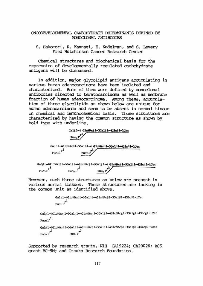
ONCODEVELOPMENTAL CARBOHYDRATE DETERMINANTS DEFINED BYMONOCLONAL ANTIBODIES
S. Hakomori, R. Kannagi, E. Nudelman, and S. LeveryFred Hutchinson Cancer Research Center
Chemical structures and biochemical basis for theexpression of developmentally regulated carbohydrateantigens will be discussed.
In addition, major glycolipid antigens accuirulating invarious human adenocarcinoma have been isolated andcharacterized. Some of them were defined by monoclonalantibodies directed to teratocarcinoma as veil as membranefraction of human adenocarcinoma. Among these, accumula-tion of three glycolipids as shown below are unique forhuman adenocarcinoma and seem to be absent in normal tissueon chemical and immunochemical basis. Those structures arecharacterized by having the common structure as shewn bybold type with underline.
Galßl-*4 GlcMftcel*3GalB1^4Glcei*lCer
FucttiyGalßl»4GlcNAcßl-<-3Galßl+4 GlcNftcEl*3Gal¡íl-MGlcl-»lCer
Fuccil' Fücal^/Galßl-»4GlcNAcßl-*3GaljSl-^4GlcNAcßl-^3Galel-^ Glct^l^3Gal6l->4Glcgl-*10ar
/3 /3Fuax]/ Fuccd'
However, such three structures as below are present invarious normal tissues. These structures are lacking inthe cornron unit as identified above.
Galgl-»4GlcNAc61-»3Galßl-»4GlcNAcßl-*-3Galßl-<-4Glcßl*lCer/3Fuccd/
Galßl*4GlcNAcßl*3Galßl-»4GlcNAcßl-3Galßl-^GlcNAcßl*3Galßl-»4Glcßl*lCer/3Fuaxl
Galß l-»4GlcNAcß l-»3Galß l-*4GlcNAcß 1-OGalß l->4GlcNAcß l-3Galß l-*4Glcß1-KLCertZ /3Fucxir Fuoar
Supported by research grants, NIH CA19224; CA20026; ACSgrant BC-9M; and Otsuka Research Foundation.
117
IMPROVED DETECTION OF HEPATITIS B VIRUS BY MONOCLONAL RADIOIM-MUNOASSAY. E. Ben-Porath, K. J. Isselbacher and J. R. Wands.Gastrointestinal Unit, Massachusetts General Hospital, Depart-ment of Medicine, Harvard Medical School, Boston, MA
The specific viral marker for acute and chronic hepatitis Bviral (HBV) infection is the presence of hepatitis B surfaceantigen (HBsAg) in the blood. However, it has been recognizedthat some individuals may transmit hepatitis B infection even
though HBsAg is undetectable by current radioimmunoassays (RIAs)which employ polyvalent antibodies. Similarly, normal individ-uals and others with acute and chronic hepatitis who are nega-tive for HBsAg by conventional RIA could also be infected withHBV if their blood contains antibodies to hepatitis B core an-
tigen (anti-HBc) and/or hepatitis B surface antigen (anti-HBs).We have developed sensitive and specific solid phase RIAsusing high affinity IgG and IgM monoclonal antibodies to HBsAg(PNAS J78:1215-1219,1981) . In the present investigation a
highly sensitive RIA was constructed with three monoclonalanti-HBs antibodies which bind to distinct and separate deter-minants on all known subtypes of HBsAg. In this assay design a
monoclonal IgM anti-HBs was linked to a solid phase supportand incubated with patient serum and the two other 125I-labeled IgG^ and IgG2a anti-HBs for 16 hr at 45° ("simultaneoussandwich" RIA). Approximately 20 pg of HBsAg could be detectedby the RIA. Serial studies on a group of patients with acuteHBV infection revealed that HBsAg was present in blood for afar longer period of time (10-60 days) when compared to conven-tional assay (Ausria II). In addition 3 dialysis patients werefollowed for 19-25 months after HBsAg was first detected intheir serum by Ausria II. We demonstrated by monoclonal RIAthat HBsAg was still present 1,9 and 15 months respectively,after it was no longer detectable by Ausria II. HBsAg was de-tectable even when anti-HBs antibodies were already presentsuggesting that the monoclonal RIA may identify "hidden" HBVdeterminants in immune complexes. We also studied 26 HBsAgnegative healthy individuals in whom anti-HBc antibodies were
the only sérologie marker of current, recent or past HBV infec-tion; six were positive by monoclonal RIA (23%). In contrast,HBsAg was undetectable by monoclonal RIA in another group of 26healthy individuals who had both anti-HBc and the protectiveanti-HBs antibodies. These studies demonstrate therefore thatHBsAg is present in the blood for a far longer period of timethan previously recognized and explains in part the late ap-pearance of the protective anti-HBs antibodies In some individ-uals. Further, patients with only anti-HBc in their blood mayhave co-existing HBV infection. The findings of extended pres-ence of HBsAg not detectable by conventional assays has impor-tant clinical and epidemiologic implications.
118


