HANDBOOK OF
DEVELOPMENTAL
COGNITIVE NEUROSCIENCE
Second Edition
Edited by
Charles A. Nelson and Monica Luciana
A BRADFORD BOOK
THE MIT PRESS
CAMBRIDGE, MASSACHUSETTS
LONDON, ENGLAND
© 2008 Massachusetts Institute of Technology
All rights reserved. No part of this book may be reproduced in any form by any electronic or mechanical means (including photocopying, recording, or information storage and retrieval) without permission in writing from the publisher.
MIT Press books may be purchased at special quantity discounts for business or sales promotional use. For information, please e-mail [email protected] or write to Special Sales Department, The MIT Press, 55 Hayward Street, Cambridge, MA 02142.
This book was set in Baskerville by SNP Best-set Typesetter Ltd., Hong Kong and was printed and bound in the United States of America.
Library of Congress Cataloging-in-Publication Data
Handbook of developmental cognitive neuroscience / edited by Charles A. Nelson and Monica Luciana.—2nd ed. p. ; cm.—(Developmental cognitive neuroscience) Includes bibliographical references and index. ISBN 978-0-262-14104-8 (hardcover : alk. paper) 1. Developmental neurobiology—Handbooks, manuals, etc. 2. Cognitive neuroscience—Handbooks, manuals, etc. I. Nelson, Charles A. (Charles Alexander) II. Luciana, Monica. III. Series. [DNLM: 1. Nervous System—growth & development. 2. Central Nervous System Diseases—physiopathology. 3. Cognition—physiology. 4. Human Development. 5. Per-ception—physiology. WL 102 H23535 2008] QP363.5.H365 2008 612.8'2—dc22
200800788610 9 8 7 6 5 4 3 2 1
1 The Formation of Axons and
Dendrites by Developing Neurons
PAUL LETOURNEAU
Introduction
The neuronal circuitry that underlies human behavior and
other neural functions develops over a prolonged period
lasting from the second fetal month through adolescent
years. These circuits arise from the extensive development
of elaborate neuronal processes, as neurons express intrinsic
morphogenetic behaviors, while interacting with other cells
and molecules of the developing nervous system. First,
immature neurons migrate from their birthplaces to the sites
where they are organized into layers, nuclei, and ganglia of
neuronal perikarya. Next, immature neurons sprout axons
and dendrites that elongate, sometimes for many centime-
ters, to make synaptic connections with target neurons or
other cells. This chapter describes intrinsic mechanisms of
morphogenesis of axons and dendrites and the extrinsic
environmental features that regulate where and when axons
and dendrites grow to create neural circuits.
The ability to extend neuronal processes, or neurites, is
intrinsic to neurons. This is demonstrated when immature
neurons, such as from prenatal hippocampus, are placed
into tissue culture. Within a few hours the neurons sprout
processes that elongate onto the substrate, each tipped by an
adherent motile structure called a growth cone. These neu-
rites mature to become axons and dendrites and form syn-
apses in vitro. These events in a neutral in vitro environment
show that the neuronal phenotype defi nes the intrinsic
behaviors that produce neuronal shape. The most signifi cant
cellular components in neuronal morphogenesis are the
protein polymers of the neuronal cytoskeleton. In the next
section the neuronal cytoskeleton and the intrinsic mecha-
nisms of neurite formation and elongation will be discussed.
In the following three sections the regulation of axonal and
dendritic growth by extrinsic molecules will be discussed.
The dynamic neuronal cytoskeleton
Neuronal morphogenesis depends on the organization and
dynamic properties of two cytoskeletal polymers, microtu-
bules and actin fi laments (Dent and Gertler, 2003; Luo,
2002). These cytoskeletal polymers are present in all cell
types, although specifi c mechanisms determine cytoskeletal
functions in neurons.
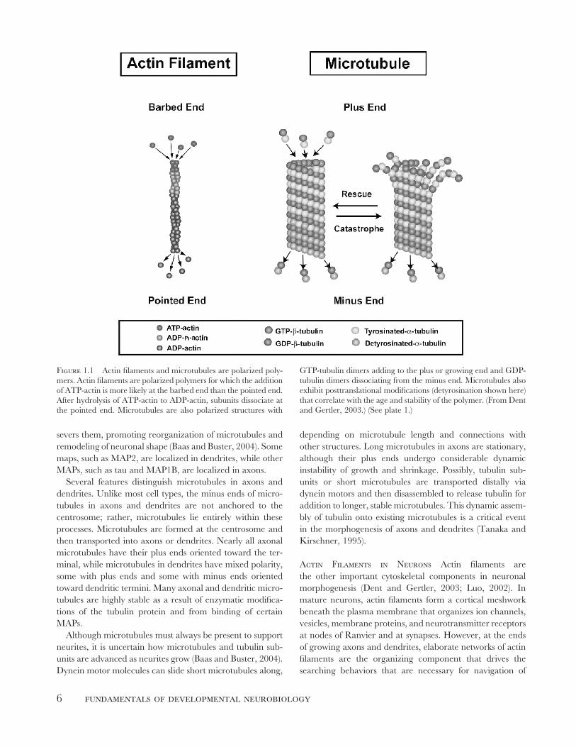
Microtubules Provide Support and a Means of Transport Microtubules are hollow cylinders 25 nm in
diameter that extend through the cytoplasm of neuronal
perikarya, axons, and dendrites (fi gures 1.1, 1.2). The wall
of a microtubule consists of subunits of highly conserved
proteins, alpha tubulin and beta tubulin. Microtubules have
no defi ned length, and single neuronal microtubules can
exceed 100 μm (Letourneau, 1982). Microtubules are rigid
and resist compression to support the elaborate extensions
of axons and dendrites. Microtubules are also the “rails”
along which organelles are transported via linkage to the
motor proteins, kinesins, and dynein (Hirokawa and
Takemura, 2004). These two functions, providing structural
support and being rails for intracellular transport, are the
functions of neuronal microtubules.
Formation of Microtubules in Cells Tubulin sub-
units polymerize by endwise addition to form microtubules.
Because of inherent asymmetry of the tubulin protein,
microtubules are polarized with a distinct molecular face at
each end. Tubulin subunits are added more rapidly at one
end, called the plus (+) end, while the less likely end for
growth is called the minus (−) end (fi gure 1.1 and plate 1).
Microtubules in neurons are formed in the centrosomal
region near the nucleus and extend throughout the
perikaryon with their minus ends anchored at the centrosome.
The plus ends of cytoplasmic microtubules undergo bouts of
growing and shrinking called dynamic instability, in which
a microtubule end may undergo rapid disassembly, either
completely or partially, which is followed by “rescue” and
renewed growth (Tanaka, Ho, and Kirschner, 1995).
Regulation of Microtubule Organization by MAPs In neurons, microtubule organization is regulated by a group
of proteins called MAPs (microtubule-associated proteins).
MAPs bind to microtubules and regulate all aspects of
their organization, including assembly and disassembly,
stability, and binding to neurofi laments, actin fi laments,
and other microtubules (Dehmelt and Halpain, 2004;
Gordon-Weeks, 2000). Motor proteins, such as kinesin, bind
to microtubules and move cargo toward microtubule plus
ends, while dynein motors move cargo toward microtubule
minus ends. The protein katanin binds microtubules and
5
6 fundamentals of developmental neurobiology
depending on microtubule length and connections with
other structures. Long microtubules in axons are stationary,
although their plus ends undergo considerable dynamic
instability of growth and shrinkage. Possibly, tubulin sub-
units or short microtubules are transported distally via
dynein motors and then disassembled to release tubulin for
addition to longer, stable microtubules. This dynamic assem-
bly of tubulin onto existing microtubules is a critical event
in the morphogenesis of axons and dendrites (Tanaka and
Kirschner, 1995).
Actin Filaments in Neurons Actin fi laments are
the other important cytoskeletal components in neuronal
morphogenesis (Dent and Gertler, 2003; Luo, 2002). In
mature neurons, actin fi laments form a cortical meshwork
beneath the plasma membrane that organizes ion channels,
vesicles, membrane proteins, and neurotransmitter receptors
at nodes of Ranvier and at synapses. However, at the ends
of growing axons and dendrites, elaborate networks of actin
fi laments are the organizing component that drives the
searching behaviors that are necessary for navigation of
Figure 1.1 Actin fi laments and microtubules are polarized poly-
mers. Actin fi laments are polarized polymers for which the addition
of ATP-actin is more likely at the barbed end than the pointed end.
After hydrolysis of ATP-actin to ADP-actin, subunits dissociate at
the pointed end. Microtubules are also polarized structures with
GTP-tubulin dimers adding to the plus or growing end and GDP-
tubulin dimers dissociating from the minus end. Microtubules also
exhibit posttranslational modifi cations (detyrosination shown here)
that correlate with the age and stability of the polymer. (From Dent
and Gertler, 2003.) (See plate 1.)
severs them, promoting reorganization of microtubules and
remodeling of neuronal shape (Baas and Buster, 2004). Some
maps, such as MAP2, are localized in dendrites, while other
MAPs, such as tau and MAP1B, are localized in axons.
Several features distinguish microtubules in axons and
dendrites. Unlike most cell types, the minus ends of micro-
tubules in axons and dendrites are not anchored to the
centrosome; rather, microtubules lie entirely within these
processes. Microtubules are formed at the centrosome and
then transported into axons or dendrites. Nearly all axonal
microtubules have their plus ends oriented toward the ter-
minal, while microtubules in dendrites have mixed polarity,
some with plus ends and some with minus ends oriented
toward dendritic termini. Many axonal and dendritic micro-
tubules are highly stable as a result of enzymatic modifi ca-
tions of the tubulin protein and from binding of certain
MAPs.
Although microtubules must always be present to support
neurites, it is uncertain how microtubules and tubulin sub-
units are advanced as neurites grow (Baas and Buster, 2004).
Dynein motor molecules can slide short microtubules along,
letourneau: formation of axons and dendrites by developing neurons 7
growth cones to their synaptic targets (fi gure 1.2 and plate
2; Letourneau, 1979, 1983; Yamada, Spooner, and Wessells,
1971).
Organization of Actin in Cells Actin fi laments are
polymers of the conserved globular protein actin (fi gure 1.1).
Actin fi laments with a diameter of about 6–7 nm are
individually not stiff, but bundles of actin fi laments have
stiffness. Unlike the cortical networks in mature neurons,
actin fi lament arrays in growth cones are extensive, especially
at the motile leading margin, where a dynamic actin fi lament
network fi lls fl attened projections, called lamellipodia, and
bundles of actin fi laments fi ll the cores of transient, fi ngerlike
projections, called fi lopodia (fi gure 1.2; Letourneau, 1983).
Like microtubule polymerization, actin fi laments poly-
merize by endwise addition of subunits. Also, like micro-
tubules, the inherent asymmetry of the actin subunit leads
to polarity of actin fi laments, in which the “barbed” end is
favored for polymerization and the “pointed” end is where
actin subunits are lost from fi laments. Again, like microtu-
bules, neurons contain many proteins, whose function is to
regulate the polymerization, stability, and interactions of
actin fi laments.
Regulation of Actin Filament Organization by ABPs Actin-binding proteins (ABPs) have numerous
functions (Dent and Gertler, 2003; Pollard and Borisy,
2003). One class of ABPs binds actin subunits, regulating
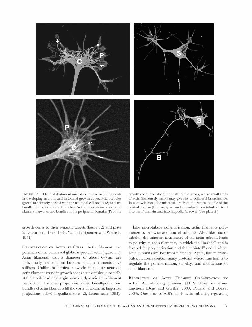
Figure 1.2 The distribution of microtubules and actin fi laments
in developing neurons and in axonal growth cones. Microtubules
(green) are densely packed with the neuronal cell bodies (S) and are
bundled in the axons and branches. Actin fi laments are arrayed in
fi lament networks and bundles in the peripheral domains (P) of the
growth cones and along the shafts of the axons, where small areas
of actin fi lament dynamics may give rise to collateral branches (B).
In a growth cone, the microtubules from the central bundle of the
central domain (C) splay apart, and individual microtubules extend
into the P domain and into fi lopodia (arrows). (See plate 2.)
8 fundamentals of developmental neurobiology
their availability for polymerization; other ABPs cross-link
actin fi laments into meshworks and bundles. ABPs that bind
the barbed and pointed ends of actin fi laments regulate the
addition and loss of actin subunits to fi laments. Several
ABPs bind actin fi laments and sever them, promoting the
remodeling of actin fi lament arrays. In growth cones, actin
fi lament barbed ends face the leading cell margin, where the
addition of actin subunits is promoted by several ABPs.
Myosins are motor molecules that bind and move cargoes
along actin fi laments. There are more than 10 myosins,
which share common features of their motor activity, but
which differ in the direction that they move cargoes along
fi laments and in cargoes that are moved (Brown and
Bridgman, 2004). Myosins in growth cones interact with
actin fi laments and generate forces to move actin fi laments,
vesicles, or other cargoes and to exert tensions on cytoskeletal
components and associated structures (Rochlin et al., 1995).
Myosin II in growth cones is particularly important in
generating forces to move components and reshape
developing axons and dendrites. In summary, ABPs are
critical to regulating the behaviors of growth cones of
developing axons and dendrites.
Regulation of Microtubule and Actin Organization and Dynamics by Cytoplasmic Signaling Pathways As noted
previously, the organization of microtubules and actin
fi laments is regulated by MAPs and ABPs. The dynamic
changes in cytoskeletal organization that drive neuronal
morphogenesis refl ect the activities of MAPs and ABPs.
Certainly, levels of these proteins are regulated by gene
transcription and protein synthesis, but in an immediate
fashion, MAPs and ABPs are regulated by intracellular
signaling and cytoplasmic second-messenger pathways.
Cytoskeletal organization can be rapidly changed by
fl uctuations in levels of small molecules such as Ca++ ions,
cAMP, cGMP, and phosphoinositides that bind MAPs and
Figure 1.3 The interwoven network of signaling molecules that
link guidance receptors with cytoskeletal dynamics underlying
growth cone motility. Membrane receptors for extracellular guid-
ance cues may function either alone or in a complex to activate
cytoplasmic adaptors and mediators. The Rho family of GTPases
may be pivotal links between guidance signals and actin-associated
proteins, which are responsible for regulating the assembly and
disassembly of actin fi laments. Similar types of molecules are rep-
resented by symbols of similar color and shape. Lines depict activa-
tion pathways that have been demonstrated experimentally in
different systems. (From Song and Poo, 2001.)
letourneau: formation of axons and dendrites by developing neurons 9
ABPs and regulate them allosterically (Dent and Gertler,
2003; Song and Poo, 2001; fi gure 1.3). The addition to and
removal of phosphate groups from MAPs and ABPs by
protein kinases and phosphates also rapidly regulate their
activities. These molecules and pathways are, in turn, regu-
lated by events at the plasma membrane, where adhesive
proteins, growth factors, and other ligands bind membrane
receptor proteins to trigger events that locally and tempo-
rally modulate the levels and activities of these regulatory
molecules. Thus cytoplasmic signaling activities that cascade
from ligand-receptor interactions at the plasma membrane
rapidly and locally regulate cytoskeletal organization during
neuronal morphogenesis (Dent and Gertler, 2003; Gallo and
Letourneau, 2004).
The Rho family of small guanosine triphosphatase
(GTPase) proteins, in particular RhoA, Rac1, and Cdc42,
are important regulatory proteins that relay signaling from
the cell surface intracellularly to the cytoskeleton (Jaffe and
Hall, 2005; fi gure 1.3). Rho GTPases bind to and regulate
MAPs and ABPs or their upstream regulators, such as
protein kinases and phosphatases. A critical feature of
GTPases is that their activity is rapidly switched on or off,
depending on whether they are bound to the nucleotides
GTP (on) or guanosine diphosphate (GDP) (off). A rich
variety of guanine nucleotide exchange factor proteins
(GEFs) selectively activate GTPases by exchanging GDP
for GTP; GTPase-activating proteins (GAPs) stimulate
hydrolysis of GTP to inactive GTPases; and GDP dissocia-
tion inhibitors (GDIs) inhibit activation of GTPases by GEFs.
These GEFs, GAPs, and GDIs are regulated by cell surface
ligand-receptor interactions. Thus by regulating GTPases
these membrane events regulate cytoskeletal proteins.
Activation of RhoA, Rac1, or Cdc42 has distinct effects
on actin fi lament organization (Jaffe and Hall, 2005). Rac1-
GTP activates several ABPs to stimulate actin polymeriza-
tion and formation of lamellipodia, while Cdc42-GTP also
stimulates actin polymerization and formation of fi lopodia.
RhoA-GTP activates the kinase ROCK, which phosphory-
lates several substrates to suppress actin polymerization and
activates the motor protein myosin II, increasing mechanical
tensions and rearrangements of actin fi laments. If RhoA
levels are highly elevated, strong contractile forces in the
growth cone can cause collapse of microtubule arrays and
signifi cant neurite retraction. All three Rho GTPases are
present in the growth cone and contribute to growth cone
motility. Microtubule organization and polymerization are
also regulated by Rho GTPases, although the mechanisms
are less well understood than for actin fi laments.
Microtubule-Actin Interactions Are Important Two particular interactions of actin fi laments, which we
will describe, are particularly important in neurite elonga-
tion and growth cone migration. As mentioned earlier,
microtubules maintain the shapes of axons and dendrites
and resist compressive forces that would collapse or withdraw
these processes. Proteins that mediate interactions between
microtubule plus ends and actin fi laments are particularly
signifi cant, because these proteins may be important in the
initiation of neurites from a spherical perikaryon or in
directing the advance of a growth cone (Rodriquez et al.,
2003). These microtubule-actin interactions link the
microtubule functions of structural support and organelle
transport to the dynamic cortical actin fi laments and
associated membrane receptors that detect extrinsic signals
and regulate the cytoskeletal activities that shape the
developing neuron (fi gure 1.4).
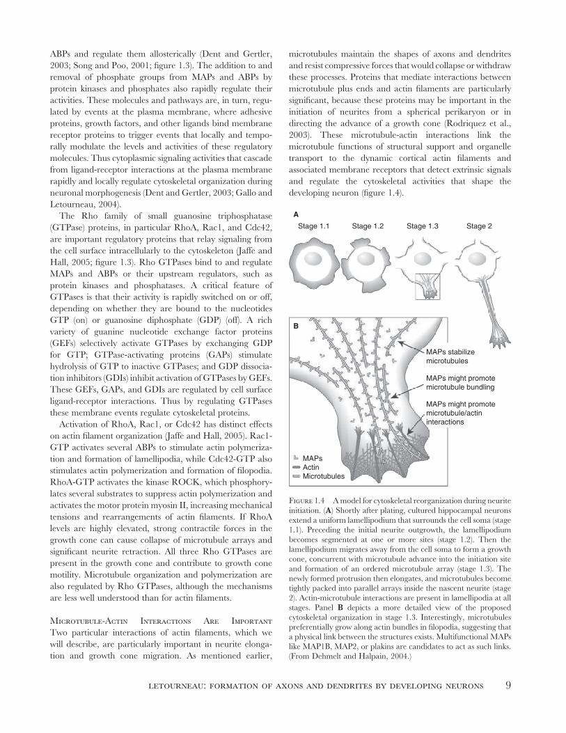
Stage 1.1 Stage 1.2 Stage 1.3 Stage 2
MAPs stabilizemicrotubules
B
A
MAPs might promotemicrotubule bundling
MAPs might promotemicrotubule/actininteractions
MAPsActinMicrotubules
Figure 1.4 A model for cytoskeletal reorganization during neurite
initiation. (A) Shortly after plating, cultured hippocampal neurons
extend a uniform lamellipodium that surrounds the cell soma (stage
1.1). Preceding the initial neurite outgrowth, the lamellipodium
becomes segmented at one or more sites (stage 1.2). Then the
lamellipodium migrates away from the cell soma to form a growth
cone, concurrent with microtubule advance into the initiation site
and formation of an ordered microtubule array (stage 1.3). The
newly formed protrusion then elongates, and microtubules become
tightly packed into parallel arrays inside the nascent neurite (stage
2). Actin-microtubule interactions are present in lamellipodia at all
stages. Panel B depicts a more detailed view of the proposed
cytoskeletal organization in stage 1.3. Interestingly, microtubules
preferentially grow along actin bundles in fi lopodia, suggesting that
a physical link between the structures exists. Multifunctional MAPs
like MAP1B, MAP2, or plakins are candidates to act as such links.
(From Dehmelt and Halpain, 2004.)

10 fundamentals of developmental neurobiology
Actin Filaments and Adhesive Contacts In addition
to interactions with microtubules, another key function of
actin fi laments involves the adhesive interactions of cells that
are mediated by membrane receptor proteins that form
noncovalent bonds between cells or between cells and
extracellular matrices (ECM). The major adhesion receptors
are the cadherins and the adhesion proteins of the
immunoglobulin-like superfamily, which mediate cell-cell
adhesions, and the integrin proteins, which mediate cell
adhesion to ECM. As cell-cell contacts are initiated by
intercellular binding, receptors cluster within the plasma
membrane to form discrete adhesive contacts. By way of
transmembrane linkage these clustered adhesion receptors
create docking sites for signaling enzymes, kinases, GEFs,
GAPs, and a number of ABPs that link actin fi laments to
the adhesive sites and induce actin polymerization. Thus
adhesive sites are loci from which regulatory signals emanate
and where actin fi lament organization and anchorage are
regulated (Zamir and Geiger, 2001).
A mechanism for neurite initiation and growth
In this section, neuritogenesis, neurite elongation, and
growth cone migration by neurons will be described, empha-
sizing the dynamic cytoskeleton of actin fi laments and
microtubules. When developing neurons are placed in
culture, the neurons settle on the substrate, and extend and
withdraw cylindrical fi lopodia and fl attened lamellipodia,
like waves lapping on a beach (see fi gure 1.4). This motility
is driven by actin fi lament polymerization, which pushes the
cell margin outward, while simultaneously myosin II, located
behind the cell margin, pulls newly formed fi laments back-
ward in a retrograde fl ow. The rearward transported fi la-
ments are severed and depolymerized, and if the protrusion
and retrograde fl ow are equal, these activities produce no
net change. Initially, microtubules remain in a loose network
around the nucleus, and any microtubules that enter the
protrusions are swept back with the retrograde fl ow of actin.
However, eventually a fi lopodium or lamellipodium thickens
and moves away from the cell body, tethered by a cylindrical
nascent neurite. The critical step that distinguishes neurite
formation from the initial protrusive activity occurs when
microtubules and associated organelles enter and remain
within a fi lopodial or lamellipodial protrusion and the pro-
trusive motility moves forward ahead of the microtubules
and organelles (Da Silva and Dotti, 2002; fi gure 1.4). Several
activities may prompt neurite initiation. An increased expres-
sion of MAPs, such as MAP2, tau, and MAP1B, may
stabilize microtubules, enhancing their resistance to the
myosin-based retrograde forces pulling actin back from the
leading margin (Dehmelt and Halpain, 2004). At sites where
protrusions make fi rm adhesive contacts with the substrate,
actin fi laments become anchored to the adhesive apparatus,
and retrograde fl ow stops, creating space into which micro-
tubules can advance. In addition, cytoplasmic signals gener-
ated at the adhesive sites may promote microtubule transport
and polymerization. Finally, actin fi laments linked to adhe-
sive sites can interact with myosin II motors and pull micro-
tubules and organelles toward the adhesive sites in opposition
to the retrograde fl ow of untethered actin fi laments (Suter
and Forscher, 2000). The signifi cance of these outwardly
directed forces in neurite initiation is illustrated by fi ndings
that neurites can be pulled out from a neuron by attaching
an adhesive bead to a neuronal surface and then pulling the
bead and attached elongating neurite away from the nerve
cell body (Fass and Odde, 2003).
Organization of Growth Cones and Growth Cone Migration A typical neurite has a central bundle
of microtubules with associated organelles and a motile
terminal expansion, the growth cone (Gordon-Weeks, 2000;
fi gures 1.2, 1.5). At the growth cone’s leading margin, called
the P-domain (peripheral), vigorous actin polymerization
pushes the cell margin forward, balanced by the myosin-
powered rearward sliding of untethered actin fi laments.
Only when the leading edge forms transient adhesive
contacts that link to actin fi laments does the retrograde
fl ow attenuate. At the base of a growth cone, microtubule-
based motor proteins move microtubules and organelles
from the neurite into the central growth cone, comprising
the C-domain (central). From the C-domain individual
microtubules extend into the P-domain, sliding forward
powered by molecular motors and elongating by adding
subunits to microtubule plus ends. Retrograde fl ow pulls
most of these microtubules back into the C-domain
(Schaefer, Kabir, and Forscher, 2002). Importantly, some
microtubules advance into fi lopodia or lamellipodia,
stabilized at adhesive sites (fi gure 1.2; Letourneau, 1979;
Suter and Forscher, 2000). If these microtubules persist and
are followed by other microtubules and organelles, the C-
domain advances and the neurite extends. To complete
the cycle of growth cone movement, actin fi laments and
membrane components that are not stabilized by adhesions
or associations with microtubules are recycled at the back
of the growth cone by the myosin II–powered retrograde
fl ow and by disassembly of actin fi laments and endocytosis
of plasma membrane.
Thus neurite elongation proceeds by three activities (Dent
and Gertler, 2003; fi gure 1.5): (1) the advance, expansion,
and adhesion of the leading margin of the growth cone,
driven by actin polymerization; (2) the advance of microtu-
bules via polymerization, transport, and linkage to actin and
adhesive sites (Letourneau, 1979); and (3) the advance of
organelles via microtubule-based transport. The coordina-
tion of actin-driven membrane expansion, formation of
adhesive contacts, and myosin II–powered exertion of
letourneau: formation of axons and dendrites by developing neurons 11
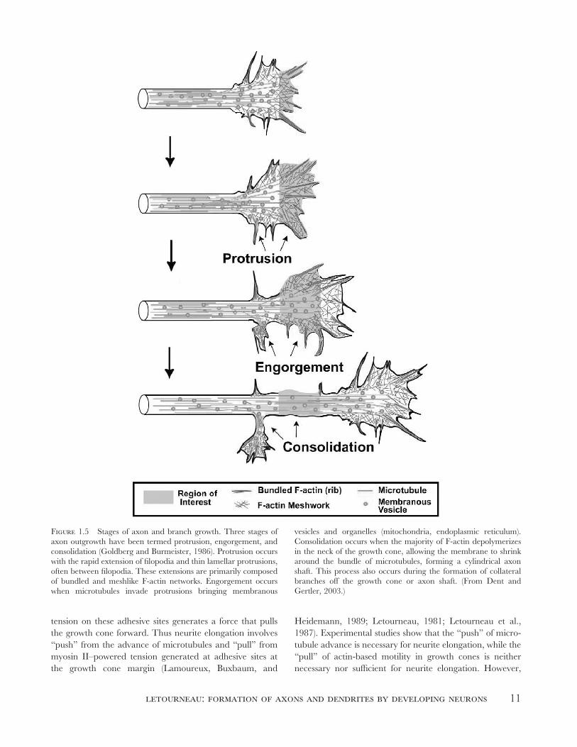
Figure 1.5 Stages of axon and branch growth. Three stages of
axon outgrowth have been termed protrusion, engorgement, and
consolidation (Goldberg and Burmeister, 1986). Protrusion occurs
with the rapid extension of fi lopodia and thin lamellar protrusions,
often between fi lopodia. These extensions are primarily composed
of bundled and meshlike F-actin networks. Engorgement occurs
when microtubules invade protrusions bringing membranous
vesicles and organelles (mitochondria, endoplasmic reticulum).
Consolidation occurs when the majority of F-actin depolymerizes
in the neck of the growth cone, allowing the membrane to shrink
around the bundle of microtubules, forming a cylindrical axon
shaft. This process also occurs during the formation of collateral
branches off the growth cone or axon shaft. (From Dent and
Gertler, 2003.)
tension on these adhesive sites generates a force that pulls
the growth cone forward. Thus neurite elongation involves
“push” from the advance of microtubules and “pull” from
myosin II–powered tension generated at adhesive sites at
the growth cone margin (Lamoureux, Buxbaum, and
Heidemann, 1989; Letourneau, 1981; Letourneau et al.,
1987). Experimental studies show that the “push” of micro-
tubule advance is necessary for neurite elongation, while the
“pull” of actin-based motility in growth cones is neither
necessary nor suffi cient for neurite elongation. However,
12 fundamentals of developmental neurobiology
growth cone “pull” accelerates neurite elongation and, as
described later, is necessary for growth cone navigation.
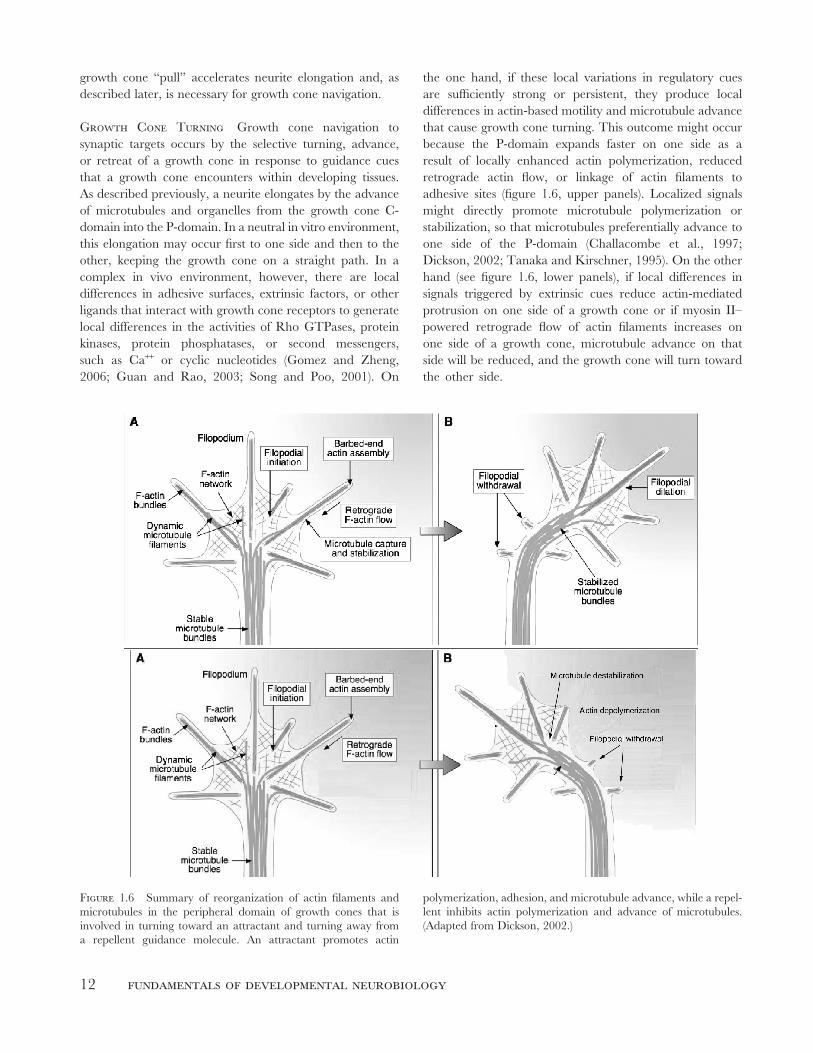
Growth Cone Turning Growth cone navigation to
synaptic targets occurs by the selective turning, advance,
or retreat of a growth cone in response to guidance cues
that a growth cone encounters within developing tissues.
As described previously, a neurite elongates by the advance
of microtubules and organelles from the growth cone C-
domain into the P-domain. In a neutral in vitro environment,
this elongation may occur fi rst to one side and then to the
other, keeping the growth cone on a straight path. In a
complex in vivo environment, however, there are local
differences in adhesive surfaces, extrinsic factors, or other
ligands that interact with growth cone receptors to generate
local differences in the activities of Rho GTPases, protein
kinases, protein phosphatases, or second messengers,
such as Ca++ or cyclic nucleotides (Gomez and Zheng,
2006; Guan and Rao, 2003; Song and Poo, 2001). On
the one hand, if these local variations in regulatory cues
are suffi ciently strong or persistent, they produce local
differences in actin-based motility and microtubule advance
that cause growth cone turning. This outcome might occur
because the P-domain expands faster on one side as a
result of locally enhanced actin polymerization, reduced
retrograde actin fl ow, or linkage of actin fi laments to
adhesive sites (fi gure 1.6, upper panels). Localized signals
might directly promote microtubule polymerization or
stabilization, so that microtubules preferentially advance to
one side of the P-domain (Challacombe et al., 1997;
Dickson, 2002; Tanaka and Kirschner, 1995). On the other
hand (see fi gure 1.6, lower panels), if local differences in
signals triggered by extrinsic cues reduce actin-mediated
protrusion on one side of a growth cone or if myosin II–
powered retrograde fl ow of actin fi laments increases on
one side of a growth cone, microtubule advance on that
side will be reduced, and the growth cone will turn toward
the other side.
Figure 1.6 Summary of reorganization of actin fi laments and
microtubules in the peripheral domain of growth cones that is
involved in turning toward an attractant and turning away from
a repellent guidance molecule. An attractant promotes actin
polymerization, adhesion, and microtubule advance, while a repel-
lent inhibits actin polymerization and advance of microtubules.
(Adapted from Dickson, 2002.)
letourneau: formation of axons and dendrites by developing neurons 13
Mechanisms of Branching Branches of neurites,
axons, or dendrites are formed in two ways: by a growth
cone splitting or by a new branch sprouting from the neurite
shaft behind a growth cone. In either case, the acquisition
of stable microtubules is key to forming a branch (fi gures 1.2,
1.5). In a growth cone, part of the P-domain and associated
C-domain may separate from the whole and establish an
independent growth cone and a new branch of the parent
neurite. This result may occur when a growth cone “pulls”
in two directions (fi gure 1.2). Branch formation along a
neurite is initiated by localized protrusion of fi lopodia or
lamellipodia (fi gure 1.2; Gallo and Letourneau, 1998). This
mechanism is particularly prevalent in the branching
morphogenesis of dendrites. This localized actin-based
motility may occur until microtubules enter an actin-fi lled
nascent branch by transport or by polymerization of
microtubules from the main neurite (Gallo and Letourneau,
1999). Microtubule ends in the main neurite may become
linked to actin fi laments of the protrusion and be pulled into
the branch. The microtubule-severing protein katanin may
promote branch formation by severing microtubules in the
neurite shaft to create microtubule ends that can be moved
into a nascent branch (Baas and Buster, 2004). Once stable
microtubules are established, the advance of microtubules
and organelles into the branch sustains its growth.
The Differentiation of Axons and Dendrites; Polarization of Neuronal Form A hippocampal
neuron in vitro initially sprouts several similar neurites that
extend slowly. After 18–24 hours one neurite expands its
growth cone and elongates signifi cantly faster than the
others. This neurite becomes the axon, and it accumulates
proteins typical of axons, such as the MAPs tau and MAP1B,
and GAP43, a protein involved in actin motility (Mandell
and Banker, 1996). Several molecules and pathways may be
critical to axonal specifi cation, including PI3 kinase, the Par
complex, and small Rho GTPases (Arimura and Kaibuchi,
2005; Wiggin et al., 2005). These molecules concentrate at
the tips of newly specifi ed axons and are implicated in
regulating key activities, such as actin fi lament organization,
microtubule polymerization or stability, and transport and
addition of plasma membrane components. It is unclear
whether axonal specifi cation always begins with the same
upstream event, such as concentration of PI3 kinase activity
in a neurite tip, or whether concentration of any of the
previously mentioned molecules or signals is suffi cient to
specify axonal character. In vitro manipulations, such as
focally pulling on a neurite or presenting adhesive proteins
to one neurite will induce a neurite to become the axon.
Thus extrinsic signals can infl uence the intrinsic mechanism
of axonal specifi cation, perhaps by locally activating PI3
kinase or other components of the mechanism. After one
neurite becomes the axon, the other neurites become
dendrites. Less is known about the mechanisms of dendrite
specifi cation. Acquisition of microtubules with mixed
polarities may be important, as well as localization of
cytoskeletal, membrane, and signaling components that
regulate dendritic characteristics.
Regulation of neuronal morphogenesis in vivo
The previous section focused on the intrinsic mechanisms of
neurite initiation and elongation, growth cone migration
and turning, neurite branching, and the specifi cation of
axons. This section will discuss the roles of extrinsic mole-
cules and signaling events in regulating neuronal mor-
phogenesis in the developing human brain. The neutral
environment of a tissue culture dish facilitates understanding
these intrinsic mechanisms. However, the in vivo environ-
ment is never neutral, and spatial and temporal patterns of
distribution of axonal guidance cues in the environment of
the developing brain shape these intrinsic morphogenetic
mechanisms to generate neural circuits (Tessier-Lavigne and
Goodman, 1996).
Neuronal Migration Immature neurons arise from
proliferation of neural precursors in the ventricular zone of
the developing brain. From their birth immature neurons
become polarized by asymmetry in local cues, including the
adhesive protein laminin in the underlying extracellular
matrix (ECM) of the ventricular layer, as well as growth
factors, morphogens, and guidance molecules, such as sonic
hedgehog and netrin, produced by the surrounding
neuroepithelial cells. These newly born neurons migrate out
of the ventricular zone of the telencephalon to establish the
cortical plate in a wave of migration between 6 and 18
gestational weeks (Ramakers, 2005). Migrating neurons
retain their initial polarization and encounter additional
cues as they migrate upward. Neural migration stops at the
outer marginal zone, where reelin, produced by Cajal-
Retzius cells of the marginal zone, triggers neurons to cease
expressing integrin adhesion receptors. Younger neurons
migrate past older neurons to reach the marginal zone, so
the upper layer II contains the youngest neurons, while the
oldest neurons inhabit the lowest layer VI.
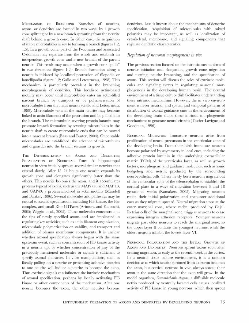
Neuronal Polarization and the Initial Growth of Axons and Dendrites Neurons sprout axons soon after
ceasing migration, as early as the seventh week in the cortex.
In a neutral tissue culture environment, it is a random
decision as to which neurite sprouted from a neuron becomes
the axon, but cortical neurons in vivo always sprout their
axon in the same direction that the axon will grow. In the
model organism, Caenorhabditis elegans, a diffusible molecule
netrin produced by ventrally located cells causes localized
activity of PI3 kinase in young neurons, which then sprout
14 fundamentals of developmental neurobiology
their axon toward the netrin source (Adler et al., 2006). PI3
kinase is involved in axonal specifi cation of mammalian
neurons, and thus localization of PI3 kinase in response to
a local cue may both specify axonal identity and regulate
actin motility to control the direction of axonal initiation.
Other factors are implicated in regulating the initial direc-
tion of cortical axonal growth. Immature cortical pyramidal
neurons fi rst extend an axon toward the ventricle, followed
by an apical dendrite, which grows toward the pial surface.
Unexpectedly, these opposite directions of axonal versus
dendritic growth are regulated by the same extracellular
molecule, semaphorin 3A (Sema3A), produced by cells near
the pial surface and released to create an extracellular gradi-
ent (Whitford et al., 2002). Axons are repelled by Sema3A,
while the subsequently formed apical dendrites of these
neurons are attracted by Sema3A. The difference in direc-
tions of these processes lies not in a local difference in expres-
sion of membrane receptors for Sema3A, but rather in a
local difference in distribution of signaling proteins that
modulate levels of the cyclic nucleotide cGMP. The combi-
nation of Sema3A signaling and high cGMP levels in the
apical dendrite promotes actin polymerization and dendritic
growth, while Sema3A signaling in the axon combined with
low cGMP activates the GTPase RhoA, which depresses
actin dynamics and activated myosin II contractility, so the
axonal growth cone migrates away from the Sema3A source.
Thus the opposite responses of axons and dendrites to
Sema3A are due to an asymmetric distribution of cytoplas-
mic signaling components in dendrites versus axons.
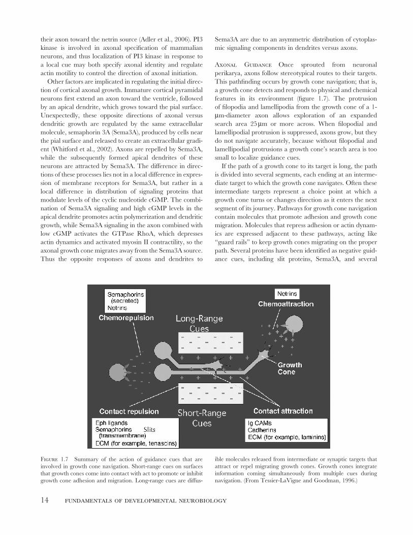
Axonal Guidance Once sprouted from neuronal
perikarya, axons follow stereotypical routes to their targets.
This pathfi nding occurs by growth cone navigation; that is,
a growth cone detects and responds to physical and chemical
features in its environment (fi gure 1.7). The protrusion
of fi lopodia and lamellipodia from the growth cone of a 1-
μm-diameter axon allows exploration of an expanded
search area 25 μm or more across. When fi lopodial and
lamellipodial protrusion is suppressed, axons grow, but they
do not navigate accurately, because without fi lopodial and
lamellipodial protrusions a growth cone’s search area is too
small to localize guidance cues.
If the path of a growth cone to its target is long, the path
is divided into several segments, each ending at an interme-
diate target to which the growth cone navigates. Often these
intermediate targets represent a choice point at which a
growth cone turns or changes direction as it enters the next
segment of its journey. Pathways for growth cone navigation
contain molecules that promote adhesion and growth cone
migration. Molecules that repress adhesion or actin dynam-
ics are expressed adjacent to these pathways, acting like
“guard rails” to keep growth cones migrating on the proper
path. Several proteins have been identifi ed as negative guid-
ance cues, including slit proteins, Sema3A, and several
Figure 1.7 Summary of the action of guidance cues that are
involved in growth cone navigation. Short-range cues on surfaces
that growth cones come into contact with act to promote or inhibit
growth cone adhesion and migration. Long-range cues are diffus-
ible molecules released from intermediate or synaptic targets that
attract or repel migrating growth cones. Growth cones integrate
information coming simultaneously from multiple cues during
navigation. (From Tessier-LaVigne and Goodman, 1996.)
letourneau: formation of axons and dendrites by developing neurons 15
ephrinA’s. Each negative cue is detected by a different spe-
cifi c receptor with specifi c signaling mechanisms, although
common features of these mechanisms include disruption of
growth cone adhesions, suppression of actin polymerization,
and activation of RhoA to stimulate myosin II–mediated
contraction, leading to growth cone collapse and sometimes
retraction of entire axonal branches or segments (Guan and
Rao, 2003). Some molecules simply mark a path as positive
or negative without providing directional information, while
other molecules are soluble, are released by navigation
targets, and are distributed in gradients that provide direc-
tional information to growth cones. At any instant a growth
cone is detecting several guidance molecules, so growth cone
migration depends on integrating the intracellular signals
simultaneously triggered from multiple receptors. The fol-
lowing section describes specifi c features of growth cone
guidance in the developing CNS. Most of the molecular
information about growth cone guidance comes from studies
of model vertebrate systems, but the timing of the events in
human brain development is included (Ramakers, 2005).
Growth cone navigation along major pathways during cerebral cortical development
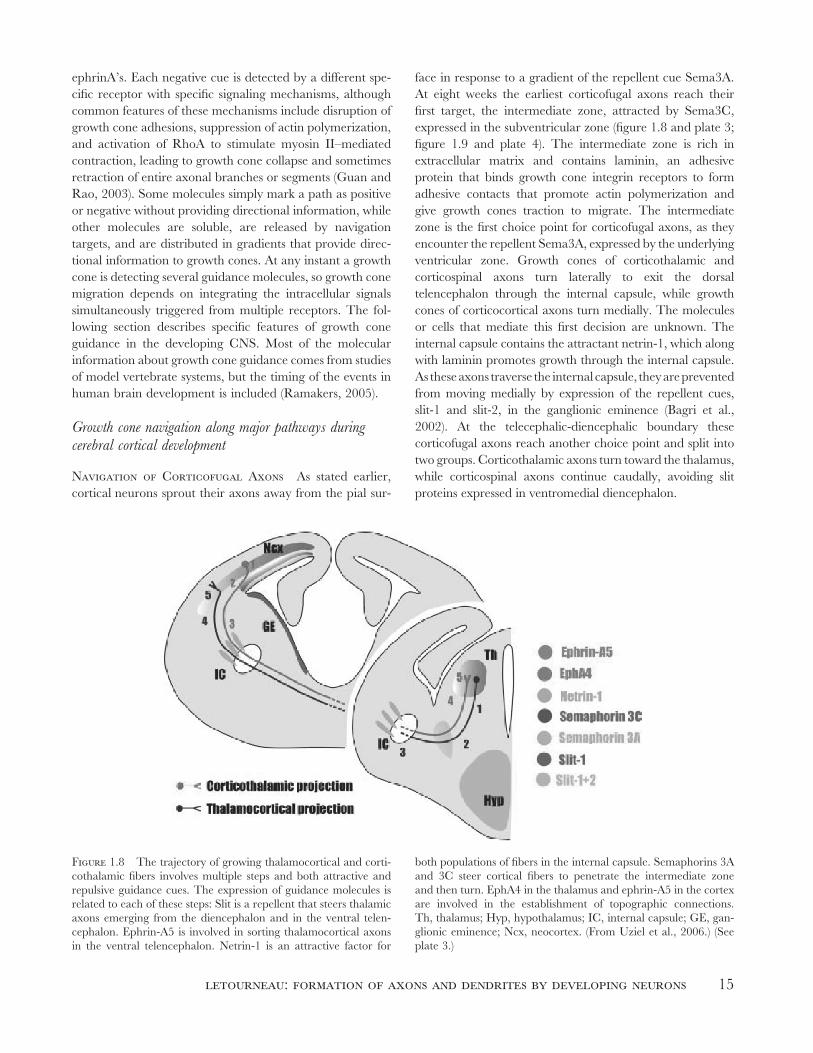
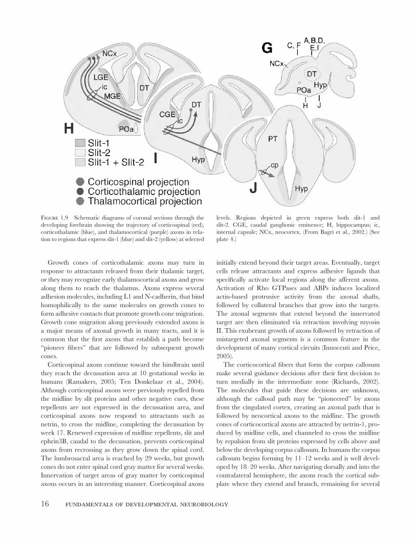
Navigation of Corticofugal Axons As stated earlier,
cortical neurons sprout their axons away from the pial sur-
face in response to a gradient of the repellent cue Sema3A.
At eight weeks the earliest corticofugal axons reach their
fi rst target, the intermediate zone, attracted by Sema3C,
expressed in the subventricular zone (fi gure 1.8 and plate 3;
fi gure 1.9 and plate 4). The intermediate zone is rich in
extracellular matrix and contains laminin, an adhesive
protein that binds growth cone integrin receptors to form
adhesive contacts that promote actin polymerization and
give growth cones traction to migrate. The intermediate
zone is the fi rst choice point for corticofugal axons, as they
encounter the repellent Sema3A, expressed by the underlying
ventricular zone. Growth cones of corticothalamic and
corticospinal axons turn laterally to exit the dorsal
telencephalon through the internal capsule, while growth
cones of corticocortical axons turn medially. The molecules
or cells that mediate this fi rst decision are unknown. The
internal capsule contains the attractant netrin-1, which along
with laminin promotes growth through the internal capsule.
As these axons traverse the internal capsule, they are prevented
from moving medially by expression of the repellent cues,
slit-1 and slit-2, in the ganglionic eminence (Bagri et al.,
2002). At the telecephalic-diencephalic boundary these
corticofugal axons reach another choice point and split into
two groups. Corticothalamic axons turn toward the thalamus,
while corticospinal axons continue caudally, avoiding slit
proteins expressed in ventromedial diencephalon.
Figure 1.8 The trajectory of growing thalamocortical and corti-
cothalamic fi bers involves multiple steps and both attractive and
repulsive guidance cues. The expression of guidance molecules is
related to each of these steps: Slit is a repellent that steers thalamic
axons emerging from the diencephalon and in the ventral telen-
cephalon. Ephrin-A5 is involved in sorting thalamocortical axons
in the ventral telencephalon. Netrin-1 is an attractive factor for
both populations of fi bers in the internal capsule. Semaphorins 3A
and 3C steer cortical fi bers to penetrate the intermediate zone
and then turn. EphA4 in the thalamus and ephrin-A5 in the cortex
are involved in the establishment of topographic connections.
Th, thalamus; Hyp, hypothalamus; IC, internal capsule; GE, gan-
glionic eminence; Ncx, neocortex. (From Uziel et al., 2006.) (See
plate 3.)
16 fundamentals of developmental neurobiology
Growth cones of corticothalamic axons may turn in
response to attractants released from their thalamic target,
or they may recognize early thalamocortical axons and grow
along them to reach the thalamus. Axons express several
adhesion molecules, including L1 and N-cadherin, that bind
homophilically to the same molecules on growth cones to
form adhesive contacts that promote growth cone migration.
Growth cone migration along previously extended axons is
a major means of axonal growth in many tracts, and it is
common that the fi rst axons that establish a path become
“pioneer fi bers” that are followed by subsequent growth
cones.
Corticospinal axons continue toward the hindbrain until
they reach the decussation area at 10 gestational weeks in
humans (Ramakers, 2005; Ten Donkelaar et al., 2004).
Although corticospinal axons were previously repelled from
the midline by slit proteins and other negative cues, these
repellents are not expressed in the decussation area, and
corticospinal axons now respond to attractants such as
netrin, to cross the midline, completing the decussation by
week 17. Renewed expression of midline repellents, slit and
ephrin3B, caudal to the decussation, prevents corticospinal
axons from recrossing as they grow down the spinal cord.
The lumbrosacral area is reached by 29 weeks, but growth
cones do not enter spinal cord gray matter for several weeks.
Innervation of target areas of gray matter by corticospinal
axons occurs in an interesting manner. Corticospinal axons
initially extend beyond their target areas. Eventually, target
cells release attractants and express adhesive ligands that
specifi cally activate local regions along the afferent axons.
Activation of Rho GTPases and ABPs induces localized
actin-based protrusive activity from the axonal shafts,
followed by collateral branches that grow into the targets.
The axonal segments that extend beyond the innervated
target are then eliminated via retraction involving myosin
II. This exuberant growth of axons followed by retraction of
mistargeted axonal segments is a common feature in the
development of many cortical circuits (Innocenti and Price,
2005).
The corticocortical fi bers that form the corpus callosum
make several guidance decisions after their fi rst decision to
turn medially in the intermediate zone (Richards, 2002).
The molecules that guide these decisions are unknown,
although the callosal path may be “pioneered” by axons
from the cingulated cortex, creating an axonal path that is
followed by neocortical axons to the midline. The growth
cones of corticocortical axons are attracted by netrin-1, pro-
duced by midline cells, and channeled to cross the midline
by repulsion from slit proteins expressed by cells above and
below the developing corpus callosum. In humans the corpus
callosum begins forming by 11–12 weeks and is well devel-
oped by 18–20 weeks. After navigating dorsally and into the
contralateral hemisphere, the axons reach the cortical sub-
plate where they extend and branch, remaining for several
Figure 1.9 Schematic diagrams of coronal sections through the
developing forebrain showing the trajectory of corticospinal (red),
corticothalamic (blue), and thalamocortical (purple) axons in rela-
tion to regions that express slit-1 (blue) and slit-2 (yellow) at selected
levels. Regions depicted in green express both slit-1 and
slit-2. CGE, caudal ganglionic eminence; H, hippocampus; ic,
internal capsule; NCx, neocortex. (From Bagri et al., 2002.) (See
plate 4.)
letourneau: formation of axons and dendrites by developing neurons 17
weeks before sprouting collateral branches at 28 weeks into
their appropriate fi nal target regions of the cortex.
Navigation of Thalamocortical Growth Cones
Thalamocortical afferent axons begin their navigation by
growing ventrally until they are stopped by repulsion from
slit proteins expressed by the underlying hypothalamus
(Lopez-Bendito and Molnar, 2003; Uziel et al., 2006; fi gure
1.8). Then the growth cones turn laterally, being attracted
by netrin-1 expressed by cells in the internal capsule. The
growth cones turn dorsally and migrate toward the cortex
within the internal capsule, keeping lateral in response to slit
proteins expressed by the ganglionic eminence (Bagri et al.,
2002). Within the internal capsule, thalamocortical axons
meet corticofugal fi bers, which they follow toward their
cortical targets. Thalamocortical axons penetrate the cortical
subplate between 9 and 18 weeks in developing humans. By
24 weeks they fi ll the upper subplate and extend branches
exploring for their correct cortical targets. Thalamocortical
axons fi nally enter the cortex between 26 and 28 weeks,
prior to the entry of callosal axons.
The preceding paragraphs have described how axons
navigate to their targets by detecting and responding to
guidance molecules that regulate growth cone motility. It
may seem that the relatively limited numbers of guidance
molecules, laminins, ephrins, semaphorins, netrins, slits, and
immunoglobulin-like adhesion molecules are too few to
account for the complexity of neural circuitry (Yu and
Bargmann, 2001). However, this diversity of axonal path-
ways arises from cell-type–specifi c differences in expression
of receptors for guidance cues, in downstream cytoplasmic
signaling activated by guidance cues, and temporal and
spatial differences in the expression of guidance cues and
their receptors by developing tissues and neuronal popula-
tions. An interesting recent fi nding is that growth cone
responses to guidance cues may depend on bursts of local
protein synthesis of receptors or signaling components within
a growth cone. For example, Sema3A rapidly stimulates
synthesis of the GTPase RhoA from mRNA within growth
cones (Wu et al., 2005). RhoA activity is necessary for
Sema3A induction of growth cone collapse. Some growth
cones cross the ventral spinal cord and only then synthesize
and express EphA receptors that mediate a repulsive response
to midline ephrins, preventing recrossing the midline (Brittis,
Lu, and Flanagan, 2002). Much remains to be learned about
how growth cones detect guidance cues and integrate
complex signals to navigate to their intermediate and fi nal
targets.
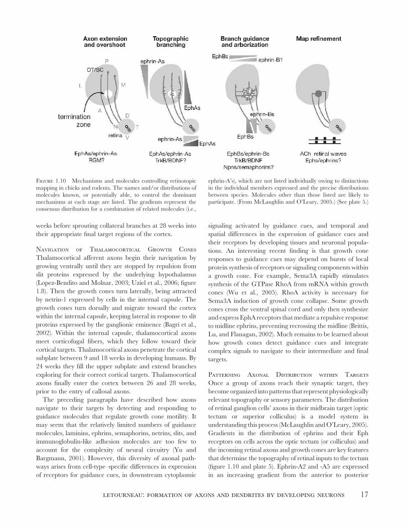
Patterning Axonal Distribution within Targets Once a group of axons reach their synaptic target, they
become organized into patterns that represent physiologically
relevant topography or sensory parameters. The distribution
of retinal ganglion cells’ axons in their midbrain target (optic
tectum or superior colliculus) is a model system in
understanding this process (McLaughlin and O’Leary, 2005).
Gradients in the distribution of ephrins and their Eph
receptors on cells across the optic tectum (or colliculus) and
the incoming retinal axons and growth cones are key features
that determine the topography of retinal inputs to the tectum
(fi gure 1.10 and plate 5). Ephrin-A2 and -A5 are expressed
in an increasing gradient from the anterior to posterior
Figure 1.10 Mechanisms and molecules controlling retinotopic
mapping in chicks and rodents. The names and/or distributions of
molecules known, or potentially able, to control the dominant
mechanisms at each stage are listed. The gradients represent the
consensus distribution for a combination of related molecules (i.e.,
ephrin-A’s), which are not listed individually owing to distinctions
in the individual members expressed and the precise distributions
between species. Molecules other than those listed are likely to
participate. (From McLaughlin and O’Leary, 2005.) (See plate 5.)
18 fundamentals of developmental neurobiology
tectum. EphA receptors bind ephrin-A ligands and trigger
decreased Rac1 and Cdc42 activities and increased RhoA
activity, stimulating growth cone repulsion. Growth cones of
temporal retinal axons express high levels of EphA receptors,
so they stop and innervate the anterior tectum, while nasal
retinal growth cones, expressing lower levels of EphA
receptors, extend to the posterior tectum, because they are
less repelled by the ephrin-A gradient. The distances that
retinal growth cones migrate along the anterior-posterior
tectal gradient of increasing ephrin-A expression are
determined by the relative levels of EphA expression by
growth cones. Retinal mapping along the medial-lateral
tectal axis involves gradients in the distribution of ephrin-Bs
and their EphB receptors among retinal axons and tectal
cells. The identifi cation of signaling activity from the
cytoplasmic domain of ephrin-B ligands indicates that both
ephrins and EphB receptors can activate cytoplasmic
signaling to regulate axonal targeting along the medial-
lateral tectal axis.
This mechanism for topographic mapping of connections
by gradients of cell surface ligands and receptors was pro-
posed by Roger Sperry (1963) as the chemoaffi nity hypoth-
esis. The discovery of gradients in expression of ephrins-A2
and -A5 confi rmed Sperry’s hypothesis. It has become clear
that the initial distributions of axons, as regulated by these
gradients, is not fi nal, and that subsequent remodeling of
axons due to further cellular interactions and physiological
activities is necessary to create more precise neural circuits.
The patterning of inputs to a target depends on activities
distributed along the afferent axons, in addition to the
growth cones. Local signaling by guidance cues or other
physiological events along axonal shafts can rapidly regulate
activities of RhoA or Rac1 and Cdc42 to regulate actin
dynamics and myosin II activity to induce retraction or addi-
tion of collateral or terminal branches along developing
axonal shafts (Gallo and Letourneau, 1998).
The accessibility and simple anatomy of the retinotectal
projection have allowed much progress in understanding the
patterning of developing neural circuits. The discovery of
gradients in the distributions of ephrin-A and EphA recep-
tors in the neocortex and thalamus, respectively, indicates
that gradients of interacting ephrins and their receptors have
similar roles in regulating axonal guidance and patterning
of thalamocortical connections to their targets in the primary
sensory regions of the cerebral cortex (Uziel et al., 2006;
fi gure 1.8). Similar mechanisms may operate in patterning
the development of circuits in other regions (Flanagan,
2006).
In addition to the development of the correct distribution
of axons within a target, axons must recognize the target
neurons with which they make synapses. Several cell
surface and extracellular molecules are expressed in a
lamina-specifi c manner in the developing cortex, including
cadherins, Eph receptors, ephrin ligands, proteoglycans, and
neurotrophins (Lopez-Bendito and Molnar, 2003). These
molecular differences may provide cues for thalamocortical
and corticocortical axons to terminate in the correct layer.
Development of dendrites
The dendritic arborization of a neuron contains the synaptic
inputs to the neuron and is where synaptic inputs are
integrated before the initiation of action potentials. Thus
dendritic arbors are critical to the processing of neural infor-
mation for behavior and other neural activities. Like the
formation of axons, dendrite formation is intrinsic to the
neuronal phenotype. In fact, different neuronal types in a
neutral tissue culture environment will form dendritic arbors
that are reminiscent of their characteristic in vivo morpholo-
gies. As described earlier, the same basic mechanisms of
actin fi lament and microtubule dynamics operate to drive
the formation of dendrites, although dendrites are more
numerous, shorter, and more elaborately branched than
axons, due to expression of dendritic-specifi c cytoskeletal,
membrane, and signaling proteins.
Generally, a neuron initiates dendrites after it is actively
engaged in axonal elongation. This lag may be several days,
and may be due to both environmental factors and intrinsic
factors, such as changes in expression of specifi c cytoskeletal
proteins. The sites of dendrite initiation from a neuron may
be determined by previous cell interactions; for example, the
apical dendrites of cerebral cortical neurons are formed
from the leading process with which immature neurons
had migrated from the ventricular lining of the cortex. As
described previously, the apical dendrites of cortical neurons
are oriented by an attractive response to Sema3A, produced
at the pial surface. Other extrinsic proteins produced by
neighboring cells or afferent axons promote the formation
of dendrites, including osteogenic protein-1 (BMP7) and
neurotrophins BDNF and NT-3 (Whitford et al., 2002).
Thus intrinsic regulation of cytoskeletal and membrane
components combined with availability of extrinsic factors,
such as osteogenic protein-1 and neurotrophins, orchestrates
the initiation and elongation of branched dendritic arbors.
However, as described in the following paragraphs, the for-
mation of dendrites is a prolonged activity, and the fi nal
shaping of dendritic arbors depends heavily on afferent
inputs and interactions with axon terminals (Van Aelst and
Cline, 2004). Visualization of the morphogenesis of individ-
ual dendrites in developing brains of living frogs and zebra
fi sh has revealed rapidly changing addition and loss of small
branches and arbors as dendrites interact with afferent
axons. Filopodia transiently extend from dendritic shafts and
termini, and if contacts are made with axonal growth cones,
the dendritic fi lopodium may be stabilized, and nascent syn-
apses may form. However, many of these contacts and
letourneau: formation of axons and dendrites by developing neurons 19
synapses are brief, and the terminal axonal and dendritic
branches may be retracted. Synaptic activity is a factor in
dendritic morphogenesis, and activation of NMDA recep-
tors at nascent synapses may regulate Rho GTPases to
modulate actin fi lament dynamics that underlie the exten-
sion and retraction of dendritic fi lopodia (Van Aelst and
Cline, 2004). The roles of these synapses in regulating den-
dritic growth may also change as the synapses mature. Post-
synaptic activation at early synapses may stimulate formation
of more dendritic fi lopodia and elaboration of dendritic
branches, while signaling at more mature synapses may gen-
erate stop-growing signals to stabilize dendritic arbors. New
excitatory synapses contain NDMA receptors only, and
AMPA receptors are added later. Addition of AMPA recep-
tors to synapses may be required for retention of synapses
and stabilization of dendritic arborizations. The fi nal shaping
of axonal terminals is also dependent on interactions with
dendrites and postsynaptic contacts. Retrograde synaptic
interactions may signal growth cones to reduce their dynamic
activity, stop, and transform to a presynaptic ending. Motor
axons growing on muscle fi bers of mice that lack the Ach-
receptor-aggregating protein, agrin, or the agrin receptor
component, MUSK, extend abnormally long distances
across muscle surfaces, implicating MUSK and agrin in an
axonal “stop signal.” The neuromuscular junction contains
a laminin isoform, S-laminin, that inhibits axonal growth.
Nitric oxide, which is released by dendrites in response to
synaptic activity, may be a retrograde signal that stops
axonal growth in synaptic regions.
Dendritogenesis in the Prenatal and Postnatal Human Brain Neurons begin to form dendrites soon
after they initiate axon formation, although dendrites are
initially short and slow growing. Apical dendrites are present
on cortical pyramidal neurons by 12–13 weeks’ gestation.
However, once innervating axons arrive in the cortical
plate at 26–28 weeks, dendrite formation accelerates as a
result of synaptic contacts, electrical stimulation by axons,
and the release of neurotrophins and other factors from
axons. In humans, most dendritic growth occurs postnatally
in conjunction with synaptogenesis and the increased
physiological experience and activity of postnatal life.
Dendrite formation in the developing human brain has been
examined most thoroughly in the visual cortex and prefrontal
cortex (Ramakers, 2005). In the visual cortex most dendritic
branches develop prenatally, and postnatal growth involves
dendritic lengthening by terminal growth of branches as
synapses are added. Total dendrite length of pyramidal
neurons in the visual cortex increases rapidly in the fi rst few
postnatal months, increasing two- or threefold and reaching
the adult levels by 1–2 years. In the prefrontal cortex,
synaptogenesis and dendritic growth proceed more slowly
than in the visual cortex. During the fi rst postnatal year the
length of dendrites increases 5- to 10-fold by branching and
elongating, while after the fi rst year most growth occurs by
elongation of branches. By two years of age the total dendritic
length per pyramidal neuron is only half the adult level. Yet,
at age two the average dendrite length per neuron in the
prefrontal cortex is longer than dendritic length in the visual
cortex, consistent with the greater dendritic and synaptic
complexity in the more integrative cortical regions, compared
to unimodal primary cortical regions.
These measures of dendrite elaboration in the developing
human brain are mostly based on anatomical studies involv-
ing Golgi staining of fi xed neurons. These data are static and
fail to account for the dynamic activities of dendritic elonga-
tion, branching, and retraction that are revealed from real-
time visualization of dendrite growth and synaptogenesis in
living embryos, as mentioned earlier. Much remains to be
learned about how axonal and dendritic shapes are sculpted
over a period of years, as the result of interactions between
genetically defi ned mechanisms of neuronal growth and
a dynamic fl ux of intercellular molecular signaling, synapto-
genesis, and the unpredictable physiological activity of
postnatal experience.
Summary
Neural circuits arise by a morphogenetic process in which
axons and dendrites are formed according to intrinsic neu-
ronal mechanisms that respond to extrinsic regulatory inter-
actions with molecules, cells, and features of the developing
organism. The driving force for axonal and dendritic growth
is the advance of microtubules and associated organelles,
while the actin-based motility of growth cones at the ends of
elongating processes allows exploration of local tissue envi-
ronments for molecular guidance cues. Binding of guidance
cues to their receptors on growth cones triggers cytoplasmic
signaling that regulates actin fi lament organization, mechan-
ical forces, and microtubule advance to locally direct growth
cone migration, turning, and branching. Axonal growth
cones reach their synaptic targets by navigating to a series
of intermediate targets, guided by positive and negative
responses to surface-bound and soluble molecular cues.
Axonal projections within a target are initially patterned
according to gradients in the expression of molecules, such
as ephrins and Eph rceptors, on axons and target cells. Syn-
aptogenesis, other cellular interactions, and physiological
activities adjust and refi ne axonal growth and branching
within a target to achieve more accurate axonal topography.
Formation of dendrites begins before afferent axons arrive
and involves interactions of intrinsic and extrinsic mecha-
nisms that regulate the orientation and rates of dendritic
growth. Dendritic growth accelerates when axons arrive and
initiate synaptogenesis. The fi nal shaping of arborizations of
dendrites and axons depends on mutual interactions, and
20 fundamentals of developmental neurobiology
physiological activity has a major role in this fi nal phase of
the formation of neural circuits. In the developing human
brain, axonal navigation to targets begins in the fi rst trimes-
ter and continues throughout the second and into the third
trimester. Dendrite growth begins in the second trimester,
accelerates in the third trimester, and continues most vigor-
ously through the fi rst 2–3 years and then for years after-
ward, as dendrites and axonal terminal arbors are sculpted
and refi ned by experience.
acknowledgments The author thanks the members of his
laboratory who have been dedicated and enthusiastic in
research on axonal growth and guidance for 30 years. The
author’s research has been supported by the National Insti-
tutes of Health, the National Science Foundation, and the
Minnesota Medical Foundation.
REFERENCES
Adler, C. E., R. D. Fetter, and C. I. Bargmann, 2006. Unc-6/
netrin induces neuronal asymmetry and defi nes the site of axon
formation. Nature Neurosci. 9:511–518.
Arimura, N., and K. Kaibuchi, 2005. Key regulators in neuronal
polarity. Neuron 48:881–884.
Baas, P. W., and D. W. Buster, 2004. Slow axonal transport
and the genesis of neuronal morphology. J. Neurobiol. 58:3–
17.
Bagri, A., O. Marin, A. S. Plump, J. Mak, S. J. Pleasure,
J. L. R. Rubenstein, and M. Tessier-Lavigne, 2002. Slit pro-
teins prevent midline crossing and determine the dorsoventral
position of major axonal pathways in the mammalian forebrain.
Neuron 33:233–248.
Brittis, P. A., Q. Lu, and J. G. Flanagan, 2002. Axonal protein
synthesis provides a mechanism for localized regulation at an
intermediate target. Cell 110:223–235.
Brown, M. E., and P. C. Bridgman, 2004. Myosin function in
nervous and sensory systems. J. Neurobiol. 58:118–130.
Challacombe, J. F., D. M. Snow, and P. C. Letourneau, 1997.
Dynamic microtubule ends are required for growth cone
turning to avoid an inhibitory guidance cue. J. Neurosci. 17:3085–
3095.
Da Silva, J. S., and C. G. Dotti, 2002. Breaking the neuronal
sphere: Regulation of the actin cytoskeleton in neuritogenesis.
Nature Rev. Neurosci. 3:694–704.
Dehmelt, L., and S. Halpain, 2004. Actin and microtubules in
neurite initiation: Are MAPs the missing link? J. Neurobiol.
58:18–33.
Dent, E. W., and F. B. Gertler, 2003. Cytoskeletal dynamics and
transport in growth cone motility and axon guidance. Neuron
40:209–227.
Dickson, B. J., 2002. Molecular mechanisms of axonal guidance.
Science 298:1959–1964.
Fass, J. N., and D. J. Odde, 2003. Tensile force–dependent neurite
elicitation via anti-β1 integrin antibody-coated magnetic beads.
Biophys. J. 85:623–636.
Flanagan, J. G., 2006. Neural map specifi cation by gradients. Curr.
Opin. Neurobiol. 16:59–66.
Gallo, G., and P. C. Letourneau, 1998. Localized sources of
neurotrophins initiate axon collateral sprouting. J. Neurosci.
18:5403–5414.
Gallo, G., and P. C. Letourneau, 1999. Different contributions
of microtubule dynamics and transport to the growth of axons
and collateral sprouts. J. Neurosci. 19:3860–3873.
Gallo, G., and P. C. Letourneau, 2004. Regulation of growth
cone actin fi laments by guidance cues. J. Neurobiol. 58:92–102.
Goldberg, D. J., and D. W. Burmeister, 1986. Stages in axon
formation: Observations of growth of aplysia axons in culture
using video-enhanced contrast-differential interference contrast
microscopy. J. Cell Biol. 103:1921–1931.
Gomez, T. M., and J. Q. Zheng, 2006. The molecular basis for
calcium-dependent axon pathfi nding. Nature Rev. Neurosci. 7:115–
125.
Gordon-Weeks, P. R., 2000. Neuronal Growth Cones. Cambridge,
UK: Cambridge University Press.
Guan, K.-L., and Y. Rao, 2003. Signaling mechanisms mediating
neuronal responses to guidance cues. Nature Rev. Neurosci. 4:941–
956.
Hirokawa, N., and R. Takemura, 2004. Molecular motors in
neuronal development, intracellular transport and diseases. Curr.
Opin. Neurobiol. 14:564–573.
Innocenti, G. M., and D. J. Price, 2005. Exuberance in the devel-
opment of cortical networks. Nature Rev. Neurosci. 6:955–965.
Jaffe, A. B., and A. Hall, 2005. Rho GTPases: Biochemistry and
biology. Annu. Rev. Cell Dev. Biol. 21:247–269.
Lamoureux, P., R. E. Buxbaum, and S. R. Heidemann, 1989.
Direct evidence that growth cones pull. Nature 340:159–162.
Letourneau, P. C., 1979. Cell-substratum adhesion of neurite
growth cones, and its role in neurite elongation. Exp. Cell Res.
124:127–138.
Letourneau, P. C., 1981. Immunocytochemical evidence for colo-
calization in neurite growth cones of actin and myosin and their
relationship to cell-substratum adhesions. Dev. Biol. 85:113–
122.
Letourneau, P. C., 1982. Analysis of microtubule number and
length in cytoskeletons of cultured chick sensory neurons.
J. Neurosci. 2:806–814.
Letourneau, P. C., 1983. Differences in the organization of actin
in the growth cones compared with the neurites of cultured
neurons from chick embryos. J. Cell Biol. 97:963–973.
Letourneau, P. C., T. A. Shattuck, and A. H. Ressler, 1987.
“Pull” and “push” in neurite elongation: Observations on the
effects of different concentrations of cytochalasin B and taxol.
Cell Motil. Cytoskeleton 8:193–209.
Lopez-Bendito, G., and Z. Molnar, 2003. Thalamocortical devel-
opment: How are we going to get there? Nature Rev. Neurosci.
4:276–289.
Luo, L., 2002. Actin cytoskeleton regulation in neuronal morpho-
genesis and structural plasticity. Annu. Rev. Cell Dev. Biol. 18:601–
635.
Mandell, J. W., and G. A. Banker, 1996. Microtubule-associated
proteins, phosphorylation gradients and the establishment of
neuronal polarity. Perspect. Dev. Neurobiol. 4:125–135.
McLaughlin, T., and D. D. M. O’Leary, 2005. Molecular gradi-
ents and development of retinotopic maps. Annu. Rev. Neurosci.
28:327–355.
Pollard, T. D., and G. G. Borisy, 2003. Cellular motility driven
by assembly and disassembly of actin fi laments. Cell 112:453–
465.
Ramakers, G. J. A., 2005. Neuronal network formation in human
cerebral cortex. Prog. Brain Res. 147:3–14.
Richards, L. J., 2002. Axonal pathfi nding mechanisms at the
cortical midline and the development of the corpus callosum.
Braz. J. Med. Biol. Res. 35:1431–1439.
letourneau: formation of axons and dendrites by developing neurons 21
Rochlin, M. W., K. Itoh, R. S. Adelstein, and P. C. Bridgman,
1995. Localization of myosin II A and B isoforms in cultured
neurons. J. Cell Sci. 108:3661–3670.
Rodriguez, O. C., A. W. Schaefer, C. A. Mandato, P. Forscher,
W. M. Bement, and C. M. Waterman-Storer, 2003. Conserved
microtubule-actin interactions in cell movement and morpho-
genesis. Nature Cell Biol. 5:599–609.
Schaefer, A. W., N. Kabir, and P. Forscher, 2002. Filopodia and
actin arcs guide the assembly and transport of two populations
of microtubules with unique dynamic parameters in neuronal
growth cones. J. Cell Biol. 158:139–152.
Song, H.-J., and M.-m. Poo, 2001. The cell biology of axonal
navigation. Nature Cell Biol. 3:E81–E88.
Sperry, R. W., 1963. Chemoaffi nity in the orderly growth of nerve
fi ber patterns and connections. Proc. Natl. Acad. Sci. USA 50:703–
710.
Suter, D. M., and P. Forscher, 2000. Substrate-cytoskeletal cou-
pling as a mechanism for regulation of growth cone motility and
guidance. J. Neurobiol. 44:97–113.
Tanaka, E., T. Ho, and M. W. Kirschner, 1995. The role of
microtubule dynamics in growth cone motility and axonal
growth. J. Cell Biol. 128:139–155.
Tanaka, E., and M. W. Kirschner, 1995. The role of microtubules
in growth cone turning at substrate boundaries. J. Cell Biol.
128:127–137.
Ten Donkelaar, H. J., M. Lammens, P. Wesseling, A. Hori, A.
Keyser, and J. Rotteveel, 2004. Development and malforma-
tions of the human pyramidal tract. J. Neurol. 251:1429–
1442.
Tessier-Lavigne, M., and C. S. Goodman, 1996. The molecular
biology of axon guidance. Science 274:1123–1133.
Uziel, D., P. Garcez, R. Lent, C. Peuckert, R. Niehage,
F. Weth, and J. Bolz, 2006. Connecting the thalamus and
cortex: The role of ephrins. Anat. Rec. A 288A–142.
Van Aelst, L., and H. T. Cline, 2004. Rho GTPases and
activity-dependent dendrite development. Curr. Opin. Neurobiol.
14:297–304.
Whitford, K. L., P. Dijkhuizen, F. Polleux, and A. Ghosh, 2002.
Molecular control of cortical dendrite development. Annu. Rev.
Neurosci. 25:127–149.
Wiggin, G. R., J. P. Fawcett, and T. Pawson, 2005. Polarity
proteins in axon specifi cation and synaptogenesis. Dev. Cell
8:803–816.
Wu, K. Y., U. Hengst, L. J. Cox, E. Z. Macosko, A. Jeromin,
E. R. Urquhart, and S. R. Jaffrey, 2005. Local transla-
tion of RhoA regulates growth cone collapse. Nature 436:1020–
1024.
Yamada, K. M., B. S. Spooner, and N. K. Wessells, 1971. Ultra-
structure and function of growth cones and axons of cultured
nerve cells. J. Cell Biol. 49:614–635.
Yu, T. W., and C. I. Bargmann, 2001. Dynamic regulation of axon
guidance. Nature Neurosci. 4:1169–1176.
Zamir, E., and B. Geiger, 2001. Molecular complexity and dynam-
ics of cell-matrix adhesions. J. Cell Sci. 114:3583–3590.


