
Guidelines on transfusion for fetuses, neonates and older children
Helen V. New1,2, Jennifer Berryman3, Paula H.B. Bolton-Maggs4, Carol Cantwell2, Elizabeth A. Chalmers5, Tony Davies6, Ruth Gottstein7, Andrea Kelleher8, Sailesh Kumar9, Sarah L Morley10, Simon J. Stanworth11 on behalf of the British Committee for Standards in Haematology
1NHS Blood and Transplant; 2Imperial College Healthcare NHS Trust, London; 3University College Hospitals NHS Trust; 4Serious Hazards of Transfusion, NHS Blood and Transplant, Manchester/TTF member; 5Royal Hospital for Sick Children, Glasgow; 6NHS Blood and Transplant, Manchester; 7St. Mary’s Hospital, Manchester/University of Manchester; 8Royal Brompton Hospital, London; 9Mater Research Institute, University of Queensland, Australia; 10Addenbrookes Hospital/NHS Blood and Transplant, Cambridge; 11Oxford University Hospitals NHS Trust/NHS Blood and Transplant, Oxford. Address for correspondence: BCSH Secretary British Society for Haematology 100 White Lion Street London N1 9PF Tel: 0207 713 0990 Fax: 0207 837 1931 e-mail [email protected]
Competing interests: the authors have no competing interests. Keywords: fetus; neonate; infant; paediatric; transfusion.

Introduction The guideline is a revision of the 2004 British Committee for Standards in Haematology
(BCSH) guideline on transfusion in neonates and older children (BCSH, 2004). Although
there has been little evidence on which to base paediatric clinical transfusion decisions in the
past, there have been a number of studies and national audits published over recent years
that contribute to decision-making in this area. In addition there have been changes to other
guidance, including the management of neonatal jaundice (NICE, 2010) and the requirement
for cytomegalovirus (CMV) seronegative components.
The clinical section focuses largely on aspects relating to transfusion indications and
administration, whereas the laboratory section contains most of the information relating to
pre-transfusion testing and component selection. Details relating to blood component
specification and typical transfusion volumes and rates may be found in Table I.
Methods The guideline writing group was selected to be representative of UK-based medical experts
including specialists from fetal medicine, neonatology, paediatric intensive care, cardiac
anaesthesia, paediatric haematology, clinical and laboratory transfusion medicine. The
guideline is based on a systematic literature search subsequent to the 2004 guideline up to
November 2014 together with other relevant papers identified. The search strategy is
presented in Appendix 1. Information from other relevant international guidelines has also
been considered. The writing group produced a draft guideline, which was subsequently
revised by consensus following comment by members of the Transfusion Task Force of the
BCSH and by a sounding board including UK haematologists, paediatricians/neonatologists.
The ‘GRADE’ system was used to quote levels and grades of evidence
(http://www.bcshguidelines.com/BCSH_PROCESS/EVIDENCE_LEVELS_AND_GRADES_
OF_RECOMMENDATION/43_GRADE.html). Recommendations entirely extrapolated from
evidence from adult studies have been given a lower grade for children.
The objective of this guideline is to provide healthcare professionals with clear guidance on
the management of transfusion in fetuses, neonates and older children. The guidelines
represent recommended UK practice. The guidance may not be appropriate for patients
with certain rare disorders and does not cover unusual procedures, such as extracorporeal
membrane oxygenation (ECMO). In all cases, individual patient circumstances may dictate
an alternative approach.
Clinical transfusion Introduction
Appropriate transfusion of fetal and paediatric patients of all ages is vital in order to balance
transfusion benefits against risks. These risks include transfusion of an incorrect blood
component due to errors, such as mistaken patient identity, or unpredictable acute
transfusion reactions (Stainsby et al, 2008). Recent studies suggest that a significant
percentage of paediatric transfusion recipients receive only one transfusion during their
admission (Slonim et al, 2008; New et al, 2014), raising the possibility that some may be
avoidable.
Specialized components are available for transfusion to different paediatric patient groups
and for different clinical indications. Plasma components have been imported for all patients
born on or after 1 January 1996 in order to reduce the risk of transfusion transmission of
variant Creutzfeldt–Jakob disease (vCJD; see Section 7). Additional component safety
measures are applied for fetal and neonatal patients, who are particularly vulnerable
recipients because of their small size and developmental immaturity and who also have the
longest potential lifespan. Information on components and their transfusion volumes is
included in Section 7 and Table I, with additional detail in the text where relevant.
Standard definitions of neonates (up to 28 days of postnatal age) and infants (> 28 days to <
1 year) are used. The definition of a child is < 18 years, but in many cases children are
admitted to adult wards from 16 years of age, and for these patients local blood transfusion
administration transfusion policies for adults may be followed. Thresholds for transfusion are
typically based on the haemoglobin concentration (Hb), platelet count and/or coagulation
screen results (Venkatesh et al, 2013). These are surrogates for clinical transfusion need
(and coagulation ranges in neonates are particularly difficult to interpret) but in most cases
are the most pragmatic solution until there is evidence for better clinical measures.
The term ‘clinically significant bleeding’ has been used for some of the recommendations in
the guideline. The most widely recognized approach to standardizing bleeding events in
transfusion is the system is based on the World Health Organization (WHO) bleeding scale,
which assigns different types and severities of bleeds to different grades between 1-4.
Significant bleeding is typically considered at grades 2-4 (for example Stanworth et al, 2013;
NICE, 2015). Although the WHO bleeding scale is more commonly used for clinical
research in adults, we suggest that a pragmatic modification may be used to help guide

transfusion decisions based on bleeding risk, taking into account the types of bleeding and
changes in haemodynamic parameters appropriate for neonatal and paediatric patients in
different clinical situations (see also Section 4 for cardiac surgery).
1 INTRAUTERINE TRANSFUSIONS 1.1 Principles Intrauterine transfusions (IUTs) are invasive procedures with a risk of fetal death of 1-3% per
procedure and up to 20% for hydropic fetuses, depending on the underlying aetiology of the
anaemia (Lee and Kaufman, 2011). IUTs are only undertaken in specialized fetal medicine
units with the requisite interventional skills and expertise. The National Clinical Reference
Group has recommended that such centres are defined as those performing at least 15
procedures per year, with a minimum of two specialists. Although technically challenging,
fetal blood sampling (FBS) and IUTs can be performed as early as 16 weeks gestation.
IUTs can be performed as late as 34-35 weeks gestation, however the increased risk/benefit
ratio must be considered with very late interventions. Complications of FBS/IUT include
miscarriage/preterm labour, fetal bradycardia, cord haematoma, vessel spasm, bleeding
from the puncture site and fetal death. The procedure is carried out under continuous
ultrasound guidance with facilities for immediate analysis of the fetal blood Hb and
haematocrit (Hct) or platelet count, allowing any decision to transfuse the fetus to be made
concurrently.
Good multidisciplinary communication is essential between fetal medicine units undertaking
the IUTs, the hospital transfusion laboratory and their counterparts in the hospital where the
baby will be delivered.
1.2 Red cell IUT Red cell IUTs are performed for the treatment of fetal anaemia, most commonly due to
haemolytic disease of the fetus and newborn (HDN) caused by anti-D, -c or -K (BCSH, 2006;
Royal College of Obstetricians and Gynaecologists, 2014), or fetal parvovirus infection.
Ultrasound monitoring using middle cerebral artery peak systolic velocities (MCA PSV) is
generally done on a weekly basis for pregnancies at risk. MCA PSV monitoring is the
standard technique for non-invasive diagnosis of fetal anaemia (Pretlove et al, 2009) and
can predict moderate or severe fetal anaemia with 88% sensitivity and a false positive rate of
18% (Oepkes et al, 2006). If MCA monitoring suggests anaemia (MCA PSV > 1.5 multiples
of the median), FBS and possibly IUT are indicated. MCA PSV monitoring should be used
with caution after 36 weeks as its sensitivity for the detection of fetal anaemia decreases. If
there are concerns beyond this gestation because of raised MCA PSV, further advice should
be sought from a fetal medicine specialist experienced in managing fetal anaemia.
IUT procedures may be required every 2-3 weeks, the frequency minimized by transfusing
red cells of high Hct and the maximum volume. The aim of each transfusion is to raise the
Hct to 0.45. In general, for red cell antibodies that could cause fetal anaemia but which have
been stable throughout pregnancy and where the MCA PSV is normal, delivery should take
place between 37 and 38 weeks of gestation. If an IUT has not been required but antibody
levels are rising and there is evidence of fetal anaemia, then consideration of earlier delivery
may be necessary. If an IUT has been required, the timing of delivery will depend on the
degree of fetal anaemia, time from IUT, rate of fall in fetal Hb/Hct and gestation. It is
important to ensure that antigen-negative blood is available at delivery for known
pregnancies with HDN if it is anticipated that the baby will be anaemic.
After delivery, neonates with HDN following IUTs may become anaemic due to haemolysis
or bone marrow suppression (Millard et al, 1990) and require monitoring for several weeks
post-delivery (see 2.2.1). Anaemia persisting for a few weeks after birth is usually the result
of passively acquired maternal antibodies causing continued haemolysis, in which case the
baby will be jaundiced and the blood film will show evidence of haemolysis. Late anaemia
may develop due to a transient suppression of neonatal erythropoiesis by transfusion.
Babies who have required several IUTs are at particular risk. All babies who have had an
IUT require admission to a neonatal unit for early phototherapy and investigation for on-
going haemolysis or anaemia.
1.2.1 Red cell transfusion and component type • Red cells for IUT are irradiated to prevent transfusion-associated graft-versus-host
disease (TA-GvHD) and have specific features (Table I). They have only a 24-hour shelf
life following irradiation and the supplying Blood Service ideally requires a minimum of 24
h notice. If an IUT is required urgently for an anaemic fetus then this should be
discussed with medical staff from the Blood Services who can expedite preparation of a
suitable pack or suggest a rapidly available alternative (see below). As with neonatal
exchange transfusion, if maternal antibodies other than anti-D, -c, -C, -E or -K are
present, additional notice is required, where possible, to ensure that suitable blood
negative for all relevant antigens is available.
• Blood for IUT should not be transfused straight from 4°C storage due to risks of fetal
bradycardia but there are no specifically designed warming systems for the small blood
volume required and the component should not be exposed to radiant heaters or sunlight
as the temperature is unmonitored and there is a risk of haemolysis.
• Transfusion volume required may be calculated based on donor and fetal Hcts and the
estimated fetoplacental blood volume (Rodeck and Deans, 2008). The fetoplacental
volume depends on gestation and fetal weight.
• In urgent situations, if IUT units are unavailable, acceptable alternatives are irradiated
red cell exchange units or irradiated paedipacks (small-volume splits of single-donor
units, Table I). These are available at all times from the Blood Services, so use of non-
irradiated blood for IUTs should be extremely rare. In emergency situations where
requesting irradiated red cells from the Blood Services would cause life-threatening
delay, it may be necessary to use a non-irradiated alternative, ideally a fresh neonatal
paedipack (before the end of Day 5 following donation, see 7.1.5) or an exchange
transfusion unit (see Appendix 2). The risk of TA-GvHD using these alternatives,
although not eliminated, is acceptable in an emergency because these components have
been leucodepleted and in most cases there will be no shared haplotype between donor
and recipient.Maternal blood should not be used for IUTs because of the significant risk
of TA-GvHD (Bolton-Maggs et al, 2013).
1.3 Platelet IUT Intrauterine platelet transfusions are usually given to correct fetal thrombocytopenia caused
by platelet alloimmunization: ‘neonatal alloimmune thrombocytopenia’ (NAIT). Alloantibodies
to human platelet antigen (HPA)-1a, HPA-5b and HPA-3a account for almost all cases of
NAIT, the commonest being anti-HPA-1a (80-90% of cases). In most cases fetal transfusion
can be avoided by treating the mother with intravenous immunoglobulin (IVIg) and/or
corticosteroids (Peterson et al, 2013). Compatible platelets should be available at the time
of diagnostic fetal sampling for NAIT, in order to prevent fetal haemorrhage if severe
thrombocytopenia is detected, the risk of which increases substantially with platelet counts
<50 x 109/l.
1.3.1 Platelet component and transfusion
• Platelets provided for IUT are HPA-compatible with maternal antibody and irradiated
• The volume transfused is calculated based on the fetal and concentrate platelet count
• Platelets should be transfused more slowly than red cells for IUT because of increased
risk of fetal circulatory stasis and stroke.
Key Practice Points
1. Fetal blood counts should be rapidly available using near patient analysers and a blood
film should subsequently be made to confirm the count and underlying diagnosis.
2. There must be good communication between the Blood Services, hospital transfusion
laboratories and clinical staff to ensure timely provision of correct blood components for
red cell and platelet IUTs. It is essential to communicate with the hospital where the
baby is subsequently delivered so that appropriate (irradiated) components can be
ordered.
Recommendations
1. Red cells specific for intrauterine transfusion (IUT) should be used whenever
possible. Fetal Medicine Units in conjunction with Hospital Transfusion teams
should develop local written protocols and provide education regarding the
hierarchy of possible alternatives for emergency IUT (Appendix 2) (1C).
2. Maternal blood should NOT be used for IUT due to the risk of transfusion-
associated graft-versus-host disease (TA-GvHD (1B).
2 TRANSFUSIONS TO NEONATES 2.1 Principles Transfusion triggers for neonates will vary depending on the clinical context, including the
gestational age at birth. Neonatal transfusion guidelines have generally been developed as
a result of neonatal studies predominantly of very low birth weight (VLBW; <1.5 kg) babies.
In neonatal intensive care units (NICUs) most transfusions are given to preterm neonates
(mostly < 32 weeks gestational age; National Comparative Audit of Blood Transfusion,
2010), some of whom will require transfusion beyond 28 days of life. In general, babies of all
gestational and postnatal ages on NICUs will tend to be transfused using the same
guidelines although there is little evidence specifically related to term babies.
2.2 Red cell transfusions The majority of extremely preterm neonates (< 28 weeks gestation) receive at least one red
cell transfusion as they frequently become anaemic, partly caused by phlebotomy losses
(note: a 0.5 ml blood sample in a 500 g infant (1 ml/kg), is roughly equivalent to a 70 ml
sample in a 70 kg adult), sometimes with sample volumes larger than required (Lin et al,
2000). Use of cord blood for initial blood tests for VLBW neonates has been advocated in
order to reduce the need for transfusion (Baer et al, 2013), but results should be interpreted
with caution if there are sampling difficulties. Neonatal transfusions are usually given as
small-volume “top-up” transfusions, to maintain the Hb above a particular threshold or
because of the presence of surrogate markers of anaemia, such as poor growth, lethargy or
increased episodes of apnoea.
Potential benefits of transfusion in this group include improved tissue oxygenation and a
lower cardiac output to maintain the same level of oxygenation (Fredrickson et al, 2011).
These benefits need to be weighed against possible adverse outcomes (Christensen and
Illstrup, 2013). In addition to the standard risks associated with transfusion, necrotizing
enterocolitis (NEC) may follow neonatal transfusion, although a causal link has not been
demonstrated (Christensen 2011, Paul et al, 2011; Mohamed and Shah, 2012). The use of
paedipacks reduces donor exposure for these multiply transfused preterm infants (Wood et
al, 1995; Fernandes da Cunha, 2005; Strauss, 2010a). Although sequential use of
paedipacks may result in the use of older blood, the Age of Red Blood Cells in Premature
Infants (ARIPI) trial reported no effects on clinical outcomes for preterm neonates using red
cells of different storage ages (Fergusson et al, 2012).
Key practice points
1. Hospitals should develop policies that help to minimize exposure of infants to multiple
donors (see 7.1.4).
2. Minimize phlebotomy where possible: agree a local policy on the frequency and types of
regular blood tests required, collecting small samples, and using small-volume
laboratory analysers and near-patient testing.
3. Hospital policies should ensure that paedipacks are available for emergency use by
maternity and neonatal units (Table I; see 7.2). The laboratory should be notified once
they have been used.
2.2.1 Exchange transfusion
Indications and aims
Exchange blood transfusion (EBT) is performed to manage a high or rapidly rising bilirubin
not responsive to intensive phototherapy or IVIg (NICE, 2010), or for severe anaemia. EBT
is mainly used in the treatment of HDN to prevent bilirubin encephalopathy by removing the
antibody-coated red cells and excess bilirubin. It may also be required for neonatal
hyperbilirubinaemia due to other causes, such glucose-6-phosphate dehydrogenase (G6PD)
deficiency.
EBT is a specialist procedure with associated risks (Ip et al, 2004; Smits-Wintjens et al,
2008) and is now infrequently performed in most neonatal units mainly as a result of the
reduction in HDN following routine antenatal anti-D prophylaxis for D-negative women

(BCSH, 2014a) and the ready availability of intensive phototherapy. EBT must take place in
an intensive care setting, with intensive physiological and biochemical monitoring carried out
by staff trained in the procedure following written informed parental consent
(www.bapm.org/publications/documents/guidelines/procedures.pdf).
A single blood volume EBT will remove 75% of the neonatal red cells, and a double volume
(160-200 ml/kg depending on gestational age) up to 85-90% red cells (Lathe, 1955; Sproul
and Smith, 1964), and up to 50% of circulating bilirubin (Forfar et al, 1958). A double-
volume exchange transfusion should be more successful in removing antibody-sensitized
neonatal red cells and reduce the need for a subsequent EBT, but there is little direct
evidence (Thayyil and Milligan, 2006).
Key practice point
Prior to and following discharge, babies who received EBT (and/or IUT) should have on-
going close monitoring, both clinically and haematologically (with full blood count,
reticulocytes, blood film and, if necessary, serum bilirubin), until the haemolysis resolves and
the Hb starts to rise (see also 1.2). While these babies still have evidence of haemolysis
they should receive folic acid supplementation.
Component and procedure specifications.
• A specific red cell component for neonatal exchange transfusion is provided by the UK
Blood Services, usually group O, and should also be compatible with any maternal
antibody. Red cell units for neonatal exchange transfusion are rarely available
immediately from the hospital transfusion laboratory and need to be requested with
sufficient notice to allow for irradiation and transportation to the hospital. When HDN is
caused by an unusual antibody, it may take longer for red cell units to be provided by
the Blood Services, and at least 24 h notice should be given if possible. In emergency
situations, it is occasionally necessary to use standard antigen-negative red cells in
saline, adenine, glucose and mannitol (SAGM) if red cells for specific exchange
transfusion cannot be provided in time. The baby will require careful biochemical
monitoring e.g. for possible rebound hypoglycaemia.
• Red cells suitable for neonatal exchange are irradiated and ‘fresh’ (before the end of
Day 5 following donation, see 7.1.5), with a 24-h shelf-life post-irradiation in order to
reduce the risk of recipient hyperkalaemia. They have a controlled Hct 0.5-0.6 (NHS
Blood and Transplant [NHSBT] 0.5-0.55), in order to reduce the risk of both post-
exchange anaemia and polycythaemia (see Table I). They are negative for high-titre
anti-A and anti-B antibodies (HT negative).
• EBT should not be undertaken with red cells straight from 4ºC storage, andan
approved/CE-marked blood-warming device can be used to avoid hypothermia (AABB
2012). However, use of a blood warmer is only appropriate if the infusion is given at a
constant rate (warming is not suited to the intermittent bolus nature of a single vessel
EBT where the “push-pull” cycle method is used). Blood warming during EBT should
not be uncontrolled, e.g. infusion lines exposed to a radiant heater (AABB, 2012),
because of the risk of red cell haemolysis.
Recommendations
1. Neonatal intensive care units (NICUs) should have local protocols for exchange
blood transfusion (EBT) procedures. There should be early contact with the local
hospital transfusion laboratory, which will contact the Blood Services to request
specific red cells suitable for neonatal exchange transfusion (1C).
2. If an exchange blood transfusion is required, a double volume procedure should
be undertaken (1C).
Haemodilution for polycythaemia (‘partial exchange transfusion’)
Polycythaemia and hyperviscosity can occur in situations of chronic fetal hypoxia, e.g.
growth restricted infants, and following twin-to-twin transfusion. Although neonatal
hyperviscosity has been implicated as a cause of long-term neurodevelopmental delay
(Delaney-Black et al, 1989; Drew et al, 1997), the use of haemodilution (described by
neonatologists as “partial exchange transfusion”) for the treatment of polycythaemia is
controversial. There is no evidence of long-term benefit and the procedure has been
associated with up to an 11-fold increase in risk of NEC (Dempsey and Barrington 2006,
Ozek et al, 2010), although the confidence intervals are wide. For the haemodilution
procedure there is minimal difference in the effectiveness of plasma, 5% albumin or
crystalloid in reducing haematocrit and no difference in viscosity or symptom relief (de Waal
et al, 2006). Therefore to minimize risks associated with use of blood products, normal
saline should be used if haemodilution is undertaken.
Recommendation
The use of haemodilution (partial exchange transfusion) for treatment of
polycythaemia is not supported by evidence, and not recommended in the
asymptomatic patient (1A). Its use in the symptomatic patient requires clinical
judgement to assess the risks and benefits (2C).
2.2.2 Small volume transfusion The majority of red cell transfusions to neonates are top-up transfusions of small volumes
(traditionally 10-20 ml/kg, typically 15 ml/kg over 4 hours) given to replace phlebotomy
losses in the context of anaemia of prematurity, particularly for preterm VLBW neonates.
There is very limited evidence to define optimal volumes for neonatal red cell transfusions,
particularly relating to long-term outcomes. Volumes greater than 20 ml/kg may increase the
risk of volume overload in non-bleeding patients. Therefore, in the context of data
supporting restrictive transfusion thresholds from patients of all age groups including
neonates, and the recommendations for older children (see 3.1) it seems prudent to use top-
up transfusion volumes of 15 ml/kg for non-bleeding neonates in most cases.
There is evidence that having a blood transfusion policy and a method of ensuring its
implementation has an impact in reducing the number of red cell transfusions (Baer et al
2011). Hb levels are widely used as a marker of need for transfusion despite the limitations
(Banerjee and Aladangady, 2014). Specific thresholds of Hb at which neonates are
transfused vary according to the cardiorespiratory status and postnatal age of the infant,
partly following the normal physiological reduction in Hb over the first few weeks of life
(Whyte and Kirpalani, 2011; National Comparative Audit of Blood Transfusion, 2010).
Since publication of the previous BCSH guidelines (BCSH, 2004), three randomized studies
addressing ‘restrictive’ vs ‘liberal’ transfusion thresholds for neonatal red cell transfusion in
VLBW babies have been published (Iowa study, Bell et al 2005; Premature Infants in Need
of Transfusion [PINT], Kirpalani et al 2006; Chen et al, 2009), and these are included in
updated systematic reviews (Whyte and Kirpalani, 2011; Venkatesh et al, 2012). Liberal
transfusion thresholds were those more typically applied in the past, by comparison to
policies describing more restricted use of red cells (at lower ‘restrictive’ thresholds by Hb or
Hct). The trials in neonates reported a small and variable reduction in the number of
transfusions with restrictive regimens. For the restrictive group (transfused at lower Hbs), at
short-term follow-up the Iowa study (Bell et al, 2005) reported an increase in episodes of
apnoea, and at 18-21 month follow-up the PINT study found a statistically significant
cognitive delay in a post-hoc analysis (Whyte et al, 2009). For the liberally transfused group,
the Iowa study patients had significantly poorer learning outcomes (McCoy, 2011) and
reduced brain volume on magnetic resonance imaging (Nopoulos, 2011). However,
information on long term outcomes is limited and contradictory and overall there is no
evidence that restrictive transfusion policies have a significant impact on mortality or major
morbidity (Whyte and Kirpalani, 2011). It should be noted that safety of Hb thresholds below
those used in the trials is unknown.
Suggested red cell transfusion thresholds for very preterm neonates are given in Table II.
They have been developed from the restrictive thresholds of the recent randomized
controlled trials of VLBW babies (gestational ages mostly < 31 weeks gestation) and are
consistent with the neonatal transfusion data from the National Comparative Audit of Blood
Transfusion (2010). The precise thresholds used will depend on the clinical situation.
Further evidence based on short-term and long-term outcomes should become available
from the multicentre randomized controlled trial (RCT) ETTNO (Effects of Transfusion
Thresholds on Neurocognitive Outcome of extremely low birth weight infants; ETTNO
Investigators, 2012), and the TOP-trial (Transfusion of Prematures trial; Clinicaltrials.gov
NCT01702805).
There is no specific evidence relating to transfusion of infants withchronic lung disease
(CLD; defined as oxygen dependency beyond 28 days of age). Ex-preterm infants with CLD
should be transfused as suggested in Table II, taking into account their clinical status. Some
clinicians may accept Hbs as low as 80 g/l with adequate reticulocytes. There is no
justification for top-up transfusion simply because the baby is about to be discharged.
Table II does not include suggested thresholds for moderate to late preterm (≥ 32 weeks
gestational age at birth) or term neonates, as there is little evidence regarding the
appropriate thresholds for this group. Clinicians may consider similar thresholds to those
used for preterm babies off oxygen.
Erythropoietin (EPO)
There are several systematic reviews and over 30 trials of EPO use in neonates (Aher and
Ohlsson, 2012, 2014; Ohlsson and Aher, 2014). EPO may reduce red cell transfusion
requirements in neonates but its effect appears to be relatively modest whether given early
or late. EPO has been suggested to have broader neuroprotection roles, but risks include
the development of retinopathy of prematurity (ROP) related to pathological
neovascularization (Aher and Ohlsson, 2014). Although underpowered for ROP, a recent
RCT of EPO and darbepoeitin alfa (a novel erythropoiesis stimulating agent) in 102 preterm
infants reported a significant reduction in transfusion requirements and donor exposures in
both the EPO and darbepoeitin alfa groups compared with placebo (Ohls et al, 2013). EPO

may be considered for preterm babies of parents who object to transfusion, e.g. Jehovah’s
Witnesses, but may not prevent the need for transfusion.
Placental transfusion including delayed cord clamping
Delayed cord clamping (DCC) of at least 1 min is recommended for the term and preterm
neonate not requiring resuscitation (Wyllie et al, 2015). Systematic reviews of DCC in term
neonates have shown significantly increased Hb after birth and decreased iron deficiency at
2-6 months of age (Hutton and Hassan, 2007; McDonald et al, 2013). There was a
significant increase in asymptomatic polycythaemia (Hct >65%) and a tendency to increased
blood viscosity following DCC (Hutton and Hassan, 2007). In preterm neonates with DCC,
the Hb is higher after birth, together with higher blood pressure and reduced red cell
transfusion requirement (Rabe et al, 2012; Ghavam et al, 2014). However, although Rabe et
al (2012) found reduction in intraventricular haemorrhage (IVH) (all grades together) the
numbers were too small to comment on the clinically significant IVHs (grade 3/4), and there
is paucity of evidence about the long-term neurodevelopmental outcomes. Further RCT
evidence is needed for DCC in the very preterm neonate and those in need of resuscitation
at birth, e.g. Australian Placental Transfusion Study (APTS);
(https://www.anzctr.org.au/Trial/Registration/TrialReview.aspx?id=335752).
Recommendations
1. Studies to date support restrictive transfusion thresholds (2B) and suggested Hb
thresholds for top-up transfusions are given in Table II.
2. Transfusion volumes of 15 ml/kg are generally recommended for non-bleeding
neonates (2C).
3. The routine use of EPO or darbepoeitin alfa is not recommended in preterm
infants to reduce transfusion (1B).
4. Where the term neonate (1B) or preterm neonate (2C) does not require
resuscitation, undertake delayed cord clamping.
2.2.3 Surgery and large volume neonatal transfusion (non-cardiac)
For surgery in neonates, the thresholds given in Table II may be used, as there is no
evidence that higher perioperative Hbs are required (for neonates on cardiopulmonary
bypass see Section 4). Large volume transfusion, defined as at least equivalent to a single
circulating blood volume (approximately 80 ml/kg for neonates) over 24 h or 50% of the
circulating volume within 3 h, may be needed for specific types of neonatal surgery, e.g.
craniofacial or liver surgery. If major blood loss (>40 ml/kg) is anticipated, consideration
should be given to the use of antifibrinolytic agents, such as tranexamic acid, although there

is little published evidence in neonates undergoing non-cardiac surgery. Cell salvage for
neonates with large volume blood loss is technically feasible and could be used to reduce
allogeneic transfusion as in older children (Section 3.2.4). For situations of massive
haemorrhage in neonates, it seems reasonable to apply the principles of the management of
major bleeding in children (Section 5) although there is little evidence for this age group
(Diab et al, 2013).
There is a risk of hyperkalaemia following large volume transfusions, particularly if infused
rapidly (Strauss, 2010b; Vraets et al, 2011; Lee et al, 2014), so it is recommended that red
cells for large volume neonatal and infant transfusions (Table I) are used before the end of
Day 5 following donation (and within 24 h of irradiation) in order to reduce this risk in the
recipient (see Sections 4.1 and 7.1.5). Rapid transfusion via a central line may represent a
particular risk, and the alternative use of large bore (greater than 23 g) peripheral lines in
small babies may not always be technically feasible. Serum electrolyte concentrations
should be monitored frequently, including calcium (to prevent hypocalcaemia secondary to
citrate overload) and potassium. All large volume transfusions should be given via a blood
warmer to avoid the development of hypothermia and the core temperature should be
monitored, as recommended for adults (NICE, 2008).
Recommendation
Transfuse red cells for large volume neonatal and infant transfusion before the end of
Day 5 following donation (1C).
2.3 Neonatal platelet transfusions The use of platelet transfusions for neonates with thrombocytopenia and active bleeding is
considered appropriate, but there is uncertainty and practice variation in the wider use of
platelet transfusions for prophylaxis in the absence of bleeding. In an evidence-based
review of the use of platelets, Lieberman et al (2014a) noted that most studies explored the
relationships between thrombocytopenia and clinical outcomes rather than the direct effects
of platelet transfusions (. In a multicentre prospective observational study of 169 neonates
with platelet counts of less than 60 x 109/l, most transfusions were prophylactic and given to
pre-term neonates, and many were given after the period when major bleeding, including
IVH, occurs most frequently. Most infants received platelet transfusions within a range of
pre-transfusion platelet counts between 25 and 50 x 109/l) (Stanworth et al, 2009). There
has been only one RCT in neonates to assess a threshold level for the effectiveness of
prophylactic platelet transfusions, (to compare prophylactic platelet thresholds of 50 vs 150 x
109/l) (Andrew et al, 1993), and the recruited patient population in that trial, conducted over

20 years ago, may be of limited relevance to current neonatal practice. A randomized trial of
prophylactic platelet thresholds is on going in the UK, Ireland and the Netherlands
(International Standard Randomized Controlled Trial Number [ISRCTN] 87736839;
www.planet-2.com; Curley A. et al, 2014). Other studies are required to address gestational
age- and postnatal age-specific effects on neonatal platelet function (Ferrer-Marin et al,
2013).
In the absence of results from RCTs in this patient group, recommendations for prophylactic
platelet transfusion are made on the basis of clinical experience. Suggested thresholds for
pre-term infants and those with NAIT are summarized in Table III. While these may also
apply to term neonates (e.g. those admitted to paediatric intensive care units (PICUs)), many
paediatricians might consider more liberal use of platelets in unstable preterm neonates and
more restrictive use in stable term infants. In the absence of specific evidence on platelet
thresholds for prophylaxis before invasive procedures, recommendations for older children
may be followed (see Table IV). Information on neonates undergoing cardiac surgery is
described later.
Neonatal alloimmune thrombocytopenia (NAIT)
NAIT results most commonly from maternally derived anti- HPA1a or 5b platelet antibodies.
All neonates with NAIT (or suspected NAIT) and thrombocytopenia after birth should be
discussed with a haematologist. Severely thrombocytopenic neonates with suspected NAIT
should receive platelet transfusions at thresholds depending on bleeding symptoms or family
history (see Table III). The suggested threshold of 25 x 109/l in the absence of bleeding is
the same as that for neonates without NAIT, but it is acknowledged that this is not evidence-
based. Results of diagnostic serological tests may not be available immediately, but the UK
Blood Services stock platelets that are negative for HPA-1a/5b antigens, antibodies to which
are responsible for over 90% of cases. A post-transfusion platelet count should be
measured to check the increment. The baby should be monitored for intracranial
haemorrhage (ICH) by cranial ultrasound and, if there is evidence of IVH, platelet
transfusions should be given to maintain platelet counts between 50 and 100 x 109/l for the
period that the baby is felt to be at highest risk of on going haemorrhage.
If HPA-1a/5b-negative platelets are unavailable or ineffective in producing a platelet rise
(Department of Health, 2008), random donor platelets and/or IVIg may be used, which may
reduce the need for platelet transfusions until spontaneous recovery in platelet count occurs
1-6 weeks after birth (see also Section 7.2).
Recommendations
1. For preterm neonates with very severe thrombocytopenia (platelet count below 25
x109/l) platelet transfusions should be administered in addition to treating the
underlying cause of the thrombocytopenia (Grade 2C). Suggested threshold
counts for platelet transfusions in different situations are given in Table III (2C).
2. Consider intravenous immunglobulin in NAIT refractory to platelets negative for
HPA-1a/5b antigens or if antigen-matched platelets are unavailable (1C).
2.4 Neonatal fresh frozen plasma (FFP) and cryoprecipitate
2.4.1 FFP
There is considerable uncertainty about appropriate use of FFP in neonates, which reflects
the lack of evidence in this area. National audits have shown high proportions of FFP
transfusions are given for prophylaxis: 42% of infant FFP transfusions in a UK audit
(Stanworth et al, 2011) and 63% in a similar Italian audit (Motta et al, 2014). Prophylactic
use of FFP, including prior to surgery, is of unproven benefit and uncertainty is compounded
by the difficulty in defining a significant coagulopathy in this age group. A large RCT
reported by the Northern Neonatal Nursing Initiative (NNNI Trial Group, 1996) reported no
benefit from prophylactic FFP given to neonates to prevent ICH, although the study did not
assess coagulopathy and the gestational age distribution of enrolled babies would not reflect
current neonatal practice. More recent non-randomized studies in preterm infants (Dani et
al, 2009; Tran et al, 2012) have shown inconsistent benefits from coagulopathy screening
and early plasma use for prevention of IVH.
Neonates have a different balance of procoagulant and anticoagulant proteins compared to
older children but overall haemostasis may be functionally adequate when defined by global
measures of haemostasis (Tripodi et al, 2008). This results in different postnatal and
gestational age-related coagulation ranges in the first months of life, particularly for the
activated partial thromboplastin time (APTT) (Andrew et al 1987, 1988; Monagle et al, 2006).
Most laboratories rely on previously published neonatal ranges due to difficulties in obtaining
locally-derived ranges in this age group but variation in reagents and analysers can make
interpretation of results difficult, and the widely quoted work is now dated. Polycythaemia
with a raised Hct may further contribute to apparent prolongation of coagulation times, in
particular the prothrombin time (PT). In older children and adults, coagulopathy is often
defined as a PT or APTT greater than 1.5 times the mid-point of normal range, but this is
more difficult to apply in neonates, especially in very preterm neonates given that the ranges
may be uncertain and broad. Moreover disseminated intravascular coagulation (DIC) is a
poorly defined entity in neonates.
Routine coagulation screening of babies admitted to NICUs may lead to increased
transfusion and it is unclear, from retrospective studies, whether mild/moderate
abnormalities are predictive of bleeding (Catford et al, 2014; Christensen et al, 2014).
Coagulation screening should therefore only be undertaken for selected neonates with
evidence of bleeding or at high risk of DIC, such as those with NEC or severe sepsis.
Although most neonatal coagulopathies will be secondary to acquired bleeding disorders,
undiagnosed congenital bleeding disorders should also be considered (see Section 3.4.8).
For transfusion management of DIC see Section 3.4.3.
Key Practice Points
1. A policy of routine coagulation screening is inappropriate as results are difficult to
interpret in neonates and routine testing may lead to increased transfusion of FFP
without benefit.
2. Wherever possible, a sample for testing should be taken prior to transfusion. Although
correction of abnormal coagulation screens by FFP is unpredictable it is good practice to
recheck tests following transfusion.
Recommendations
1. There is no evidence to support the routine use of fresh frozen plasma (FFP) to try
to correct abnormalities of the coagulation screen alone in non-bleeding neonates
(1C).
2. FFP may be of benefit in neonates with clinically significant bleeding (including
massive blood loss) or prior to invasive procedures with a risk of significant
bleeding, and who have an abnormal coagulation profile, defined as a PT or APTT
significantly above the normal gestational and postnatal age-related reference
range (taking into account local reference ranges where available) (2C).
3. FFP should not be used for simple volume replacement or routinely for prevention
of IVH (1B).
2.4.2 Purpura fulminans secondary to severe homozygous deficiency of protein C or
protein S
Neonatal purpura fulminans (PF) may be the presenting feature of a severe deficiency of
either protein C (PC) or, less commonly, protein S (PS) (Chalmers et al 2011; Price et al
2011). These deficiencies are due to pathological mutations in the PROC and PROS1
genes respectively. Neonatal PF is a haematological emergency characterized by skin
necrosis and DIC that may progress rapidly to multi-organ failure. Early recognition is crucial
to reduce morbidity and mortality. While PC concentrate has better efficacy in the
management of PC deficiency, early empiric FFP (15-20 ml/kg given 8-12 hourly) is likely to
be required until the diagnosis is confirmed and PC concentrate is made available (Dreyfus,
1995). FFP is the only available treatment for severe PS deficiency (Mahasandana et al,
1996).
Recommendations
1. FFP is appropriate for the early management of severe hereditary protein C
deficiency but should not be used in preference to protein C concentrate if this is
available (2B).
2. FFP should be used for the management of severe hereditary protein S deficiency
(2C).
2.4.3 Neonatal cryoprecipitate
Overall, the management of low fibrinogen is the same in neonates as in children. Severe
congenital hypofibrinogenaemia (see Section 3.4.8) may present in the neonatal period but
neonatal hypofibrinogenaemia is most likely to be acquired, secondary to DIC (see Section
3.4.3) or liver dysfunction (see Section 3.4.4). Cryoprecipitate may also be indicated in
neonatal cardiac surgery and major haemorrhage (see Sections 4 and 5).
2.4.4 Vitamin K deficiency bleeding
Vitamin K deficiency bleeding (VKDB) may occur and require urgent treatment if major
bleeding occurs in neonates or children. Four factor prothrombin complex concentrate
(PCC) is preferable to FFP, although there is little published data on this indication. Vitamin
K is recommended for every newborn infant, and bleeding may occur after missed
prophylaxis (Clarke and Shearer, 2007).
2.5 Neonatal granulocyte transfusions A recent Cochrane review identified 4 RCTs which addressed the effect of granulocyte or
buffy coat transfusions as adjuncts to antibiotics after confirmed or suspected sepsis in
neutropenic neonates (Pammi and Brocklehurst, 2011). The authors concluded that the
evidence from RCTs was insufficient to support or refute the routine use of granulocyte
transfusions in septic neutropenic neonates.
Recommendation
There is insufficient evidence to recommend the routine use of granulocyte
transfusions for neonates (Grade 2C).
2.6 T-activation T-activation occurs when sialic acid residues are stripped from the red cell surface by
neuraminidase producing organisms, exposing the T-cryptantigen. It can occur in infants
with NEC and children with S. pneumoniae infection, including pneumococcus-associated
haemolytic uraemic syndrome (pHUS) (Crookston et al, 2000). T-activation can be detected
using a lectin panel. Anti-T antibodies are naturally occurring IgM antibodies in adult
plasma, developing during infancy and absent in neonates. A causal role for anti-T
antibodies in post-transfusion haemolysis of T-activated red cells or in the pathogenesis of
pHUS has not been established (Eder and Manno, 2001, Ramathesu and Luban, 2001,
Crookston et al, 2000, Johnson and Waters, 2012). Investigation for T-activation in infants
with NEC in whom haemolysis has occurred following transfusion and in children with
suspected pHUS should include a lectin test for T-activation (for further information see
Massey 2011).
If transfusion is required for neonates with T-activation (usually in the context of NEC) and
haemolysis following previous transfusion, red cells in SAGM are suitable as these contain
little plasma. If platelets or FFP are clinically indicated (see Sections 2.3 and 2.4.1),
‘washed’ platelets in platelet suspension medium, or low-titre anti-T FFP (Table Id) may be
used. There is no consensus as to the need for routine provision of these platelet and FFP
components for children with pHUS (who are usually old enough to have developed anti-T)
or for neonates with T-activation but no transfusion-related haemolysis.
Key practice point
The provision of special blood products for neonates with suspected T-activation and
transfusion-related haemolysis requires close liaison between neonatologists and
haematologists, including with the Blood Services. The time taken to provide special rather
than standard components should be balanced against the urgency of transfusion. The
causes of haemolysis should be investigated and other measures to treat coagulopathy,
such as use of vitamin K, employed where appropriate.
3 INFANTS AND CHILDREN
This section relates to infants and children, excluding neonates.
3.1 Principles of red cell transfusion The recent National Comparative Audit of Blood Transfusion of paediatric red cell
transfusions reported that more than half of paediatric transfusions on non-neonatal wards
were given to haematology/oncology patients (New et al, 2014). Other frequently transfused
groups include those on PICU or undergoing cardiac surgery or with ECMO. A significant
proportion of children are transfused on general rather than specialist paediatric wards.
Transfused children often have only a single transfusion during their admission (Slonim et al,
2008; New et al, 2014), and indications for transfusions should be followed carefully to
ensure that they are not given unnecessarily. RCTs of different red cell transfusion policies
have mostly been conducted in adults and systematic reviews indicate that liberal
transfusion thresholds are not associated with benefit and may be associated with harm
(Carson et al, 2012, Hébert and Carson, 2014; Rohde et al, 2014).
Most recent research has related to transfusion thresholds rather than optimal volumes for
transfusion. Nonetheless, in the context of the evidence favouring restrictive thresholds,
transfusions of single red cell units have been recommended for non-bleeding adults (BCSH,
2012a; NICE, 2015). In the absence of evidence to the contrary, this guideline recommends
that the volume of red cells transfused should also be minimized for infants and children,
taking into account the likelihood of requiring subsequent transfusions.
All children starting regular transfusions should be vaccinated against hepatitis B as early as
possible (Sickle Cell Society, 2008). Those on chronic transfusion regimens should have an
extended red cell phenotype/genotype (Section 8.4), particularly those with
haemoglobinopathies, but also those with congenital dyserythropoietic anaemia, aplastic
anaemia and other bone marrow failure syndromes. This should be performed prior to, or as
soon as possible after, commencing regular transfusions. For chronically transfused
paediatric patients, monitoring growth and development are important outcome measures of
efficacy.
Key practice point
Transfusion volumes for non-bleeding infants and children, excluding those on chronic
transfusion programmes, should generally be calculated to take the post-transfusion Hb to
no more than 20 g/l above the transfusion threshold (see Section 6.1.2 for calculation),
usually a maximum of one unit. Where arterial or central venous access is available (e.g. in
theatres) use regular Hb estimation to ensure the smallest necessary volume is transfused.
3.2 Red cell transfusion 3.2.1 Paediatric Intensive Care
Transfusion indications in children are largely extrapolated from adult studies. However, the
TRIPICU study of red cell transfusions in stable critically ill children on PICU (Lacroix et al,
2007) compared a restrictive Hb transfusion threshold (70 g/l) vs a liberal (95 g/l). The more
restrictive transfusion practice (mean Hb 87 g/l versus 108 g/l in the liberal group) was
associated with reduced blood use and no significant increase in adverse outcomes. The
findings were similar by subgroup analysis of patients including those with sepsis, non-
cardiac surgery, and respiratory dysfunction (Lacroix et al, 2012). A transfusion threshold of
70 g/l in stable, non-cyanotic, patients on PICU is therefore considered reasonable based on
current evidence in children. This threshold also concurs with the recommended threshold
for most adult red cell transfusions following systematic reviews and an increasing evidence
base (Carson et al, 2012; BCSH, 2013a; Hébert and Carson 2014; NICE, 2015). For
cyanotic patients see Section 4.
As on NICU, phlebotomy losses on PICU may contribute to anaemia, are associated with
increased transfusion requirements (Fowler and Berenson, 2003, Bateman et al, 2008) and
may be partially avoidable (Valentine and Bateman, 2012).
Key Practice Point
In order to reduce the requirement for red cell transfusions in intensive care, minimize blood
sampling and use near patient testing where possible as for neonates.
Recommendation Use an Hb threshold of 70 g/l pre-transfusion in stable non-cyanotic patients (1B). If
the child is unstable or has symptomatic anaemia a higher threshold may be
considered (2C).
3.2.2 Stem cell transplant/oncology
For paediatric haemopoietic stem cell transplant (HSCT) and oncology patients, there is no
specific evidence to guide the optimum Hb transfusion threshold although current practice
would suggest that a threshold between 70-80 g/l may be reasonable. In the acute setting,
the Transfusion Requirements in the Pediatric Intensive Care Unit (TRIPICU) study supports
a threshold of 70 g/l. This threshold has been reported (Lightdale et al, 2012; Bercovitz and
Quinones, 2013), and is also implied by the median pre-transfusion Hb of 74 g/l for oncology
patients in the UK National Comparative Audit of Blood Transfusion (New et al, 2014) and of
72 g/l at a Canadian oncology centre (Lieberman et al, 2014b). A Canadian multicentre RCT
in paediatric HSCT randomized between Hb triggers of 120 g/l and 70 g/l but was closed
after enrolling only 6 patients: those in the higher Hb arm developed veno-occlusive disease
but those in the lower Hb arm did not (Robitaille et al, 2013). The authors recommend a
threshold of 70 g/l as the standard of care. The results of a restrictive vs liberal transfusion
RCT in adults undergoing HSCT are awaited (Tay et al, 2011).
For children undergoing HSCT for thalassaemia, some centres use hypertransfusion (for
example keeping the Hb > 130 g/l) during the peri-transplant period to try to reduce the
incidence of donor chimerism (Amrolia et al, 2001) with the rationale that bone marrow
hyperplasia may be associated with a decreased chance of successful transplant (Shen et
al, 2008). However, there is insufficient evidence to make a specific recommendation.
There is little evidence to guide best practice for red cell transfusion in the setting of chronic
anaemia other than in haemoglobinopathy patients (BCSH unpublished guideline). A
threshold of 70 g/l may be insufficient in the long-term to support normal growth and
development in non-haemoglobinopathy children with chronic anaemia. Practice is
consensus-based, and for patients with Diamond-Blackfan anaemia, transfusion to keep the
Hb above 80 g/l has been recommended (Vlachos et al, 2008). The management of iron
overload and chelation is beyond the scope of this guideline.
Recommendations 1. There is insufficient evidence to make recommendations for pre-transfusion Hb
thresholds in paediatric haematology/oncology patients and those undergoing
stem cell transplantation (2C).
2. Patients with chronic anaemia due to red cell aplasia may require an Hb threshold
of 80 g/l (2C).
3.2.3 Haemoglobinopathies
The new BCSH guidelines on transfusion in haemoglobinopathy patients bring together
guidance for both adults and children and should be referred to for this group of patients
(BCSH unpublished guideline, see also Section 8.4).
3.2.4 Surgery (non-cardiac)
Major blood loss in paediatric surgery mostly occurs in craniofacial, scoliosis and cardiac
surgery (see Section 4, and also Section 2.2.3 for large volume infant transfusion). Prior to
elective surgery, the preoperative Hb should be optimised by treating iron deficiency
anaemia, which is common in children (Brotanek et al, 2008). With the exception of children
with sickle cell disease (Howard et al 2013), there is no evidence to suggest that children
undergoing elective non-cardiac surgery require a higher Hb transfusion threshold than
those on PICU (70 g/l; for cyanotic children see Section 4.1). Evidence from a subgroup
analysis of 124 paediatric general surgery patients in the TRIPICU study (Rouette et al,
2010) supported a threshold of 70 g/l for stable postoperative patients, and this threshold
has been also reported in paediatric scoliosis surgery (van Popta et al, 2014).
There is evidence that antifibrinolytics, such as tranexamic acid, reduce blood loss
(Neilipovitz et al, 2001; Sethna et al, 2005; Tzortzopoulou et al, 2008; Verma et al, 2014),
the amount of blood transfused (Song et al, 2013) or both (Goobie et al, 2011) in children
undergoing craniosynostosis and scoliosis surgery. This is broadly consistent with evidence
from adult surgery (Henry et al, 2011; Ker et al, 2013). However the appropriate dose is
unclear (Royal College of Paediatrics and Child Health, 2012; Goobie, 2013;), as is the
incidence of serious side effects. Large well-designed RCTs are required to address these
issues.
Cell salvage can significantly reduce allogeneic blood transfusion in adults (Carless et al,
2010) and with the development of small bowls, is feasible in infants as well as older children
(Seyfried et al, 2014).Contraindications include sickle cell disease and other conditions
characterized by red cell fragility. A careful risk assessment is essential in malignancy and
abdominal injury when the salvaged blood may contain a high concentration of malignant
cells or bacteria (Association of Anaesthetists of Great Britain and Ireland [AAGBI] Safety
Guideline, 2009).
Key Practice Point
Cell salvage should be supported by a programme of staff training, accreditation and audit in
order to ensure a product of a consistently high quality (AAGBI Safety Guideline 2009).
Recommendations
1. The preoperative Hb should be optimised by treating iron deficiency anaemia (1C).
2. A perioperative Hb transfusion threshold of 70 g/l should be used in stable
patients without major co-morbidity or bleeding (1C).
3. Tranexamic acid should be considered in all children undergoing surgery where
there is risk of significant bleeding (1B).
4. Red cell salvage should be considered in all children at risk of significant bleeding
undergoing surgery and where transfusion may be required, providing there are
appropriately trained staff (2C).
3.3 Platelet transfusion

Most platelet transfusions are given to critically ill children in PICU, haemato-oncology
patients and those undergoing cardiac surgery. Children may also bleed during recovery
from HSCT and frequently receive prophylactic platelet transfusions. A recent systematic
review summarized the effect of platelet transfusions on platelet count increment, bleeding
and mortality and aimed to formulate recommendations for the use of platelet transfusions
for non-bleeding critically ill children with severe thrombocytopenia (platelet count <50 x109/l;
Lieberman et al, 2014a). Only one study relevant to critically ill children was identified
(prospective cohort, n=138) which reported no difference in mortality between transfused
and non-transfused children in adjusted analyses (Agrawal et al, 2008). There are very few descriptive data on patterns of bleeding and use of platelet transfusions
in children with haematological malignancies. In a (post-hoc) subgroup analysis of a RCT of
different platelet doses in patients with haematological malignancies (PLADO), higher rates
of bleeding were noted in children, although the reasons for this difference compared to
adults was not clear (Josephson et al, 2012). The optimal safe platelet count for routine
lumbar punctures (LPs) for children on treatment for leukaemia is also uncertain. One of the
few (and largest) case series to report on outcomes of children treated for acute
lymphoblastic leukaemia undergoing LP reported no haemorrhagic complications in 941
procedures performed in children with platelet counts <50 x 109/l who had not received a
prophylactic platelet transfusion (Howard et al, 2000; Astwood and Vora, 2011). A recent
survey of UK paediatric oncology centres showed that prior to LP, there was variation in
accepted platelet transfusion threshold between 10 and 70-80 x 109/l (E. Chalmers
unpublished observation). For insertion of central venous catheters in patients with
thrombocytopenia, a retrospective study in adults with acute leukaemia by Zeidler et al
(2011) showed an increased risk of non-severe bleeding only in patients with platelet counts
< 20 x 109/l.
Overall, there is insufficient evidence in children to significantly change recommendations
made in the previous BCSH guidelines (BCSH, 2004). Suggested thresholds are shown in
Table IV. The precise platelet threshold used for individual patients or patient groups will
depend on the presence of other clinical risk factors. Indications for platelet transfusion in
children are consensus-based; in general, a platelet count of 10 x 109/l can be used as a
transfusion trigger in non-infected well children, but higher thresholds are used for children
who are unstable and/or bleeding. Patients with aplastic anaemia may be best managed
without routine prophylactic platelet transfusions in order to reduce the risk of
alloimmunization, apart from situations of increased risk of bleeding.

Platelet transfusions are not given on the basis of a low count alone in immune
thrombocytopenias, such as immune thrombocytopenia (ITP), or in the thrombotic disorders
heparin-induced thrombocytopenia (HIT) and thrombotic thrombocytopenic
purpura/haemolytic uraemic syndrome (TTP/HUS). Platelets should only be used where
there is life-threatening bleeding in HIT and TTP/HUS as there is a risk of exacerbating
thrombosis (BCSH, 2012b; George and Al-Nouri, 2012; Balestracci et al, 2013; BCSH,
2012c; Goel et al, 2015).
Recommendations
1. Given a lack of studies in paediatrics, recommendations for platelet transfusions
in critically ill children or those with haematological/oncological malignancies
who develop severe thrombocytopenia are drawn from the wider adult literature
and recommendations (2C) (BCSH unpublished guideline; see Table IV for
suggested thresholds).
2. As pragmatic guidance, it is suggested that for most stable children prophylactic
platelet transfusions should be administered when the platelet count is below 10
x 109/l, excluding patients with immune thrombocytopenia, thrombotic
thrombocytopenic purpura/haemolytic uraemic syndrome and heparin-induced
thrombocytopenia who should only be transfused with platelets for life-
threatening bleeding (2B).
3.4 FFP and cryoprecipitate
3.4.1 Principles
FFP and cryoprecipitate may be administered either therapeutically for the management of
bleeding or prophylactically. There is very little evidence of benefit from FFP administration
in many settings where it is currently used (Stanworth et al, 2004; Yang et al, 2012) and
significant variation in practice is seen. As a result it appears there is frequent inappropriate
use of FFP (Stanworth et al, 2011). Although there is little direct evidence in children
relating to the appropriate FFP transfusion volume, for example in patients with significant
bleeding, higher doses are likely to have a greater effect on reducing the abnormality of
coagulation tests.
In the UK, the main source of concentrated fibrinogen is cryoprecipitate, although FFP also
contains fibrinogen. Fibrinogen concentrate is only licensed in the UK for treatment of
congenital deficiency although it is sometimes used for acquired deficiency on an individual
patient basis. There is no evidence of a benefit from prophylactic use of cryoprecipitate.
The major indications for cryoprecipitate transfusion in infants and children are DIC with
bleeding, bleeding following cardiac surgery and major haemorrhage. There remains
controversy over the fibrinogen transfusion threshold for cryoprecipitate transfusion. There
is no evidence to alter the previously recommended fibrinogen threshold of 1.0 g/l outside
the setting of major bleeding. Fibrinogen threshold levels of 1.0 g/l are recommended for
inherited hypofibrinogenaemia (BCSH, 2014b) but where there is rapid consumption e.g. in
DIC or major haemorrhage, higher target thresholds for therapy may be recommended
(Sections 3.4.3, 4.4 and 5).
There is increasing interest in point-of-care testing results, such as
thromboelastography/thromboelastometry, but there is limited evidence as to how/whether
these should guide transfusion in children in the absence of bleeding (see also Section
4.4.1).
Key practice points
1. Transfuse FFP volumes of 15-20 ml/kg, using the higher volumes particularly in
bleeding patients, and ensure monitoring of clinical outcome. However, care should be
taken to avoid volume overload, particularly in vulnerable patients.
2. Transfuse cryoprecipitate volumes of 5-10 ml/kg, using the higher volumes particularly
in bleeding patients, and ensure monitoring of clinical outcome and fibrinogen levels.
3.4.2 Correction of minor acquired coagulation abnormalities in non-bleeding
patients (excluding DIC)
One of the commonest reasons for the administration of FFP in both children and adults is
for the correction of minor/moderate abnormalities of the PT/International Normalized Ratio
(INR) in non-bleeding patients (Stanworth et al, 2011), often done prior to surgery or other
invasive procedures. There is accumulating evidence that this approach is incorrect and that
much of this FFP use is likely to be inappropriate and exposes patients to unnecessary risk.
Minor abnormalities of the PT or INR are poorly predictive of surgical bleeding (Segal and
Dzik, 2005; BCSH, 2008) and the effect of FFP in normalizing the PT/INR is poor. Two
studies in adults and children assessing the effect of FFP in patients with INRs 1.1–1.6 and
1.1–1.85 found that FFP failed to significantly improve the INR in the majority of cases and
also noted no relationship with bleeding (Holland and Brooks, 2006; Abdel-Wahab et al,
2006). Abnormalities of the PT or APTT should however be appropriately investigated.
Cryoprecipitate similarly should not be given to correct mild degrees of hypofibrinogenaemia
in non-bleeding patients.

Recommendations 1. Prophylactic FFP should not be administered to non-bleeding children with minor
prolongation of the prothrombin time[PT] (2B)/activated partial thromboplastin
time [APTT], including prior to surgery, although it may be considered for surgery
to critical sites (2C).
2. Prophylactic cryoprecipitate should not be routinely administered to non-bleeding
children with decreased fibrinogen including prior to surgery. It may be
considered for fibrinogen < 1 g/l for surgery at risk of significant bleeding or to
critical sites (2C).
3.4.3 Disseminated intravascular coagulation
Data on blood product support in children with DIC are limited and there are no guidelines
for paediatric practice. Recommendations are therefore largely extrapolated from adult
practice. The primary aim should be reversal of the underlying cause. Recent guidance
published by the Scientific and Standardization Committee on DIC of the International
Society on Thrombosis and Haemostasis harmonizes guidelines published from the UK, Italy
and Japan (Wada et al, 2013). These guidelines state that FFP may be useful in patients
who are actively bleeding and who have either a prolonged PT/APTT (> 1.5 times midpoint
of normal range) or a decreased fibrinogen (< 1.5 g/l) and that FFP should also be
considered in patients with similar laboratory abnormalities prior to invasive procedures. The
evidence for these recommendations is of low quality. Similar recommendations can be
applied to children with DIC. For children, evidence for a fibrinogen level of 1.0 vs 1.5 g/l as
a threshold for transfusion remains unclear. In practice, it is necessary to take into account
clinical factors including the rate of fall of fibrinogen and severity of bleeding. FFP contains
all the coagulation factors and fibrinogen, so is used in the first instance for DIC with
bleeding, reserving cryoprecipitate for persistent hypofibrinogenaemia despite FFP.
However, consideration may be given to giving cryoprecipitate as the initial treatment prior to
FFP when the fibrinogen is very low (e.g. 0.5 g/l), dropping rapidly, or if there is major
haemorrhage.
FFP and cryoprecipitate should not be administered on the basis of laboratory tests alone
but should be restricted to those with signs of bleeding or where invasive procedures are
planned. A possible exception in clinical practice is children presenting with acute
promyelocytic leukaemia, who may be at particularly high risk of developing bleeding
problems and may require more aggressive initial support as part of their leukaemia
management protocol (Breen et al, 2011). When thrombocytopenia co-exists with a
coagulopathy in DIC, platelets should be administered to maintain a platelet count > 50 x
109/l (see Table IV). Patients should also be treated with vitamin K if deficiency is
suspected.
Purpura fulminans (PF)
PF in children may occur in both inherited (see Section 2.4.2) and acquired deficiencies of
protein C and S (Chalmers et al, 2011; Price et al, 2011) and requires urgent investigation to
determine the most likely cause. Where inherited PC or PS deficiency is suspected
(sometimes in combination with sepsis), initial treatment is usually with FFP as for neonates.
Protein C concentrate is the treatment of choice for on-going management of severe
homozygous protein C deficiency (see Section 2.4.2). In acquired PF, management of the
underlying cause is crucial. There is much less evidence to support the use of PC and PS
supplementation in PF due to sepsis although FFP is frequently used for this indication. PC
concentrate has been reported to be of benefit in some studies (Veldman et al, 2010), but is
not currently licensed for this indication.
Key Practice Points
1. Make sure that patients are vitamin K replete; this may mean giving it routinely to sick
children.
2. FFP (15-20ml/kg given 8-12 hourly) may be used as first line therapy to treat acquired
PF in association with PC or PS deficiency while the underlying cause is being
investigated. The underlying cause should be treated, and it may be helpful to monitor
PC/PS levels.
Recommendation
FFP may be beneficial in children with DIC who have a significant coagulopathy
(PT/APTT >1.5 times midpoint of normal range or fibrinogen <1.0 g/l) associated with
clinically significant bleeding or prior to invasive procedures (2C). Cryoprecipitate
may be given if the fibrinogen is < 1.0g/l despite FFP, or in conjunction with FFP for
very low or rapidly falling fibrinogen (2C).
3.4.4 Liver disease
Liver disease may be associated with a variable degree of coagulopathy. Severe liver failure
is usually accompanied by profound coagulation derangement, including
hypofibrinogenaemia. Lesser degrees of liver dysfunction may also be associated with
abnormal coagulation but recent evidence shows that the haemostatic system is reset with
an accompanying reduction in the natural anticoagulants associated with an increased risk
of thrombosis (Weeder et al, 2014). No RCTs have addressed the use of FFP or
cryoprecipitate in this setting although the use of blood product support may have a role in
patients with bleeding and prior to interventions with clinically significant bleeding risk.
Key practice point
In liver disease the standard coagulation tests may be misleading and do not reflect bleeding
risk. They should generally not be used alone to trigger transfusion with FFP or
cryoprecipitate.
3.4.5 Warfarin anticoagulation reversal Most children on long-term warfarin therapy have underlying congenital heart disease.
Emergency reversal of over-anticoagulation is occasionally required to treat major bleeding,
or bleeding in critical sites. High quality evidence from adult studies shows that FFP
produces suboptimal correction of coagulation defects compared with PCCs (Makris et al,
1997; Goldstein et al, 2015). A dose of 25-50 iu/kg of a four factor PCC (containing factors
II, VII, IX and X) together with vitamin K administration is now the treatment of choice
(BCSH, 2011a). FFP should only be used if four factor PCC is not available. Treatment
options for bleeding in association with use of new oral anti-coagulants are beyond the
scope of this guideline.
Recommendation FFP should not be used for urgent warfarin reversal unless four factor prothrombin
complex concentrate is unavailable (1B). 3.4.6 Vitamin K deficiency bleeding
See Section 2.4.4.
3.4.7 Thrombotic Thrombocytopenic Purpura and Haemolytic Uraemic Syndrome
TTP, with pathological features caused by microangiopathic thrombosis, results from a
deficiency of the ADAMTS13 enzyme. This may be secondary to anti-ADAMTS13
antibodies, or due to an inherited deficiency in congenital TTP (Loirat et al, 2013).
Acquired TTP
TTP should be considered in the differential diagnosis in children presenting with
microangiopathic haemolytic anaemia (MAHA) and thrombocytopenia. It is a serious
disease with a high mortality if not treated promptly (BCSH, 2012b). Urgent (within 6 h)
plasma exchange (PEX) using solvent detergent (SD) treated FFP is mandatory and is
superior to plasma infusion alone. Methylene blue (MB) FFP has been associated with a
need for increased numbers of PEX and with a longer hospital stay in TTP (de la Rubia et al,
2001; del Río-Garma et al, 2008). Urgent PEX is also recommended for some forms of
atypical HUS although not routinely for diarrhoea-associated HUS (Schwartz et al, 2013) or
pneumococcus-associated HUS (Spinale et al, 2013; Schwartz et al, 2013). It may be
considered for HUS with cerebral symptoms. Note that platelet transfusions are generally
avoided in TTP or HUS unless there is life-threatening bleeding due to concerns that they
may worsen the clinical situation (see Section 3.3).
Congenital TTP
Congenital TTP is a rare disorder that can present at any age (e.g. triggered by pregnancy),
with more severe forms usually presenting early in life. Congenital TTP is managed with
either SD FFP or with intermediate purity FVIII concentrate, which contains ADAMTS13 (e.g.
BPL 8Y) and which may also be used for prophylaxis. For further information see BCSH
guidelines (BCSH, 2012b).
Recommendations 1. Urgent plasma exchange with SD FFP is indicated for TTP (1B) and some forms of
atypical HUS (2C).
2. SD FFP infusion (in the acute phase) and intermediate purity Factor VIII (e.g. BPL
8Y) should be used to treat congenital TTP (1C).
3.4.8 Inherited bleeding disorders
Where specific coagulation factor concentrates are available, these are the treatment of
choice for patients with inherited bleeding disorders. FFP and cryoprecipitate should not be
used (United Kingdom Haemophilia Centre Doctors' Organization [UKHCDO], 2008; BCSH,
2014b). Factor (F) V deficiency is the only single factor deficiency where a factor
concentrate does not currently exist; in this situation, pathogen-inactivated plasma, e.g. SD
FFP is recommended. This can also be used together with FVIII concentrate in the
management of combined FV & FVIII deficiency. In FXI deficiency, pathogen-inactivated
plasma FFP may be preferred in certain situations due to prothrombotic risks associated with
FXI concentrate (Pike and Bolton-Maggs, 2015). This is less likely to be an issue in children
where the overall risk of thrombosis is low but SD FFP may be used if replacement therapy
is required urgently and FXI concentrate is not immediately available.
In certain situations, while awaiting confirmation of a suspected inherited factor deficiency,
FFP may be used for acute management. In suspected haemophilia, doses of 20 ml/kg are

often recommended but will only result in a relatively small increase in the FVIII or FIX and
should not be used once a specific factor deficiency is confirmed.
Recommendations
1. FFP should not be used in the management of inherited factor deficiencies other
than in a few exceptional circumstances where specific factor concentrates are
not available (1B).
2. Cryoprecipitate should not be used for congenital hypofibrinogenaemia unless
fibrinogen concentrate is unavailable (1C).
3.5 Granulocytes In the UK, granulocytes for transfusion are produced using one of two means: by apheresis
or as a component derived from whole blood donations (Bashir et al, 2008). Granulocyte
transfusions may be requested for use in neutropenic haematology/oncology/immunology
patients with refractory infection or at high risk of developing severe infection (Strauss,
2012). Most patients prescribed granulocyte transfusions are those with cancer-related
neutropenia, who are receiving myeloablative chemotherapy with or without haemopoietic
stem cell rescue. Recent studies with variable or promising, but overall inconclusive, results
have been reported both in adults (Oza et al, 2006; Seidel et al, 2008) and children (Sachs
et al, 2006). The exact role of granulocyte transfusions (whether derived from whole blood
or collected by apheresis) therefore remains unclear. In the UK, a recent study reported on
the safety of the use of a component derived from whole blood donations, and recruitment
included 13 children (Massey et al, 2012). The reaction profile was similar to that with other
granulocyte components and all the children recovered.
Recommendation
Granulocyte transfusions may be considered for treatment of refractory infections in
children with severe neutropenia (2C)
4 CARDIAC SURGERY Approximately 13% of red cell transfusions to children in the UK are to support cardiac
surgery (New et al, 2014). The factors contributing to this high blood use include the nature
of the surgery and the coagulopathy associated with cardiopulmonary bypass (CPB).
Clinically significant bleeding associated with paediatric cardiac surgery may be defined as
WHO grade 3-4. Certain congenital cardiac conditions are associated with T-cell
immunodeficiency (including Di George syndrome) and, if suspected, irradiated cellular

blood components should be provided until the syndrome is excluded by diagnostic testing
(BCSH, 2011b).
4.1 Red Blood Cells Red blood cells (RBCs) are required during cardiac surgery with CPB, both as part of the
priming solution for the bypass circuit to counter the effects of haemodilution and following
CPB to replace losses. The primary transfusion threshold for red cells in paediatric cardiac
surgery remains the Hb. The optimum Hb thresholds are not clear and there is variation in
practice (Mazine et al, 2015).
A recent Cochrane review of children requiring surgery for congenital disorders (Wilkinson et
al, 2014) including 862 patients in 11 RCTs found insufficient evidence to assess the impact
of different red cell transfusion strategies due to the small size and heterogeneity of the
trials. It is argued that oxygen delivery in cyanotic heart disease is reduced and to
compensate for this such children require a higher Hb than children with non-cyanotic heart
disease. The evidence to support this is limited and does not take into account the multiple
compensatory physiological mechanisms that help support adequate oxygen delivery in
progressive anaemia and desaturation (Wang & Klein, 2010). However current practice is
that children with cyanotic heart disease are treated differently to those with non-cyanotic
disease (Du Pont-Thibodeau et al, 2014).
It is recommended that red cells for neonates and infants receiving large volume red cell
transfusions for cardiac surgery should be used before the end of Day 5 (see Sections 2.2.3
and 7.1.5), although there is not strong evidence to support this strategy. Electrolyte
changes, such as hypocalcaemia, must be closely monitored and corrected and there is
concern that older or irradiated blood may be associated with cardiac arrest at the start of
CPB in small children due to high serum potassium concentrations. It is also possible that
some rare units might have particularly high levels of potassium if the donor has a mutation
for familial pseudohyperkalaemia, resulting in red cells that leak potassium more rapidly at
the low temperatures of red cell storage (Bawazir et al, 2014). If the concentration of
potassium in a unit of red cells is high, it is possible to wash the red cells in a cell saver prior
to addition to the circuit (Hall et al, 1993; Lee et al, 2014).
Cardiopulmonary bypass
The volume of red cells required in the priming solution depends upon the mismatch
between the volume of the circuit and the weight of the child, together with the pre-bypass
and target Hb. Although experience with miniaturized circuits is reported, reducing the need
for red cells, (Redlin et al, 2012) their use is uncommon and currently red cells are usually
required for priming standard circuits for neonates.
For non-cyanotic children during CPB, an RCT reported by De Gast-Bakker et al (2013)
showed that a transfusion threshold of 80 g/l was safe both on bypass and in the
postoperative period for low risk non-neonatal patients. During CPB in children with cyanotic
heart disease, better outcomes were shown in three small randomized trials including
neurodevelopmental outcome at 1 year when the haematocrit on bypass was maintained
above 0.25 (Hb approximately 85 g/l) (Jonas et al, 2003; Wypij et al, 2008; Newburger et al,
2008). Adult evidence (Curley, 2014) may also be used to guide red cell usage in low risk
patients. However, the current level of evidence in children precludes making firm
recommendations.
There is no evidence to guide appropriate transfusion thresholds in neonates during CPB;
current practice generally follows the guidance for cyanotic non-neonatal patients.
Post-cardiopulmonary bypass
Data derived from cardiac patients in the TRIPICU study showed that in 125 stable non-
neonatal, non-cyanotic patients, a restrictive red-cell transfusion strategy with a threshold of
70 g/l in the postoperative period was not associated with a statistically significant change in
rates of organ dysfunction when compared with a more liberal threshold of 95 g/l (Willems et
al, 2010). De Gast-Bakker et al (2013) compared a restrictive (80 g/l) and liberal (108 g/l)
transfusion strategy in non-neonatal, non-cyanotic children undergoing cardiac surgery and
found that the restrictive group had a shorter length of hospital stay, suggesting that a
threshold of 80 g/l throughout the perioperative course was safe. It remains unclear whether
a higher threshold for transfusion is required for unstable non-cyanotic patients (Lacroix et
al, 2012).
In a small postoperative study of 60 children with single ventricle (cyanotic) physiology, 30
were randomized to a restrictive strategy (threshold 90 g/l) and 30 to a liberal strategy (130
g/l) (Cholette et al, 2011). The two groups showed no difference in outcomes including
lactate concentration, arteriovenous and arteriocerebral oxygen content and length of
hospital stay. This suggests that a transfusion threshold of 90 g/l may be safe for stable
cyanotic children following cardiac surgery but the study was small and insufficient to
support a recommendation.
In unstable or actively bleeding cyanotic or non-cyanotic patients in the post-CPB period, in
addition to Hb, overt signs of inadequate oxygen delivery, such as tachycardia, hypotension,
a rising lactate concentration or decreasing mixed venous or cerebral regional oxygen
saturation, may provide additional information to support transfusion (Guzzetta, 2011).
Recommendations
1. There is insufficient evidence to make a recommendation regarding an appropriate
transfusion threshold during cardiopulmonary bypass (CPB) for non-cyanotic or
cyanotic patients (2C).
2. For stable children with non-cyanotic heart disease, a restrictive transfusion
threshold of 70 g/l following CPB is recommended (2B). There is insufficient
evidence to make a recommendation for children with cyanotic heart disease (2C).
3. In neonates (both cyanotic and non-cyanotic) or actively bleeding or unstable
children following CPB, a higher Hb threshold may be appropriate (see Table II for
general neonatal guidance), and signs of inadequate oxygen delivery can provide
additional information to support transfusion (2C).
4. Blood used for cardiac surgery in neonates and infants should be used before the
end of Day 5 (see 2.2.3) (1C)
5. Potassium concentrations should be checked in the bypass fluid before
connecting to the patient to ensure that they are within the normal range.
Individual paediatric cardiac surgery units should have their own internal
guidance on the maximum acceptable potassium concentration in the circuit prior
to commencing CPB, and measures to adjust the level if necessary, such as
washing or ultrafiltration of the prime. If the bypass circuit potassium levels are
noted to be unusually high such that they cannot be adjusted by normal
procedures, an alternative red cell unit should be requested (with appropriate
specification dependent on availability if the situation is urgent) (1C).
4.2 Cell Salvage Cell salvage including collection and washing of the residual bypass circuit contents is
commonly used during cardiac surgery in both neonates and children. In addition to
reducing allogeneic transfusion in the first 48 h following surgery (Cholette et al, 2013), cell
salvage is associated with a lower incidence of postoperative renal failure, a higher
postoperative haematocrit and no increase in chest tube drainage. (Ye et al, 2013). The
transfusion thresholds described in the previous section apply to allogeneic blood; cell
salvage is frequently reinfused in theatre at Hbs above these thresholds in order to reduce
subsequent allogeneic transfusion.
Key practice point
It is reasonable to re-infuse salvaged cells even if the patient’s Hb is above the
recommended transfusion threshold as this may reduce subsequent allogeneic transfusion
and additional donor exposure. The risks are low, but adequately trained staff are essential.
Recommendation Red cell salvage is recommended for all neonates and children undergoing cardiac
surgery with CPB. (1B).
4.3 Antifibrinolytics and other strategies to reduce blood loss Tranexamic acid significantly reduces bleeding and blood transfusion following paediatric
cardiac surgery (Zonis et al, 1996; Chauhan, 2003; Faraoni et al, 2012), with most evidence
from patients with cyanotic heart disease. However, although several studies are reported,
most are small and poorly designed with a marked variation in dosing (from 10-100 mg/kg as
a bolus dose, 0-200 mg/kg during CPB and 0-15 mg/kg/h). A systematic review found that
due to the heterogeneity of the studies, the benefit to risk ratio of tranexamic acid for
paediatric cardiac surgery could not be adequately defined, and therefore current evidence
to support its routine use in these patients is weak (Faraoni et al, 2012). The optimum dose
of tranexamic acid for different age groups remains unclear, but recent pharmacokinetic
analysis (Wesley et al, 2015) suggests that a bolus dose should be followed by an infusion.
In view of increasing evidence to support the use of tranexamic acid in non-cardiac surgery,
there is an urgent need for large well-designed randomized trials to also clarify its possible
role in cardiac surgery.
Aprotinin, an alternative antifibrinolytic agent, also reduces bleeding and blood transfusion
following paediatric cardiac surgery (Breuer et al, 2009; Guzzetta et al, 2009; Arnold et al,
2006). The adverse outcomes reported in some adults, including acute kidney injury, have
not been reported in children. A recent multicentre comparative analysis of aprotinin and
other antifibrinolytics in 22,258 children reported that aprotinin vs no drug was associated
with a reduction in bleeding, reoperation and mortality without an increase in the need for
dialysis (Pasquali et al, 2012). There was, however, no observed benefit of aprotinin in
neonates. Conversely, tranexamic acid vs aprotinin showed improved benefits for
tranexamic acid in all ages including in neonates, apart from a re-do sternotomy subgroup.

This large but observational study was limited by the lack of data on comparative dosing.
Overall, UK paediatric cardiac surgery practice in the use of antifibrinolytics is variable due to
a persisting lack of clarity on appropriate dosing (Arnold, 2013).
Modified ultrafiltration immediately following separation from CPB has been shown to reduce
dilutional coagulopathy, increase Hb, and decrease postoperative bleeding and transfusion
(Friesen et al, 1997).
Fibrin sealants are increasingly used in paediatric cardiac surgery. There is some evidence
from adult studies and a limited number of small randomized controlled trials in children to
suggest that these may have some additional benefits in reducing bleeding and blood
transfusion following cardiac surgery (Carless et al, 2003; Codispoti and Mankad, 2002).
Recommendation
Consider using antifibrinolytic therapy in neonates and children undergoing cardiac
surgery at high risk of significant bleeding (1B).
4.4 Haemostasis Pre-operative haemostasis should be optimised, e.g. by ensuring adequate vitamin K
replacement. In addition, children may have been prescribed oral anticoagulants or
antiplatelet agents following previous cardiac surgery; these must be discontinued and, if
necessary, bridged with unfractionated or low molecular weight heparin (Jain and
Vaidyanathan, 2010; Mohanty and Vaidyanathan, 2013). Pre-operative prophylactic
transfusion of FFP or cryoprecipitate is not indicated for minor coagulation abnormalities,
particularly as the patients will be anticoagulated with heparin prior to CPB. If there is post-
operative bleeding and the APTT is prolonged it is important to ensure that heparin has been
adequately reversed. The recommendations below refer to transfusion for clinically
significant bleeding post-CPB.
CPB results in reduced platelet numbers and impairs function, predisposing to increased
postoperative bleeding. If the patient is bleeding and a surgical source cannot be identified
platelet transfusions are frequently prescribed when the platelet count is less than 100 x
109/l (Table IV). CPB in neonates and children may result in marked reduction of
coagulation factors including fibrinogen, due to haemodilution, loss from the circuit and
consumption. In a patient with significant bleeding following cardiac surgery, FFP may be of
benefit when the PT is greater than 1.5 times normal. A number of recent studies have

correlated fibrinogen levels with blood loss following adult (Kindo et al, 2014) and paediatric
cardiac surgery (Moganasundram et al, 2010; Faraoni et al, 2014). A fibrinogen level of 1.5
g/l is commonly used as the transfusion threshold for cryoprecipitate in line with major
haemorrhage guidelines (BCSH, 2015). There has been increasing interest in the role of
fibrinogen concentrate, but a recent systematic review by Lunde (2014) concluded the
quality of the currently available evidence was insufficient to support this. This guidance
may change in the light of future high quality RCTs. Fibrinogen concentrate is not licensed
for this use the UK.
Recommendation
For clinically significant bleeding following CPB and platelet count < 100 x 109/l, PT or
APTT > 1.5 times midpoint of normal range, fibrinogen < 1.5 g/l specific component
replacement may be warranted (2C).
4.4.1 The Role of Point of Care Testing
Thrombelastometry and thromboelastography may support early appropriate treatment of
the coagulopathy associated with CPB and haemorrhage in paediatric cardiac surgery
(Moganasundram et al, 2010). It remains unclear whether the correlation between
thromboelastography and postoperative bleeding is better than with conventional laboratory
testing (Pekelharing et al, 2014), but results may be available more quickly, allowing earlier
intervention. Several small randomized controlled trials have suggested that the
development of algorithms based on this technology may reduce blood loss and transfusion,
however the predictive value has still not been fully validated, and large scale studies are
required (Romlin et al, 2011; Nakayama et al, 2015). In addition, there are few data on
reference ranges for point of care testing in neonates (Chan et al, 2007).
Key Practice Point Point-of-care testing in paediatric cardiac surgery may support a rational approach to
coagulopathy following CPB. Further developments in this area must be supported by a
critical evaluation of developing evidence and an on-going programme of audit and quality
assurance.
5 MAJOR HAEMORRHAGE 5.1 Massive blood boss in infants and children
Massive blood loss (MBL) related to trauma is uncommon in children. Major bleeding is
more common in the surgical setting. The total blood volume in children ranges from 90
ml/kg in term infants down to 70-80 ml/kg in later childhood/adolescence. For simplicity, a

figure of 80 ml/kg could reasonably be applied for all children. Massive blood loss may be
defined as either 80 ml/kg in 24 h, 40 ml/kg in 3 h or 2-3 ml/kg/min. In clinical practice,
haemodynamic changes compatible with hypovolaemia accompanying evidence or
suspicion of serious haemorrhage are the usual triggers.
The principles of management of massive blood loss in adults should be broadly applied to
the care of children (Spahn et al, 2013; BCSH, 2015). There is little evidence available to
guide paediatric care (Diab et al, 2013).
Key principles in MBL are:
• Early recognition of children at risk of MBL using clinical parameters
• Education of staff to understand when to activate/trigger the local major haemorrhage
protocol and to seek specialist assistance as appropriate
• Active resuscitation and control of bleeding
• Seek specialist assistance (with paediatric expertise)
• Rapid provision of O D-negative or group-specific red cells
• Prescribe all transfused components in ml/kg bodyweight (for children < 50 kg) and
not as units
• Anticipate and treat coagulopathy and thrombocytopenia in trauma with early use of
FFP and consideration of platelets and cryoprecipitate in on-going bleeding
• Use tranexamic acid in trauma (see below)
• Avoid hypothermia, hypocalcaemia, acidosis and hyperkalaemia
Good communication with the hospital transfusion laboratory is essential and should be
clearly defined in a massive haemorrhage protocol (MHP), which should include a section
adapted for children. Education about the core principles of MHP activation and
management in children should be targeted at paediatric trainees and staff in Emergency
Departments and theatres. Audit and review of management of all cases of massive blood
loss/activation of protocols in children should be planned.
5.2 Component use
Transfuse age-appropriate components where possible (Section 7). If a child has life-
threatening haemorrhage and no suitable paediatric component is available, then the next
best adult component should be provided until the situation is stabilized or the laboratory
receives age-appropriate components.
Because unit sizes vary for children, the recommended component ratios should be
pragmatically given on a volume basis rather than as units. Initial immediate transfusion of

20 ml/kg RBCs should be given (up to 4 adult units) O D-negative or ABO and D-specific.
The recent Pragmatic Randomized Optimal Platelet and Plasma Ratios (PROPPR)
randomized clinical trial (Holcomb et al, 2015) in adults reported that there was no difference
in overall survival between early administration of plasma, platelets and RBCs in a 1:1:1 ratio
and in a 1:1:2 ratio. However fewer patients in the 1:1:1 group died due to exsanguination
by 24 h.Therefore, early use of FFP and platelets should be considered prior to the results
of coagulation tests where bleeding is on-going.
A ratio of at least 1 FFP:2 RBC is recommended in early resuscitation of major haemorrhage
(in major trauma clinicians may consider aiming for a ratio of 1 FFP:1 RBC). Platelets and
cryoprecipitate must be considered if active bleeding persists after initial resuscitation.
Appropriate aliquots to be transfused are as follows:
• RBCs 20 ml/kg aliquots (maximum 4 adult units), O D-negative or ABO and D-
specific (ideally, cross-matched)
• FFP in 20 ml/kg aliquots (maximum 4 adult units)
• Platelets in 15-20 ml/kg aliquots (maximum 1 adult therapeutic dose) to be
considered after every 40 ml/kg RBCs
• Cryoprecipitate 10 ml/kg (maximum 2 pools)
These aliquots should be repeated in recommended ratios as necessary until bleeding is
controlled. Ratios should be modified accordingly once laboratory parameters are available.
The therapeutic aims should be Hb 80 g/l, fibrinogen >1.5 g/l, PT ratio <1.5, platelet count
>75 x 109/l. Careful monitoring for adequacy of resuscitation and for circulatory overload is
essential. See Appendix 3 for an example of a major blood loss algorithm.
Key Practice Points
1. Each hospital that may encounter children with massive blood loss should agree and
operate a dedicated children’s massive blood loss guideline and algorithm including
transfusion and clinical guidance. Surgical and trauma teams should have immediate
access to emergency RBCs and transfusion laboratories should have plans in place to
ensure rapid provision of components for children.
2. Early use of FFP, platelets and cryoprecipitate is recommended in order to reduce
coagulopathy and thrombocytopenia.
Early use of tranexamic acid has been shown to reduce mortality in adult trauma (CRASH-2
trial collaborators, 2010) and this beneficial effect may also apply in children. An initial dose

of 15 mg/kg (maximum 1000 mg) intravenously over 10 min, given as soon as possible and
within 3 ho of trauma followed by 2 mg/kg/h for at least 8 h or until the bleeding stops has
been recommended by the RCPCH and the Neonatal and Paediatric Pharmacists Group
(RCPCH, 2012).
Recommendation
Tranexamic acid should be used where massive blood loss is anticipated in children
presenting with major traumatic injuries, according to the timing and dosage
recommended by the Royal College of Paediatrics and Child Health (2012) (Grade 2C).
6 PRESCRIPTION AND ADMINISTRATION The recommendations of the BCSH Guideline on the administration of blood components
(BCSH 2009) should be followed. However, there are a number of circumstances that may
place infants and children at particular risk and where particular care is required. The
Serious Hazards of Transfusion reporting scheme has shown that there were a
disproportionate number of transfusion errors in the paediatric age group (Stainsby et al,
2008) and the paediatric red cell National Comparative Audit of Blood Transfusion reported a
significant proportion of transfusions prescribed as units, and with volumes transfused > 20
ml/kg (New et al, 2014). Education should be targeted at all clinical staff on all paediatric
wards.
6.1 Key areas for caution in paediatric administration and prescribing
6.1.1 Patient identification
There may be confusion over maternal and baby samples, multiple births (especially using
consecutive identification numbers), babies without first names, failure to apply wristbands,
removal of wristbands by children and/or parents or during procedures and failure to make
wristbands accessible during surgery (note alternatives may be used (National Comparative
Audit of Blood Transfusion, 2011). For these reasons, the practice of requiring a second
sample collected at a different time for confirmation of the ABO group of a first time patient
prior to crossmatching is advocated (BCSH, 2013b; see also Section 8.2.3) unless secure
electronic patient identification systems are in place, as long as this does not delay urgent
transfusion. In order to reduce neonatal blood testing it is acceptable to use a cord sample
as the first grouping sample.
6.1.2 Transfusion volumes (See Table I for further details including transfusion rates)
In order to prevent over-transfusion of blood components all prescriptions should be ordered
and prescribed in millilitres rather than units although some hospitals may have local
protocols allowing transfusion in units for larger, older children. The maximum prescribed
volume should not be greater than the volume for equivalent adult transfusions.
Calculation of red cell transfusion volume in non-bleeding patients
In a non-bleeding infant or child it is important to take into account the pre-transfusion Hb in
relation to the transfusion threshold, and it is recommended that a post-transfusion Hb no
more that 20 g/l above the threshold be aimed for.
Volume to transfuse (ml) = Desired Hb (g/l) - Actual Hb (g/l) x Weight (kg) x Factor
10
• This transfusion formula does not provide a precise prediction of the rise in Hb for a
given transfused volume due to variation in the clinical situation and Hct of transfused
red cells. Factors between 3-5 have been recommended (see New et al, 2014). It is
reasonable to use a factor of 4 in order to avoid over-transfusion but this should be
assessed on an individual patient basis. 4 ml/kg approximates to a one unit
transfusion for a 70-80 kg adult, typically giving an Hb increment of 10 g/l (BCSH,
2012a)
Note: the formula has been adapted to the harmonized units for Hb in g/l (previously
usually quoted as Hb in g/dl), which requires that the calculation includes a step of
division by 10. As this is a change from previous practice, in order to prevent over-
transfusion it is recommended that clinicians double-check that the final volume
calculated is not more than 20 ml/kg for top-up transfusions.
Blood components will be provided by hospital transfusion laboratories as units, and it is
good practice to liaise with the laboratory in order to ensure that donor exposure is
minimized and that the volume ordered and prescribed is up to the maximum normally
prescribed for an adult in a similar situation e.g.
• Platelets – 1 pack (approx. 200 ml for apheresis platelets)
• Red cells –1 unit (approx. 270 ml) for a paediatric top-up transfusion
Note: this is particularly relevant for children > 50 kg in weight
Consideration should be given to a dedicated prescription chart for blood components in
neonatal units and paediatric wards, allowing for the inclusion of prompts for correct
prescribing and space for recording multiple units of blood for a single transfusion episode.
Key Practice Points
1. Hospitals should have clear guidelines on transfusion thresholds for different paediatric
patient groups
2. Hospitals are recommended to develop paediatric prescription charts to aid correct
prescribing of blood components.
3. Monitoring during the transfusion process is essential, especially as neonates and
younger children may be less able to communicate symptoms of a transfusion reaction.
Recommendations
1. Prescription of blood components for paediatric transfusion should be in
millilitres unless there are local risk-assessed protocols for prescribing in units
for older children, and the maximum volume should not be greater than prescribed
for adults (1C). Prescribers must take particular care in calculating paediatric
transfusion volumes using a transfusion formula, noting particularly the recent
changes to reporting Hb (1C).
2. As for recommendations in adults (BCSH, 2013b), a second sample collected at a
different time should be tested for confirmation of the ABO group of a first time
patient prior to transfusion unless secure electronic patient identification systems
are in place, as long as this does not delay urgent transfusion (1C).
6.1.3 Consent
Formal signed consent by the patient (or parent / carer) is not required for blood transfusion
(SaBTO [Advisory Committee on the Safety of Blood, Tissues and Organs], 2011), but the
issues surrounding transfusion must be discussed with the parent/carer and patient (where
age-appropriate) and valid consent taken and documented prior to transfusion wherever
possible (Akinkugbe et al, 2016). Parent/child information leaflets are published by NHSBT
(http://hospital.blood.co.uk/patient-services/patient-blood-management/patient-information-
leaflets).The British Association of Perinatal Medicine recommends formal written consent
for neonatal exchange transfusion (Section 2.2.1).
For children whose parents refuse to consent to transfusion, for example Jehovah’s
witnesses, a full and timely discussion between the consultant and the family is crucial. The
discussion should include optimising any cardiovascular or respiratory disease, investigation
and correction of anaemia, and nutritional advice including information on ensuring adequate
iron in the diet. Other measures are use of erythropoietin and iron therapy where
appropriate to maximize the Hb, stopping non-steroidal anti-inflammatory drugs between 10
days and two weeks prior to surgery, and making a perioperative management plan for
children who are on warfarin. Blood components have been administered in order to save
life, despite parental refusal or refusal of the child, and individual cases should always be
discussed with the Trust/Health Board legal department where possible.For further details
see BCSH (2009) and the UK Handbook of Transfusion Medicine (Norfolk 2013).
Blood components and pre-transfusion testing
7 BLOOD COMPONENTS AND SPECIFICATIONS In the UK, blood components and their specifications are described in the UK ‘Guidelines for
the Blood Transfusion Services’ (http://www.transfusionguidelines.org.uk/red-book) and the
NHSBT components portfolio (http://hospital.blood.co.uk/products). In order to reduce the
risk of transfusion transmission of vCJD, it is recommended that non-UK plasma from
countries with a low risk of vCJD is used for all patients born on or after 1 January 1996
(thus including all children)
(http://hospital.blood.co.uk/media/26824/plasma_components_paed.pdf; SaBTO, 2012a)
and that apheresis platelets should be provided for this age group whenever possible. MB
FFP, MB cryoprecipitate and SD FFP (commercially available) are non-UK sourced and
have additional pathogen inactivation steps to reduce the risk of viral transmission due to
differences in baseline viral infectivity levels between countries.
7.1 Fetal/neonatal/infant components Blood components provided for the fetal/neonatal/infant age group in the UK have a
particular specification with additional safety features, as these recipients are a vulnerable
group due to factors including immunological and neurodevelopmental immaturity and small
circulating blood volumes.
Blood components with fetal/neonatal/infant specification are suitable for all recipients under
1 year of age. Individual component types have additional special features, which are
described in more detail in Table I. For example, red cells for intrauterine and neonatal
exchange transfusion are suspended in citrate-phosphate dextrose to reduce the theoretical
risk of toxicity of adenine and mannitol to this age group. Red cells for neonatal exchange
transfusion and other large volume neonatal and infant red cell transfusions need to be
‘fresh’ (used before midnight of Day 5; see Section 7.1.5 and Table I) in order to reduce the
risk of hyperkalaemia.
Fetal/neonatal/infant specification components include the following, details of which can be
found in Table I:
• Intra-uterine transfusion (IUT) red cells and platelets
• Neonatal small volume red cells (‘paedipacks’)
• Neonatal large volume red cells (‘LVT’s)
• Neonatal exchange red cells
• Neonatal platelets
Note: MB FFP and MB cryoprecipitate, SD FFP, granulocytes and low titre anti-T fresh
frozen plasma may be used for neonates and infants but are not of specific
fetal/neonatal/infant specification.
7.1.1 Donor microbiological testing
Components with fetal/neonatal/infant specification are prepared from blood donated by
donors who have given at least one previous donation within the previous 2 years, which
was negative for all mandatory microbiological markers (unless the components have been
treated with a validated pathogen inactivation process). SaBTO
https://www.gov.uk/government/groups/advisory-committee-on-the-safety-of-blood-tissues-
and-organs) recommended in 2013 that components for infants under one year old should
continue to be manufactured from donors who have donated at least once previously.
Hepatitis E virus (HEV) transfusion transmission has been reported in the UK and other
countries (Hewitt et al, 2014). Although transfusion-transmitted HEV infection rarely causes
acute morbidity, in some immunosuppressed recipients hepatitis E infection can become
persistent. As a result, the introduction of HEV RNA testing of blood components for solid
organ and stem cell transplant recipients has been recommended (SaBTO 2015,) and some
Blood Services may also provide these components for infants under one year old.
7.1.2 CMV seronegativity
SaBTO (2012b) recommended that CMV seronegative components are required for IUTs
and neonates up to 28 days post-expected date of delivery (i.e. 44 weeks corrected
gestational age). Once an infant is greater than 4 weeks after their expected date of
delivery, they no longer require CMV-negative components. Due to the difficulty in
communicating the corrected gestational age for every neonate, issuing CMV-negative
components up to 6 months post-gestational age would provide a safety net to comply with
the SaBTO recommendations. However, all cellular blood components of
fetal/neonatal/infant specification for use up to 1 year of age are currently CMV negative, so
are compliant with the SaBTO recommendation.

Granulocytes should be CMV negative for neonates up to 28 days post-expected date of
delivery or recipients who otherwise require CMV-negative components (see Section 9.3).
7.1.3 Additional antibody screening
Red cell and platelet components with fetal/neonatal/infant specification have been tested by
the UK Blood Services and found to be negative for high titre (HT) anti-A and anti-B
antibodies. This is in order to minimize risk of haemolysis due to transfusion of ABO non-
identical plasma. However, the selection of HT negative platelet components does not
totally eliminate the risk of haemolysis. Note that MB FFP and MB cryoprecipitate are not
tested for HT antibodies, therefore appropriate group selection of components within the
laboratory must also be undertaken (Table V).
An additional indirect antiglobulin test is performed to screen donor blood for clinically
significant red cell antibodies. This is sometimes known as PANTS (‘paediatric antibody
test’) testing.
7.1.4 Minimizing donor exposure
Hospital transfusion laboratories should liaise with neonatal units to develop policies and
procedures that help to reduce exposure of recipients to components from multiple donors
by using paedipacks (see Section 2.2). For neonatal top-up transfusions paedipacks can be
transfused until the expiry date (end of Day 35); ideally, the first paedipack allocation should
have a long expiry date so that the multiple packs from the same donor can be used for the
neonate as required (see Appendix 4). These measures further reduce the risk of
transmission of infectious agents via the blood supply.
7.1.5 Minimizing risk of hyperkalaemia
For some neonatal/infant transfusions “fresh” blood is recommended in order to reduce the
risk of hyperkalaemia: red cell IUTs, neonatal exchange transfusion and neonatal/infant
large volume transfusions. The red cells should be less than 5 days old at the time of
transfusion. This means if the collection date is Day 0, the component must be transfused
before midnight of Day 5.
Irradiation of red cell units affects the expiry date of the unit due to increases in potassium
levels, which occur rapidly following irradiation, reaching levels normally seen at end of
storage within a few days post irradiation (Serrano et al, 2014). For IUT, exchange
transfusions and neonatal/infant large volume transfusions, irradiated red cells must be
given within 24 h of irradiation. Red cells for top-up transfusions given at standard flow rates
may be used up to 14 days following irradiation (BCSH, 2011b).
Emergency paedipacks intended for neonatal resuscitation (up to 20 ml/kg) should ideally be
less than 14 days old to reduce the risk of hyperkalaemia although this is not evidence-
based. It is considered good practice for hospitals to have a robust stock rotation
mechanism to ensure that the freshest paedipack units are available for resuscitation,
especially if they are irradiated.
7.2 Emergency situations
Emergency blood should be available for maternity and specialist neonatal units. Group O
D-negative paedipacks should be available for emergency neonatal use. However, O D-
negative red cells are incompatible with anti-c/cE and relevant antigen-negative red cells
must be used for babies with these maternal antibodies. Two paedipacks should provide a
sufficient volume for neonatal resuscitation (up to 20 ml/kg). Standard “adult” units are not
suitable as a standby for neonatal resuscitation except in an extreme emergency as they
lack the additional safety specification of neonatal components, including HT negative
status. If maternal and neonatal blood units are stored in the same refrigerator, they should be
separated and clearly labelled to prevent accidental selection of the wrong component.
Alternative components in emergency
In emergency situations it may not be possible to meet all the standard neonatal/paediatric
specifications and the risk of delays in transfusion have to be balanced against the risk of
using components of alternative specification. This includes the use of D-negative units for
babies of mothers with non-D antibodies (e.g. anti-c/cE). There should be a locally agreed
concessionary release policy for acceptable alternatives for emergency use including a
process for communication between the clinical area, the laboratory and the Blood Services
(see also BCSH, 2015).
Alternatives are dependent upon the reason for transfusion, availability of components
routinely held in stock, timescales for delivery from the Blood Centre and proximity of the
local blood storage to the clinical area. A hierarchy for consideration is:
1. ABO compatibility with mother and infant
2. Antigen-negative for maternal antibodies
3. Age of unit
4. Irradiation status
5. CMV negativity: there is acceptance that, in an emergency situation, leucodepleted
components may be provided for recipients who would normally receive CMV-
negative components
6. A component that satisfies the neonatal specification e.g. multi-satellite packs, MB
FFP, HT negative red cells.
It should be noted that in the situation of emergency large volume transfusion for a neonate
or infant < I year with no neonatal/infant specification red cells available in the hospital, if a
non-group O neonate/infant was given an adult group O unit with unknown HT antibody
status there is a very low risk of haemolysis from HT antibodies given the small volume of
plasma in SAGM units.
Recommended alternatives for emergency intrauterine red cell transfusion can be found in
Appendix 2.
Use of D-positive platelets for D-negative female recipients in an emergency
If it is necessary to transfuse a D-negative female recipient with D-positive platelets in an
emergency where the appropriate component is unavailable, the recipient should be given
anti-D prophylaxis following BCSH recommendations (BCSH, 2014a). This is particularly
likely if HPA-1a/5b negative platelets are transfused in suspected NAIT, as HPA antigen
negativity would have higher priority than D-type.
Key Practice Points 1. Allocate a set of paedipacks when the first top-up transfusion is requested. They can be
used up to 35 days after donation (see Appendix 4).
2. Hospital transfusion laboratories should ensure that maternity and neonatal units should
have access to emergency O D-negative paedipacks (see Section 2.2).
3. Hospitals should agree a protocol outlining the hierarchy for acceptable alternatives if
specific components are not available in an emergency and the communication pathway
between the clinical area, the hospital transfusion laboratory and the Blood Services.
Recommendations 1. It is recommended that recipients under 1 year of age be transfused with
components with neonatal/infant specification (1C).
2. In order to avoid delays in blood provision, if specific components are not
available in an emergency, use pre-agreed hierarchies of alternative components
and communication pathways (1C).
8 KEY PRINCIPLES FOR PRE-TRANSFUSION TESTING AND SELECTION OF RED CELLS FOR NEONATES AND INFANTS LESS THAN 4 MONTHS OF AGE 8.1 Principles
Fetal and neonatal ABO grouping differs from adult ABO grouping because:
• Fetal/neonatal ABO red cell antigens may be poorly expressed (Klein and Anstee,
2005)
• Due to the naivety of the fetal/neonatal immune system, the corresponding ABO red
cell antibodies are not usually well-developed
• Maternal IgG ABO antibodies may be detectable in the fetal /neonatal plasma
(Shaikh and Sloan, 2011; Roseff, 2011).
The in-built laboratory double-check for ABO blood grouping cannot be used for
fetal/neonatal samples because the red cell antigen (forward) group cannot be confirmed by
the plasma antibody (reverse) group.
Fetal/neonatal antibody screening differs from adult antibody grouping because:
• Red cell antibodies are not usually produced within the first four months of life even
after multiple transfusions (Floss et al, 1986; Ludvigsen et al, 1987; Klein and
Anstee, 2005).
• Maternal IgG antibodies are actively transported across the placenta during the
second trimester onwards (Saji et al, 1999) providing acquired immunity to the fetus
and neonate. These can include clinically significant red cell antibodies and
prophylactic anti-D if administered during pregnancy.
Due to these factors, antibody screening of a fetus/neonate represents the maternal
antibody status rather than the fetal/neonatal antibody status.
8.2 Pre-transfusion testing for neonates and infants less than four months of age 8.2.1 Why use the maternal sample?
Within the first four months, wherever possible, samples from both mother and infant should
be obtained for initial ABO and D group determination. The antibody screen should be
undertaken on the maternal sample when available. A maternal sample is preferred for
antibody testing for the following reasons:
• If maternal antibody has bound to fetal cells in vivo, the resulting lower concentration
of antibody in neonatal plasma could lead to a false negative antibody screen result.
• It is easier to obtain a sufficiently large sample from the mother to allow for screening
and antibody identification if required.
• Sample collection from the infant exacerbates the anaemia of prematurity.
The maternal sample should be collected within 3 days pre-delivery or collected post-
delivery.
8.2.2 Determining the maternal transfusion history
If the maternal sample is unavailable or the baby was born in another hospital, the maternal
group and antibody status and the transfusion history of both mother and baby should be
sought from the referring hospital transfusion laboratory. It is vital to remember that sick
neonates may be transferred between multiple hospitals: a full transfusion and testing history
should be obtained. All information regardless of source should be relayed to the hospital
transfusion laboratory, particularly if an IUT has been given, when the infant would require
irradiated cellular blood components until 6 months after the expected date of delivery
(BCSH, 2011b). Hospitals should use agreed procedures for obtaining clinical information
(see Appendix 5 for example proforma), and for management of compatibility testing if the
mother remains at a separate hospital following an ex-utero transfer.
8.2.3 Sample Testing
All reagents and sample testing processes should be in accordance with BCSH guidelines
for Pre-Transfusion Compatibility Testing (BCSH, 2013b).
Investigations on the maternal sample:
• ABO and D groups (BCSH, 2013b)
• Screen for the presence of atypical red cell antibodies
• Identification of the antibody/antibodies if the antibody screen is positive
Investigations on the infant sample:
• ABO and D forward group: if transfusion is required or likely to be required the
infant’s blood group should be verified on two samples (unless a secure electronic
patient identification system is in place) collected at different times, where this does
not impede the delivery of urgent red cells or other components (BCSH, 2013b).
One of these samples can be a cord blood sample. Prior transfusion can affect blood
group interpretation so any transfusion history needs to be taken into account.
• Direct antiglobulin test (DAT) should be performed when haemolysis/HDN is
suspected or where the mother has had clinically significant red cell antibodies.
DATs should not be routinely performed in other situations, including on cord
samples sent from neonates of D-negative mothers (BCSH, 2006).
• In the absence of maternal plasma, screen the infant’s plasma for atypical antibodies.
8.2.4 Interpretation of test results and further investigations
Caution when interpreting neonatal ABO grouping is required because fetal or neonatal
transfusion prior to sample collection may lead to mixed field results, or misinterpretation of
the blood group due to presence of transfused cells. Ensure that the neonate’s transfusion
history is considered when interpreting and reporting ABO and D grouping.
If the DAT (if indicated) and antibody screen are negative and the confirmation ABO and D
groups are not anomalous, then no further pre-transfusion testing is required for 4 months.
If there is an atypical red cell antibody in the maternal or neonatal plasma and/or a positive
DAT on the neonate’s red cells further investigations should be undertaken to identify the
following:
• Has the maternal antibody the potential to cause HDN?
• Is the neonate antigen-positive for the maternal antibody?
• Is there ABO incompatibility between mother and infant?
• Has the mother received prophylactic anti-D?
When the neonatal DAT is positive an elution may be performed if there is haemolysis and
diagnostic uncertainty but is otherwise not generally required. A flowchart for a summary
algorithm of testing decisions is shown in Appendix. The likelihood of HDN based on the
clinical significance of the implicated antibody should be reported and appropriate blood
selected for transfusion (see Section 8.3.2).
Note: care must be taken when interpreting a DAT result. It can sometimes be negative
during acute haemolysis or be positive for no obvious clinical or serological reason. It may
be positive due to anti-D given to D-negative mothers as part of routine antenatal
prophylaxis.
8.2.5 Clinical Special Requirements
Special requirements may be due to clinical factors not known to the laboratory e.g. IUT,
immunodeficiency, transplantation. There should be local and shared care procedures for
communicating this information to the laboratory (see Appendix 5). The laboratory should
have a procedure for recording and managing this information in the form of rules for
selection of suitable blood components, e.g. in a Laboratory Information Management
System (LIMS).
8.2.6 Neonatal name change
There should be a local policy in place regarding the management of temporary names for
neonates e.g. ‘Baby’ to ‘Clare’. The local policy should identify whether a repeat sample is
required when the baby's name is changed in the hospital patient administration system.
8.3 Red cell selection for neonates and infants less than four months of age It is important to take the following into consideration:
• Red cells for IUT or neonatal transfusion must be ABO and D compatible with both
maternal and neonatal groups, and must be IAT crossmatch-compatible with
clinically significant red cell antibodies present in maternal or neonatal plasma.
• If mother and infant are not ABO identical and maternal anti-A or anti-B is present in
the infant’s plasma, transfused blood that is ABO identical to the infant might
haemolyse due to stronger ABO antigen expression on adult donor cells. This is why
units that are ABO compatible with both mother and baby must be selected even if
the pre-transfusion DAT is negative.
• In general, group O D-negative red cells are used for most neonatal top-up and
exchange transfusions. If hospitals use group-specific red cells, most commonly for
elective large volume transfusions, they must be ABO and D compatible with both
maternal and neonatal groups. It is good practice to use group identical units for
elective large volume transfusions in infants in order to minimize use of group O D-
negative red cells where possible. • It is important to minimize donor exposure. Hospital transfusion laboratories may use
algorithms that include information about the likelihood of transfusion and age of red
cells to guide allocation of paedipacks for top-up transfusion, see Appendix 4 for an
example.
8.3.1 Red Cell Selection: no maternal antibodies present
Select appropriate group and correct neonatal specification red cells. Group O D-negative
red cells may be issued electronically without serological crossmatch. If the laboratory does
not universally select group O D-negative red cells for neonatal transfusions, group selection
should either be controlled by the LIMS to prevent issue of an incorrect ABO group of red
cells, or an IAT crossmatch should be performed using maternal or neonatal plasma to
serologically confirm ABO compatibility.
8.3.2 Red Cell Selection: maternal antibodies present
Select appropriate group red cells, compatible with maternal alloantibody/ies. An IAT
crossmatch should be performed using the maternal plasma. If it is not possible to obtain a
maternal sample it is acceptable to crossmatch antigen-negative units against the infant’s
plasma.
In cases where paedipacks are being issued from one donor unit it is only necessary to
crossmatch the first split as the crossmatch result will be representative of all the satellite
units from that donor unit. Subsequent packs from this multi-satellite unit can be
automatically issued without further crossmatch until the unit expires or the infant is older
than four months. If packs from a different donor are required, an IAT crossmatch should be
performed.
Blood that is compatible with maternal antibodies should be provided until the maternal
antibody is undetectable in the neonate. However, it is not always practical to repeatedly
collect neonatal samples to perform antibody screening so antigen-negative blood
crossmatched against maternal plasma is usually provided for up to four months. If there is
no maternal plasma sample left, repeat testing can either be performed against a fresh
maternal or a neonatal sample. If the neonate’s antibody screen and DAT become negative,
no further crossmatching is required.
Transfusion laboratories should consider how electronic rules for red cell selection and issue
are controlled given that the presence of maternal antibody in the neonatal circulation is
transient and not neonatal in origin.
Key Practice Point
It is vital to communicate the need for special transfusion requirements (e.g. irradiated
components post IUT) to the laboratory, with shared care hospitals, or internally with other
wards.
Recommendations
1. Obtain the neonatal and maternal transfusion history (including fetal transfusions)
for all new neonatal admissions. Obtain a maternal sample for initial testing when
possible and use this for crossmatching if required (1C).
2. Laboratory control measures are required, ideally controlled by the LIMS, to
ensure that units are ABO, D compatible with both mother and baby, and antigen-
negative for clinically-significant maternal antibodies (1C).
8.4 Pre-transfusion testing and red cell selection for infants and children from four months of age. For infants and children from four months of age, pre-transfusion testing and compatibility
procedures should be performed as recommended for adults (BCSH, 2013b). This includes
the recommendation that children with sickle cell disease should have extended red cell
phenotyping or genotyping (D, C, c, E, e, K, Fya, Fyb, Jka, Jkb, M, N, S and s) prior to
transfusion and, as a minimum, red cells should be matched for Rh (D, C, c, E, e) and K
antigens. It is considered good practice for these same recommendations to apply to
children on chronic transfusion programmes, such as those with thalassaemia and bone
marrow failure syndromes.
Recipients of allogeneic haemopoietic stem cell transplantations present blood grouping
complexities with associated red cell selection problems. Blood component group selection
for these patients should be performed as recommended for adults (BCSH, 2013b).
9 SELECTION OF OTHER BLOOD COMPONENTS For further details see Table V.
9.1 Selection of Platelets Platelets should match the recipient ABO blood group wherever possible, but it may be
necessary to use alternative groups as in Table V. D-negative paediatric recipients should
not receive D-positive platelets because of the risk of allo-immunization to the D antigen. If
D-positive platelets must be given in emergency (see Section 7.2), prophylactic anti-D
should be considered if the recipient is female.
When NAIT is suspected and results of diagnostic tests are not available, order platelets
negative for HPA-1a/5b antigens from the Blood Services until the tests either confirm or
exclude the presence of NAIT.
9.2 Selection of Plasma
Plasma components should be ABO compatible with the recipient’s blood group. In
emergencies it may be necessary to use alternative groups, but note that MB FFP and MB
cryoprecipitate components are not tested for HT antibodies. Information on HT antibodies
is unavailable for SD FFP and ABO compatible SD FFP is recommended
(www.octapharma.co.uk).
D compatibility is irrelevant for FFP and cryoprecipitate due to negligible residual red cells.
Rules for group and specification of suitable plasma components should be managed by the
LIMS (BCSH, 2014c).
9.3 Selection of Granulocytes CMV-negative granulocytes should be selected for CMV seronegative recipients.
Granulocytes are irradiated to prevent TA-GvHD. Granulocyte pools are contaminated with
RBCs (Hct < 0.20) and, as such, should be selected by blood group, crossmatched if
necessary or electronically issued based on the same rules as for red cells (for further
information see Table I and Elebute et al, 2016).
ACKNOWLEDGEMENTS The authors wish to thank the BCSH Transfusion Task Force, BSH sounding board, and
fetal medicine, neonatal, paediatric and transfusion colleagues in the wider sounding board
for review of this guideline. Sylvia Hennem contributed to the initial drafts. Suzanne
Gilardoni provided invaluable administrative support for the guideline preparation.
AUTHOR CONTRIBUTIONS AND DECLARATIONS OF INTEREST HN chaired the writing group and assembled the final draft. All authors took an active role in
drafting and reviewing sections of the guidelines and in approving the full guidelines in a
series of meetings, discussions and correspondence. The BCSH paid the expenses
incurred during the writing of the guidelines. All authors have made a declaration to the
BCSH and Task Force Chairs, which may be reviewed on request.
DISCLAIMER

While the advice and information in these guidelines is believed to be true and accurate at
the time of going to press, neither the authors, the British Society for Haematology nor the
publishers accept any legal responsibility for the content of these guidelines.

REFERENCES
AABB (2012) Neonatal transfusion guidance. AABB, Bethesda, MD, USA. ISBN 978-1-56395-848-9
Abdel-Wahab, O.I., Healy, B. & Dzik, W.H. (2006) Effect of fresh-frozen plasma transfusion on prothrombin time and bleeding in patients with mild coagulation abnormalities. Transfusion, 46, 1279-1285. Agrawal, S., Sachdev, A., Gupta, D. & Chugh, K. (2008) Platelet counts and outcome in the pediatric intensive care unit. Indian Journal of Critical Care Medicine, 12, 102-108.
Aher, S.M. & Ohlsson, A. (2012) Early versus late erythropoietin for preventing red blood cell transfusion in preterm and/or low birth weight infants. Cochrane Database of Systematic Reviews, Issue 10. Art. No.: CD004865. DOI: 10.1002/14651858.CD004865.pub3
Aher, S.M. & Ohlsson, A. (2014) Late erythropoietin for preventing red blood cell transfusion in preterm and/or low birth weight infants. Cochrane Database of Systematic Reviews Issue 4. Art. No.CD004868.DOI:10.1002/14651858.CD004868.pub4 Akinkugbe, O., Inwald, D. & New H. (2016) Patient consent (children). In: All Blood Counts – a manual for blood conservation and patient blood management. Eds D. Thomas, J. Thompson, B.M.F. Ridler. TFM Publishing Ltd. Shrewsbury, UK. Chapter 9: 127-140.. Amrolia, P.,J., Vulliamy, T., Vassiliou, G., Lawson, S., Bryon, J., Kaeda, J., Dokal, I.,Johnston, R., Veys, P., Darbyshire, P. & Roberts, I.A. (2001) Analysis of chimaerism in thalassaemic children undergoing stem cell transplantation. British Journal of Haematology, 114, 219-225. Andrew, M., Paes, B., Milner, R., Johnston, M., Mitchell, L., Tollefsen, D.M. & Powers, P. (1987) Development of the human coagulation system in the full-term infant. Blood, 70,165-172. Andrew, M., Paes, B., Milner, R., Johnston, M., Mitchell, L., Tollefsen, D.M., Castle, V. & Powers, P. (1988) Development of the human coagulation system in the healthy premature infant. Blood, 72, 1651-1657. Andrew, M., Vegh, P., Caco, C., Kirpalani, H., Jefferies, A., Ohlsson, A., Watts, J., Saigal,S., Milner, R. & Wang, E. (1993) A randomized, controlled trial of platelet transfusions in thrombocytopenic premature infants. Journal of Pediatrics, 123, 285-291. Arnold, D.M., Fergusson, D.A., Chan, A.K., Cook, R.J., Fraser, G.A., Lim, W., Blajchman, M.A. & Cook, D.J. (2006) Avoiding transfusions in children undergoing cardiac surgery: a meta-analysis of randomized trials of aprotinin. Anesthesia and Analgesia, 102, 731-737. Arnold P.D. (2014) Coagulation and the surgical neonate. Paediatric Anaesthia, 24, 89-97. AAGBI (2009) AAGBI Safety Guideline. Blood Transfusion and the Anaesthetist Intra-operative Cell Salvage. Association of Anaesthetists of Great Britain and Ireland, London. http://www.aagbi.org/sites/default/files/cell%20_salvage_2009_amended.pdf Astwood, E. & Vora, A. (2011) Personal practice: how we manage the risk of bleeding and thrombosis in children and young adults with acute lymphoblastic leukaemia. British Journal of Haematology, 152, 505-511.
Baer, V.L., Henry, E., Lambert, D.K., Stoddard, R.A., Wiedmeier, S.E., Eggert, L.D., Ilstrup, S. & Christensen, R.D. (2011) Implementing a program to improve compliance with neonatal intensive care unit transfusion guidelines was accompanied by a reduction in transfusion rate: a pre-post analysis within a multihospital health care system. Transfusion, 51, 264-269. Baer, V.L., Lambert, D.K., Carroll, P.D., Gerday, E. & Christensen, R.D. (2013) Using umbilical cord blood for the initial blood tests of VLBW neonates results in higher hemoglobin and fewer RBC transfusions. Journal of Perinatology, 33, 363-365. Balestracci, A., Martin, S.M., Toledo, I., Alvarado, C. & Wainsztein, R.E. (2013) Impact of platelet transfusions in children with post-diarrheal hemolytic uremic syndrome. Pediatric Nephrology, 28, 919-925. Banerjee, J. & Aladangady, N. (2014) Biomarkers to decide red blood cell transfusion in newborn infants. Transfusion, 54, 2574-2582. Bashir, S., Stanworth, S., Massey, E., Goddard, F. & Cardigan, R. (2008) Neutrophil function is preserved in a pooled granulocyte component prepared from whole blood donations. British Journal of Haematology, 140, 701-711. Bateman, S.T., Lacroix, J., Boven, K., Forbes, P., Barton, R., Thomas, N.J., Jacobs, B., Markovitz, B., Goldstein, B., Hanson, J.H., Li, H.A. & Randolph, A.G.; Pediatric Acute Lung Injury and Sepsis Investigators Network. (2008) Anemia, blood loss, and blood transfusions in North American children in the intensive care unit. American Journal of Respiratory Critical Care Medicine, 178, 26-33. Bawazir, W.M., Flatt, J.F., Wallis, J.P., Rendon, A., Cardigan, R.A., New H.V., Wiltshire, M., Page, L., Chapman, C.E., Stewart, G.W. & Bruce, L.J. (2014) Familial pseudohyperkalemia in blood donors: a novel mutation with implications for transfusion practice. Transfusion, 54, 3043-3050. Bell, E.F., Strauss, R.G., Widness, J.A., Mahoney, L.T., Mock, D.M., Seward, V.J., Cress, G.A., Johnson, K.J., Kromer, I.J. & Zimmerman, M.B. (2005) Randomized trial of liberal versus restrictive guidelines for red blood cell transfusion in preterm infants. Pediatrics, 115, 1685-1691. Bercovitz, R.S. & Quinones, R.R. (2013) A survey of transfusion practices in pediatric hematopoietic stem cell transplant patients. Journal of Pediatric Haematology/ Oncology, 35, e60-63. Bolton-Maggs, P.H.B., (Ed), Poles, D., Watt, A., Thomas, D. & Cohen, H., on behalf of the Serious Hazards of Transfusion (SHOT) Steering Group. (2013) The 2012 Annual SHOT Report.http://www.shotuk.org/wp-content/uploads/2013/08/SHOT-Annual-Report-2012.pdf Breen, K.A., Grimwade, D. & Hunt, B.J. (2012) The pathogenesis and management of the coagulopathy of acute promyelocytic leukaemia. British Journal of Haematology, 156, 24-36. Breuer, T., Martin, K., Wilhelm, M., Wiesner, G., Schreiber, C., Hess, J., Lange, R. & Tassani, P. (2009) The blood sparing effect and the safety of aprotinin compared to tranexamic acid in paediatric cardiac surgery. European Journal of Cardio-thoracic Surgery, 35, 167-171.

British Committee for Standards in Haematology. (2004) Transfusion guideline for neonates and older children. British Journal of Haematology, 124, 433-453. British Committee for Standards in Haematology. (2005) Addendum to BCSH Transfusion guidelines for neonates and older children. http://www.bcshguidelines.com/documents/transfusion_neonates_amendments_bcsh__091205.pdf British Committee for Standards in Haematology. (2006) Guideline for Blood Grouping and Antibody Testing in Pregnancy. http://www.bcshguidelines.com/documents/antibody_testing_pregnancy_bcsh_07062006.pdf British Committee for Standards in Haematology. (2008) Guidelines on the assessment of bleeding risk prior to surgery or invasive procedures. British Journal of Haematology, 140, 496-504. British Committee for Standards in Haematology. (2009) Guideline on the Administration of Blood Components. http://www.bcshguidelines.com/documents/Admin_blood_components_bcsh_05012010.pdf British Committee for Standards in Haematology. (2011a) Guidelines on oral anticoagulation with warfarin - fourth edition. British Journal of Haematology, 154, 311-324. British Committee for Standards in Haematology. (2011b) Guidelines on the use of irradiated blood components. British Journal of Haematology, 152, 35–51.
British Committee for Standards in Haematology. (2012a) Guideline on the Administration of Blood Components. Addendum: Avoidance of Transfusion Associated Circulatory Overload (TACO) and Problems Associated with Over-transfusion. http://www.bcshguidelines.com/documents/BCSH_Blood_Admin_-_addendum_August_2012.pdf British Committee for Standards in Haematology. (2012b) Guidelines on the diagnosis and management of thrombotic thrombocytopenic purpura and other thrombotic microangiopathies. British Journal of Haematology, 158, 323-335.
British Committee for Standards in Haematology. (2012c) Guidelines on the diagnosis and management of heparin-induced thrombocytopenia: second edition. British Journal of Haematology, 159, 528-540. British Committee for Standards in Haematology. (2013a) Guidelines on the management of anaemia and red cell transfusion in adult critically ill patients. British Journal of Haematology, 160, 445-464. British Committee for Standards in Haematology. (2013b) Guidelines for pre-transfusion compatibility procedures in blood transfusion laboratories. Transfusion Medicine, 23, 3–35. British Committee for Standards in Haematology. (2014a) Guideline for the use of anti-D immunoglobulin for the prevention of haemolytic disease of the fetus and newborn. Transfusion Medicine, 24, 8–20. British Committee for Standards in Haematology. (2014b) Guideline for the diagnosis and management of the rare coagulation disorders: a United Kingdom Haemophilia Centre Doctors' Organization guideline on behalf of the British Committee for Standards in Haematology. British Journal of Haematology, 167, 304-326.

British Committee for Standards in Haematology. (2014c) Guidelines for the specification, implementation and management of information technology (IT) systems in hospital transfusion laboratories. http://www.bcshguidelines.com/documents/IT_guidelinesAug14_final.pdf British Committee for Standards in Haematology. (2015) A practical guideline for the haematological management of major haemorrhage. British Journal of Haematology, 170, 788-803. Brotanek, J.M., Gosz, J., Weitzman, M. & Flores, G. (2008) Secular trends in the prevalence of iron deficiency among US toddlers, 1976-2002. Archives in Pediatric and Adolescent Medicine, 162, 374-381. Cardigan, R., Philpot, K., Cookson, P. & Luddington, R. (2009) Thrombin generation and clot formation in methylene blue-treated plasma and cryoprecipitate. Transfusion, 49, 696-703. Carless, P.A., Henry, D.A. & Anthony, D.M. (2003) Fibrin sealant use for minimising peri-operative allogeneic blood transfusion. Cochrane Database of Systematic Reviews, Issue 1. Art. No.: CD004171. DOI: 10.1002/14651858.CD004171. Carless, P.A., Henry, D.A., Moxey, A.J., O'Connell, D., Brown, T. & Fergusson, D.A. (2010) Cell salvage for minimising perioperative allogeneic blood transfusion. Cochrane Database of Systematic Reviews, Issue 4. Art. No.: CD001888. DOI: 10.1002/14651858.CD001888.pub4. Carson, J.L., Carless, P.A. & Hebert, P.C. (2012) Transfusion thresholds and other strategies for guiding allogeneic red blood cell transfusion. Cochrane Database of Systematic Reviews, Issue 4. Art. No.: CD002042. DOI: 10.1002/14651858.CD002042.pub3. Catford, K., Muthukumar, P., Reddy, C., Al Atrash, H., Clarke, P., Venkatesh, V. & Curley, A.E. (2014) Routine neonatal coagulation testing increases use of fresh-frozen plasma. Transfusion, 54, 1444-1445. Chalmers, E., Cooper, P., Forman, K., Grimley, C., Khair, K., Minford, A., Morgan, M. & Mumford, A.M. (2011) Purpura fulminans: recognition, diagnosis and management. Archives of Disease in Childhood – Fetal and Neonatal Edition, 96, 1066–1071. Chan, K.L., Summerhayes, R.G., Ignjatovic, V., Horton, S.B. & Monagle, P.T. (2007) Reference values for kaolin-activated thromboelastography in healthy children. Anesthesia and Analgesia, 105, 1610-1613. Chauhan S., Bisoi A., Modi R., Gharde P. & Rajesh M.R. (2003) Tranexamic acid in paediatriccardiac surgery. Indian Journal of Medical Research, 118, 86-89. Chen, H.L., Tseng, H.I., Lu, C.C., Yang, S.N., Fan, H.C. & Yang, R.C. (2009) Effect of blood transfusions on the outcome of very low body weight preterm infants under two different transfusion criteria. Journal of Pediatrics and Neonatology, 50, 110-116. Cholette, J.M., Rubenstein, J.S., Alfieris, G.M., Powers, K.S., Eaton, M. & Lerner, N.B. (2011) Children with single-ventricle physiology do not benefit from higher hemoglobin levels post cavopulmonary connection: results of a prospective, randomized, controlled trial of a restrictive versus liberal red-cell transfusion strategy. Pediatric Critical Care Medicine, 12, 39-45.

Cholette, J.M., Powers, K.S., Alfieris, G.M., Angona, R., Henrichs, K.F., Masel, D., Swartz, M.F., Daugherty, L.E., Belmont, K. & Blumberg, N. (2013) Transfusion of cell saver salvaged blood in neonates and infants undergoing open heart surgery significantly reduces RBC and coagulant product transfusions and donor exposures: results of a prospective, randomized, clinical trial. Pediatric Critical Care Medicine, 14, 137-147. Christensen, R.D. (2011) Association between red blood cell transfusions and necrotizing enterocolitis. Journal of Pediatrics, 158, 349-350. Christensen, R.D. & Ilstrup, S. (2013) Recent advances toward defining the benefits and risks of erythrocyte transfusions in neonates. Archives of Disease in Childhood – Fetal and Neonatal Edition, 98, 365-372. Christensen, R.D., Baer, V.L., Lambert, D.K., Henry, E., Ilstrup, S.J. & Bennett, S.T. (2014) Reference intervals for common coagulation tests of preterm infants (CME). Transfusion, 54, 627-632. Clarke, P. & Shearer, M.J. (2007) Vitamin K deficiency bleeding after missed prophylaxis: rapid synergistic effect of vitamin K therapy on hemostasis. Southern Medical Journal , 100, 612-3. Codispoti, M. & Mankad, P.S. (2002) Significant merits of a fibrin sealant in the presence of coagulopathy following paediatric cardiac surgery: randomised controlled trial. European Journal of Cardiothoracic Surgery, 22, 200-205. CRASH-2 trial collaborators. (2010) Effects of tranexamic acid on death, vascular occlusive events, and blood transfusion in trauma patients with significant haemorrhage (CRASH-2): a randomised, placebo-controlled trial. Lancet. 376, 23-32. Crookston, K.P., Reiner, A.P., Cooper, L.J., Sacher, R.A., Blajchman, M.A. & Heddle, N.M. (2000) RBC T activation and hemolysis: implications for pediatric transfusion management. Transfusion, 40, 801-812. Curley, A., Venkatesh, V., Stanworth, S., Clarke, P., Watts, T., New, H., Willoughby, K., Khan, R., Muthukumar, P. & Deary, A. (2014) Platelets for neonatal transfusion - study 2: a randomised controlled trial to compare two different platelet count thresholds for prophylactic platelet transfusion to preterm neonates. Neonatology, 106, 102-106. Curley, G.F., Shehata, N., Mazer, C.D., Hare, G.M. & Friedrich, J.O. (2014) Transfusion triggers for guiding RBC transfusion for cardiovascular surgery: a systematic review and meta-analysis. Critical Care Medicine, 42, 2611-2624. Dani, C., Poggi, C., Ceciarini, F., Bertini, G., Pratesi, S. & Rubaltelli, F.F. (2009) Coagulopathy screening and early plasma treatment for the prevention of intraventricular hemorrhage in preterm infants. Transfusion, 49, 2637-2644. de Gast-Bakker, D.H., de Wilde, R.B., Hazekamp, M.G., Sojak, V., Zwaginga, J.J., Wolterbeek, R., de Jonge, E. & Gesink-van der Veer, B.J. (2013) Safety and effects of two red blood cell transfusion strategies in pediatric cardiac surgery patients: a randomized controlled trial. Intensive Care Medicine, 39, 2011-2019. de la Rubia, J., Arriaga, F., Linares, D., Larrea, L., Carpio, N., Marty, M.L. & Sanz, M.A. (2001) Role of methylene blue-treated or fresh-frozen plasma in the response to plasma

exchange in patients with thrombotic thrombocytopenic purpura. British Journal of Haematology, 114, 721-723. de Waal, K.A., Baerts, W. & Offringa, M. (2006) Systematic review of the optimal fluid for dilutional exchange transfusion in neonatal polycythaemia. Archives of Disease in Childhood. Fetal and Neonatal Edition, 91, F7-10. del Río-Garma, J., Alvarez-Larrán, A., Martínez, C., Muncunill, J., Castellà, D., de la Rubia, J., Zamora, C., Corral, M., Viejo, A., Peña, F., Rodríguez-Vicente, P., Contreras, E., Arbona, C., Ramírez, C., Garcia-Erce, J.A., Alegre, A., Mateo, J. & Pereira, A. (2008) Methylene blue-photoinactivated plasma versus quarantine fresh frozen plasma in thrombotic thrombocytopenic purpura: a multicentric, prospective cohort study. British Journal of Haematology, 143, 39-45. Delaney-Black, V., Camp, B.W., Lubchenco, L.O., Swanson, C., Roberts, L., Gaherty, P. & Swanson, B. (1989) Neonatal hyperviscosity association with lower achievement and IQ scores at school age. Pediatrics, 83, 662-667. Dempsey, E.M. & Barrington, K. (2006) Short and long term outcomes following partial exchange transfusion in the polycythaemic newborn: a systematic review. Archives of Disease in Childhood. Fetal and Neonatal Edition, 91, F2-6. Department of Health. (2008) Clinical Guidelines for Immunoglobulin Use: Second Edition. https://www.gov.uk/government/publications/clinical-guidelines-for-immunoglobulin-use-second-edition-update. Diab, Y.A., Wong, E.C. & Luban, N.L. (2013) Massive transfusion in children and neonates. British Journal of Haematology, 161, 15-26. Drew, J.H., Guaran, R.L., Cichello, M. & Hobbs, J.B. (1997) Neonatal whole blood hyperviscosity: the important factor influencing later neurologic function is the viscosity and not the polycythemia. Clinical Hemorheology Microcirculation, 17, 67-72. Dreyfus, M., Masterson, M., David, M., Rivard,G.E., Müller, F.M., Kreuz, W., Beeg, T., Minford, A., Allgrove, J., Cohen, J.D., Christoph, J., Bergmann, F., Mitchell, V.E., Haworth, C., Nelson, K. & Schwarz, H.P. (1995) Replacement therapy with a monoclonal antibody purified protein C concentrate in newborns with severe congenital protein C deficiency. Seminars in Thrombosis and Hemostasis, 21, 37–81. Du Pont-Thibodeau, G., Harrington, K. & Lacroix, J. (2014) Anemia and red blood cell transfusion in critically ill cardiac patients. Annals of Intensive Care, 4, 16. doi: 10.1186/2110-5820-4-16. Eder, A.F. & Manno, C.S. (2001) Does red-cell T activation matter? British Journal of Haematology, 114, 25-30. Elebute, M., Massey, E., Benjamin, S., Stanworth, S., Navarette, C. & Lucas, G. (2016). Clinical guidelines for the use of granulocyte transfusions. NHS Blood and Transplant information document INF276/3. http://hospital.blood.co.uk/media/28261/inf2764-clinical-guidelines-for-the-use-of-granulocyte-transfusions.pdf
ETTNO Investigators. (2012) The 'Effects of Transfusion Thresholds on Neurocognitive Outcome of Extremely Low Birth-Weight Infants (ETTNO)’ Study: Background, Aims, and Study Protocol. Neonatology, 101, 301-305.
Faraoni, D., Willems, A., Melot, C., De Hert, S. & Van der Linden, P. (2012) Efficacy of tranexamic acid in paediatric cardiac surgery: a systematic review and meta-analysis. European Journal of Cardio-thoracic Surgery, 42, 781-786. Faraoni, D., Willems, A., Savan, V., Demanet, H., De Ville, A. & Van der Linden, P. (2014) Plasma fibrinogen concentration is correlated with postoperative blood loss in children undergoing cardiac surgery. A retrospective review. European Journal of Anaesthesiology, 31, 317-326. Fergusson, D.A., Hébert, P., Hogan, D.L., LeBel, L., Rouvinez-Bouali, N., Smyth, J.A., Sankaran, K., Tinmouth, A., Blajchman, M.A., Kovacs, L., Lachance, C., Lee, S., Walker, C.R., Hutton, B., Ducharme, R., Balchin, K., Ramsay, T., Ford, J.C., Kakadekar, A., Ramesh, K. & Shapiro, S. (2012) Effect of fresh red blood cell transfusions on clinical outcomes in premature, very low-birth-weight infants: the ARIPI randomized trial. Journal of the American Medical Association, 308, 1443-1451. Fernandes da Cunha, D.H., Nunes dos Santos, A.M., Kopelman, B.I., Areco, K.N., Guinsburg, R., de Araújo Peres, C., Chiba, A.K., Kuwano, S.T., Terzian, C.C.N. & Bordin, J.O. (2005) Transfusions of CPDA-1 red blood cells stored for up to 28 days decrease donor exposures in very low birth weight premature infants. Transfusion Medicine, 15, 467-473. Ferrer-Marin, F., Stanworth, S., Josephson, C. & Sola-Visner, M. (2013) Distinct differences in platelet production and function between neonates and adults: implications for platelet transfusion practice. Transfusion, 53, 2814-2821; quiz 2813. Floss, A.M., Strauss, R.G., Goeken, N. & Knox, L. (1986) Multiple transfusion fail to provoke antibodies against blood cell antigens in human infants. Transfusion, 26, 419-422. Forfar, J.O., Keay, A.J., Elliot, W.D. & Cumming, R.A. (1958) Exchange transfusion in neonatal hyperbilirubinemia. Lancet, 2, 1131-1137. Fowler, R.A., & Berenson, M. (2003) Blood conservation in the intensive care unit. Critical Care Medicine, 31, S715-720. Fredrickson, L.K., Bell, E.F., Cress, G.A., Johnson, K.J., Zimmerman, M.B., Mahoney, L.T., Widness, J.A. & Strauss, R.G. (2011) Acute physiological effects of packed red blood cell transfusion in preterm infants with different degrees of anaemia. Archives of Disease in Childhood. Fetal and Neonatal Edition, 96, F249-253. Friesen, R.H., Campbell, D.N., Clarke, D.R. & Tornabene, M.A. (1997) Modified ultrafiltration attenuates dilutional coagulopathy in pediatric open heart operations. Annals of Thoracic Surgery, 64, 1787-1789. George, J.N. & Al-Nouri, Z.L. (2012) Diagnostic and therapeutic challenges in the thrombotic thrombocytopenic purpura and hemolytic uremic syndromes. Hematology American Society of Hematology Education Program, 2012, 604-609. Ghavam, S., Batra, D., Mercer. J., Kugelman, A., Hosono, S., Oh, W., Rabe, H. & Kirpalani, H. (2014) Effects of placental transfusion in extremely low birthweight infants: meta-analysis of long- and short-term outcomes. Transfusion, 54, 1192–1198.

Goel, R., Ness, P.M., Takemoto, C.M., Krishnamurti, L., King, K.E. & Tobian, A.A. (2015) Platelet transfusions in platelet consumptive disorders are associated with arterial thrombosis and in-hospital mortality. Blood, 125,1470-1476.
Goldstein, J.N., Refaai, M.A., Milling,T.J.Jr., Lewis, B., Goldberg-Alberts, R., Hug, B.A. & Sarode, R. (2015) Four-factor prothrombin complex concentrate versus plasma for rapid vitamin K antagonist reversal in patients needing urgent surgical or invasive interventions: a phase 3b, open-label, non-inferiority, randomised trial. Lancet, 385, 2077-2087. Goobie S. (2013) The case for the use of tranexamic acid. Paediatric Anaesthesia, 23, 281-284. Goobie, S.M., Meier, P.M., Pereira, L.M., McGowan, F.X., Prescilla, R.P., Scharp, L.A., Rogers, G.F., Proctor, M.R., Meara, J.G., Soriano, S.G., Zurakowski, D. & Sethna, N.F. (2011) Efficacy of tranexamic acid in pediatric craniosynostosis surgery: a double-blind, placebo-controlled trial. Anesthesiology, 114, 862-871. National Blood Service (2013) Guidelines for the Blood Transfusion Services in the UK. The Stationary Office, London. Guzzetta, N.A. (2011) Benefits and risks of red blood cell transfusion in pediatric patients undergoing cardiac surgery. Paediatric Anaesthesia, 21, 504-511. Guzzetta, N.A., Evans, F.M., Rosenberg, E.S., Fazlollah, T.M., Baker, M.J., Wilson, E.C., Kaiser, A.M., Tosone, S.R. & Miller, B.E. (2009) The impact of aprotinin on postoperative renal dysfunction in neonates undergoing cardiopulmonary bypass: a retrospective analysis. Anesthesia and Analgesia, 108, 448-455. Hall, T.L., Barnes, A., Miller, J.R., Bethencourt, D.M. & Nestor, L. (1993) Neonatal mortality following transfusion of red cells with high plasma potassium levels. Transfusion, 33, 606-609. Hébert, P.C. & Carson, J.L. (2014) Transfusion threshold of 7 g per deciliter--the new normal. New England Journal of Medicine, 371, 1459-1461. Henry, D.A., Carless, P.A., Moxey, A.J., O'Connell, D., Stokes, B.J., Fergusson, D.A. & Ker, K. (2011) Anti-fibrinolytic use for minimising perioperative allogeneic blood transfusion. Cochrane Database of Systematic Reviews, Issue 1. Art. No.: CD001886. DOI: 10.1002/14651858.CD001886.pub3. Hewitt, P.E., Ijaz, S., Brailsford, S.R., Brett, R., Dicks, S., Haywood, B., Kennedy, I.T., Kitchen, A., Patel, P., Poh, J., Russell, K., Tettmar, K.I., Tossell, J., Ushiro-Lumb, I. & Tedder, R.S. (2014) Hepatitis E virus in blood components: a prevalence and transmission study in southeast England. Lancet, 384, 1766-73. Holcomb, J.B., Tilley, B.C., Baraniuk, S., Fox, E.E., Wade, C.E., Podbielski, J.M., del Junco, D.J., Brasel, K.J., Bulger, E.M., Callcut, R.A., Cohen, M.J., Cotton, B.A., Fabian, T.C., Inaba, K., Kerby, J.D., Muskat, P., O'Keeffe, T., Rizoli, S., Robinson, B.R., Scalea, T.M., Schreiber, M.A., Stein, D.M., Weinberg, J.A., Callum, J.L., Hess, J.R., Matijevic, N., Miller, C.N., Pittet, J.F., Hoyt, D.B., Pearson, G.D., Leroux, B. & van Belle, G.; PROPPR Study Group. (2015) Transfusion of plasma, platelets, and red blood cells in a 1:1:1 vs a 1:1:2 ratio and mortality in patients with severe trauma: the PROPPR randomized clinical trial. The Journal of the American Medical Association, 313, 471-482.
Holland, L.L. & Brooks, J.P. (2006) Toward rational fresh frozen plasma transfusion: the effect of plasma transfusion on coagulation test results. American Journal of Clinical Pathology, 126, 133-139. Howard, S.C., Gajjar, A., Ribeiro, R.C., Rivera, G.K., Rubnitz, J.E., Sandlund J.T., Harrison, P.L., de Armendi, A., Dahl, G.V. & Pui, C.H. (2000) Safety of lumbar puncture for children with acute lymphoblastic leukemia and thrombocytopenia. Journal of the American Medical Association, 284, 2222-2224. Howard, J., Malfroy, M., Llewelyn, C., Choo, L., Hodge, R., Johnson, T., Purohit, S., Rees, D.C., Tillyer, L., Walker, I., Fijnvandraat, K., Kirby-Allen, M., Spackman, E., Davies, S.C. & Williamson, L.M. (2013) The Transfusion Alternatives Preoperatively in Sickle Cell Disease (TAPS) study: a randomised, controlled, multicentre clinical trial. Lancet, 381, 930-938. Hutton, E.K. & Hassan, E.S. (2007) Late vs early clamping of the umbilical cord in full term neonates, systematic review and meta-analysis of controlled trials. Journal of the American Medical Association, 297, 1241-1252. Ip, S., Chung, M., Kulig, J., O'Brien, R., Sege, R., Glicken, S., Maisels, M.J. & Lau, J. (2004) American Academy of Pediatrics Subcommittee on Hyperbilirubinemia. An evidence-based review of important issues concerning neonatal hyperbilirubinemia. Pediatrics, 114, 130-153. Jain, S. & Vaidyanathan, B. (2010) Oral anticoagulants in pediatric cardiac practice: A systematic review of the literature. Annals of Pediatric Cardiology, 3, 31-34. Johnson, S. & Waters, A. (2012) Is complement a culprit in infection-induced forms of haemolytic uraemic syndrome? Immunobiology, 217, 235-243. Jonas, R.A., Wypij, D., Roth, S.J., Bellinger, D.C., Visconti, K.J., du Plessis, A.J., Goodkin, H., Laussen, P.C., Farrell, D.M., Bartlett, J., McGrath, E., Rappaport, L.J., Bacha, E.A., Forbess, J.M., del Nido, P.J., Mayer, J.E.Jr. & Newburger, J.W. (2003) The influence of hemodilution on outcome after hypothermic cardiopulmonary bypass: results of a randomized trial in infants. Journal of Thoracic Cardiovascular Surgery, 126, 1765-1774. Josephson, .CD., Granger. S., Assmann, S.F., Castillejo, M.I., Strauss, R.G., Slichter, S.J., Steiner M.E., Journeycake, J.M., Thornburg, C.D., Bussel, J., Grabowski, E.F., Neufeld, E.J, Savage, W. & Sloan, S.R. (2012) Bleeding risks are higher in children versus adults given prophylactic platelet transfusions for treatment-induced hypoproliferative thrombocytopenia. Blood, 120, 748-760. Ker, K., Prieto-Merino. D. & Roberts. I. (2013) Systematic review, meta-analysis and meta-regression of the effect of tranexamic acid on surgical blood loss. British Journal of Surgery, 100, 1271-1279. Kindo, M., Hoang Minh, T., Gerelli, S., Perrier, S., Meyer, N., Schaeffer, M., Bentz, J., Announe, T., Mommerot, A., Collange, O., Pottecher, J., Cristinar, M., Thiranos, J.C., Gros, H., Mertes, P.M., Billaud, P. & Mazzucotelli, J.P. (2014 ) Plasma fibrinogen level on admission to the intensive care unit is a powerful predictor of postoperative bleeding after cardiac surgery with cardiopulmonary bypass. Thrombosis Research, 134, 360-368. Kirpalani, H., Whyte, R.K., Andersen, C., Asztalos, E.V., Heddle, N., Blajchman, M.A., Peliowski, A., Rios, A., LaCorte, M., Connelly, R., Barrington, K. & Roberts, R.S. (2006) The Premature Infants in Need of Transfusion (PINT) study: a randomized, controlled trial of a restrictive (low) versus liberal (high) transfusion threshold for extremely low birth weight infants. Journal of Pediatrics, 149, 301-307.

Klein, H.G. & Anstee, D.J., editors. (2005) Mollison’s blood transfusion in clinical medicine. 11th ed. Oxford, UK: Blackwell Publishing Ltd.;. p. 71-2, 118-9, 528-535. Lacroix, J., Hebert, P.C., Hutchison, J.S., Hume, H.A., Tucci, M., Ducruet, T., Gauvin, F., Collet, J.P., Toledano, B.J., Robillard, P., Joffe, A., Biarent, D., Meert, K. & Peters, M.J. (2007) TRIPICU Investigators; Canadian Critical Care Trials Group; Pediatric Acute Lung Injury and Sepsis Investigators Network. Transfusion strategies for patients in pediatric intensive care units. New England Journal of Medicine, 356, 1609-1619. Lacroix, J., Demaret, P. & Tucci, M. (2012) Red blood cell transfusion: decision making in pediatric intensive care units. Seminars in Perinatology, 36, 225-231. Lathe, G.H. (1955) Exchange transfusion as a means of removing bilirubin in haemolytic disease of newborn. British Medical Journal, 22, 192-196. Lee, A.C., Reduque, L..L., Luban, N.L., Ness, P.M., Anton, B. & Heitmiller, E.S. (2014) Transfusion associated hyperkalaemic cardiac arrest in paediatric patients receiving massive transfusion. Transfusion, 54, 244-254. Lee, A.I. & Kaufman, R.M. (2011) Transfusion medicine and the pregnant patient. Hematology Oncology Clinics of North America. 25, 393-413.
Lieberman, L., Bercovitz, R.S., Sholapur, N.S., Heddle, N.M., Stanworth, S.J. & Arnold, D.M. (2014a) Platelet transfusions for critically ill patients with thrombocytopenia. Blood, 123, 1146-1151.
Lieberman, L., Liu, Y., Portwine, C., Barty, R. L. & Heddle, N.M. (2014b) An epidemiologic cohort study reviewing the practice of blood product transfusions among a population of pediatric oncology patients. Transfusion, 54, 2736-2744. Lightdale, J.R., Randolph, A.G., Tran, C.M., Jiang, H., Colon, A., Houlahan, K., Billet, A., Sloan, S. & Lehmann, L.E. (2012) Impact of a conservative red blood cell transfusion strategy in children undergoing hematopoietic stem cell transplantation. Biology of Blood and Marrow Transplantion, 18, 813-817. Lin., J,C., Strauss, R.G., Kulhavy, J.C., Johnson, K.J., Zimmerman, M.B., Cress, G.A., Connolly, N.W. & Widness, J.A. (2000) Phlebotomy overdraw in the neonatal intensive care nursery. Pediatrics, 106, E19. Loirat, C., Coppo, P. & Veyradier, A. (2013) Thrombotic thrombocytopenic purpura in children. Current Opinion in Pediatrics, 25, 216–224. Luban, N.L., Strauss, R.G. & Hume, H.A. (1991) Commentary on the safety of red cells preserved in extended-storage media for neonatal transfusions. Transfusion, 31, 229-35. Ludvigsen, C.W. Jr, Swanson, J.L., Thompson, T.R. & McCullough, J. (1987) The failure of neonates to form red blood cell alloantibodies in response to multiple transfusions. American Journal of Clinical Pathology, 87, 250-251. Lunde, J., Stensballe, J., Wikkelsø, A., Johansen, M. & Afshari, A. (2014) Fibrinogen concentrate for bleeding--a systematic review. Acta Anaesthesiologica Scandinavica, 58, 1061-1074.

Mahasandana, C., Veerakul, G., Tanphaichitr, V.S., Suvatte, V., Opartkiattikul, N. & Hathaway, W.E. (1996) Homozygous protein S deficiency: 7-year follow-up. Thrombosis and Haemostasis, 76, 1122. Makris, M., Greaves, M., Phillips, W.S., Kitchen, S., Rosendaal, F.R. & Preston, E.F. (1997) Emergency oral anticoagulant reversal: the relative efficacy of infusions of fresh frozen plasma and clotting factor concentrate on correction of the coagulopathy. Journal of Thrombosis and Haemostasis. 77, 477-80. Massey, E. (2011) Diagnosis and management of T-antigen activation. NHS Blood and Transplant specification SPN204/1.1. http://hospital.blood.co.uk/media/2178/f62ef923-2fa0-4bea-82c1-c43989b7e111.pdf . Massey, E., Harding, K., Kahan, B.C., Llewelyn, C., Wynn, R., Moppett, J., Robinson, S.P., Green, A., Lucas, G., Sadani, D., Liakopoulou, E., Bolton-Maggs, P., Marks, D.I. & Stanworth, S. (2012) The granulocytes in neutropenia 1 (GIN 1) study: a safety study of granulocytes collected from whole blood and stored in additive solution and plasma. Transfusion Medicine, 22, 277-284. Mazine, A., Rached-D'Astous, S., Ducruet, T., Lacroix, J. & Poirier, N.; Pediatric Acute Lung Injury and Sepsis Investigators Network. (2015) Blood transfusions after pediatric cardiac operations: a North American multicenter prospective study. Annals of Thoracic Surgery, 100, 671-677. McCoy, T., Conrad, A.L., Richman, L., Lindgren, S., Nopoulos, P. & Bell, E.F. (2011) Neurocognitive Profiles of Preterm Infants Randomly Assigned to Lower or Higher Hematocrit Thresholds for Transfusion. Child Neuropsychology, 17, 347–367 McDonald, S.J., Middleton, P., Dowswell, T. & Morris, P.S. (2013) Effect of timing of umbilical cord clamping of term infants on maternal and neonatal outcomes. Cochrane Database of Systematic Reviews, Issue 7. Art. No.: CD004074. DOI: 10.1002/14651858.CD004074.pub3 Millard, D.D., Gidding, S.S., Socol, M.L., MacGregor, S.N., Dooley, S.L., Ney, J.A. & Stockman, J.A, 3rd. (1990) Effects of intravascular, intrauterine transfusion on prenatal and postnatal hemolysis and erythropoiesis in severe fetal isoimmunization. Journal of Pediatrics, 117, 447–454. Mohamed, A. & Shah, P.S. (2012) Transfusion associated necrotizing enterocolitis: a meta-analysis of observational data. Pediatrics, 129, 529-540.
Moganasundram, S., Hunt, B.J., Sykes, K., Holton, F., Parmar, K., Durward, A., Murdoch, I.A., Austin, C., Anderson, D. & Tibby, S.M. (2010) The relationship among thromboelastography, hemostatic variables, and bleeding after cardiopulmonary bypass surgery in children. Anesthesia and Analgesia, 110, 995-1002. Mohanty, S. & Vaidyanathan, B. (2013) Anti-platelet agents in pediatric cardiac practice. Annals of Pediatric Cardiology, 6, 59-64. Monagle, P., Barnes, C., Ignjatovic, V., Furmedge, J., Newall, F., Chan, A., De Rosa, L., Hamilton, S., Ragg, P., Robinson, S., Auldist, A., Crock, C., Roy, N. & Rowlands, S. (2006) Developmental haemostasis. Impact for clinical haemostasis laboratories. Journal of Thrombosis and Haemostasis, 95, 362-372.
Motta, M., Del, Vecchio, A., Perrone, B., Ghirardello, S. & Radicioni, M. (2014) Fresh frozenplasma use in the NICU: a prospective, observational, multicentred study. Archives of Disease in Childhood. Fetal and Neonatal Edition, 99, F303-308. Nakayama, Y., Nakajima, Y., Tanaka, K.A., Sessler, D.I., Maeda, S., Iida, J., Ogawa, S. & Mizobe, T. (2015) Thromboelastometry-guided intraoperative haemostatic management reduces bleeding and red cell transfusion after paediatric cardiac surgery. British Journal of Anaesthesia, 114, 91-102. National Comparative Audit of Blood Transfusion (2010) National Comparative Audit of the use of red cells in neonates and children. http://hospital.blood.co.uk/media/26872/nca-red_cells_neonates_children.pdf National Comparative Audit of Blood Transfusion. (2011) 2011 Re-audit of bedside transfusion practice. Appendix H (page 68). http://hospital.blood.co.uk/media/26863/nca_2011_bedside_tx_re-audit_report.pdf. National Institute for Healthcare and Excellence (NICE) (2008) CG 65 Hypothermia: prevention and management in adults having surgery. London: NICE www.nice.org.uk/guidance/cg65 National Institute for Health and Clinical Excellence (NICE) (2010) CG 98 Neonatal jaundice. London: NICE. www.nice.org.uk/guidance/CG98. National Institute for Health and Clinical Excellence (NICE) (2015) NG 24 Blood Transfusion. London: NICE. www.nice.org.uk/guidance/ng24 Neilipovitz. D,T., Murto, K., Hall, L., Barrowman, N.J. & Splinter, W.M. (2001) A randomized trial of tranexamic acid to reduce blood transfusion for scoliosis surgery. Anesthesia and Analgesia, 93, 82-87.
New, H.V., Grant-Casey, J., Lowe, D., Kelleher, A., Hennem, S. & Stanworth, S.J. (2014) Red blood cell transfusion practice in children: current status and areas for improvement? A study of the use of red blood cell transfusions in children and infants. Transfusion, 54, 119-127.
Newburger, J.W., Jonas, R.A., Soul, J., Kussman, B.D., Bellinger, D.C., Laussen, P.C., Robertson, R., Mayer, J.E., Jr, del Nido, P.J., Bacha, E.A., Forbess, J.M., Pigula, F., Roth, S.J., Visconti, K.J., du Plessis, A.J., Farrell, D.M., McGrath, E., Rappaport, L.A. & Wypij, D. (2008) Randomized trial of hematocrit 25% versus 35% during hypothermic cardiopulmonary bypass in infant heart surgery. Journal of Thoracic Cardiovascular Surgery, 135, 347-354. Nopoulos, P.C., Conrad, A.L., Bell, E.F., Strauss, R.G., Widness, J.A., Magnotta, V.A., Zimmerman, M.B., Georgieff, M.K., Lindgren, S.D. & Richman, L.C. (2011) Long-term outcome of brain structure in premature infants: effects of liberal vs restricted red blood cell transfusions. Archives of Pediatrics & Adolescent Medicine, 165, 443-450. Norfolk, D. (Ed) (2014) Handbook of Transfusion Medicine, 5th Edition. United Kingdom Blood Services. The Stationary Office, London. http://www.transfusionguidelines.org.uk/document-library/documents/transfusion-handbook-5th-edition/download-file/5th%20Handbook%20of%20Transfusion%20Medicine.pdfhttp://www.transfusionguidelines.org.uk/transfusion-handbook
Northern Neonatal Nursing Initiative [NNNI] Trial Group. (1996) A randomized trial comparing the effect of prophylactic intravenous fresh frozen plasma, gelatin or glucose on early mortality and morbidity in preterm babies. European Journal of Pediatrics, 155, 580-588. Octapharma (2012) Octaplas LG Summary of Product Characteristics. Octapharma Web Site [Online] Available from http://www.octapharma.co.uk/fileadmin/user_upload/Octapharma_UK_New/OPL1202.pdf [Accessed 5 Jul 2014] Oepkes D., Seaward P.G., Vandenbussche F.P., Windrim R., Kingdom J., Beyene J., Kanhai H.H., Ohlsson A. & Ryan G.; DIAMOND Study Group. (2006) Doppler ultrasonography versus amniocentesis to predict fetal anemia. New England Journal of Medicine, 355, 156-164. Ohls, R.K., Christensen, R.D., Kamath-Rayne, B.D., Rosenberg, A., Wiedmeier, S.E., Roohi, M., Backstrom, Lacy, C., Lambert , D.K., Burnett, J.J., Pruckler, B., Schrader, R. & Lowe, J.R. (2013) A randomized masked placebo-controlled study of Darbepoetin Alfa in Preterm Infants. Pediatrics, 132, 119-127.
Ohlsson, A. & Aher, S.M. (2014) Early erythropoietin for preventing red blood cell transfusion in preterm and/or low birth weight infants. Cochrane Database of Systematic Reviews Issue 4. Art. No.: CD004863. DOI: 10.1002/14651858.CD004863.pub4
Oza, A., Hallemeier, C., Goodnough, L., Khoury, H., Shenoy, S., Devine, S., Augustin, K., Vij., R., Trinkaus, K., Dipersio, J.F. & Adkins, D. (2006) Granulocyte-colony-stimulating factor-mobilized prophylactic granulocyte transfusions given after allogeneic peripheral blood progenitor cell transplantation result in a modest reduction of febrile days and intravenous antibiotic usage. Transfusion, 46, 14-23. Özek, E., Soll, R. & Schimmel, M.S. (2010) Partial exchange transfusion to prevent neurodevelopmental disability in infants with polycythaemia. Cochrane Database of Systematic Reviews, Issue 1. Art. No.: CD005089.DOI: 10.102/14651858.CD005089.pub2. Pammi, M. & Brocklehurst, P. (2011) Granulocyte transfusions for neonates with confirmed or suspected sepsis and neutropenia. Cochrane Database of Systematic Reviews, Issue 10. Art. No.: CD003956. DOI: 10.1002/14651858.CD003956.pub2. Pasquali, S.K., Li, J.S., He, X., Jacobs, M.L., O'Brien, S.M., Hall, M., Jaquiss, R.D., Welke, K.F., Peterson, E.D., Shah, S.S. & Jacobs, J.P. (2012) Comparative analysis of antifibrinolytic medications in pediatric heart surgery. Journal of Thoracic and Cardiovascular Surgery, 143, 550-557. Paul, D.A., Mackley, A., Novitsky, A., Zhao, Y., Brooks, A. & Locke, R.G. (2011) Increased odds of necrotizing enterocolitis after transfusion of red blood cells in premature infants. Pediatrics, 127, 635-641. Pekelharing, J., Furck, A., Banya, W., Macrae, D. & Davidson, S.J. (2014) Comparison between thromboelastography and conventional coagulation tests after cardiopulmonary bypass surgery in the paediatric intensive care unit. International Journal of Laboratory Hematology, 36, 465-471. Peterson, J.A., McFarland, J.G., Curtis, B.R. & Aster, R.H. (2013) Neonatal alloimmune thrombocytopenia: pathogenesis, diagnosis and management. British Journal of Haematology, 161, 3-14.
Pike, G.N. & Bolton-Maggs, P.H.B. (2015) Factor XI-related thrombosis and the role of concentrate treatment in factor XI deficiency. Haemophilia, 21, 477–480. Pretlove S.J., Fox C.E., Khan K.S. & Kilby M.D. (2009) Noninvasive methods of detecting fetalanaemia: a systematic review and meta-analysis. British Journal of Obstetrics and Gynaecology, 116, 1558-1567. Price, V.E., Ledingham, D.L, Krümpel, A. & Chan, A.K. (2011) Diagnosis and management of neonatal purpura fulminans. Seminars in Fetal & Neonatal Medicine 16, 318-322. Prowse C. (2009) Properties of pathogen-inactivated plasma components. Transfusion Medicine Reviews, 23, 124-133. Rabe, H., Diaz-Rossello, J.L., Duley, L. & Dowswell, T. (2012) Effect of timing of umbilical cord clamping and other strategies to influence placental transfusion at preterm birth on maternal and infant outcomes. Cochrane Database of Systematic Reviews, Issue 8. Art. No.: CD003248. DOI: 10.1002/14651858.CD003248.pub3. Ramasethu, J. & Luban, N. (2001) T activation. British Journal of Haematology, 112, 259-263. Redlin, M., Habazettl, H., Boettcher, W., Kukucka, M., Schoenfeld, H., Hetzer, R. & Huebler, M. (2012) Effects of a comprehensive blood-sparing approach using body weight–adjusted miniaturized cardiopulmonary bypass circuits on transfusion requirements in pediatric cardiac surgery. Journal of Thoracic and Cardiovascular Surgery, 144, 493-499. Robitaille, N., Lacroix, J., Alexandrov, L., Clayton, L., Cortier, M., Schultz, K.R., Bittencourt, H. & Duval, M. (2013) Excess of veno-occlusive disease in a randomized clinical trial on a higher trigger for red blood cell transfusion after bone marrow transplantation: a Canadian blood and marrow transplant group trial. Biology of Blood and Marrow Transplantion, 19, 468-473. Rodeck, C.H. & Deans, A. (2008) Red cell alloimmunisation. In: Fetal Medicine: Basic Science and Clinical Practice (2nd ed. by C.H.Rodeck and M.J.Whittle), pp. P559–P577. Churchill Livingstone, London. Rohde, J.M., Dimcheff, D.E., Blumberg, N., Saint, S., Langa, K.M., Kuhn, L., Hickner, A. & Rogers, M.A. (2014) Health care-associated infection after red blood cell transfusion: a systematic review and meta-analysis. Journal of the American Medical Association, 311, 1317-1326. Romlin, B.S., Wåhlander, H., Berggren, H., Synnergren, M., Baghaei, F., Nilsson, K. & Jeppsson, A. (2011) Intraoperative thromboelastometry is associated with reduced transfusion prevalence in pediatric cardiac surgery. Anesthesia and Analgesia, 112, 30-36. Roseff, S.D. (2011) Neonatal transfusion practice: should our policies mature with our patients? Transfusion, 51, 908-913. Rouette, J., Trottier, H., Ducruet ,T., Beaunoyer, M., Lacroix, J. & Tucci, M.; Canadian Critical Care Trials Group; PALISI Network. (2010) Red blood cell transfusion threshold in postsurgical pediatric intensive care patients: a randomized clinical trial. Annals of Surgery, 251, 421-427.

Royal College of Obstetricians and Gynaecologists (2014) The management of women with red cell antibodies during pregnancy. Green-top Guideline No. 65 https://www.rcog.org.uk/globalassets/documents/guidelines/rbc_gtg65.pdf Royal College of Paediatrics and Child Health (RCPCH) (2012) Evidence Statement: Major trauma and the use of tranexamic acid in children. http://www.rcpch.ac.uk/system/files/protected/page/Major%20Trauma%20and%20the%20Use%20of%20Tranexamic%20Acid%20in%20Children%20-%20Evidence%20Statement%202012-11.pdf SaBTO (2011) Patient Consent for Blood Transfusion. Advisory Committee on the Safety of Blood, Tissues and Organs. https://www.gov.uk/government/uploads/system/uploads/attachment_data/file/216586/dh_130715.pdf SaBTO (2012a) Minutes of the sixteenth meeting, 9th March 2012. Advisory Committee on the Safety of Blood, Tissues and Organs. http://webarchive.nationalarchives.gov.uk/20130402145952/http://media.dh.gov.uk/network/261/files/2012/04/sabto-16-draft-minutes-final-for-pubn-redaction-amended-june2012.pdf SaBTO (2012b) Cytomegalovirus tested blood components – position statement. Advisory Committee on the Safety of Blood, Tissues and Organs. https://www.gov.uk/government/uploads/system/uploads/attachment_data/file/215125/dh_133086.pdf SaBTO (2015) Minutes of the Extraordinary Meeting 7th July 2015. Advisory Committee on the Safety of Blood, Tissues and Organs. https://app.box.com/s/m6or0zdspah90u6kg3r9/1/4217161119/34764863333/1 Sachs, U.J., Reiter, A., Walter, T., Bein, G. & Woessmann, W. (2006) Safety and efficacy of therapeutic early onset granulocyte transfusions in pediatric patients with neutropenia and severe infections. Transfusion, 46, 1909-1914. Saji, F., Samejima, Y., Kamiura, S. & Koyama, M. (1999) Dynamics of immunoglobulins at the feto-maternal interface. Reviews of Reproduction, 4, 81–89. Schwartz, J., Winters, J.L., Padmanabhan, A., Balogun, R.A., Delaney, M., Linenberger, M.L., Szczepiorkowski, Z.M., Williams, M.E., Wu, Y. & Shaz, B.H. (2013) Guidelines on the use of therapeutic apheresis in clinical practice-evidence-based approach from the Writing Committee of the American Society for Apheresis: the sixth special issue. Journal of Clinical Apheresis, 28, 145-284. Segal, J.B. & Dzik, W.H. (2005) Paucity of studies to support that abnormal coagulation test results predict bleeding in the setting of invasive procedures: an evidence-based review. Transfusion, 45, 1413-1425. Seidel, M.G., Peters, C., Wacker, A., Northoff, H., Moog, R., Boehme, A., Silling, G, Grimminger, W. & Einsele, H. (2008) Randomized phase III study of granulocyte transfusions in neutropenic patients. Bone Marrow Transplantation, 42, 679-684. Serrano, K., Chen, D., Hansen, A.L., Levin, E., Turner, T.R., Kurach, J.D., Acker, J.P. & Devine, D.V. (2014) The effect of timing of gamma-irradiation on hemolysis and potassium release in leukoreduced red cell concentrates stored in SAGM. Vox Sanguinis, 106, 379-81.
Sethna, N.F., Zurakowski, D., Brustowicz, R.M., Bacsik, J., Sullivan, L.J. & Shapiro, F. (2005) Tranexamic acid reduces intraoperative blood loss in pediatric patients undergoing scoliosis surgery. Anesthesiology, 102, 727-732. Seyfried, T., Breu, A., Gruber, M., Reipert, J. & Hansen, E. (2014) Processing of small volumes in blood salvage devices. Transfusion, 54, 2775-2781. Shaikh, S. & Sloan, S.R. (2011) Clearance of maternal isohemagglutinins from infant circulation (CME). Transfusion, 51, 938-942. Shen, J., Griffith, J.F., Cheng, L.N., Duan, X.H., Liang, B.L. & Xu, H.G. (2008) Bone marrow MR imaging as predictors of outcome in hemopoietic stem cell transplantation. European Radiology, 18, 1884-1891. Sickle Cell Society (2008) Standards for the clinical care of adults with sickle cell disease in the UK.http://sicklecellsociety.org/wp-content/uploads/2016/02/Standards-for-the-Clinical-Care-of-Adults-with-Sickle-Cell-Disease-in-the-UK.pdf Slonim, A.D., Joseph, J.G., Turenne, W.M., Sharangpani, A. & Luban, N.L. (2008) Blood transfusions in children: a multi-institutional analysis of practices and complications. Transfusion, 48, 73-80. Smits-Wintjens V.E., Walther, F.J. & Lopriore, E. (2008) Rhesus haemolytic disease of the newborn: Postnatal management, associated morbidity and long-term outcome. Semininars in Fetal and Neonatal Medicine, 13, 265-271. Song, G., Yang, P., Zhu, S., Luo, E., Feng, G., Hu, J., Li, J. & Li, Y. (2013) Tranexamic Acid reducing blood transfusion in children undergoing craniosynostosis surgery. Journal of Craniofacial Surgery, 24, 299-303. Spahn, D.R., Bouillon, B., Cerny, V., Coats, T.J., Duranteau, J., Fernandez-Mondejar, E., Filipescu, D., Hunt, B.J., Komadina, R., Nardi, G., Neugebauer, E., Ozier, Y., Riddez, L., Schultz, A., Vincent, J.L. & Rossaint, R. (2013) Management of bleeding and coagulopathy following major trauma: an updated European guideline. Critical Care, 17, R76. Spinale J.M., Ruebner R.L., Kaplan B.S. & Copelovitch L. (2013) Update on Streptococcus pneumoniae associated hemolytic uremic syndrome. Current Opinion in Pediatrics, 25, 203-208. Sproul, A. & Smith, L. (1964) Bilirubin equilibration during exchange transfusion in hemolytic disease of newborn. Journal of Pediatrics, 65,12-26. Stainsby, D., Jones, H., Wells, A.W., Gibson, B. & Cohen, H.; SHOT Steering Group. (2008) Adverse outcomes of blood transfusion in children: analysis of UK reports of the serious hazards of transfusion scheme 1996-2005. British Journal of Haematology, 141, 73-79. Stanworth, S.J., Brunskill, S.J., Hyde, C.J., McClelland, D.B. & Murphy, M.F. (2004) Is fresh frozen plasma clinically effective? A systematic review of randomized controlled trials. British Journal of Haematology, 126, 139-152. Stanworth, S.J., Clarke, P., Watts, T., Ballard, S., Choo, L., Morris, T., Murphy, M.F. & Roberts, I.; Platelets and Neonatal Transfusion Study Group. (2009) Prospective, observational study of outcomes in neonates with severe thrombocytopenia. Pediatrics, 124, e826-34.
Stanworth, S.J., Grant-Casey, J., Lowe, D., Laffan, M., New, H., Murphy, M.F. & Allard, S. (2011) The use of fresh-frozen plasma in England: high levels of inappropriate use in adults and children. Transfusion, 51, 62-70. Stanworth S.J., Estcourt L.J., Powter G., Kahan B.C., Dyer C., Choo L., Bakrania L., Llewelyn C., Littlewood T., Soutar R., Norfolk D., Copplestone A., Smith N., Kerr P., Jones G., Raj K., Westerman D.A., Szer J., Jackson N., Bardy P.G., Plews D., Lyons S., Bielby L., Wood E.M. & Murphy M.F; TOPPS Investigators. (2013) A no-prophylaxis platelet-transfusion strategy for hematologic cancers. New England Journal of Medicine, 368, 1771-80. Strauss, R.G. (2010a) Anaemia of prematurity: pathophysiology and treatment. Blood Reviews, 24, 221-225. Strauss, R.G. (2010b) RBC storage and avoiding hyperkalemia from transfusions to neonates & infants. Transfusion, 50, 1862-1865. Strauss R.G. (2012) Role of granulocyte/neutrophil transfusions for haematology/oncology patients in the modern era. British Journal of Haematology, 158, 299-306. Tay, J., Tinmouth, A., Fergusson, D. & Allan, D. (2011) Transfusion of red cells inhematopoietic stem cell transplantation (TRIST): study protocol for a randomized controlled trial. Trials, 12, 207. doi: 10.1186/1745-6215-12-207.
Thayyil, S. & Milligan, D.W. (2006) Single versus double volume excahange transfusion in jaundiced newborn infants.Cochrane Database of Systematic Reviews. Issue 4. Art. No.: CD004592. DOI: 10.1002/14651858.CD004592.pub2.
Tran, T.T., Veldman, A. & Malhotra, A. (2012) Does risk-based coagulation screening predict intraventricular haemorrhage in extreme premature infants? Blood Coagulation and Fibrinolysis, 23, 532-536. Tripodi, A., Ramenghi, L.A., Chantarangkul, V., De Carli, A., Clerici, M., Groppo, M., Mosca, F. & Mannucci, P.M. (2008) Normal thrombin generation in neonates in spite of prolonged conventional coagulation tests. Haematologica, 93, 1256-1259. Tzortzopoulou, A., Cepeda, M.S., Schumann, R. & Carr, D.B. (2008) Antifibrinolytic agents for reducing blood loss in scoliosis surgery in children. Cochrane Database of Systematic Reviews, Issue 3. Art. No.: CD006883. DOI: 10.1002/14651858.CD006883.pub2. United Kingdom Haemophilia Centre Doctors' Organisation (UKHCDO) guideline approved by the British Committee for Standards in Haematology. (2008) Guideline on the selection and use of therapeutic products to treat haemophilia and other hereditary bleeding disorders. Haemophilia, 14, 671-684. Valentine, S.L. & Bateman, S.T. (2012) Identifying factors to minimize phlebotomy-induced blood loss in the pediatric intensive care unit. Pediatric Critical Care Medicine. 13, 22-27. van Popta, D., Stephenson, J., Patel, D. & Verma, R. (2014) The pattern of blood loss in adolescent idiopathic scoliosis. Spine Journal, 14, 2938-2945. Veldman, A., Fischer, D., Wong, F.Y., Kreuz, W., Sasse, M., Eberspächer, B., Mansmann, U. & Schosser, R. (2010) Human protein C concentrate in the treatment of purpura

fulminans: a retrospective analysis of safety and outcome in 94 pediatric patients. Critical Care, 14, R156. Venkatesh, V., Khan, R., Curley, A., Hopewell, S., Doree, C. & Stanworth, S. (2012) The safety and efficacy of red cell transfusions in neonates: a systematic review of randomized controlled trials. British Journal of Haematology, 158, 370-385. Venkatesh, V., Khan, R., Curley, A., New, H. & Stanworth, S. (2013) How we decide when a neonate needs a transfusion. British Journal of Haematology, 160, 421-433. Verma K., Errico T., Diefenbach C., Hoelscher C., Peters A., Dryer J., Huncke T., Boenigk K. & Lonner B.S. (2014) The relative efficacy of antifibrinolytics in adolescent idiopathic scoliosis: a prospective randomized trial. Journal of Bone and Joint Surgery, 96, e80. Vlachos, A., Ball, S., Dahl, N., Alter, B.P., Sheth, S., Ramenghi, U., Meerpohl, J., Karlsson, S., Liu, J.M., Leblanc, T., Paley, C., Kang, E.M., Leder, E.J., Atsidaftos, E., Shimamura, A., Bessler, M., Glader, B., Lipton, J.M. & Participants of Sixth Annual Daniella MariaArturi International Consensus Conference. (2008) Diagnosing and treating DiamondBlackfan anaemia: results of an international clinical consensus conference. British Journal of Haematology, 142, 859-876. Vraets, A., Lin, Y. & Callum, J.L. (2011) Transfusion-associated hyperkalemia. Transfusion Medicine Reviews, 25, 184-196. Wada, H., Thachil, J., Di Nisio, M., Mathew, P., Kurosawa, S., Gando, S., Kim, HK., Nielsen, J.D., Dempfle, C-E., Levi, M. & Toh, C.H. (2013) Guidance for diagnosis and treatment of disseminated intravascular coagulation from harmonization of the recommendations from three guidelines. Journal of Thrombosis and Haemostasis, 11, 761–767. Wang, J.K. & Klein, H.G. (2010) Red blood cell transfusion in the treatment and management of anaemia: the search for the elusive transfusion trigger. Vox Sanguinis, 98 2-11.
Weeder, P.D., Porte, R.J. & Lisman, T. (2014) Hemostasis in liver disease: implications of new concepts for perioperative management. Transfusion Medicine Reviews, 28, 107-113. Wesley, M.C., Pereira, L.M., Scharp, L.A., Emani, S.M., McGowan, F.X. Jr & DiNardo, J.A. (2015) Pharmacokinetics of tranexamic acid in neonates, infants, and children undergoing cardiac surgery with cardiopulmonary bypass. Anesthesiology, 122, 746-758. Whyte, R. & Kirpalani, H. (2011) Low versus high haemoglobin concentration threshold for blood transfusion for preventing morbidity and mortality in very low birth weight infants. Cochrane Database of Systematic Reviews, Issue 11. Art. No.: CD000512. DOI:10.1002/14651858.CD000512.pub2. Whyte, R.K., Kirpalani, H., Asztalos, E.V., Andersen, C., Blajchman, M., Heddle, N., LaCorte, M., Robertson, C.M., Clarke, M.C., Vincer, M.J., Doyle, L.W., Roberts, R.S. & PINTOS Study Group. (2009) Neurodevelopmental outcome of extremely low birth weight infants randomly assigned to restrictive or liberal hemoglobin thresholds for blood transfusion. Pediatrics. 123, 207-213.
Wilkinson, K.L., Brunskill, S.J., Doree, C., Trivella, M., Gill, R. & Murphy, M.F. (2014) Red cell transfusion management for patients undergoing cardiac surgery for congenital heart
disease. Cochrane Database of Systematic Reviews, Issue 2. Art. No.: CD009752. DOI: 10.1002/14651858.CD009752.pub2. Willems, A., Harrington, K., Lacroix, J., Biarent, D., Joffe, A.R., Wensley, D., Ducruet T., Hébert, P.C., Tucci, M. & TRIPICU investigators; Canadian Critical Care Trials Group; Pediatric Acute Lung Injury and Sepsis Investigators (PALISI) Network. (2010) Comparison of two red-cell transfusion strategies after pediatric cardiac surgery: a subgroup analysis. Critical Care Medicine. 38, 649-656.
Wilsher, C., Garwood, M., Sutherland, J., Turner, C. & Cardigan, R. (2008) The effect of storing whole blood at 22°C for up to 24 hours with and without rapid cooling on the quality of red cell concentrates and fresh-frozen plasma. Transfusion, 48, 2338-2347. Wood, A., Wilson, N., Skacel, P., Thomas, R., Tidmarsh, E., Yale, C. & de Silva M. (1995) Reducing donor exposure in preterm infants requiring multiple blood transfusions. Archives of Disease in Childhood. Fetal Neonatal Edition, 72, F29-33. Wyllie, J., Perlman, J.M., Kattwinkel, J., Wyckoff, M.H., Aziz, K., Guinsburg, R., Kim, H.S., Liley, H.G., Mildenhall, L., Simon, W.M., Szyld, E., Tamura, M. & Velaphi, S.; Neonatal Resuscitation Chapter Collaborators. (2015) Part 7: Neonatal resuscitation: 2015 International Consensus on Cardiopulmonary Resuscitation and Emergency Cardiovascular Care Science with Treatment Recommendations. Resuscitation, 95, e169-201.
Wypij, D., Jonas, R.A., Bellinger, D.C., Del Nido, P.J., Mayer, J.E.Jr., Bacha, E.A., Forbess, J.M., Pigula, F., Laussen, P.C. & Newburger, J.W. (2008) The effect of hematocrit during hypothermic cardiopulmonary bypass in infant heart surgery: results from the combined Boston hematocrit trials. Journal of Thoracic and Cardiovascular Surgery, 135, 355-360. Yang, L., Stanworth, S., Hopewell, S., Doree, C. & Murphy, M. (2012) Is fresh-frozen plasma clinically effective? An update of a systematic review of randomized controlled trials. Transfusion, 52, 1673-1686. Ye, L., Lin, R., Fan, Y., Yang, L., Hu, J. & Shu, Q. (2013) Effects of circuit residual volume salvage reinfusion on the postoperative clinical outcome for pediatric patients undergoing cardiac surgery. Pediatric Cardiology, 34, 1088-1093. Zeidler, K., Arn, K., Senn, O., Schanz, U. & Stussi, G. (2011) Optimal preprocedural platelet transfusion threshold for central venous catheter insertions in patients with thrombocytopenia. Transfusion, 51, 2269-2276. Zonis Z., Seear M., Reichert C., Sett S. & Allen, C. (1996) The effect of preoperative tranexamic acid on blood loss after cardiac operations in children. Journal of Thoracic and Cardiovascular Surgery, 111, 982-987.
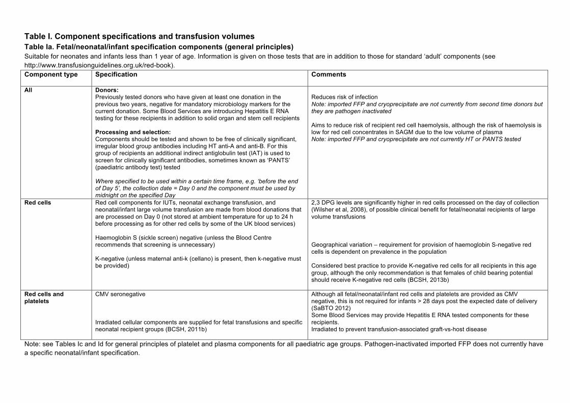
Table I. Component specifications and transfusion volumes Table Ia. Fetal/neonatal/infant specification components (general principles) Suitable for neonates and infants less than 1 year of age. Information is given on those tests that are in addition to those for standard ‘adult’ components (see http://www.transfusionguidelines.org.uk/red-book). Component type Specification Comments
All Donors:
Previously tested donors who have given at least one donation in the previous two years, negative for mandatory microbiology markers for the current donation. Some Blood Services are introducing Hepatitis E RNA testing for these recipients in addition to solid organ and stem cell recipients Processing and selection: Components should be tested and shown to be free of clinically significant, irregular blood group antibodies including HT anti-A and anti-B. For this group of recipients an additional indirect antiglobulin test (IAT) is used to screen for clinically significant antibodies, sometimes known as ‘PANTS’ (paediatric antibody test) tested Where specified to be used within a certain time frame, e.g. ‘before the end of Day 5’, the collection date = Day 0 and the component must be used by midnight on the specified Day
Reduces risk of infection Note: imported FFP and cryoprecipitate are not currently from second time donors but they are pathogen inactivated Aims to reduce risk of recipient red cell haemolysis, although the risk of haemolysis is low for red cell concentrates in SAGM due to the low volume of plasma Note: imported FFP and cryoprecipitate are not currently HT or PANTS tested
Red cells Red cell components for IUTs, neonatal exchange transfusion, and neonatal/infant large volume transfusion are made from blood donations that are processed on Day 0 (not stored at ambient temperature for up to 24 h before processing as for other red cells by some of the UK blood services) Haemoglobin S (sickle screen) negative (unless the Blood Centre recommends that screening is unnecessary) K-negative (unless maternal anti-k (cellano) is present, then k-negative must be provided)
2,3 DPG levels are significantly higher in red cells processed on the day of collection (Wilsher et al, 2008), of possible clinical benefit for fetal/neonatal recipients of large volume transfusions Geographical variation – requirement for provision of haemoglobin S-negative red cells is dependent on prevalence in the population
Considered best practice to provide K-negative red cells for all recipients in this age group, although the only recommendation is that females of child bearing potential should receive K-negative red cells (BCSH, 2013b)
Red cells and platelets
CMV seronegative Irradiated cellular components are supplied for fetal transfusions and specific neonatal recipient groups (BCSH, 2011b)
Although all fetal/neonatal/infant red cells and platelets are provided as CMV negative, this is not required for infants > 28 days post the expected date of delivery (SaBTO 2012) Some Blood Services may provide Hepatitis E RNA tested components for these recipients. Irradiated to prevent transfusion-associated graft-vs-host disease
Note: see Tables Ic and Id for general principles of platelet and plasma components for all paediatric age groups. Pathogen-inactivated imported FFP does not currently have a specific neonatal/infant specification.
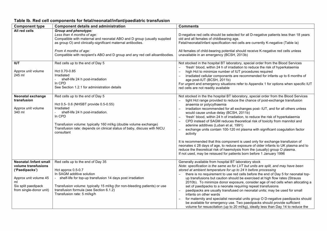
Table Ib. Red cell components for fetal/neonatal/infant/paediatric transfusion Component type Component details and administration Comments All red cells Group and phenotype:
Less than 4 months of age: Compatible with maternal and neonatal ABO and D group (usually supplied as group O) and clinically-significant maternal antibodies.
From 4 months of age: Compatible with recipient’s ABO and D group and any red cell alloantibodies.
D-negative red cells should be selected for all D-negative patients less than 18 years old and all females of childbearing age. Fetal/neonatal/infant specification red cells are currently K-negative (Table Ia) All females of child-bearing potential should receive K-negative red cells unless unavailable in an emergency (BCSH, 2013b)
IUT Approx unit volume 245 ml
Red cells up to the end of Day 5 Hct 0.70-0.85 Irradiated - shelf-life 24 h post-irradiation In CPD See Section 1.2.1 for administration details
Not stocked in the hospital BT laboratory, special order from the Blood Services − ‘fresh’ blood, within 24 h of irradiation to reduce the risk of hyperkalaemia − high Hct to minimize number of IUT procedures required − irradiated cellular components are recommended for infants up to 6 months of
age post-IUT (BCSH, 2011b) For urgent and emergency situations refer to Appendix 1 for options when specific IUT red cells are not readily available
Neonatal exchange transfusion Approx unit volume 340 ml
Red cells up to the end of Day 5 Hct 0.5- 0.6 (NHSBT provide 0.5-0.55) Irradiated - shelf-life 24 h post-irradiation. In CPD
Transfusion volume: typically 160 ml/kg (double volume exchange) Transfusion rate: depends on clinical status of baby, discuss with NICU consultant
Not stocked in the the hospital BT laboratory, special order from the Blood Services − tight Hct range provided to reduce the chance of post-exchange transfusion
anaemia or polycythaemia − irradiation recommended for all exchanges post- IUT, and for all others unless
would cause undue delay (BCSH, 2011b) − ‘fresh’ blood, within 24 h of irradiation, to reduce the risk of hyperkalaemia − CPD instead of SAGM reduces theoretical risk of toxicity from mannitol and
adenine additives (Luban et al, 1991) − exchange units contain 100-120 ml plasma with significant coagulation factor
activity
It is recommended that this component is used only for exchange transfusion of neonates ≤ 28 days of age, to reduce exposure of older infants to UK plasma and to reduce the theoretical risk of haemolysis from the (usually) group O plasma.If not used, may be reissued for patients born before 1 January 1996
Neonatal /infant small volume transfusions (‘Paedipacks’) Approx unit volume 45 ml Six split paedipack from single-donor unit)
Red cells up to the end of Day 35 Hct approx 0.5-0.7 In SAGM additive solution- shelf-life for top-up transfusion 14 days post irradiation Transfusion volume: typically 15 ml/kg (for non-bleeding patients) or use transfusion formula (see Section 6.1.2) Transfusion rate: 5 ml/kg/h
Generally available from hospital BT laboratory stock Note: specification is the same as for LVT but units are split, and may have been stored at ambient temperature for up to 24 h before processing − there is no requirement to use red cells before the end of Day 5 for neonatal top-
up transfusions but caution should be exercised at high flow rates (Strauss 2010b). To minimize donor exposure, consider age of red cells when allocating a set of paedipacks to a neonate requiring repeat transfusions
− paedipacks are usually transfused on neonatal units; may be used for small infants on other wards
− for maternity and specialist neonatal units group O D-negative paedipacks should be available for emergency use. Two paedipacks should provide sufficient volume for resuscitation (up to 20 ml/kg), ideally less than Day 14 to reduce the
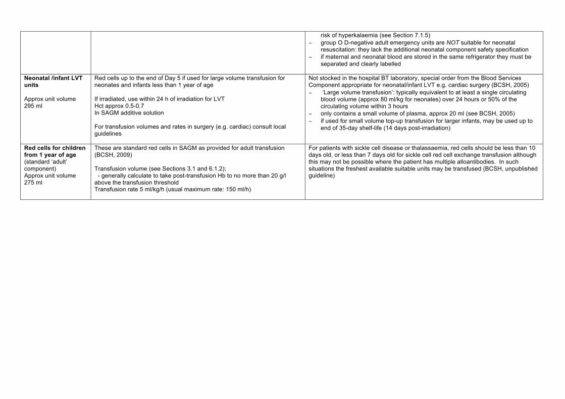
risk of hyperkalaemia (see Section 7.1.5) − group O D-negative adult emergency units are NOT suitable for neonatal
resuscitation: they lack the additional neonatal component safety specification − if maternal and neonatal blood are stored in the same refrigerator they must be
separated and clearly labelled
Neonatal /infant LVT units Approx unit volume 295 ml
Red cells up to the end of Day 5 if used for large volume transfusion for neonates and infants less than 1 year of age If irradiated, use within 24 h of irradiation for LVT Hct approx 0.5-0.7 In SAGM additive solution For transfusion volumes and rates in surgery (e.g. cardiac) consult local guidelines
Not stocked in the hospital BT laboratory, special order from the Blood Services Component appropriate for neonatal/infant LVT e.g. cardiac surgery (BCSH, 2005) − ‘Large volume transfusion’: typically equivalent to at least a single circulating
blood volume (approx 80 ml/kg for neonates) over 24 hours or 50% of the circulating volume within 3 hours
− only contains a small volume of plasma, approx 20 ml (see BCSH, 2005) − if used for small volume top-up transfusion for larger infants, may be used up to
end of 35-day shelf-life (14 days post-irradiation)
Red cells for children from 1 year of age (standard ‘adult’ component) Approx unit volume 275 ml
These are standard red cells in SAGM as provided for adult transfusion (BCSH, 2009) Transfusion volume (see Sections 3.1 and 6.1.2): - generally calculate to take post-transfusion Hb to no more than 20 g/l above the transfusion threshold Transfusion rate 5 ml/kg/h (usual maximum rate: 150 ml/h)
For patients with sickle cell disease or thalassaemia, red cells should be less than 10 days old, or less than 7 days old for sickle cell red cell exchange transfusion although this may not be possible where the patient has multiple alloantibodies. In such situations the freshest available suitable units may be transfused (BCSH, unpublished guideline)

Table Ic. Platelets for fetal/neonatal/infant/paediatric transfusion Component type Component details and administration
Comments
IUT platelets Approx unit volume 75 ml
Group A, D-negative (if ABO D group unknown) or group specific/compatible with maternal antibody HPA-compatible with maternal antibody for NAIT (HPA1a,5b negative/as required) Obtained by apheresis from a single donor Hyperconcentrated to a platelet count of at least 2000 x 109/l Irradiated See Section 1.3.1 for administration details
Special order from Blood Services, requiring several days notice − group O platelets should not normally be selected for non-O or unknown
group recipients, however the availability of HPA antigen-negative platelets may override ABO group selection considerations
− for HPA-matched platelets, donors are negative for clinically significant HLA and HPA antibodies
− hyperconcentrated to optimise platelet count and minimize volume load Irradiated cellular components are recommended for infants up to 6 months of age post- IUT (BCSH, 2011b)
Neonatal platelets Approx unit volume 55 ml
ABO- and D- identical or compatible with recipient (see Appendix 6) HPA-compatible with maternal platelet antibody for neonates with NAIT (as for IUT platelets) Obtained by apheresis from a single donor, split into four smaller units Typical transfusion volume: 10-20 ml/kg Typical administration rate: 10-20 ml/kg/h
HPA-matched platelets require special order from Blood Services, but HPA1a/5b- negative usually available ‘off the shelf’ depending on the geographical location Suitable for neonatal and infant transfusion
Platelets for children from 1 year of age (standard ‘adult’ apheresis platelets) Approx unit volume 200 ml
ABO- and D-identical or compatible with recipient (see Appendix 6) Obtained by apheresis from a single donor where possible Typical transfusion volume: − 10 – 20 ml/kg for children < 15 kg, or a single pack for children ≥ 15 kg. − maximum volume 1 pack. Transfusion rate: 10-20 ml/kg/h
These differ from ‘neonatal’ platelets by not having fetal/neonatal/infant specification. − recipients born on or after 1 January 1996 should be provided with apheresis
platelets when possible, as a vCJD risk reduction measure
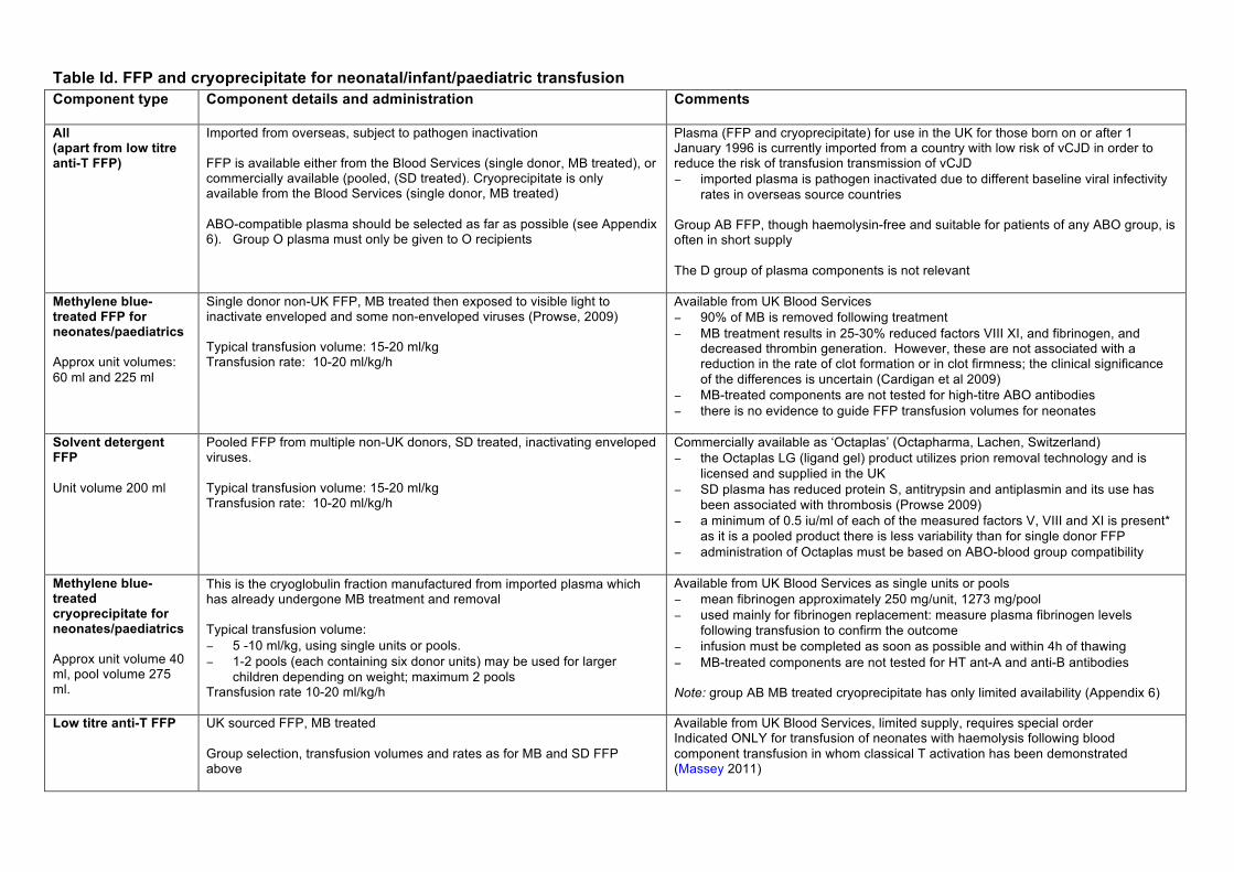
Table Id. FFP and cryoprecipitate for neonatal/infant/paediatric transfusion Component type
Component details and administration Comments
All (apart from low titre anti-T FFP)
Imported from overseas, subject to pathogen inactivation FFP is available either from the Blood Services (single donor, MB treated), or commercially available (pooled, (SD treated). Cryoprecipitate is only available from the Blood Services (single donor, MB treated) ABO-compatible plasma should be selected as far as possible (see Appendix 6). Group O plasma must only be given to O recipients
Plasma (FFP and cryoprecipitate) for use in the UK for those born on or after 1 January 1996 is currently imported from a country with low risk of vCJD in order to reduce the risk of transfusion transmission of vCJD − imported plasma is pathogen inactivated due to different baseline viral infectivity
rates in overseas source countries Group AB FFP, though haemolysin-free and suitable for patients of any ABO group, is often in short supply The D group of plasma components is not relevant
Methylene blue-treated FFP for neonates/paediatrics Approx unit volumes: 60 ml and 225 ml
Single donor non-UK FFP, MB treated then exposed to visible light to inactivate enveloped and some non-enveloped viruses (Prowse, 2009) Typical transfusion volume: 15-20 ml/kg Transfusion rate: 10-20 ml/kg/h
Available from UK Blood Services − 90% of MB is removed following treatment − MB treatment results in 25-30% reduced factors VIII XI, and fibrinogen, and
decreased thrombin generation. However, these are not associated with a reduction in the rate of clot formation or in clot firmness; the clinical significance of the differences is uncertain (Cardigan et al 2009)
− MB-treated components are not tested for high-titre ABO antibodies − there is no evidence to guide FFP transfusion volumes for neonates
Solvent detergent FFP Unit volume 200 ml
Pooled FFP from multiple non-UK donors, SD treated, inactivating enveloped viruses. Typical transfusion volume: 15-20 ml/kg Transfusion rate: 10-20 ml/kg/h
Commercially available as ‘Octaplas’ (Octapharma, Lachen, Switzerland) − the Octaplas LG (ligand gel) product utilizes prion removal technology and is
licensed and supplied in the UK − SD plasma has reduced protein S, antitrypsin and antiplasmin and its use has
been associated with thrombosis (Prowse 2009) − a minimum of 0.5 iu/ml of each of the measured factors V, VIII and XI is present*
as it is a pooled product there is less variability than for single donor FFP − administration of Octaplas must be based on ABO-blood group compatibility
Methylene blue-treated cryoprecipitate for neonates/paediatrics Approx unit volume 40 ml, pool volume 275 ml.
− This is the cryoglobulin fraction manufactured from imported plasma which has already undergone MB treatment and removal Typical transfusion volume: − 5 -10 ml/kg, using single units or pools. − 1-2 pools (each containing six donor units) may be used for larger
children depending on weight; maximum 2 pools Transfusion rate 10-20 ml/kg/h
Available from UK Blood Services as single units or pools − mean fibrinogen approximately 250 mg/unit, 1273 mg/pool − used mainly for fibrinogen replacement: measure plasma fibrinogen levels
following transfusion to confirm the outcome − infusion must be completed as soon as possible and within 4h of thawing − MB-treated components are not tested for HT ant-A and anti-B antibodies Note: group AB MB treated cryoprecipitate has only limited availability (Appendix 6)
Low titre anti-T FFP UK sourced FFP, MB treated Group selection, transfusion volumes and rates as for MB and SD FFP above
Available from UK Blood Services, limited supply, requires special order Indicated ONLY for transfusion of neonates with haemolysis following blood component transfusion in whom classical T activation has been demonstrated (Massey 2011)
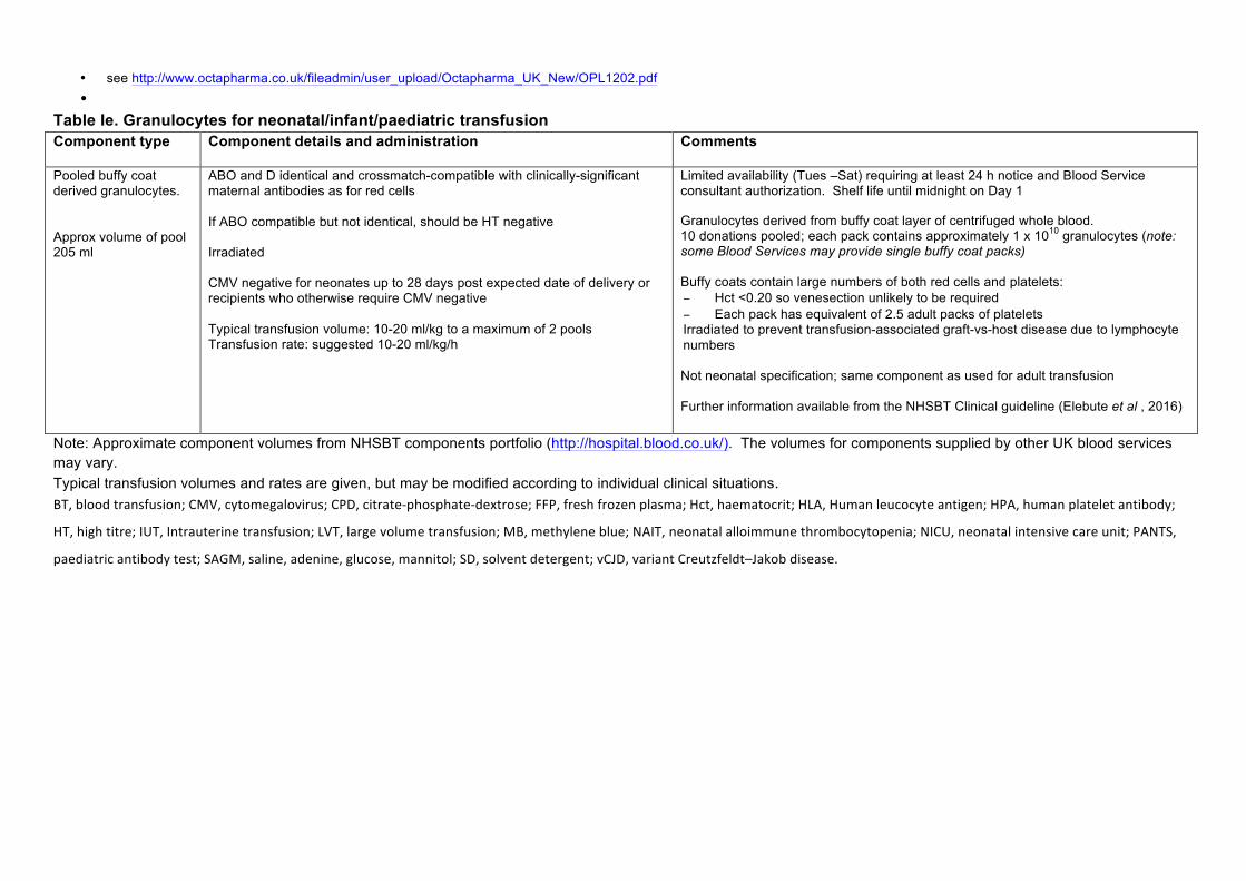
• see http://www.octapharma.co.uk/fileadmin/user_upload/Octapharma_UK_New/OPL1202.pdf •
Table Ie. Granulocytes for neonatal/infant/paediatric transfusion Component type Component details and administration
Comments
Pooled buffy coat derived granulocytes. Approx volume of pool 205 ml
ABO and D identical and crossmatch-compatible with clinically-significant maternal antibodies as for red cells If ABO compatible but not identical, should be HT negative Irradiated CMV negative for neonates up to 28 days post expected date of delivery or recipients who otherwise require CMV negative Typical transfusion volume: 10-20 ml/kg to a maximum of 2 pools Transfusion rate: suggested 10-20 ml/kg/h
Limited availability (Tues –Sat) requiring at least 24 h notice and Blood Service consultant authorization. Shelf life until midnight on Day 1 Granulocytes derived from buffy coat layer of centrifuged whole blood. 10 donations pooled; each pack contains approximately 1 x 10¹⁰ granulocytes (note: some Blood Services may provide single buffy coat packs) Buffy coats contain large numbers of both red cells and platelets: − Hct <0.20 so venesection unlikely to be required − Each pack has equivalent of 2.5 adult packs of platelets Irradiated to prevent transfusion-associated graft-vs-host disease due to lymphocyte numbers Not neonatal specification; same component as used for adult transfusion Further information available from the NHSBT Clinical guideline (Elebute et al , 2016)
Note: Approximate component volumes from NHSBT components portfolio (http://hospital.blood.co.uk/). The volumes for components supplied by other UK blood services may vary. Typical transfusion volumes and rates are given, but may be modified according to individual clinical situations. BT,bloodtransfusion;CMV,cytomegalovirus;CPD,citrate-phosphate-dextrose;FFP,freshfrozenplasma;Hct,haematocrit;HLA,Humanleucocyteantigen;HPA,humanplateletantibody;
HT,hightitre;IUT,Intrauterinetransfusion;LVT,largevolumetransfusion;MB,methyleneblue;NAIT,neonatalalloimmunethrombocytopenia;NICU,neonatalintensivecareunit;PANTS,
paediatricantibodytest;SAGM,saline,adenine,glucose,mannitol;SD,solventdetergent;vCJD,variantCreutzfeldt–Jakobdisease.

Table II. Suggested transfusion thresholds for preterm neonates*
Postnatal age Suggested transfusion threshold Hb (g/l)
Ventilated On oxygen/ NIPPV† Off oxygen
First 24 h
< 120 < 120 < 100
≤ week 1 (day 1-7)
< 120 < 100 < 100
week 2 (day 8 -14) ≥ week 3 (day 15 onwards)
< 100 < 95 < 85
< 75**
* Standard definition of preterm is <37 weeks gestational age at birth but table applies to very preterm neonates (< 32 weeks). ** It is accepted that clinicians may use up to 85 g/l depending on clinical situation † NIPPV, non-invasive positive pressure ventilation
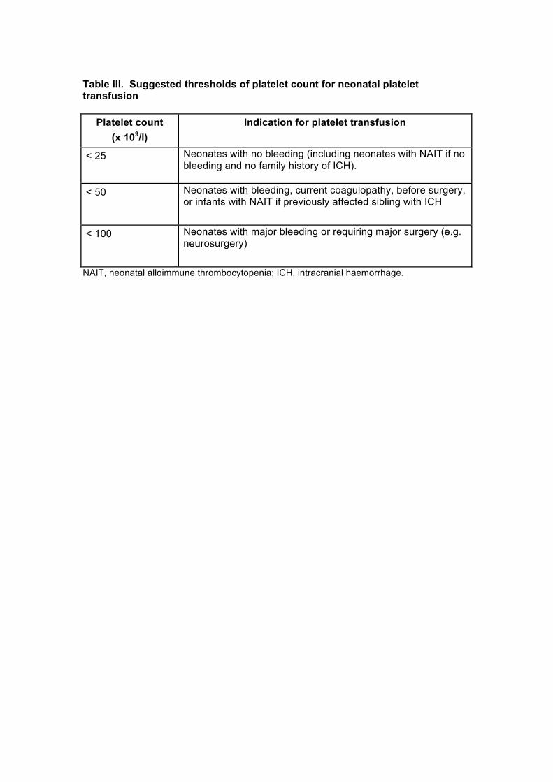
Table III. Suggested thresholds of platelet count for neonatal platelet transfusion
Platelet count (x 109/l)
Indication for platelet transfusion
< 25 Neonates with no bleeding (including neonates with NAIT if no bleeding and no family history of ICH).
< 50 Neonates with bleeding, current coagulopathy, before surgery, or infants with NAIT if previously affected sibling with ICH
< 100 Neonates with major bleeding or requiring major surgery (e.g. neurosurgery)
NAIT, neonatal alloimmune thrombocytopenia; ICH, intracranial haemorrhage.

Table IV. Suggested thresholds of platelet counts for platelet transfusion in
children
Platelet count (x 109/l) Clinical situation to trigger platelet transfusion
< 10 Irrespective of signs of haemorrhage (excluding ITP, TTP/HUS, HIT)
< 20 Severe mucositis Sepsis Laboratory evidence of DIC in the absence of bleeding* Anticoagulant therapy Risk of bleeding due to a local tumour infiltration Insertion of a non-tunnelled central venous line
< 40 Prior to lumbar puncture**
< 50 Moderate haemorrhage (e.g. gastrointestinal bleeding) including bleeding in association with DIC Surgery, unless minor (except at critical sites) - including tunnelled central venous line insertion
< 75 -100 Major haemorrhage or significant post-operative bleeding (e.g. post cardiac surgery) Surgery at critical sites: central nervous system including eyes
* note: routine screening by standard coagulation tests not advocated without clinical indication; for laboratory evidence of DIC see Section 3.4.3 ** It is accepted that prior to lumbar puncture some clinicians will transfuse platelets at higher counts (e.g. 50 x 109/l) in clinically unstable children, non ALL patients, or for the first LP in newly-diagnosed ALL patients to avoid haemorrhage and cerebrospinal fluid contamination with blasts, or at lower counts (≤ 20 x 109/l) in stable patients with ALL, depending on the clinical situation. These practices emphasise the importance of considering the clinical setting and patient factors. ALL, acute lymphoblastic leukaemia; DIC, disseminated intravascular coagulation; HIT, heparin-induced thrombocytopenia; HUS, haemolytic uraemic syndrome; ITP, immune thrombocytopenia; LP, lumbar puncture; TTP, thrombotic thrombocytopenic purpura
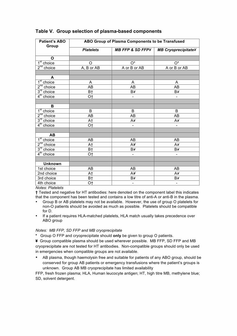
Table V. Group selection of plasma-based components
Patient’s ABO Group
ABO Group of Plasma Components to be Transfused
Platelets MB FFP & SD FFP¥ MB Cryoprecipitate¥
O 1st choice O O* O* 2nd choice A, B or AB A or B or AB A or B or AB
A 1st choice A A A 2nd choice AB AB AB 3rd choice B† B¥ B¥ 4th choice O† - -
B 1st choice B B B 2nd choice AB AB AB 3rd choice A† A¥ A¥ 4th choice O† - -
AB 1st choice AB AB AB 2nd choice A† A¥ A¥ 3rd choice B† B¥ B¥ 4th choice O† - -
Unknown 1st choice AB AB AB 2nd choice A† A¥ A¥ 3rd choice B† B¥ B¥ 4th choice O† - -
Notes: Platelets † Tested and negative for HT antibodies: here denoted on the component label this indicates that the component has been tested and contains a low titre of anti-A or anti-B in the plasma. • Group B or AB platelets may not be available. However, the use of group O platelets for
non-O patients should be avoided as much as possible. Platelets should be compatible for D.
• If a patient requires HLA-matched platelets, HLA match usually takes precedence over ABO group
Notes: MB FFP, SD FFP and MB cryoprecipitate * Group O FFP and cryoprecipitate should only be given to group O patients. ¥ Group compatible plasma should be used wherever possible. MB FFP, SD FFP and MB cryoprecipitate are not tested for HT antibodies. Non-compatible groups should only be used in emergencies when compatible groups are not available. • AB plasma, though haemolysin free and suitable for patients of any ABO group, should be
conserved for group AB patients or emergency transfusions where the patient’s groups is unknown. Group AB MB cryoprecipitate has limited availability
FFP, fresh frozen plasma; HLA, Human leucocyte antigen; HT, high titre MB, methylene blue; SD, solvent detergent.

Appendix 1 Guideline literature search terms
(((Blood Transfusion[mh:exp]) OR (transfus*[TI] OR pretransfus*[TI] OR retransfus*[TI] OR red cell*[TI] OR red blood cell*[TI] OR RBC*[TI] OR PRBC*[TI] OR FFP[TI] OR fresh plasma[TI] OR frozen plasma[TI] OR maternal plasma[TI] OR platelets[TI] OR platelet concentrate*[TI] OR granulocytes[TI] OR cryoprecipitate[TI] OR blood component*[TI] OR blood product*[TI] OR cell salvage[TI] OR blood salvage[TI] OR cell saver*[TI] OR TRALI[TI]) OR (exchange transfusion*[Title/Abstract] OR plasma exchange[Title/Abstract] OR plasmapheresis[Title/Abstract] OR in utero transfusion*[Title/Abstract] OR intrauterine transfusion*[Title/Abstract] OR maternal transfusion*[Title/Abstract] OR placental transfusion*[Title/Abstract] OR partial exchange[Title/Abstract] OR neonatal exchange[Title/Abstract] OR disseminated intravascular coagulation[Title/Abstract] OR DIC[Title] OR T-activation[Title/Abstract] OR coagulopath*[Title/Abstract] OR ((transfus*[Title/Abstract] OR retransfus*[Title/Abstract] OR red cell*[Title/Abstract] OR red blood cell*[Title/Abstract] OR RBC*[Title/Abstract] OR PRBC*[Title/Abstract] OR FFP[Title/Abstract] OR plasma[Title/Abstract] OR platelet*[Title/Abstract]) AND (trigger*[Title/Abstract] OR threshold*[Title/Abstract]))) AND ((Child[mh:exp]) OR (Pediatrics[mh:exp]) OR (Infant[mh:exp]) OR (Adolescent[mh]) OR (low birth weight*[Title/Abstract]) OR (child[Title/Abstract] OR children[Title/Abstract] OR paediatric[Title/Abstract] OR pediatric*[Title/Abstract] OR infant*[Title/Abstract] OR infancy[Title/Abstract] OR neonat*[Title/Abstract] OR newborn*[Title/Abstract] OR babies[Title/Abstract] OR adolescen*[Title/Abstract] OR teen*[Title/Abstract])))) AND (random* OR blind* OR control group OR groups OR placebo* OR controlled trial OR controlled study OR guideline* OR trials OR systematic review OR meta-analysis OR metaanalysis OR literature search OR medline OR cochrane OR embase)
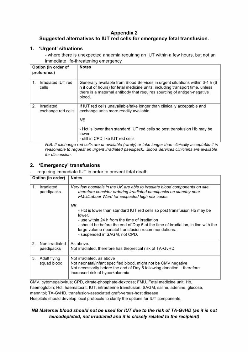
Appendix 2 Suggested alternatives to IUT red cells for emergency fetal transfusion.
1. ‘Urgent’ situations - where there is unexpected anaemia requiring an IUT within a few hours, but not an immediate life-threatening emergency
Option (in order of preference)
Notes
1. Irradiated IUT red cells
Generally available from Blood Services in urgent situations within 3-4 h (6 h if out of hours) for fetal medicine units, including transport time, unless there is a maternal antibody that requires sourcing of antigen-negative blood.
2. Irradiated exchange red cells
If IUT red cells unavailable/take longer than clinically acceptable and exchange units more readily available
NB
- Hct is lower than standard IUT red cells so post transfusion Hb may be lower - still in CPD like IUT red cells
N.B. If exchange red cells are unavailable (rarely) or take longer than clinically acceptable it is reasonable to request an urgent irradiated paedipack. Blood Services clinicians are available for discussion.
2. ‘Emergency’ transfusions - requiring immediate IUT in order to prevent fetal death Option (in order) Notes
1. Irradiated paedipacks
Very few hospitals in the UK are able to irradiate blood components on site, therefore consider ordering irradiated paedipacks on standby near FMU/Labour Ward for suspected high risk cases.
NB - Hct is lower than standard IUT red cells so post transfusion Hb may be lower. - use within 24 h from the time of irradiation - should be before the end of Day 5 at the time of irradiation, in line with the large volume neonatal transfusion recommendations. - suspended in SAGM, not CPD.
2. Non irradiated paedipacks
As above. Not irradiated, therefore has theoretical risk of TA-GvHD.
3. Adult flying squad blood
Not irradiated, as above Not neonatal/infant specified blood, might not be CMV negative Not necessarily before the end of Day 5 following donation – therefore increased risk of hyperkalaemia
CMV, cytomegalovirus; CPD, citrate-phosphate-dextrose; FMU, Fetal medicine unit; Hb, haemoglobin; Hct, haematocrit; IUT, intrauterine transfusion; SAGM, saline, adenine, glucose, mannitol; TA-GvHD, transfusion-associated graft-versus-host disease Hospitals should develop local protocols to clarify the options for IUT components. NB Maternal blood should not be used for IUT due to the risk of TA-GvHD (as it is not
leucodepleted, not irradiated and it is closely related to the recipient)
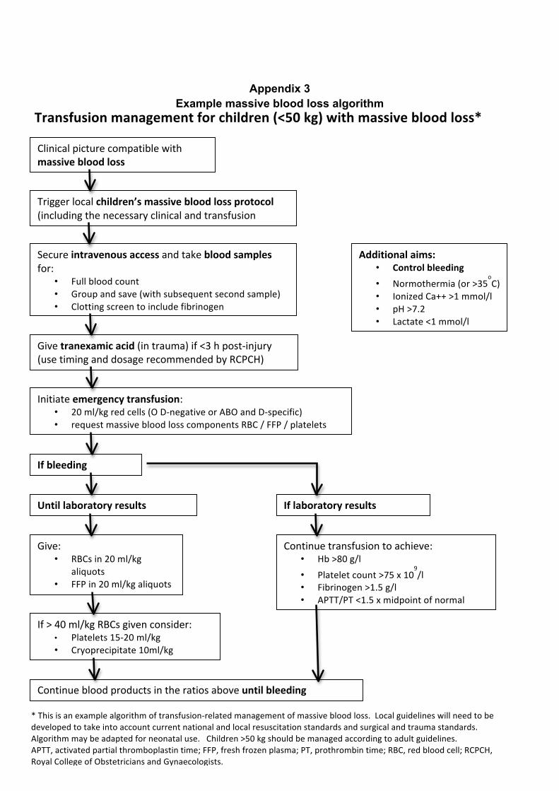
Appendix 3
Example massive blood loss algorithm
Transfusionmanagementforchildren(<50kg)withmassivebloodloss*
Clinicalpicturecompatiblewithmassivebloodloss
Triggerlocalchildren’smassivebloodlossprotocol (includingthenecessaryclinicalandtransfusionteams)
Secureintravenousaccessandtakebloodsamplesfor:
• Fullbloodcount• Groupandsave(withsubsequentsecondsample)• Clottingscreentoincludefibrinogen• Nearpatienttesting(bloodgas,electrolytes)
Givetranexamicacid(intrauma)if<3hpost-injury (usetiminganddosagerecommendedbyRCPCH)
Initiateemergencytransfusion: • 20ml/kgredcells(OD-negativeorABOandD-specific)• requestmassivebloodlosscomponentsRBC/FFP/platelets
Ifbleedingcontinues
Untillaboratoryresultsavailable
Give: • RBCsin20ml/kg
aliquots• FFPin20ml/kgaliquots
ToachieveRBC:FFPratio1:1
If>40ml/kgRBCsgivenconsider: • Platelets15-20ml/kg• Cryoprecipitate10ml/kg
Iflaboratoryresultsavailable:
Continuetransfusiontoachieve: • Hb>80g/l• Plateletcount>75x10
9/l
• Fibrinogen>1.5g/l• APTT/PT<1.5xmidpointofnormal
range
Continuebloodproductsintheratiosaboveuntilbleedingcontrolled
Additionalaims: • Controlbleeding• Normothermia(or>35
oC)
• IonizedCa++>1mmol/l• pH>7.2• Lactate<1mmol/l
*Thisisanexamplealgorithmoftransfusion-relatedmanagementofmassivebloodloss.Localguidelineswillneedtobedevelopedtotakeintoaccountcurrentnationalandlocalresuscitationstandardsandsurgicalandtraumastandards.Algorithmmaybeadaptedforneonataluse.Children>50kgshouldbemanagedaccordingtoadultguidelines.APTT,activatedpartialthromboplastintime;FFP,freshfrozenplasma;PT,prothrombintime;RBC,redbloodcell;RCPCH,RoyalCollegeofObstetriciansandGynaecologists.
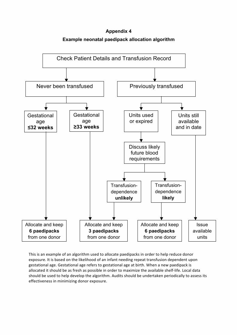
Appendix 4
Example neonatal paedipack allocation algorithm
Thisisanexampleofanalgorithmusedtoallocatepaedipacksinordertohelpreducedonorexposure.Itisbasedonthelikelihoodofaninfantneedingrepeattransfusiondependentupongestationalage.Gestationalagereferstogestationalageatbirth.Whenanewpaedipackisallocateditshouldbeasfreshaspossibleinordertomaximizetheavailableshelf-life.Localdatashouldbeusedtohelpdevelopthealgorithm.Auditsshouldbeundertakenperiodicallytoassessitseffectivenessinminimizingdonorexposure.
Gestational age
≤32 weeks
Gestational age
≥33 weeks
Units used or expired
Units still available
and in date
Discuss likely future blood
requirements
Transfusion-dependence
unlikely
Transfusion-dependence
likely
Allocate and keep 6 paedipacks from one donor
Allocate and keep 3 paedipacks from one donor
Allocate and keep 6 paedipacks from one donor
Issue available
units
Check Patient Details and Transfusion Record
Never been transfused Previously transfused
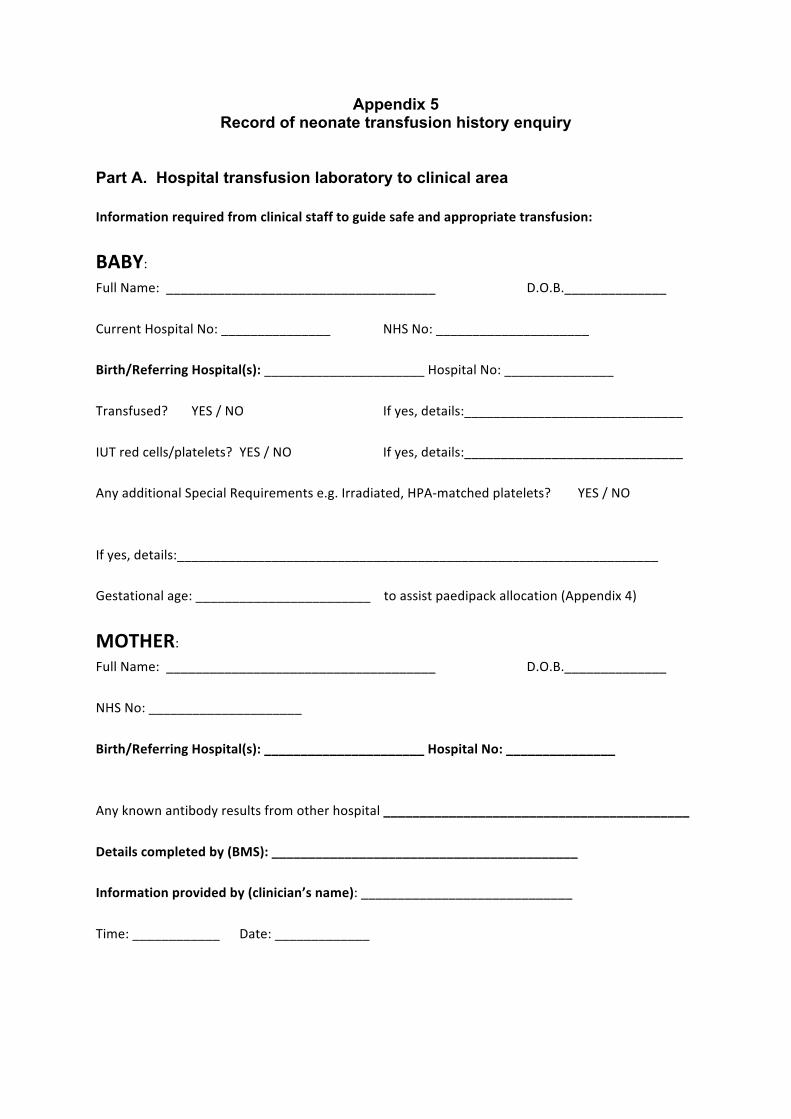
Appendix 5 Record of neonate transfusion history enquiry
Part A. Hospital transfusion laboratory to clinical area Informationrequiredfromclinicalstafftoguidesafeandappropriatetransfusion:
BABY:FullName:_____________________________________ D.O.B.______________CurrentHospitalNo:_______________ NHSNo:_____________________Birth/ReferringHospital(s):______________________HospitalNo:_______________ Transfused? YES/NO Ifyes,details:______________________________IUTredcells/platelets? YES/NO Ifyes,details:______________________________ AnyadditionalSpecialRequirementse.g.Irradiated,HPA-matchedplatelets? YES/NOIfyes,details:__________________________________________________________________Gestationalage:________________________toassistpaedipackallocation(Appendix4)
MOTHER:
FullName:_____________________________________ D.O.B.______________NHSNo:_____________________Birth/ReferringHospital(s):______________________HospitalNo:_______________ Anyknownantibodyresultsfromotherhospital__________________________________________Detailscompletedby(BMS):__________________________________________Informationprovidedby(clinician’sname):_____________________________Time:____________ Date:_____________
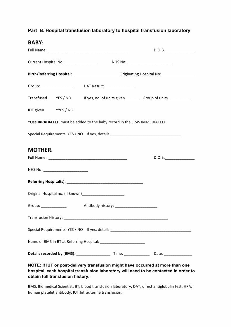
Part B. Hospital transfusion laboratory to hospital transfusion laboratory BABY:FullName:_____________________________________ D.O.B.______________CurrentHospitalNo:_______________ NHSNo:_____________________Birth/ReferringHospital:______________________OriginatingHospitalNo:_______________ Group:_______________ DATResult:______________Transfused YES/NO Ifyes,no.ofunitsgiven_______Groupofunits__________IUTgiven *YES/NO *UseIRRADIATEDmustbeaddedtothebabyrecordintheLIMSIMMEDIATELY.SpecialRequirements:YES/NOIfyes,details:_________________________________
MOTHER:
FullName:_____________________________________ D.O.B.______________NHSNo:_____________________ReferringHospital(s):____________________________________OriginalHospitalno.(ifknown)____________________Group:____________ Antibodyhistory:____________________TransfusionHistory:_________________________________________________SpecialRequirements:YES/NOIfyes,details:______________________________________NameofBMSinBTatReferringHospital:_____________________Detailsrecordedby(BMS):________________Time:____________ Date:_____________NOTE: If IUT or post-delivery transfusion might have occurred at more than one hospital, each hospital transfusion laboratory will need to be contacted in order to obtain full transfusion history.
BMS,BiomedicalScientist:BT,bloodtransfusionlaboratory;DAT,directantiglobulintest;HPA,humanplateletantibody;IUTIntrauterinetransfusion.
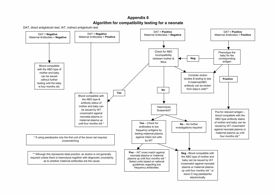
Appendix 6 Algorithm for compatibility testing for a neonate
DAT, direct antiglobulin test; IAT, indirect antiglobulin test
DAT = Positive
Maternal Antibodies = Negative DAT = Negative Maternal Antibodies = Positive
** Although this represents ideal practice, an elution is not generally required unless there is haemolysis together with diagnostic uncertainty
as to whether maternal antibodies are the cause.
* If using paedipacks only the first unit of the donor set requires crossmatching
DAT = Negative Maternal Antibodies = Negative
Blood compatible with the ABO type of
mother and baby can be issued without further
testing until the baby is four months old.
Blood compatible with the ABO type &
antibody status of mother and baby can
be issued by IAT crossmatch against neonatal plasma or maternal plasma up
until four months old *
YesNo
Check for ABO incompatibility
between mother & fetus
Haemolysis Suspected?
Consider elution studies & testing to see
if maternal/ABO antibody can be eluted
from baby’s cells**
Neg - Blood compatible with the ABO type of mother and baby can be issued by IAT
crossmatch against neonatal plasma or maternal plasma up until four months old * or
issue O neg paedipacks electronically
Pos – IAT cross match against neonatal plasma or maternal
plasma up until four months old * Select units based on national
guidelines regarding low frequency antibodies.
Yes – Check for antibodies to low
frequency antigens by testing maternal plasma against infant red cells
by IAT
No – No further investigations required
DAT = Positive Maternal Antibodies = Positive
Phenotype the baby for the
corresponding antigen
Pos for relevant antigen – blood compatible with the ABO type antibody status
of mother and baby can be issued by IAT crossmatch
against neonatal plasma or maternal plasma up until
four months old *
Positive
Neg


