Group A Streptococcus Secreted Esterase HydrolyzesPlatelet-Activating Factor to Impede NeutrophilRecruitment and Facilitate Innate Immune EvasionMengyao Liu1., Hui Zhu1,2., Jinquan Li1,3, Cristiana C. Garcia4, Wenchao Feng1, Liliya N. Kirpotina1,
Jonathan Hilmer5, Luciana P. Tavares4, Arthur W. Layton6, Mark T. Quinn1, Brian Bothner5,
Mauro M. Teixeira4, Benfang Lei1*
1 Department of Immunology and Infectious Diseases, Montana State University, Bozeman, Montana, United States of America, 2 Department of Physiology, Harbin
Medical University, Harbin, People’s Republic of China, 3 State Key Laboratory of Agricultural Microbiology, Huazhong Agricultural University, Wuhan, People’s Republic of
China, 4 Laboratory of Immunopharmacology, Federal University of Minas Gerais, Belo Horizonte, Brazil, 5 Department of Chemistry and Biochemistry, Montana State
University, Bozeman, Montana, United States of America, 6 Montana Veterinary Diagnostic Laboratory, Bozeman, Montana, United States of America
Abstract
The innate immune system is the first line of host defense against invading organisms. Thus, pathogens have developedvirulence mechanisms to evade the innate immune system. Here, we report a novel means for inhibition of neutrophilrecruitment by Group A Streptococcus (GAS). Deletion of the secreted esterase gene (designated sse) in M1T1 GAS strainswith (MGAS5005) and without (MGAS2221) a null covS mutation enhances neutrophil ingress to infection sites in the skin ofmice. In trans expression of SsE in MGAS2221 reduces neutrophil recruitment and enhances skin invasion. The sse deletionmutant of MGAS5005 (DsseMGAS5005) is more efficiently cleared from skin than the parent strain. SsE hydrolyzes the sn-2 esterbond of platelet-activating factor (PAF), converting biologically active PAF into inactive lyso-PAF. KM and kcat of SsE forhydrolysis of 2-thio-PAF were similar to those of the human plasma PAF acetylhydrolase. Treatment of PAF with SsEabolishes the capacity of PAF to induce activation and chemotaxis of human neutrophils. More importantly, PAF receptor-deficient mice significantly reduce neutrophil infiltration to the site of DsseMGAS5005 infection. These findings identify the firstsecreted PAF acetylhydrolase of bacterial pathogens and support a novel GAS evasion mechanism that reduces phagocyterecruitment to sites of infection by inactivating PAF, providing a new paradigm for bacterial evasion of neutrophilresponses.
Citation: Liu M, Zhu H, Li J, Garcia CC, Feng W, et al. (2012) Group A Streptococcus Secreted Esterase Hydrolyzes Platelet-Activating Factor to Impede NeutrophilRecruitment and Facilitate Innate Immune Evasion. PLoS Pathog 8(4): e1002624. doi:10.1371/journal.ppat.1002624
Editor: Michael R. Wessels, Children’s Hospital Boston, United States of America
Received September 26, 2011; Accepted February 21, 2012; Published April 5, 2012
Copyright: � 2012 Liu et al. This is an open-access article distributed under the terms of the Creative Commons Attribution License, which permits unrestricteduse, distribution, and reproduction in any medium, provided the original author and source are credited.
Funding: This work was supported in part by NIH Grants R01AI095704 from the National Institute of Allergy and Infectious Diseases, P20 RR-020185 from theNational Center for Research Resources, and GM103500-09 from the National Institute of General Medical Sciences, USDA NRI/CSREES grant 2007-35204-18306,and the Montana State University Agricultural Experimental Station. The work done at UFMG was financed by Conselho Nacional de Desenvolvimento Cientifico eTecnologio (CNPq, Brazil) and Fundacao do Amparo a Pesquisas do Estado de Minas Gerais (FAPEMIG). The work done at Harbin Medical School was supported bygrant LC2011C02 from Natural Science Foundation of Heilongjiang Province, China. JL was supported by the PhD student exchange scholarship of the Ministry ofEducation, China. The MSU Mass Spectrometry Facility receives support from the Murdock Charitable trust, NIH INBRE MT grant Number P20 RR-16455-08 and NIHgrant P20 1P20RR024237. The funders had no role in study design, data collection and analysis, decision to publish, or preparation of the manuscript.
Competing Interests: The authors have declared that no competing interests exist.
* E-mail: [email protected]
. These authors contributed equally to this work.
Introduction
Neutrophils are one of the first responders of innate inflamma-
tory cells to migrate towards the site of infecting agents. Evasion of
the neutrophil microbicidal response is critical for survival,
dissemination, and infectability of bacterial pathogens. Bacterial
pathogens evade the neutrophil responses by multiple mecha-
nisms, including inhibition of neutrophil infiltration, antiphagocy-
tosis, and killing of neutrophils. Group A Streptococcus (GAS) causes
a variety of diseases, ranging from relatively mild pharyngitis to
potentially lethal invasive infections, such as necrotizing fasciitis
[1]. The success of GAS as a pathogen is based, in part, on its
ability to evade the innate immune system. GAS expresses
extracellular peptidases ScpA and SpyCEP/ScpC to inhibit
neutrophil recruitment by degrading the chemotactic C5a peptide
and IL-8/CXC chemokines, respectively [2,3,4,5]. The hyaluro-
nic acid capsule and surface M protein made by GAS confer
resistance to opsonophagocytosis and phagocytosis by neutrophils
[6,7]. Secreted DNase Sda1 helps GAS escape from neutrophil
extracellular traps [8]. Mac/IdE inhibits opsonophagocytosis
[9,10]. Streptolysin S and streptolysin O kill and induce apoptosis
of neutrophils [11,12].
GAS pathogenesis is mediated by many virulence factors, and
alteration in regulation of virulence factors greatly affects clinical
outcomes. The two component regulatory system CsrRS/CovRS
negatively regulates many virulence factor genes of GAS,
including most of the virulence factors involved in the innate
immune evasion [13,14]. Nonsense and missense mutations in
csrRS/covRS occur during human infections and are epidemiolog-
ically linked to severe GAS infections [15]. Selection of hypervirulent
PLoS Pathogens | www.plospathogens.org 1 April 2012 | Volume 8 | Issue 4 | e1002624
strains with csrRS/covRS mutations during experimental invasive
infections in mice further highlights the critical role of csrRS/covRS
mutations in progression of invasive GAS infections [16,17,18]. Loss
of SpeB and enhanced production of the hyaluronic acid capsule
contribute to the progression of invasive GAS infections [19,20].
Enhanced production of the virulence factors in the innate immune
evasion as a result of csrRS/covRS mutations plays a key role in
selection for hypervirulent csrRS/covRS mutants. The DNase Sda1
helps GAS escape neutrophil extracellular traps and provides
selection pressure for csrRS/covRS mutations [8]. Neutrophil
infiltration to infection sites is almost completely inhibited in some
necrotizing fasciitis patients and during experimental severe soft
tissue infections in primates and mice [2,21,22,23]. Enhanced
production of SpyCEP/ScpC and ScpA as a result of csrRS/covRS
mutations are believed to contribute to the enhanced inhibition of
neutrophil recruitment in severe invasive infections.
It is not known whether SpyCEP/ScpC and ScpA are entirely
responsible for the dramatic inhibition of neutrophil recruitment
by hypervirulent GAS strains with csrRS/covRS mutations. Platelet-
activating factor (PAF) also has chemotactic activity for inflam-
matory cells. PAF is a phospholipid mediator with the chemical
structure of 1-O-alkyl-2-acetyl-sn-glycero-3-phosphorylcholine
[24]. PAF is produced by endothelial cells, neutrophils, macro-
phages, and eosinophils in responses to proinflammatory cyto-
kines, phagocytosis, and/or other stimuli [25]. This important
phospholipid mediator has diverse and potent biological activities,
including participation in normal physiological processes, such as
inflammation, hemostasis, and reproduction, and contribution to
pathological responses, including asthma, ischemia, gastric and
pulmonary distress, allergy, and shock [26]. Particularly, PAF can
activate platelets [27] and neutrophils [28]. The biological
activities of PAF are mediated by a G protein-linked receptor
(PAFR) that is expressed on the surface of various cell types
[29,30].
The biological activities of PAF are regulated by PAF
acetylhydrolases that hydrolyze the sn-2 acetyl ester bond,
converting PAF into acetic acid and lyso-PAF. Four mammal
PAF acetylhydrolases, secreted or plasma, two intracellular type I,
and intracellular type II PAF acetylhydrolases, have been
described [31,32,33,34]. The plasma and intracellular type II
PAF acetylhydrolases belong to group VII of phospholipases A2,
and the type I PAF acetylhydrolases are classified as group VIII
phospholipases A2 [35]. Group VIII PAF acetylhydrolases are
completely specific for PAF whereas the plasma and type II PAF
acetylhydrolases hydrolyze unmodified sn-2 fatty acyl residues up
to 5 or 6 carbon atoms long and longer sn-2 acyl residues with
modification by oxidation [35]. PAF acetylhydrolase activity has
been also detected in bacteria and fungus. An intracellular yeast
group VII PAF acetylhydrolase enhances the viability of yeast
under oxidative stress [36]. The spirochete Leptospira interrogans
produces a PAF acetylhydrolase [37]. An apparently intracellular
esterase Est13 from an earthworm gut-associated microorganism
inhibits PAF-induced platelet aggregation [38]. Both L. interrogans
PAF acetylhydrolase and Est13 share sequence homology with the
a1 subunit of the type intracellular I mammalian PAF acetylhy-
drolase. The function of these bacterial PAF acetylhydrolases is
not known. These yeast and bacterial PAF acetylhydrolases are
intracellular proteins.
The esterase secreted by GAS (designated SsE) is a protective
antigen [39] and is regulated by CsrRS/CovRS and required for
GAS virulence and dissemination [40]. The basis for the
contribution of SsE to GAS virulence and dissemination is
unknown. Identification of the esterase target is essential for
elucidating the functional mechanism of SsE. The homologue of
SsE in the horse pathogen Streptococcus equi possesses optimal
activity to acetyl esters [41]. We hypothesize that SsE targets PAF
and is involved in evasion of the innate immune system. Here, we
report on studies designed to test this hypothesis. Our findings
demonstrate that SsE is indeed a potent PAF acetylhydrolase and
is required for inhibition of neutrophil infiltration. We also present
evidence for one of mechanisms for SsE to evade the neutrophil
response by targeting PAF, identifying a new novel virulence
factor for innate immune evasion.
Results
PAF Acetylhydrolase Activity of SsEIdentification of the esterase target is essential for elucidating the
mechanism by which GAS uses SsE to contribute to GAS
virulence and dissemination. Since the homologue of SsE in
Streptococcus equi has optimal activities to acetyl esters [41], we
considered whether the target of SsE is a molecule with a short-
chain acyl ester group. PAF appears to be a good candidate as the
target of SsE since it has an acetyl group and is an inflammatory
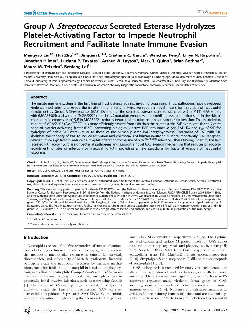
mediator and chemoattractant [28,42]. PAF was incubated with
SsE, and the reaction was analyzed by thin layer chromatography
(TLC), which could resolve PAF and lyso-PAF because PAF
migrates much faster (Figure 1). SsE-treated PAF migrated the
same distance as lyso-PAF, indicating that PAF was hydrolyzed by
SsE. To confirm that PAF hydrolysis was due to the enzymatic
activity of SsE, we performed a control experiment using SsES178A
mutant protein. This mutant lacks the catalytic residue, Ser178,
and, therefore, lacks enzymatic activity [39]. Indeed, SsES178A-
treated PAF and untreated PAF had the same migration rate.
These results indicate that SsE hydrolyzes the acetyl ester bond in
PAF, resulting in lyso-PAF.
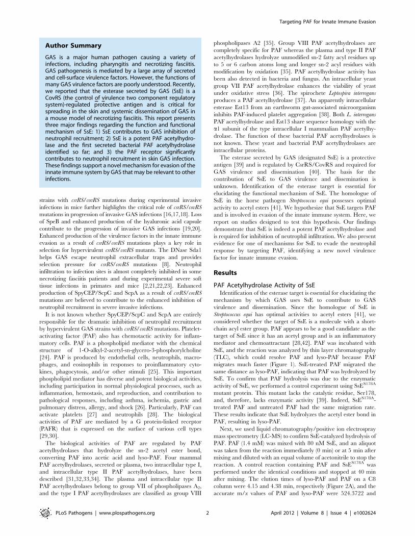
Next, we used liquid chromatography/positive ion electrospray
mass spectrometry (LC-MS) to confirm SsE-catalyzed hydrolysis of
PAF. PAF (1.4 mM) was mixed with 80 nM SsE, and an aliquot
was taken from the reaction immediately (0 min) or at 5 min after
mixing and diluted with an equal volume of acetonitrile to stop the
reaction. A control reaction containing PAF and SsES178A was
performed under the identical conditions and stopped at 40 min
after mixing. The elution times of lyso-PAF and PAF on a C8
column were 4.15 and 4.38 min, respectively (Figure 2A), and the
accurate m/z values of PAF and lyso-PAF were 524.3722 and
Author Summary
GAS is a major human pathogen causing a variety ofinfections, including pharyngitis and necrotizing fasciitis.GAS pathogenesis is mediated by a large array of secretedand cell-surface virulence factors. However, the functions ofmany GAS virulence factors are poorly understood. Recently,we reported that the esterase secreted by GAS (SsE) is aCovRS (the control of virulence two component regulatorysystem)-regulated protective antigen and is critical forspreading in the skin and systemic dissemination of GAS ina mouse model of necrotizing fasciitis. This report presentsthree major findings regarding the function and functionalmechanism of SsE: 1) SsE contributes to GAS inhibition ofneutrophil recruitment; 2) SsE is a potent PAF acetylhydro-lase and the first secreted bacterial PAF acetylhydrolaseidentified so far; and 3) the PAF receptor significantlycontributes to neutrophil recruitment in skin GAS infection.These findings support a novel mechanism for evasion of theinnate immune system by GAS that may be relevant to otherinfections.
Targeting PAF for Innate Immune Evasion
PLoS Pathogens | www.plospathogens.org 2 April 2012 | Volume 8 | Issue 4 | e1002624
482.3600, respectively (Figure 2B). We found that 57% and 100% of
PAF was converted into lyso-PAF for the SsE-treated PAF samples
obtained at 0 and 5 min after mixing, respectively (Figure 2B and
2C), whereas no PAF was hydrolyzed into lyso-PAF at 40 min after
mixing PAF with inactive SsES178A (Figure 2D). These results
unambiguously demonstrate that SsE catalyzes the conversion of
PAF into lyso-PAF. Thus, SsE is a PAF acetylhydrolase.
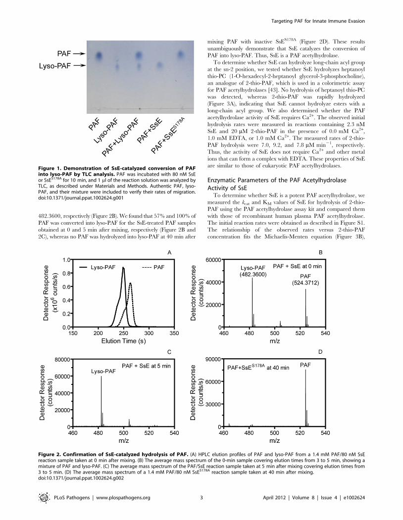
To determine whether SsE can hydrolyze long-chain acyl group
at the sn-2 position, we tested whether SsE hydrolyzes heptanoyl
thio-PC (1-O-hexadecyl-2-heptanoyl glycerol-3-phosphocholine),
an analogue of 2-thio-PAF, which is used in a colorimetric assay
for PAF acetylhydrolases [43]. No hydrolysis of heptanoyl thio-PC
was detected, whereas 2-thio-PAF was rapidly hydrolyzed
(Figure 3A), indicating that SsE cannot hydrolyze esters with a
long-chain acyl group. We also determined whether the PAF
acetylhydrolase activity of SsE requires Ca2+. The observed initial
hydrolysis rates were measured in reactions containing 2.3 nM
SsE and 20 mM 2-thio-PAF in the presence of 0.0 mM Ca2+,
1.0 mM EDTA, or 1.0 mM Ca2+. The measured rates of 2-thio-
PAF hydrolysis were 7.0, 9.2, and 7.8 mM min21, respectively.
Thus, the activity of SsE does not require Ca2+ and other metal
ions that can form a complex with EDTA. These properties of SsE
are similar to those of eukaryotic PAF acetylhydrolases.
Enzymatic Parameters of the PAF AcetylhydrolaseActivity of SsE
To determine whether SsE is a potent PAF acetylhydrolase, we
measured the kcat and KM values of SsE for hydrolysis of 2-thio-
PAF using the PAF acetylhydrolase assay kit and compared them
with those of recombinant human plasma PAF acetylhydrolase.
The initial reaction rates were obtained as described in Figure S1.
The relationship of the observed rates versus 2-thio-PAF
concentration fits the Michaelis-Menten equation (Figure 3B),
Figure 1. Demonstration of SsE-catalyzed conversion of PAFinto lyso-PAF by TLC analysis. PAF was incubated with 80 nM SsEor SsES178A for 10 min, and 1 ml of the reaction solution was analyzed byTLC, as described under Materials and Methods. Authentic PAF, lyso-PAF, and their mixture were included to verify their rates of migration.doi:10.1371/journal.ppat.1002624.g001
Figure 2. Confirmation of SsE-catalyzed hydrolysis of PAF. (A) HPLC elution profiles of PAF and lyso-PAF from a 1.4 mM PAF/80 nM SsEreaction sample taken at 0 min after mixing. (B) The average mass spectrum of the 0-min sample covering elution times from 3 to 5 min, showing amixture of PAF and lyso-PAF. (C) The average mass spectrum of the PAF/SsE reaction sample taken at 5 min after mixing covering elution times from3 to 5 min. (D) The average mass spectrum of a 1.4 mM PAF/80 nM SsES178A reaction sample taken at 40 min after mixing.doi:10.1371/journal.ppat.1002624.g002
Targeting PAF for Innate Immune Evasion
PLoS Pathogens | www.plospathogens.org 3 April 2012 | Volume 8 | Issue 4 | e1002624
yielding a kcat of 69.6 s21 and an apparent KM of 7.0 mM for SsE.
In comparison, kcat and KM of recombinant human plasma PAF
acetylhydrolase were determined to be 15.4 s21 and 8.0 mM,
respectively. These measurements indicate that SsE has similar
KM with and higher kcat than the human enzyme.
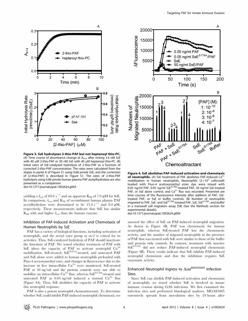
Inhibition of PAF-Induced Activation and Chemotaxis ofHuman Neutrophils by SsE
PAF has a variety of biological functions, including activation of
neutrophils, and the acetyl ester group at sn-2 is critical for its
activities. Thus, SsE-catalyzed hydrolysis of PAF should inactivate
the functions of PAF. We tested whether treatment of PAF with
SsE alters the capacity of PAF to activate neutrophil Ca2+
mobilization. SsE-treated, SsES187A-treated, and untreated PAF
and SsE alone were added to human neutrophils preloaded with
Fluo-4 acetoxymethyl ester, and changes in fluorescence due to the
increase in free intracellular Ca2+ were monitored. SsE-treated
PAF at 50 ng/ml and the protein controls were not able to
mobilize an intracellular Ca2+ flux, whereas SsES178A-treated and
untreated PAF at 0.05 ng/ml induced a normal Ca2+ flux
(Figure 4A). Thus, SsE abolishes the capacity of PAF to activate
this neutrophil response.
PAF is also a potent neutrophil chemoattractant. To determine
whether SsE could inhibit PAF-induced neutrophil chemotaxis, we
assessed the effect of SsE on PAF-induced neutrophil migration.
As shown in Figure 4B, PAF was chemotactic for human
neutrophils, whereas SsE-treated PAF lost the chemotactic
activity, and the number of migrated neutrophils in the presence
of PAF that was treated with SsE were similar to those of the buffer
and protein only controls. In contrast, treatment with inactive
SsES178A did not reduce PAF-induced neutrophil chemotaxis
(Figure 4B). These results indicate that SsE inhibits PAF-induced
neutrophil chemotaxis and that the inhibition requires SsE
enzymatic activity.
Enhanced Neutrophil Ingress to DsseMGAS5005 InfectionSites
Since SsE can abolish PAF-induced activation and chemotaxis
of neutrophils, we tested whether SsE is involved in innate
immune evasion during GAS infections. We first examined the
infection sites and performed histological analyses. MGAS5005
extensively spreads from inoculation sites by 24 hours after
Figure 3. SsE hydrolyzes 2-thio-PAF but not heptanoyl thio-PC.(A) Time course of absorbance change at A414 after mixing 3.4 nM SsEwith 40 mM 2-thio-PAF or 30 nM SsE with 40 mM heptanoyl thio-PC. (B)Initial rates of SsE-catalyzed hydrolysis of 2-thio-PAF as a function ofcorrected 2-thio-PAF concentration. The rates were calculated from theslopes in panel A of Figure S1 using 0.68 pmole SsE, and the correctionof [2-thio-PAF] is described in Figure S1. The rates of 2-thio-PAFhydrolysis using 0.86 pmole human plasma PAF acetylhydrolase are alsopresented as a comparison.doi:10.1371/journal.ppat.1002624.g003
Figure 4. SsE abolishes PAF-induced activation and chemotaxisof neutrophils. (A) SsE treatment of PAF abolishes PAF-induced Ca2+
mobilization in human neutrophils. Neutrophils (26105 cells/well)loaded with Fluo-4 acetoxymethyl ester dye were mixed with0.05 ng/ml PAF, 0.05 ng/ml SsES178A-treated PAF, 50 ng/ml SsE-treatedPAF, or SsE alone control, and Ca2+ flux was recorded. Presented aretime courses of the fluorescence intensity after addition of PAF, SsE-treated PAF, or SsE or buffer controls. (B) Number of neutrophilsmigrated to PAF, SsE- and SsES178A-treated PAF, SsE, SsES178A, and bufferin a transwell cell migration assay [58] (See the Methods section forexperimental details).doi:10.1371/journal.ppat.1002624.g004
Targeting PAF for Innate Immune Evasion
PLoS Pathogens | www.plospathogens.org 4 April 2012 | Volume 8 | Issue 4 | e1002624
inoculation (Figure S2A) whereas the DsseMGAS5005 mutant
remained at the inoculation site (Figure S2B). The histological
analyses of the skin infection sites with the Gram and hematoxylin
and eosin (H&E) stains reveal distinct patterns of inflammatory cell
infiltration between MGAS5005 and DsseMGAS5005 sites at 24 h
after inoculation. Inflammatory cells and amorphous materials
were kept away from GAS at the MGAS5005 inoculation site, and
few neutrophils were found at the spread area of MGAS5005
(Figure S3A and S3B). In contrast, inflammatory cells were present
throughout the inoculation site with more inflammatory cells
surrounding the infection site (Figure S3C and S3D). The distinct
details of these patterns are more evident at a higher magnifica-
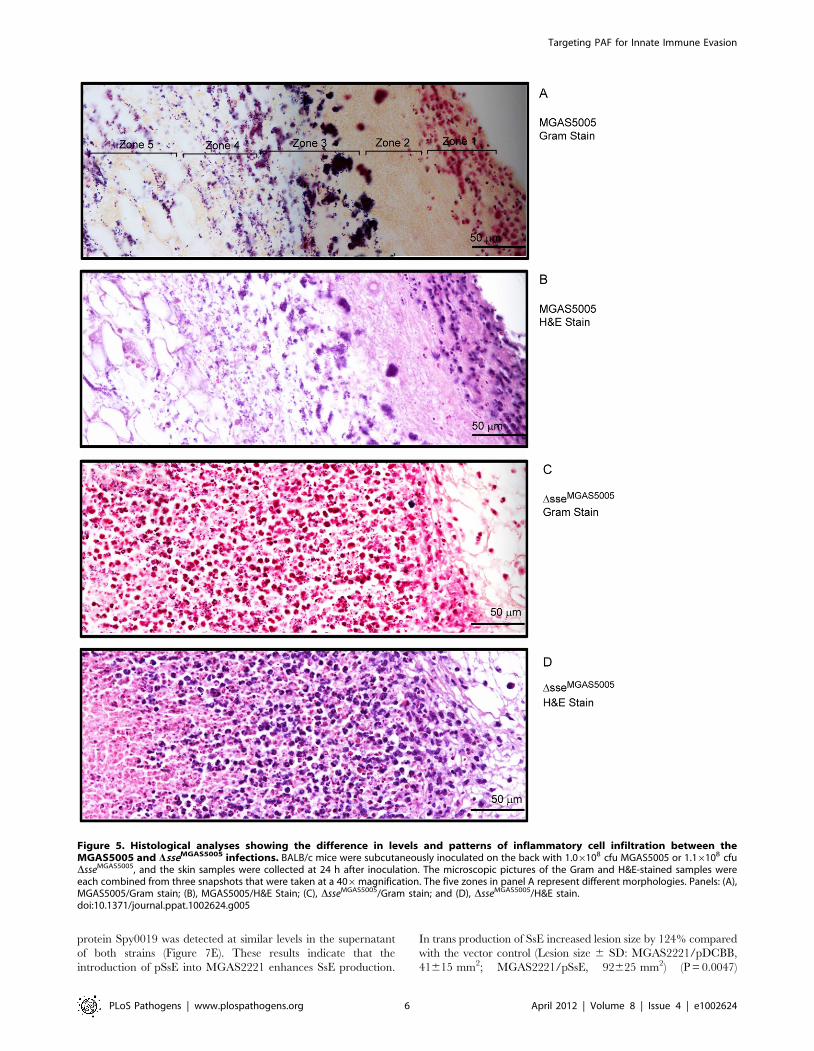
tion. There are five morphological zones at an end of the
MGAS5005 inoculation site starting from the interior side of the
skin (the right side in panels A and B of Figure 5): Zone 1,
neutrophils and other inflammatory cells without GAS; Zone 2,
amorphous host materials lack of GAS; Zone 3, a few
inflammatory cells that could reach the boundary of the GAS
territory were victimized by and associated with massive amount
of GAS; Zone 4, necrotized adipose tissue and GAS without
inflammatory cells; and Zone 5, invasion of GAS along the
interstitial space of the adipose cells (Figure 5A and 5B). Thus,
MGAS5005 not only reduces infiltration of neutrophils but also
keep inflammatory cells away. However, inflammatory cells and
DsseMGAS5005 bacteria were mingled throughout the infection site
(Figure 5C and 5D). Similar results were obtained in CD-1 Swiss
mice.
Next, we used the myeloperoxidase assay [44] to quantify
neutrophil ingress to the skin infection sites of MGAS5005 and
DsseMGAS5005 at 24 h after subcutaneous infection of BALB/c
mice. The mean neutrophil number 6 SD of the Dsse infection site
was (1.160.12)6106/mm2, which was 19.6 and 346-fold greater
than the neutrophil number at the MGAS5005 inoculation site
[(5.462.3)6104 neutrophils/mm2] and at the spread infection
area of MGAS5005 [(3.160.87)6103 neutrophils/mm2]. Reverse
complementation of DsseMGAS5005 with the sse gene (Dsse-sse)
restored the inhibition of neutrophil recruitment [(5.661.0)6104
neutrophils/mm2]. The difference is significant between the
DsseMGAS5005 sample and each of the other samples but
insignificant among the other samples in one way ANOVA
analysis using the Tukey’s Multiple Comparison Test (Figure 6A).
Reduction of Neutrophil Ingress to DsseMGAS5005 Sites inPAF Receptor-Deficient Mice
The receptor of PAF (PAFR) is a G protein-coupled receptor
that mediates the biological activities of PAF. We used PAFR-
deficient mice [30] to test whether SsE inhibits neutrophil
infiltration by hydrolyzing PAF. MGAS5005 induced low and
similar levels of neutrophil recruitment in both BALB/c and
PAFR2/2 mice. However, the mean number of recruited
neutrophils at the DsseMGAS5005 infection site was reduced by
47% in PAFR2/2 mice compared with BALB/c mice (Figure 6B).
The reduction of neutrophil influx due to the absence of the PAF
receptor was 52.7% of the enhancement of neutrophil influx as a
result of the sse deletion. These results suggest that targeting PAF
by SsE is an equally important mechanism as an PAF-independent
mechanism. These results strongly suggest that PAF plays a
significant role in neutrophil infiltration in GAS infections and that
SsE-mediated hydrolysis of PAF contributes to the observed
reduction in neutrophil infiltration.
Efficient Clearance of DsseMGAS5005 by NeutrophilsSince Dsse bacteria were associated with high levels of neutrophils,
these bacteria should be killed by recruited neutrophils. Indeed, the
numbers of viable DsseMGAS5005 at 24 and 48 hours post-inoculation
were 8.3% and 4.8% of those found at 1 h after inoculation,
respectively; whereas the numbers of MGAS5005 at 24 and 48 h
post-inoculation were 70% and 128% of those found at 1 h after
inoculation, respectively (Figure 6C), suggesting that DsseMGAS5005 is
cleared more efficiently than MGAS5005 at skin infection sites.
No Detrimental Effects of sse Deletion on Transcriptionof spyCEP, scpA, and Other CsrRS/CovRS- andMga-Regulated Genes
In a transcription profiling analysis for MGAS5005 and
DsseMGAS5005 using the NimbleExpress Streptococcus pyogenes arrays,
the transcription levels of the genes regulated by the multiple gene
regulator of GAS (Mga) and CsrRS/CovRS in DsseMGAS5005 were
70% to 135% of those in MGAS5005 at the mid-exponential
growth phase except that sse transcript was not detected in
DsseMGAS5005 (Figure S4). These results rule out the possibility that
the phenotype of DsseMGAS5005 is caused by alteration in
transcription of the scpA, spyCEP/scpC, sda1/sdaD2, slo, sagA, hasA,
speB, and emm genes, which are involved in innate immune evasion
by GAS.
Effects of sse Deletion on Virulence, Soft Tissue Invasion,and Neutrophil Recruitment in MGAS2221 Infection
MGAS5005 has a natural null covS deletion, which enhances
expression of sse and many other virulence genes [18,40]. To test
whether SsE contributes to pathogenesis and inhibition of
neutrophil recruitment in GAS with the wild-type csrRS/covRS
genes, we deleted the sse gene in MGAS2221. Fifty seven percent
of BALB/c mice infected subcutaneously with 1.56108 cfu of
MGAS2221 were dead whereas all mice infected with 1.66108 cfu
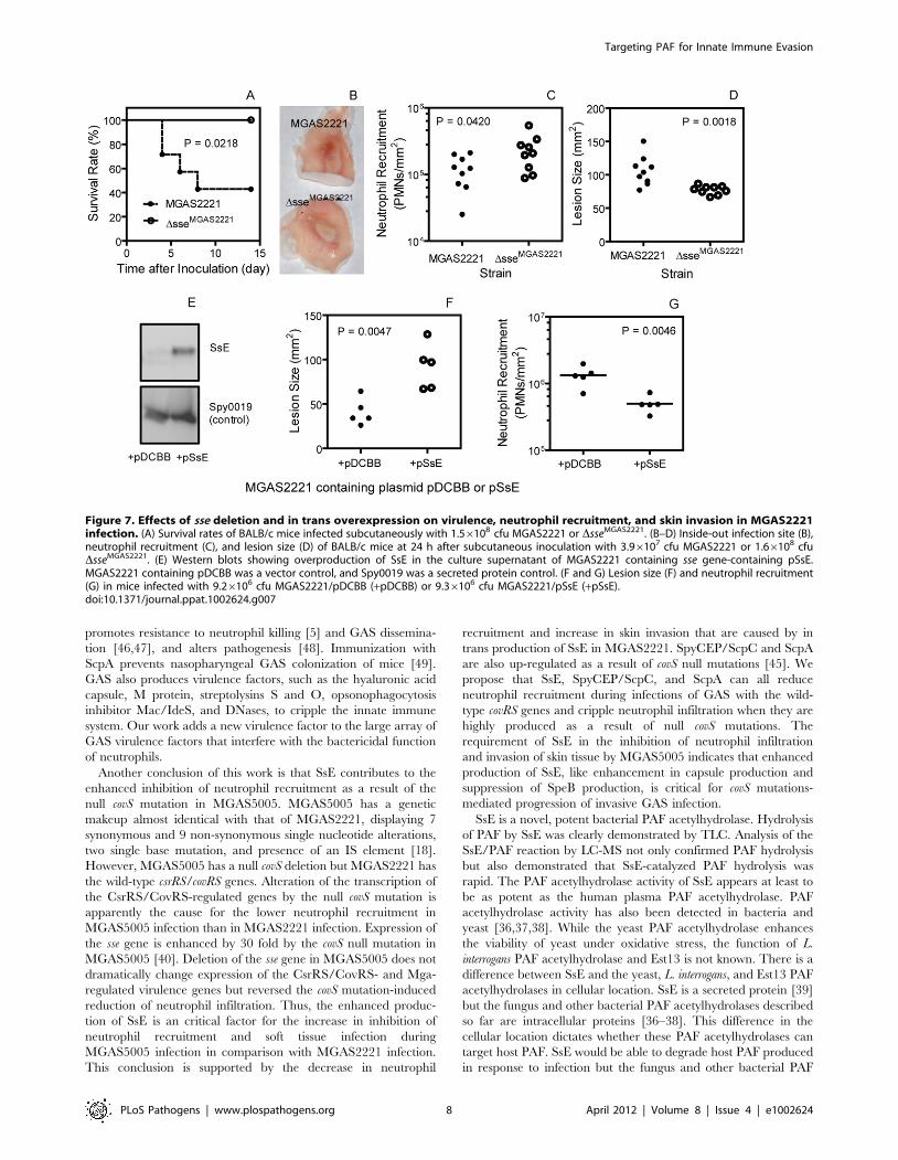
DsseMGAS2221 survived (P = 0.0218) (Figure 7A). In a separate
experiment, 3.96107 cfu MGAS2221 or 1.66108 cfu DsseM-
GAS2221 bacteria were inoculated into BALB/c mice. The lesion
appearance was obviously different between the wt and mutant
infection sites (Figure 7B). The number of neutrophils at the
DsseMGAS2221 site was significantly higher than that at the
MGAS2221 site (mean neutrophil number 6 SD: DsseMGAS2221,
(2.461.4)6105/mm2; MGAS2221, (1.260.6)6105/mm2) (P =
0.0420) (Figure 7C). Conversely, the size of the DsseMGAS2221 site
was significant smaller than that of the MGAS2221 site
(mean size 6 SD: MGAS2221, 106620 mm2; DsseMGAS2221,
7766 mm2) (P = 0.0014) (Figure 7D). It should be stressed that the
significant role of SsE in the invasion of skin tissue and inhibition
of neutrophil recruitment was observed with a dose of
DsseMGAS2221 that was 3 times higher than that of MGAS2221.
The results using the higher dose of the mutant suggest that the
mutant phenotype is not caused by a growth defect. Thus, SsE can
reduce neutrophil recruitment and enhances soft tissue invasion in
infection with a representative M1T1 strain with the wild-type
csrRS/covRS background.
Inhibition of Neutrophil Recruitment and Enhancementof Soft Tissue Invasion by In Trans Expression of SsE inMGAS2221
The effects of in trans expression of SsE on neutrophil
recruitment and lesion size during subcutaneous MGAS2221
infection of mice further confirm the role of SsE in inhibition of
neutrophil recruitment and enhancement of soft tissue invasion by
GAS. The sse gene was cloned into pDCBB [45], yielding pSsE. At
the early growth phase (OD600 = 0.2), SsE was detected in the
supernatant of MGAS2221/pSsE but not MGAS2221/pDCBB
(vector control) by Western blotting analysis, whereas the secreted
Targeting PAF for Innate Immune Evasion
PLoS Pathogens | www.plospathogens.org 5 April 2012 | Volume 8 | Issue 4 | e1002624
protein Spy0019 was detected at similar levels in the supernatant
of both strains (Figure 7E). These results indicate that the
introduction of pSsE into MGAS2221 enhances SsE production.
In trans production of SsE increased lesion size by 124% compared
with the vector control (Lesion size 6 SD: MGAS2221/pDCBB,
41615 mm2; MGAS2221/pSsE, 92625 mm2) (P = 0.0047)
Figure 5. Histological analyses showing the difference in levels and patterns of inflammatory cell infiltration between theMGAS5005 and DsseMGAS5005 infections. BALB/c mice were subcutaneously inoculated on the back with 1.06108 cfu MGAS5005 or 1.16108 cfuDsseMGAS5005, and the skin samples were collected at 24 h after inoculation. The microscopic pictures of the Gram and H&E-stained samples wereeach combined from three snapshots that were taken at a 406magnification. The five zones in panel A represent different morphologies. Panels: (A),MGAS5005/Gram stain; (B), MGAS5005/H&E Stain; (C), DsseMGAS5005/Gram stain; and (D), DsseMGAS5005/H&E stain.doi:10.1371/journal.ppat.1002624.g005
Targeting PAF for Innate Immune Evasion
PLoS Pathogens | www.plospathogens.org 6 April 2012 | Volume 8 | Issue 4 | e1002624
(Figure 7F). Inversely, in trans production of SsE reduced neutrophil
recruitment by 72% (mean neutrophil number 6 SD: MGAS2221/
pDCBB, (6.260.28)6105/mm2; MGAS2221/pSsE, 1.760.11)6105/mm2) (P = 0.0111) (Figure 7G).
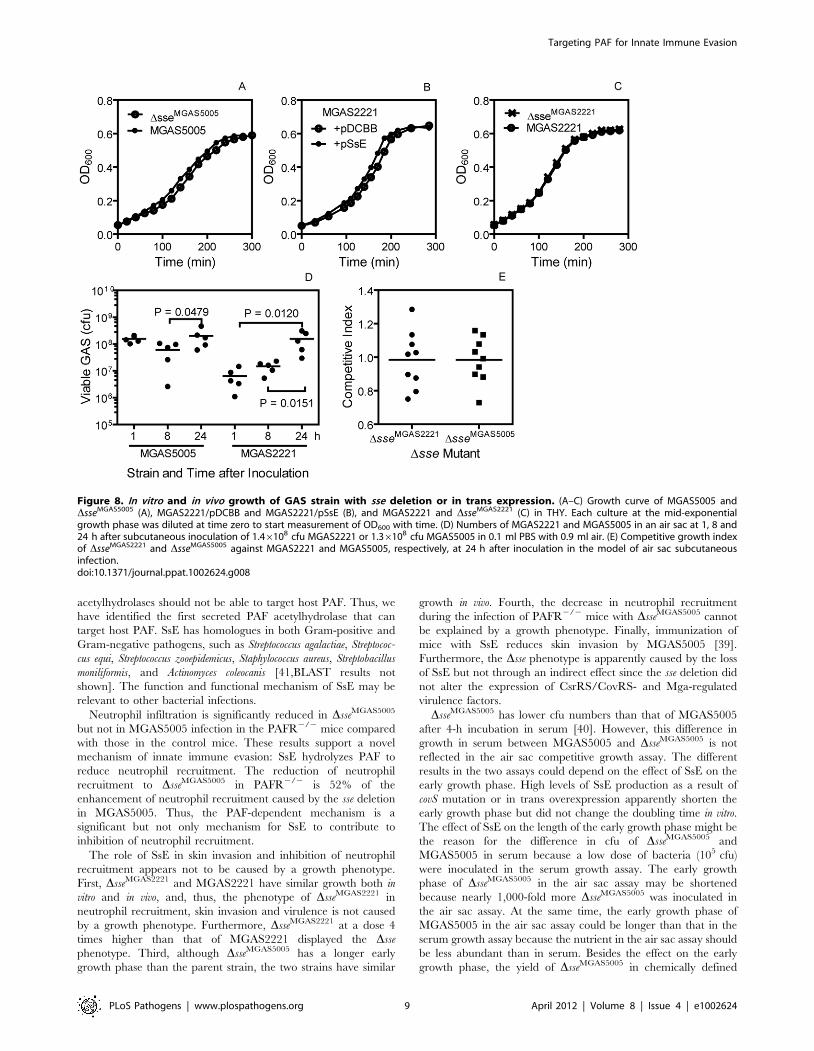
In Vitro and In Vivo Growth of DsseMGAS5005 andDsseMGAS2221
The DsseMGAS5005 mutant has a longer early growth phase by
about 15 min (Figure 8A) and about 10% more viable CFU per
OD600 at the exponential growth phase (data not shown) than its
parent strain in Todd-Hewitt broth supplemented with 0.2% yeast
extract (THY). Consistent with this result, in trans overexpression
of SsE in MGAS2221 shows a 20-min shorter early growth phase
than the vector control (Figure 8B). However, MGAS2221 and
DsseMGAS2221 have identical growth curves in THY (Figure 8C).
Thus, the effect of sse expression on the length of early growth
phase is obvious when SsE is highly produced.
To examine the growth of the mutants in vivo, we performed a
competitive growth assay using an air sac infection model. A 1:1
DsseMGAS2221:MGAS2221 or DsseMGAS5005:MGAS5005 mixture
was injected with air in the subcutis of mice, and, 24 h later, the
air sac was lavaged after the mice were euthanized. The lavage
samples were plated, and the Dsse:wt GAS ratio of the GAS
colonies was determined by PCR analysis. The Dsse:wt GAS ratio
in the inoculum was measured by plating the individual GAS
suspension prior to mixing. The mean number of MGAS5005 and
MGAS2221 at 24 h was 11 and 3 times as the corresponding
number at 8 h, respectively (Figure 8D), indicating that GAS grew
in the air sac. The competitive index, the Dsse:wt ratio in the
lavage sample/the ratio in the inoculum, for both DsseMGAS2221
and DsseMGAS5005 has a mean value of about 1 (Figure 8E),
indicating that each mutant and its parent strain have similar
growth in vivo. These data indicate that the phenotype of
DsseMGAS5005 and DsseMGAS2221 is not caused by a growth
phenotype.
Discussion
This study presents three major findings regarding evasion of
the innate immune system by GAS. First, SsE significantly
contributes to GAS inhibition of neutrophil recruitment. Second,
SsE is a potent PAF acetylhydrolase and the first secreted bacterial
PAF acetylhydrolase identified so far. Third, SsE inactivates the
ability of PAF to induce activation and migration of neutrophils,
and the PAF receptor significantly contributes to neutrophil
recruitment in skin GAS infection. These findings identify a new
means for evasion of the innate immune system by GAS and
support a novel paradigm for bacterial inhibition of neutrophil
recruitment and function in which neutrophil recruitment is
reduced by inactivating PAF.
One conclusion of this work is that SsE is required for the severe
inhibition of neutrophil recruitment by MGAS5005 in the mouse
model of necrotizing fasciitis. This nearly complete inhibition of
neutrophil infiltration is similar to that of severe GAS infections in
some human patients and experimental animal infections
[2,21,22,23]. In addition, SsE is critical for the virulence and
dissemination of MGAS5005 and is a protective antigen [39,40].
SsE also reduces neutrophil recruitment and enhances virulence
and skin tissue invasion in infection with MGAS2221. Thus, SsE is
a significant contributor to the innate immune evasion and tissue
invasion by GAS with or without covRS mutations. It is well known
that GAS produces C5a peptidase ScpA and IL-8/CXC peptidase
SpyCEP/ScpC to reduce neutrophil recruitment. SpyCEP/ScpC
reduces neutrophil infiltration in soft tissue infections of mice [2,3],
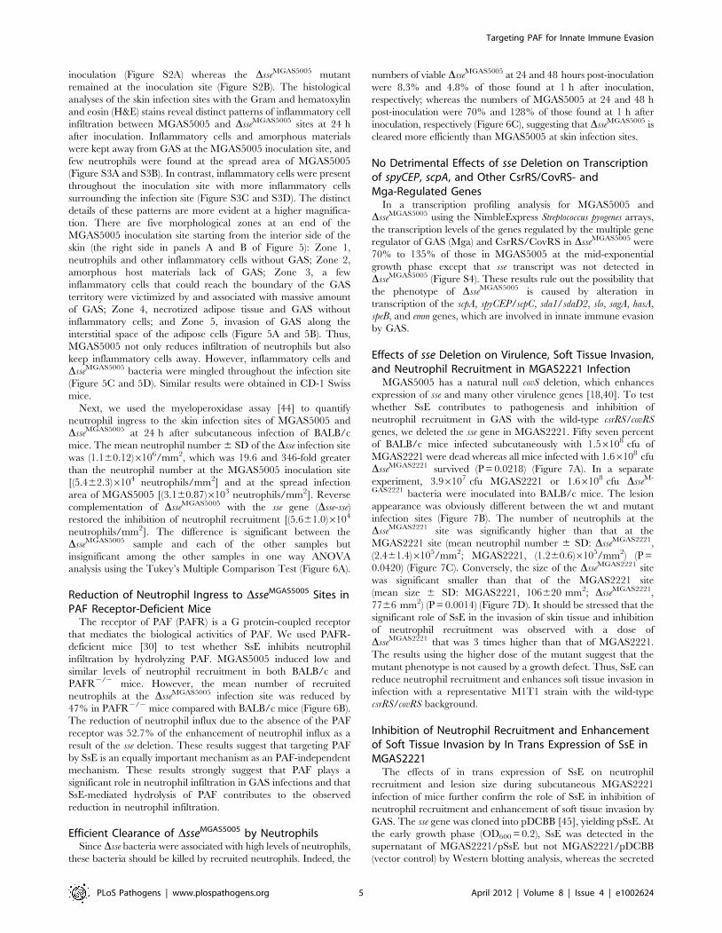
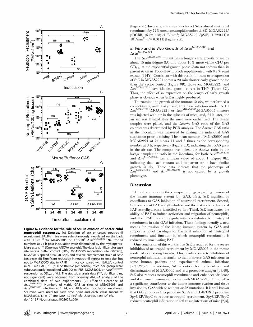
Figure 6. Evidence for the role of SsE in evasion of bactericidalneutrophil responses. (A) Deletion of sse enhances neutrophilrecruitment. BALB/c mice were subcutaneously inoculated on the backwith 1.06108 cfu MGAS5005 or 1.16108 DsseMGAS5005. Neutrophilnumbers at 24 h post-inoculation were determined by the myeloperox-idase assay. *** (One-way ANOVA analysis): The data is significant for Dssesite versus buffer control (PBS), MGAS5005 inoculation site (5005inj),MGAS5005 spread area (5005sp), and reverse-complement strain of Dsse(Dsse-sse). (B) Significant reduction in neutrophil ingress to Dsse site, butnot to MGAS5005 site, in PAFR2/2 mice compared with BALB/c controlmice. Five PAFR2/2 (KO) or BALB/c (wt control) mice per group weresubcutaneously inoculated with 0.2 ml PBS, MGAS5005, or DsseMGAS5005
suspension at OD600 of 0.8. The statistic analysis data (***, significant; ns,not significant) were obtained from one-way ANOVA analysis of thecombined data of two experiments. (C) Efficient clearance ofDsseMGAS5005. Numbers of viable GAS at sites of MGAS5005 andDsseMGAS5005 infection at 1, 24, and 48 h after inoculation are shown.Six mice were used for each time point and each strain. Inoculum:MGAS5005, 1.16108 cfu; Dsse, 1.26108 cfu; Dsse-sse, 1.06108 cfu.doi:10.1371/journal.ppat.1002624.g006
Targeting PAF for Innate Immune Evasion
PLoS Pathogens | www.plospathogens.org 7 April 2012 | Volume 8 | Issue 4 | e1002624
promotes resistance to neutrophil killing [5] and GAS dissemina-
tion [46,47], and alters pathogenesis [48]. Immunization with
ScpA prevents nasopharyngeal GAS colonization of mice [49].
GAS also produces virulence factors, such as the hyaluronic acid
capsule, M protein, streptolysins S and O, opsonophagocytosis
inhibitor Mac/IdeS, and DNases, to cripple the innate immune
system. Our work adds a new virulence factor to the large array of
GAS virulence factors that interfere with the bactericidal function
of neutrophils.
Another conclusion of this work is that SsE contributes to the
enhanced inhibition of neutrophil recruitment as a result of the
null covS mutation in MGAS5005. MGAS5005 has a genetic
makeup almost identical with that of MGAS2221, displaying 7
synonymous and 9 non-synonymous single nucleotide alterations,
two single base mutation, and presence of an IS element [18].
However, MGAS5005 has a null covS deletion but MGAS2221 has
the wild-type csrRS/covRS genes. Alteration of the transcription of
the CsrRS/CovRS-regulated genes by the null covS mutation is
apparently the cause for the lower neutrophil recruitment in
MGAS5005 infection than in MGAS2221 infection. Expression of
the sse gene is enhanced by 30 fold by the covS null mutation in
MGAS5005 [40]. Deletion of the sse gene in MGAS5005 does not
dramatically change expression of the CsrRS/CovRS- and Mga-
regulated virulence genes but reversed the covS mutation-induced
reduction of neutrophil infiltration. Thus, the enhanced produc-
tion of SsE is an critical factor for the increase in inhibition of
neutrophil recruitment and soft tissue infection during
MGAS5005 infection in comparison with MGAS2221 infection.
This conclusion is supported by the decrease in neutrophil
recruitment and increase in skin invasion that are caused by in
trans production of SsE in MGAS2221. SpyCEP/ScpC and ScpA
are also up-regulated as a result of covS null mutations [45]. We
propose that SsE, SpyCEP/ScpC, and ScpA can all reduce
neutrophil recruitment during infections of GAS with the wild-
type covRS genes and cripple neutrophil infiltration when they are
highly produced as a result of null covS mutations. The
requirement of SsE in the inhibition of neutrophil infiltration
and invasion of skin tissue by MGAS5005 indicates that enhanced
production of SsE, like enhancement in capsule production and
suppression of SpeB production, is critical for covS mutations-
mediated progression of invasive GAS infection.
SsE is a novel, potent bacterial PAF acetylhydrolase. Hydrolysis
of PAF by SsE was clearly demonstrated by TLC. Analysis of the
SsE/PAF reaction by LC-MS not only confirmed PAF hydrolysis
but also demonstrated that SsE-catalyzed PAF hydrolysis was
rapid. The PAF acetylhydrolase activity of SsE appears at least to
be as potent as the human plasma PAF acetylhydrolase. PAF
acetylhydrolase activity has also been detected in bacteria and
yeast [36,37,38]. While the yeast PAF acetylhydrolase enhances
the viability of yeast under oxidative stress, the function of L.
interrogans PAF acetylhydrolase and Est13 is not known. There is a
difference between SsE and the yeast, L. interrogans, and Est13 PAF
acetylhydrolases in cellular location. SsE is a secreted protein [39]
but the fungus and other bacterial PAF acetylhydrolases described
so far are intracellular proteins [36–38]. This difference in the
cellular location dictates whether these PAF acetylhydrolases can
target host PAF. SsE would be able to degrade host PAF produced
in response to infection but the fungus and other bacterial PAF
Figure 7. Effects of sse deletion and in trans overexpression on virulence, neutrophil recruitment, and skin invasion in MGAS2221infection. (A) Survival rates of BALB/c mice infected subcutaneously with 1.56108 cfu MGAS2221 or DsseMGAS2221. (B–D) Inside-out infection site (B),neutrophil recruitment (C), and lesion size (D) of BALB/c mice at 24 h after subcutaneous inoculation with 3.96107 cfu MGAS2221 or 1.66108 cfuDsseMGAS2221. (E) Western blots showing overproduction of SsE in the culture supernatant of MGAS2221 containing sse gene-containing pSsE.MGAS2221 containing pDCBB was a vector control, and Spy0019 was a secreted protein control. (F and G) Lesion size (F) and neutrophil recruitment(G) in mice infected with 9.26106 cfu MGAS2221/pDCBB (+pDCBB) or 9.36106 cfu MGAS2221/pSsE (+pSsE).doi:10.1371/journal.ppat.1002624.g007
Targeting PAF for Innate Immune Evasion
PLoS Pathogens | www.plospathogens.org 8 April 2012 | Volume 8 | Issue 4 | e1002624
acetylhydrolases should not be able to target host PAF. Thus, we
have identified the first secreted PAF acetylhydrolase that can
target host PAF. SsE has homologues in both Gram-positive and
Gram-negative pathogens, such as Streptococcus agalactiae, Streptococ-
cus equi, Streptococcus zooepidemicus, Staphylococcus aureus, Streptobacillus
moniliformis, and Actinomyces coleocanis [41,BLAST results not
shown]. The function and functional mechanism of SsE may be
relevant to other bacterial infections.
Neutrophil infiltration is significantly reduced in DsseMGAS5005
but not in MGAS5005 infection in the PAFR2/2 mice compared
with those in the control mice. These results support a novel
mechanism of innate immune evasion: SsE hydrolyzes PAF to
reduce neutrophil recruitment. The reduction of neutrophil
recruitment to DsseMGAS5005 in PAFR2/2 is 52% of the
enhancement of neutrophil recruitment caused by the sse deletion
in MGAS5005. Thus, the PAF-dependent mechanism is a
significant but not only mechanism for SsE to contribute to
inhibition of neutrophil recruitment.
The role of SsE in skin invasion and inhibition of neutrophil
recruitment appears not to be caused by a growth phenotype.
First, DsseMGAS2221 and MGAS2221 have similar growth both in
vitro and in vivo, and, thus, the phenotype of DsseMGAS2221 in
neutrophil recruitment, skin invasion and virulence is not caused
by a growth phenotype. Furthermore, DsseMGAS2221 at a dose 4
times higher than that of MGAS2221 displayed the Dsse
phenotype. Third, although DsseMGAS5005 has a longer early
growth phase than the parent strain, the two strains have similar
growth in vivo. Fourth, the decrease in neutrophil recruitment
during the infection of PAFR2/2 mice with DsseMGAS5005 cannot
be explained by a growth phenotype. Finally, immunization of
mice with SsE reduces skin invasion by MGAS5005 [39].
Furthermore, the Dsse phenotype is apparently caused by the loss
of SsE but not through an indirect effect since the sse deletion did
not alter the expression of CsrRS/CovRS- and Mga-regulated
virulence factors.
DsseMGAS5005 has lower cfu numbers than that of MGAS5005
after 4-h incubation in serum [40]. However, this difference in
growth in serum between MGAS5005 and DsseMGAS5005 is not
reflected in the air sac competitive growth assay. The different
results in the two assays could depend on the effect of SsE on the
early growth phase. High levels of SsE production as a result of
covS mutation or in trans overexpression apparently shorten the
early growth phase but did not change the doubling time in vitro.
The effect of SsE on the length of the early growth phase might be
the reason for the difference in cfu of DsseMGAS5005 and
MGAS5005 in serum because a low dose of bacteria (105 cfu)
were inoculated in the serum growth assay. The early growth
phase of DsseMGAS5005 in the air sac assay may be shortened
because nearly 1,000-fold more DsseMGAS5005 was inoculated in
the air sac assay. At the same time, the early growth phase of
MGAS5005 in the air sac assay could be longer than that in the
serum growth assay because the nutrient in the air sac assay should
be less abundant than in serum. Besides the effect on the early
growth phase, the yield of DsseMGAS5005 in chemically defined
Figure 8. In vitro and in vivo growth of GAS strain with sse deletion or in trans expression. (A–C) Growth curve of MGAS5005 andDsseMGAS5005 (A), MGAS2221/pDCBB and MGAS2221/pSsE (B), and MGAS2221 and DsseMGAS2221 (C) in THY. Each culture at the mid-exponentialgrowth phase was diluted at time zero to start measurement of OD600 with time. (D) Numbers of MGAS2221 and MGAS5005 in an air sac at 1, 8 and24 h after subcutaneous inoculation of 1.46108 cfu MGAS2221 or 1.36108 cfu MGAS5005 in 0.1 ml PBS with 0.9 ml air. (E) Competitive growth indexof DsseMGAS2221 and DsseMGAS5005 against MGAS2221 and MGAS5005, respectively, at 24 h after inoculation in the model of air sac subcutaneousinfection.doi:10.1371/journal.ppat.1002624.g008
Targeting PAF for Innate Immune Evasion
PLoS Pathogens | www.plospathogens.org 9 April 2012 | Volume 8 | Issue 4 | e1002624
medium is lower than that of MGAS5005 [40], suggesting that
SsE may be able to recycle metabolites or surface structures. These
differential in vitro growth features of DsseMGAS5005 and
MGAS5005 appear not to be displayed in vivo, suggesting that
the in vitro difference does not represent a genuine growth defect.
Nonetheless, the in vitro growth data indicate that SsE can act on
the GAS bacteria. This action could be the basis for a PAF-
independent mechanism, in addition to the PAF-dependent
mechanism, for the innate immune evasion by SsE.
Neutrophil influx to DsseMGAS5005 sites in the PAFR2/2 mice
was half of that in the control mice. This is the first demonstration
for the importance of the PAF receptor in neutrophil recruitment
in response to a bacterial infection. The PAF receptor is not
critical for neutrophil infiltration in pulmonary Klebsiella pneumonia,
Pseudomonas aeruginosa, Streptococcus pneumoniae infections and poly-
microbial sepsis caused by cecum ligation and puncture
[50,51,52,53]. This difference suggests that PAF may play a
critical role in neutrophil recruitment in skin infection but not in
pulmonary infections. It is also possible that these pathogens, like
MGAS5005, can inactivate PAF.
Hermoso et al. have found that the protein Pce of Streptococcus
pneumoniae hydrolyzes the phosphocholine group of PAF and
hypothesized that Pce has the capacity to interact with and
hydrolyze PAF in the bloodstream in vivo, impacting on
pathogenesis [54]. Apparently, bacterial pathogens have evolved
different enzymatic activities to eliminate PAF, supporting an
important role of PAF in host responses against bacterial
infections.
PAF can be involved in innate immune responses in different
ways. Administration of PAF can lead to neutrophil infiltration in
the lung and skin [55], and PAF may participate in the
inflammatory responses during GAS infections. IL-12-induced
chemotaxis of NK cells and neutrophils is mediated by PAF [56].
PAF can activate neutrophils and induce migration of isolated
neutrophils [28]. Treatment of PAF with SsE abolishes the ability
of PAF to activate and induce migration of neutrophils. PAF can
function as a chemoattractant in the neutrophil responses during
GAS infection. It is also possible that PAF plays a role in both the
inflammatory response and chemotaxis during GAS infection.
PAF also activates platelets in human and some animals. However,
the inhibition of the PAF-induced activation of platelets does not
play a role in the phenotype of the Dsse mutants in the mouse
infections since murine platelets do not produce the PAF receptor
according to Dr. Guy Zimmerman at University of Utah. We will
examine how PAF contributes to the neutrophil response during
GAS infections in our follow-up studies.
Materials and Methods
Declaration of Ethical ApprovalAll animal experimental procedures were carried out in strict
accordance with the recommendations in the Guide for the Care
and Use of Laboratory Animals of the National Institutes of
Health. The protocols for the experiments performed at Montana
State University (MSU) and Federal University of Minas Gerais
(UFMG) were approved by the Institutional Animal Care and Use
Committee at MSU (Permit number: 2009-09) and the Animal
Ethics Committee of Instituto de Ciencias Biologicas (Permit
number: 168/11) (Belo Horizonte, Brazil), respectively. Blood was
collected from healthy donors in accordance with a protocol
approved by the Institutional Review Board at MSU (Protocol
No. BL031109). Written informed consent was provided by study
participants and/or their legal guardians.
MaterialsPAF (1-O-hexadecyl-2-acetyl-sn-glycero-3-phosphorylcholine),
lyso-PAF C-16 (1-O-hexadecyl-sn-glycero-3-phosphocholine), hu-
man recombinant plasma PAF acetylhydrolase, heptanoyl thio-
PC, and the PAF acetylhydrolase assay kit using 2-thio-PAF as the
substrate were purchased from Cayman Chemical (An Harbor,
MI, USA). Whatman LK6D Silica Gel 60A thin layer chroma-
tography plates were purchased from Whatman International
LLC (Clifton, NJ, USA). Recombinant wild-type and S178A
mutant SsE proteins were prepared, as previously described [39].
Bacterial Strains and GrowthMGAS5005 is a hypervirulent M1T1 GAS strain isolated from
an invasive case in Ontario [9]. MGAS2221 is a M1T1 GAS
strain isolated from a scarlet fever patient [48]. MGAS5005 and
MGAS2221 have almost identical genetic contents but the former
has a null covS 1-bp deletion [18]. DsseMGAS5005, an in-frame sse
deletion mutant of MGAS5005 missing amino acids 55–261 of
SsE and Dsse-sse, a reverse complement strain of Dsse, have been
described [40]. The same sse deletion procedure was followed to
obtain DsseMGAS2221. These bacteria for experiments conducted at
MSU were grown to mid-exponential phase at 37uC in 5% CO2 in
THY. GAS bacteria used in the PAFR2/2 experiment at UFMG
were grown in brain heart infusion broth (BHI). Tryptose agar
with 5% sheep blood, THY agar, and BHI agar were used as the
solid media. GAS bacteria used for the animal experiments were
harvested at the exponential growth phase and washed three times
with and resuspended in pyrogen-free phosphate-buffered saline
(PBS) to desired doses.
Assays for PAF Acetylhydrolase Activity of SsESsE-catalyzed hydrolysis of PAF was monitored by TLC and
LC-MS analyses and a colorimetric assay. For TLC analysis,
1.4 mM PAF was mixed with 0.08 mM wild-type SsE or SsES178A
in 50 ml of 20 mM Tris-HCl, pH 8.0, and the reaction was
stopped by adding 50 ml acetonitrile containing 1% formic acid
after 10-min incubation at room temperature. Two ml of the
reaction samples, untreated PAF, lyso-PAF, and PAF/lyso-PAF
mixtures were spotted on a TLC plate, and these compounds were
resolved using a methanol/chloroform/water (65:30:6 by volume)
mixture as the mobile phase. After chromatography, PAF and
lyso-PAF were visualized by spraying with 5% ammonium
molybdate sulfate and heating. Protein concentrations were
determined using the modified Lowry protein assay kit from
Pierce with bovine serum albumin as a standard.
For LC-MS analysis, PAF hydrolysis reactions were performed
as in the TLC analysis and stopped at 0 and 5 min after mixing for
the wild-type SsE/PAF reaction and 40 min for the SsES178A/PAF
reaction. The samples were diluted with 5% acetonitrile
containing 1% formic acid, and 1 ml of the diluted samples were
analyzed by reverse-phase liquid chromatography and positive ion
mass spectroscopy using an Agilent 1100 HPLC with autosampler
(Agilent Technologies, Inc., Santa Clara, CA, USA) and a Bruker
micrOTOF mass spectrometer (Bruker Daltonik GmbH, Bremen,
German). The reverse-phase chromatography consisted of a 3.2-
ml gradient between H2O and 95% acetonitrile, both with 0.1%
formic acid, using a Michrom Bioresources C8 column (861 mm).
The LC/MS data were analyzed using DataAnalysis 4.0 software
(Bruker Daltonik GmbH). The mass spectrometer was calibrated
using the peaks between 118 and 922 m/z of the Agilent G2421A
electrospray calibrant solution infused directly to the source at a
rate of 180 ml/h. PAF and lyso-PAF compounds were identified
via high mass accuracy with positive control samples with m/z
values of 482.3600 and 524.3722, respectively (actual masses of
Targeting PAF for Innate Immune Evasion
PLoS Pathogens | www.plospathogens.org 10 April 2012 | Volume 8 | Issue 4 | e1002624
482.3605 and 524.3711, errors of 21 ppm and +0.2 ppm,
respectively). PAF and lyso-PAF were evaluated for carry-over
on the C8 column with blank runs, but the C8 column with the
described chromatography had no detectable carry-over between
runs.
The colorimetric assay used the PAF acetylhydrolase assay kit
from Cayman Chemical. The reactions were initiated by mixing
100 ml 20 mM Tris-HCl, pH 8.0, containing 2-thio-PAF at
various concentrations and 100 ml Tris-HCl containing 4.3 nM
SsE and 0.5 mM DTNB at room temperature in a 96-well plate.
Absorbance at 414 nm (A414) of the reaction mixture was recorded
every 6 s using a SPECTRAMax 384 Plus spectrophotometer
(Molecular Devices, Sunnyvale, CA, USA) and was used to
determine initial rates of hydrolysis of 2-thio-PAF as described in
the Results section.
Isolation of Human NeutrophilsNeutrophils were isolated from the blood using dextran
sedimentation, followed by Histopaque 1077 gradient separation
and hypotonic lysis of red blood cells, as described previously [57].
Isolated neutrophils were washed twice and resuspended in HBSS
without Ca2+ and Mg2+ for Ca2+ mobilization or with Ca2+ and
Mg2+ for chemotaxis measurement. Neutrophil preparations were
.95% pure, as determined by light microscopy, and .98%
viable, as determined by trypan blue exclusion.
Ca2+ Mobilization AssayChanges in free intracellular [Ca2+] were measured with a
FlexStation II Scanning Fluorometer (Molecular Devices) using
Fluo-4 acetoxymethyl ester (Invitrogen), as previously described
[58]. Briefly, human neutrophils, suspended in Hanks’ balanced
salt solution (HBSS) containing 10 mM HEPES, were loaded with
Fluo-4 acetoxymethyl ester dye (1.25 ıg/ml final concentration) for
30 min in the dark at 37uC. After dye loading, the cells were
washed with HBSS containing 10 mM HEPES, resuspended in
HBSS containing 10 mM HEPES and Ca2+ and Mg2+, and
separated into aliquots, which were inserted into the wells of flat-
bottomed, half-area-well black microtiter plates (26105 cells/well).
After addition of untreated or SsE-treated PAF, changes in
fluorescence were monitored (lex = 485 nm, lem = 538 nm) every
5 s for 240 s at room temperature.
Chemotaxis AssayThe chemotaxis assay was performed using the ChemoTx
Disposable Chemotaxis System in a 96 well microplate format
(Neuro Probe, Inc., Gaithersburg, MD, USA) and the CellTitr-
Glo Luminescent Cell Viability Assay (Promega, Madison, WI,
USA), as described previously [58]. PAF (1.4 mM) was incubated
with 0.08 mM SsE or SsES178A in 50 ml PBS, pH 7.0, at room
temperature for 30 min, and the reaction was stopped by adding
an equal volume of acetonitrile. Untreated and treated PAF were
diluted to desired concentrations with HBSS containing 10 mM
HEPES, Ca2+, Mg2+, and 0.1% BSA (HBSS/BSA). The protein
control reaction samples were diluted by the same fold of the
dilution as the treated PAF samples. The samples were added to
wells of the assay plate at 30 ml/well in 4 replicates. The plate was
covered with the filter, and 46104 neutrophils/well were placed
on the top of the filter. The plate was incubated at 37uC for 1 h.
Neutrophils that did not migrate were removed, and 20 ml/well of
2.5 mM EDTA was added. After incubating for 10 min at 4uC,
the EDTA solution was removed, the plate was centrifuged at
600 rpm for 5 min, and 20 ml/well of CellTitr-Glo Luminescent
Cell Viability Assay reagent was added. Luminescence from each
well was monitored using a Fluoroscan Ascent FL Luminometer
(Thermo Electron Corporation). The number of migrated cells
was determined based on a standard curve using known numbers
of neutrophils.
Mouse InfectionsGroups of five-week-old female inbred BALB/c and outbred
CD-1 Swiss mice (Charles River Laboratory) were subcutaneously
infected with 0.2 ml of an OD600 of 0.8 of GAS suspension in PBS
or at indicated doses. Inocula were determined by plating. Mice
were sacrificed at 24 h to collect skin samples for histological
analyses and measurement of neutrophil infiltration and GAS
CFU. Infected mice in virulence studies were monitored twice a
day to get survival rates.
The PAFR2/2 mouse experiment was similarly performed at
Dr. Mauro Teixeira’s laboratory at UFMG, Brazil. BALB/c mice
(8 to 12 week-old) were obtained from CEBIO (Bioterism Center)
of UFMG, and PAFR2/2 mice (8 to 12 week-old) were generated
as previously described and backcrossed at least 10 generations
into a BALB/c background [30,53]. Mice were housed in standard
conditions and had free access to commercial chow and water.
Quantification of Neutrophil InfiltrationWhole infection area in the skin was recognized by the
boundary of the inflammation after the skin around the infection
site was peeled off (Figure S2). The skin containing the infection
area was excised and traced on a paper, which was used to
measure the area of infection sites by weighing the traced paper.
Numbers of recruited neutrophils in the excised skin were
estimated by the myeloperoxidase assay, as described previously
[44]. Skin samples were grinded in 0.5% hexadecyltrimethylam-
monium bromide in 50 mM potassium and sonicated on ice for
15 seconds to extract myeloperoxidase. The samples were frozen
and thawed for 3 times, sonicated, and centrifuged at 16,000 g for
5 min. The myeloperoxidase activity in the supernatant obtained
was measured colormetrically in 0.2 ml of 50 mM phosphate
buffer, pH 6.0, containing the supernatant, 0.167 mg/ml o-
dianisidine dihydrochloride, and 0.001% hydrogen peroxide.
The change in absorbance at 460 nm (DA460) was recorded with
time with a SPECTRAmax 384 Plus spectrophotometer (Molecular
Devices). The myeloperoxidase activity, DA460/min, was convert-
ed into the number of neutrophils using a stand curve of
myeloperoxidase activities versus known numbers of murine
neutrophils, which were isolated from the bone marrow of mice,
as previously described [57].
Histological AnalysesSkin samples were excised with a wide margin around the
infection site after the skin was peeled off and fixed in 10% neutral
buffered formalin for 24 h. The samples were dehydrated with
ethanol, cleared with xylene, and infiltrated with paraffin using a
Tissue Embedding Console System (Sakura Finetek, Inc.). The
paraffin blocks was processed to obtain 4-mm sections, which were
stained with H&E or with a tissue Gram stain kit from Richard-
Allan Scientific according to the manufacturer’s protocol. The
stained slides were examined using a Nikon ECLIPSE 80i
microscope.
GAS Clearance and Competitive Growth AssayClearance of GAS in the skin was measured by determining the
numbers of viable GAS at infection sites. The skin around the
infection sites was peeled off, excised, and grinded in 2 ml of PBS
to recover bacteria, and the samples at appropriate dilution were
plated on tryptose agar with 5% sheep blood to count cfu as the
Targeting PAF for Innate Immune Evasion
PLoS Pathogens | www.plospathogens.org 11 April 2012 | Volume 8 | Issue 4 | e1002624
number of viable GAS. In the competitive growth assay, 0.2 ml
of a 1:1 DsseMGAS5005:MGAS5005 or DsseMGAS2221:MGAS2221
mixture with 0.8 ml air was injected subcutaneously into mice.
The mice were euthanized at 24 h after inoculation, and the air
sac was lavaged with 1 ml PBS. The lavage samples at appropriate
dilution were plated on THY agar plates. The Dsse/wt GAS ratio
in the lavage samples was determined by analyzing 96 colonies of
each sample with colony PCR using primers 59-ATAACATTTA-
CATTAAGGAGATAC-39 and 59-CAGATTTGGTGTTT-
GAAAAAG-39, which yielded 1232-bp and 611-bp PCR products
for the wt and Dsse strains respectively. The Dsse/wt GAS ratio in
the inoculum was determined by plating the individual GAS
suspension prior to mixing. The competitive index is calculated by
dividing the Dsse/wt GAS ratio in the lavage samples by the ratio
in the inoculum.
In Trans Overexpression of SsEThe sse gene of MGAS5005 was PCR cloned into pDCBB [45]
at the XbaI and EcoRI sites using primers 59-ATCTAGAATAA-
CATTTACATTAAGGAGATAC-39 and 59-AGAATTCCA-
GATTTGGTGTTT-39, yielding pSsE. MGAS2221 was trans-
formed with pSsE for in trans SsE overexpression and with
pDCBB for vector control. Levels of SsE in the culture supernatant
of MGAS2221/pDCBB and MGAS2221/pSsE were compared
using Western blotting, as previously described [39].
Statistic AnalysesStatistic analyses of the data of the animal experiments were
performed using the GraphPad Prism software with the following
tests: Log-rank (Mantel-Cox) Test for the survival data in
Figure 7A; one way ANOVA Tukey’s Multiple Comparison Test
for the data in Figure 6; and one-tailed, unpaired t test for
Figures 7C, 7D, 7F, 7G, and 8D.
Supporting Information
Figure S1 Kinetic analysis of SsE-catalyzed hydrolysisof 2-thio-PAF. (A) Time course of absorbance change at A414
after mixing 3.4 nM SsE with 2-thio-PAF at 10, 20, 30, 40, 50, 60,
70, 110, and 200 mM, which correspond to the curves from
bottom to top. (B) Linear regression of the DA414 data up to the
0.6-min time point in panel A to obtain initial rates in hydrolysis of
2-thio-PAF. The rate of the hydrolysis reaction was fast, and 2-
thio-PAF was consumed rapidly, even when nM of SsE was used.
Thus, the absorbance data in the first 36 s of the reaction was used
to calculate initial reaction rates at different 2-thio-PAF concen-
trations for Figure 3B. Because significant portions of the substrate
had been hydrolyzed when the measurement started at time zero,
we corrected substrate concentrations for Figure 3B by subtracting
the hydrolyzed amounts from the total added substrate concen-
trations using the A414 readings at time zero and e414 of
7.16 mM21 for a light path of 0.53 cm under the assay conditions.
(TIF)
Figure S2 Inside-out images of the MGAS5005 andDsseMGAS5005 infection site. BALB/c mice were subcutane-
ously inoculated on the back with 1.06108 cfu MGAS5005 or
1.16108 cfu DsseMGAS5005, and the skin around the infection site
was collected at 24 h after inoculation. (A) Infection site of
MGAS5005. GAS spread in the skin from the inoculation site,
which is circled, toward the stomach area, and the spread area
indicated by the arrow was inflamed and red in color. (B) Infection
site of DsseMGAS5005. The sse deletion mutant did not substantially
invade the surrounding skin tissue.
(TIF)
Figure S3 Histological images of the MGAS5005 andDsseMGAS5005 infection site. BALB/c mice were subcutane-
ously inoculated on the back with 1.06108 cfu MGAS5005 or
1.16108 cfu DsseMGAS5005, and the skin samples were collected
24 h post-inoculation. (A and B) Images of H&E (A)- and Gram
(B)-stained dissection of a part of the MGAS5005 site. (C and D)
Images of H&E (C)- and Gram (D)-stained dissection of the whole
DsseMGAS5005 infection site. The images were each combined from
three snapshots that were taken at a 46magnification. Scale bar:
500 mm. The boxes indicate the loci that are shown in Figure 5 at
a 406magnification.
(TIF)
Figure S4 No detrimental effect of the sse deletion onexpression of virulence genes. The expression levels of Mga
and CovRS/CsrRS regulons and the gyrA gene were assessed by
microarray analysis using NimbleExpress Streptococcus pyogenes
MGAS5005 arrays, as we previously described (Liu M, et al.
Microbiology 152: 967–978). Because of limited resources, no
replicates were performed. Presented are fluorescence intensities of
the genes that were normalized with per chip per gene median
polishing.
(TIF)
Author Contributions
Conceived and designed the experiments: ML HZ BL. Performed the
experiments: ML HZ JL CCG WF LNK JH LPT BL. Analyzed the data:
ML HZ CCG LNK JH AWL MTQ MMT BL. Contributed reagents/
materials/analysis tools: BB. Wrote the paper: ML HZ BL.
References
1. Carapetis JR, Steer AC, Mulholland EK, Weber M (2005) The global burden of
group A streptococcal diseases. Lancet Infect Dis 5: 685–694.
2. Hidalgo-Grass C, Mishalian I, Dan-Goor M, Belotserkovsky I, Eran Y, et al.
(2006) A streptococcal protease that degrades CXC chemokines and impairs
bacterial clearance from infected tissues. EMBO J 25: 4628–4637.
3. Edwards RJ, Taylor GW, Ferguson M, Murray S, Rendell N, et al. (2005)
Specific C-terminal cleavage and inactivation of interleukin-8 by invasive disease
isolates of Streptococcus pyogenes. J Infect Dis 192: 783–790.
4. Wexler DE, Chenoweth DE, Cleary PP (1985) Mechanism of action of the
group A streptococcal C5a inactivator. Proc Natl Acad Sci U S A 82:
8144–8148.
5. Zinkernagel AS, Timmer AM, Pence MA, Locke JB, Buchanan JT, et al. (2008)
The IL-8 protease SpyCEP/ScpC of group A Streptococcus promotes resistance
to neutrophil killing. Cell Host Microbe 4: 170–178.
6. Perez-Casal J, Caparon MG, Scott JR (1992) Introduction of the emm6 gene
into an emm-deleted strain of Streptococcus pyogenes restores its ability to resist
phagocytosis. Res Microbiol 143: 549–558.
7. Ashbaugh CD, Moser TJ, Shearer MH, White GL, Kennedy RC, et al. (2000)
Bacterial determinants of persistent throat colonization and the associated
immune response in a primate model of human group A streptococcal
pharyngeal infection. Cell Microbiol 2: 283–292.
8. Walker MJ, Hollands A, Sanderson-Smith ML, Cole JN, Kirk JK, et al. (2007)
DNase Sda1 provides selection pressure for a switch to invasive group A
streptococcal infection. Nat Med 13: 981–985.
9. Lei B, DeLeo FR, Hoe NP, Graham MR, Mackie SM, et al. (2001) Evasion of
human innate and acquired immunity by a bacterial homolog of CD11b that
inhibits opsonophagocytosis. Nat Med 7: 1298–1305.
10. von Pawel-Rammingen U, Johansson BP, Bjorck L (2002) IdeS, a novel
streptococcal cysteine proteinase with unique specificity for immunoglobulin G.
EMBO J 21: 1607–1615.
11. Timmer AM, Timmer JC, Pence MA, Hsu LC, Ghochani M, et al. (2009)
Streptolysin O promotes group A Streptococcus immune evasion by accelerated
macrophage apoptosis. J Biol Chem 284: 862–871.
12. Miyoshi-Akiyama T, Takamatsu D, Koyanagi M, Zhao J, Imanishi K, et al.
(2005) Cytocidal effect of Streptococcus pyogenes on mouse neutrophils in vivo and the
critical role of streptolysin S. J Infect Dis 192: 107–116.
13. Heath A, DiRita VJ, Barg NL, Engleberg NC (1999) A two-component
regulatory system, CsrR-CsrS, represses expression of three Streptococcus pyogenes
Targeting PAF for Innate Immune Evasion
PLoS Pathogens | www.plospathogens.org 12 April 2012 | Volume 8 | Issue 4 | e1002624
virulence factors, hyaluronic acid capsule, streptolysin S, and pyrogenic exotoxin
B. Infect Immun 67: 5298–5305.
14. Federle MJ, McIver KS, Scott JR (1999) A Response Regulator That Represses
Transcription of Several Virulence Operons in the Group A Streptococcus.
J Bacteriol 181: 3649–3657.
15. Ikebe T, Ato M, Matsumura T, Hasegawa H, Sata T, et al. (2010) Highly
frequent mutations in negative regulators of multiple virulence genes in group A
streptococcal toxic shock syndrome isolates. PLoS Pathog 6: e1000832.
16. Engleberg NC, Heath A, Miller A, Rivera C, DiRita VJ (2001) Spontaneous
mutations in the CsrRS two-component regulatory system of Streptococcus pyogenes
result in enhanced virulence in a murine model of skin and soft tissue infection.
J Infect Dis 183: 1043–1054.
17. Cole JN, McArthur JD, McKay FC, Sanderson-Smith ML, Cork AJ, et al.
(2006) Trigger for group A streptococcal M1T1 invasive disease. FASEB J 20:
1745–1747.
18. Sumby P, Whitney AR, Graviss EA, DeLeo FR, Musser JM (2006) Genome-
Wide Analysis of Group A Streptococci Reveals a Mutation That Modulates
Global Phenotype and Disease Specificity. PloS Pathog 2: 41–49.
19. Aziz RK, Pabst MJ, Jeng A, Kansal R, Low DE, et al. (2004) Invasive M1T1
group A Streptococcus undergoes a phase-shift in vivo to prevent proteolytic
degradation of multiple virulence factors by SpeB. Mol Microbiol 51: 123–134.
20. Engleberg NC, Heath A, Vardaman K, DiRita VJ (2004) Contribution of CsrR-
regulated virulence factors to the progress and outcome of murine skin infections
by Streptococcus pyogenes. Infect Immun 72: 623–628.
21. Bakleh M, Wold LE, Mandrekar JN, Harmsen WS, Dimashkieh HH, et al.
(2005) Correlation of histopathologic findings with clinical outcome in
necrotizing fasciitis. Clin Infect Dis 40: 410–414.
22. Cockerill FR, 3rd, Thompson RL, Musser JM, Schlievert PM, Talbot J, et al.
(1999) Molecular, serological, and clinical features of 16 consecutive cases of
invasive streptococcal disease. Clin Infect Dis 26: 1448–1458.
23. Taylor FB, Jr., Bryant AE, Blick KE, Hack E, Jansen PM, et al. (1999) Staging of
the baboon response to group A streptococci administered intramuscularly: a
descriptive study of the clinical symptoms and clinical chemical response
patterns. Clin Infect Dis 29: 167–177.
24. Hanahan DJ, Demopoulos CA, Liehr J, Pinckard RN (1980) Identification of
platelet activating factor isolated from rabbit basophils as acetyl glyceryl ether
phosphorylcholine. J Biol Chem 255: 5514–5516.
25. Chao W, Olson MS (1993) Platelet-activating factor: receptors and signal
transduction. Biochem J 292: 617–629.
26. Venable ME, Zimmerman GA, McIntyre TM, Prescott SM (1993) Platelet-
activating factor: a phospholipid autacoid with diverse actions. J Lipid Res 34:
691–702.
27. Benveniste J, Henson PM, Cochrane CG (1972) Leukocyte-dependent histamine
release from rabbit platelets. The role of IgE, basophils, and a platelet-activating
factor. J Exp Med 136: 1356–1377.
28. Shaw JO, Pinckard RN, Ferrigni KS, McManus LM, Hanahan DJ (1981)
Activation of human neutrophils with 1-O-hexadecyl/octadecyl-2-acetyl-sn-
glycerol-3-phosphorylcholine (platelet activating factor). J Immunol 127:
1250–1255.
29. Honda Z, Nakamura M, Miki I, Minami M, Watanabe T, et al. (1991) Cloning
by functional expression of platelet-activating factor receptor from guinea-pig
lung. Nature 349: 342–346.
30. Ishii S, Kuwaki T, Nagase T, Maki K, Tashiro F, et al. (1998) Impaired
anaphylactic responses with intact sensitivity to endotoxin in mice lacking a
platelet-activating factor receptor. J Exp Med 187: 1779–1788.
31. Tjoelker LW, Wilder C, Eberhardt C, Stafforini DM, Dietsch G, et al. (1995)
Anti-inflammatory properties of a platelet-activating factor acetylhydrolase.
Nature 374: 549–553.
32. Hattori K, Adachi H, Matsuzawa A, Yamamoto K, Tsujimoto M, et al. (1996)
cDNA cloning and expression of intracellular platelet-activating factor (PAF)
acetylhydrolase II: Its homology with plasma PAF acetylhydrolase. J Biol Chem
271: 33032–33038.
33. Hattori M, Arai H, Inoue K (1993) Purification and characterization of bovine
brain platelet-activating factor acetylhydrolase. J Biol Chem 268: 18748–18753.
34. Stafforini DM, Prescott SM, Zimmerman GA, McIntyre TM (1996)
Mammalian platelet-activating factor acetylhydrolases. Biochim Biophys Acta
1301: 161–173.
35. Stafforini DM, McIntyre TM, Zimmerman GA, Prescott SM (1997) Platelet-
activating factor acetylhydrolases. J Biol Chem 272: 17895–17898.
36. Foulks JM, Weyrich AS, Zimmerman GA, McIntyre TM (2008) A yeast PAF
acetylhydrolase ortholog suppresses oxidative death. Free Radic Biol Med 45:
434–442.
37. Yang J, Zhang Y, Xu J, Geng Y, Chen X, et al. (2009) Serum activity of platelet-
activating factor acetylhydrolase is a potential clinical marker for leptospirosispulmonary hemorrhage. PLoS One 4: e4181.
38. Navarro-Fernandez J, Nechitaylo TY, Guerrero JA, Golyshina OV, Garcıa-
Carmona F, et al. (2011) A novel platelet-activating factor acetylhydrolasediscovered in a metagenome from the earthworm-associated microbial
community. Environ Microbiol 13: 3036–3046.39. Liu M, Zhu H, Zhang J, Lei B (2007) Active and passive immunizations with the
streptococcal esterase Sse protect mice against subcutaneous infection with
group A streptococci. Infect Immun 75: 3651–3657.40. Zhu H, Liu M, Sumby P, Lei B (2009) The secreted esterase of group a
streptococcus is important for invasive skin infection and dissemination in mice.Infect Immun 77: 5225–5232.
41. Xie G, Liu M, Zhu H, Lei B (2008) Esterase SeE of Streptococcus equi ssp. equiis a novel nonspecific carboxylic ester hydrolase. FEMS Microbiol Lett 289:
181–186.
42. Yost CC, Weyrich AS, Zimmerman GA (2010) The platelet activating factor(PAF) signaling cascade in systemic inflammatory responses. Biochimie 92:
692–697.43. Aarsman AJ, Neys FW, Van den Bosch H (1991) Catabolism of platelet-
activating factor and its acyl analog. Differentiation of the activities of
lysophospholipase and platelet-activating-factor acetylhydrolase. Eur J Biochem200: 187–193.
44. Bradley PP, Priebat DA, Christensen RD, Rothstein G (1982) Measurement ofcutaneous inflammation: estimation of neutrophil content with an enzyme
marker. J Invest Dermatol 78: 206–209.45. Trevino J, Perez N, Ramirez-Pena E, Liu Z, Shelburne SA, et al. (2009) CovS
simultaneously activates and inhibits the CovR-mediated repression of distinct
subsets of group A Streptococcus virulence factor-encoding genes. Infect Immun77: 3141–3149.
46. Kurupati P, Turner CE, Tziona I, Lawrenson RA, Alam FM, et al. (2010)Chemokine-cleaving Streptococcus pyogenes protease SpyCEP is necessary and
sufficient for bacterial dissemination within soft tissues and the respiratory tract.
Mol Microbiol 76: 1387–1397.47. Turner CE, Kurupati P, Wiles S, Edwards RJ, Sriskandan S (2009) Impact of
immunization against SpyCEP during invasive disease with two streptococcalspecies: Streptococcus pyogenes and Streptococcus equi. Vaccine 27:
4923–4929.48. Sumby P, Zhang S, Whitney AR, Falugi F, Grandi G, et al. (2008) A chemokine-
degrading extracellular protease made by group A Streptococcus alters
pathogenesis by enhancing evasion of the innate immune response. InfectImmun 76: 978–985.
49. Ji Y, Carlson B, Kondagunta A, Cleary PP (1997) Intranasal immunization withC5a peptidase prevents nasopharyngeal colonization of mice by the group A
Streptococcus. Infect Immun 65: 2080–2087.
50. van Zoelen MA, Florquin S, Meijers JC, de Beer R, de Vos AF, et al. (2008)Platelet-activating factor receptor contributes to host defense against Pseudo-
monas aeruginosa pneumonia but is not essential for the accompanyinginflammatory and procoagulant response. J Immunol 180: 3357–3365.
51. Branger J, Wieland CW, Florquin S, Maris NA, Pater JM, et al. (2004) Platelet-activating factor receptor-deficient mice show an unaltered clearance of
nontypeable Haemophilus influenzae from their respiratory tract. Shock 22:
543–547.52. Rijneveld AW, Weijer S, Florquin S, Speelman P, Shimizu T, et al. (2004)
Improved host defense against pneumococcal pneumonia in platelet-activatingfactor receptor-deficient mice. J Infect Dis 189: 711–716.
53. Moreno SE, Alves-Filho JC, Rios-Santos F, Silva JS, Ferreira SH, et al. (2006)
Signaling via platelet-activating factor receptors accounts for the impairment ofneutrophil migration in polymicrobial sepsis. J Immunol 177: 1264–1271.
54. Hermoso JA, Lagartera L, Gonzalez A, Stelter M, Garcıa P, et al. (2005) Insightsinto pneumococcal pathogenesis from the crystal structure of the modular
teichoic acid phosphorylcholine esterase Pce. Nat Struct Mol Biol 12: 533–538.
55. Lee YM, Hybertson BM, Cho HG, Repine JE (2002) Platelet-activating factorinduces lung inflammation and leak in rats: hydrogen peroxide production along
neutrophil-lung endothelial cell interfaces. J Lab Clin Med 140: 312–319.56. Bussolati B, Mariano F, Cignetti A, Guarini A, Cambi V, et al. (1998) Platelet-
activating factor synthesized by IL-12-stimulated polymorphonuclear neutro-phils and NK cells mediates chemotaxis. J Immunol 161: 1493–1500.
57. Siemsen DW, Schepetkin IA, Kirpotina LN, Lei B, Quinn MT (2007)
Neutrophil isolation from nonhuman species. Methods Mol Biol 412: 21–34.58. Kirpotina LN, Khlebnikov AI, Schepetkin IA, Ye RD, Rabiet MJ, et al. (2010)
Identification of novel small-molecule agonists for human formyl peptidereceptors and pharmacophore models of their recognition. Mol Pharmacol 77:
159–170.
Targeting PAF for Innate Immune Evasion
PLoS Pathogens | www.plospathogens.org 13 April 2012 | Volume 8 | Issue 4 | e1002624


