Download - Gall bladder cancer

GALL BLADDER CANCERDr. Zeeshan

OVERVIEW GB cancer is rare – traditionally incurable Late presentation Disseminated disease Dismal prognosis and lack of effective therapy
Blalock – “ In malignancy of GB, when a diagnosis can be made without exploration, no operation should be performed, inasmuch as it only shortens the patient’s life”

TENDENCY TO SPREAD
Lymphatics Hematogenous Peritoneal Along biopsy tracts and wounds
Overall 5 year survival : 5% Median survival : < 6 months
Treatment : Complete surgical resection

EPIDEMIOLOGY
Highest incidence:- Females in India : (21.5 per 100,000)- Females in Pakistan : (13.8 per 100,000) In USA : Females ( 2 per 100,000)
Female : male – 3:1 Increase in age : increase in incidence Obesity : BMI 30 – 34.9 vs 18.5 – 24.9 ---RR of
death from CA GB 2.13

ETIOLOGY
Most consistent risk factor : Cholelithiasis with chronic inflammation (75-90%)
RR of CA GB with stone >3cm – 10.1
Possibility of stone formation and CA sharing same risk factors
Stones may prompt a radiological workup / cholecystectomy resulting in detection


CHRONIC INFLAMMATION
Biliary enteric fistulas Typhoid infections Pancreaticobiliary malfunctions
Calcification : PORCELAIN GB- Type of calcification – degree of risk Stippled >>>> Diffuse intramural calcification


CHEMICALS
OCP Methyl Dopa INH Rubber industry

??ADENOMA- CARCINOMA SEQUENCE
Poor association No increased risk of malignancy in polyps
ANATOMY OF GALL BLADDER
GB partially intraperitoneal structure – attached to liver on segment IV b and V
Side of GB attached to liver bed – no peritoneal covering
“Cystic plate” – fibrous lining
In simple cholecystectomy – Plane between muscularis of GB and cystic plate dissected ---INADEQUATE FOR CA GB

ANATOMY
Body and fundus : Lies at a distance from major inflow structures
Limited segmental resection (Segment IV b and V) adequate
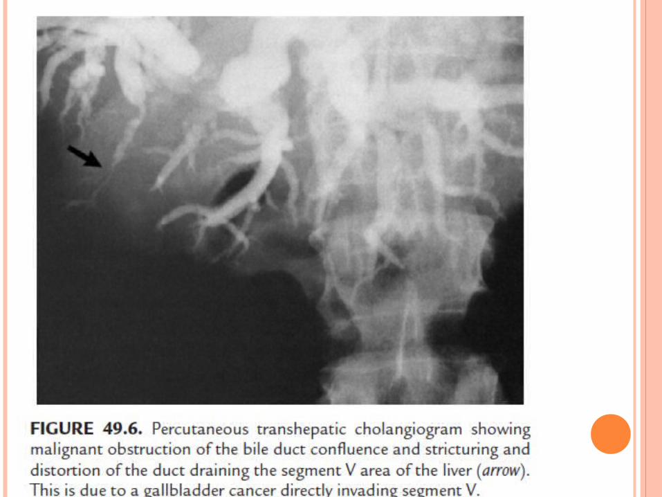
Infundibulum : Encroaches onto the porta hepatis
Tumors of this area – involves porta Prepare to perform bile duct resection/ major
hepatic resection

LYMPHATICS
PATHOLOGY AND STAGING
Fundus – 60% of tumors Body – 30% of tumors Neck – 10% of tumors
Gross findings:- Typical of chronic cholecystitis- Tumors in lower end of GB obstructing –
HYDROPS- Advanced tumors in neck/infundibulum –
jaundice / vascular invasion/ hepatic atrophy

GROSS DESCRIPTIONS
Infiltrative Nodular Combined nodular infiltrative Papillary - Better prognosis Combined papillary infiltrative

PAPILLARY ADENOCARCINOMA

HISTOLOGY
Adenocarcinoma – 89.4% Squamous / Adenosquamous – 4% Neuroendocrine – 3% Sarcoma/Adenosarcoma – 1.6% Melanoma - <1%
CLINICAL PRESENTATION
SCENARIOS:1. Final pathology after routine cholecystectomy
identifies CA GB
2. GB cancer discovered intraoperatively
3. GB cancer suspected before surgery

HISTORY
Constant RUQ pain – rather than episodic crampy pain of biliary colic
Elderly patients Weight loss Anorexia Jaundice
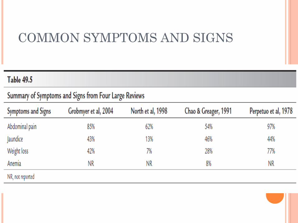
COMMON SYMPTOMS AND SIGNS
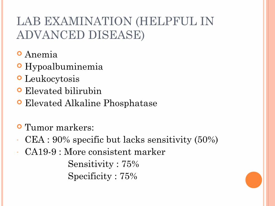
LAB EXAMINATION (HELPFUL IN ADVANCED DISEASE) Anemia Hypoalbuminemia Leukocytosis Elevated bilirubin Elevated Alkaline Phosphatase
Tumor markers:- CEA : 90% specific but lacks sensitivity (50%)- CA19-9 : More consistent marker Sensitivity : 75% Specificity : 75%
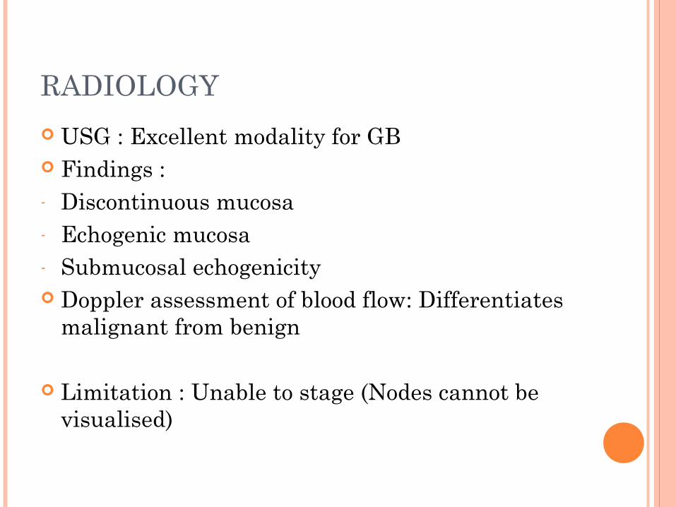
RADIOLOGY
USG : Excellent modality for GB Findings :- Discontinuous mucosa- Echogenic mucosa- Submucosal echogenicity Doppler assessment of blood flow: Differentiates
malignant from benign
Limitation : Unable to stage (Nodes cannot be visualised)

CT/MRI
Can assess extent of disease Detects presence of distant metastases
MC finding : Mass in GB
Assessment of LN:- Size > 1cm- Ring like heterogenous enhancement

CT/MRI
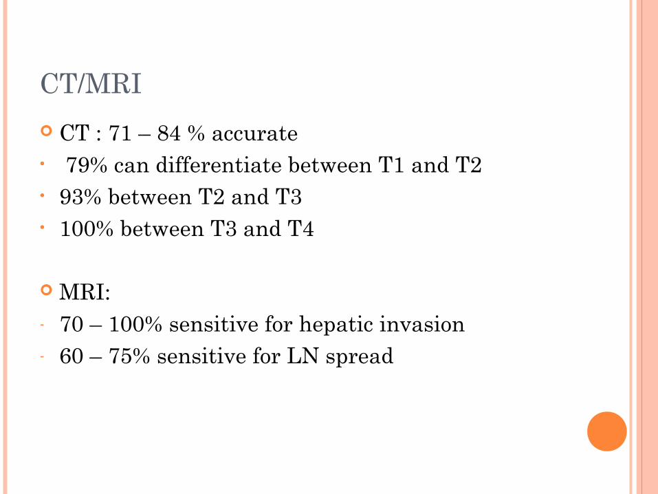
CT : 71 – 84 % accurate• 79% can differentiate between T1 and T2• 93% between T2 and T3• 100% between T3 and T4
MRI:- 70 – 100% sensitive for hepatic invasion- 60 – 75% sensitive for LN spread

FDG PET scan :
- More accurate than CT in diagnosing metastatic disease
- Poor in differentiating benign inflammatory state vs malignancy


PRE-OPERATIVE PATHOLOGICAL DIAGNOSIS
If CA-GB suspected on clinical and radiological grounds – Histological diagnosis NOT necessary
Biopsy increases risk of seeding
If concern for GB malignancy significant – Unwise to perform simple cholecystectomy
For unresectable disease – Percutaneous needle biopsy – 90% accurate

BILE CYTOLOGY
Less risky way of making diagnosis without risk of peritoneal seeding.
Justifiable in patients undergoing ERCP/PTC
If NOT - unwarranted

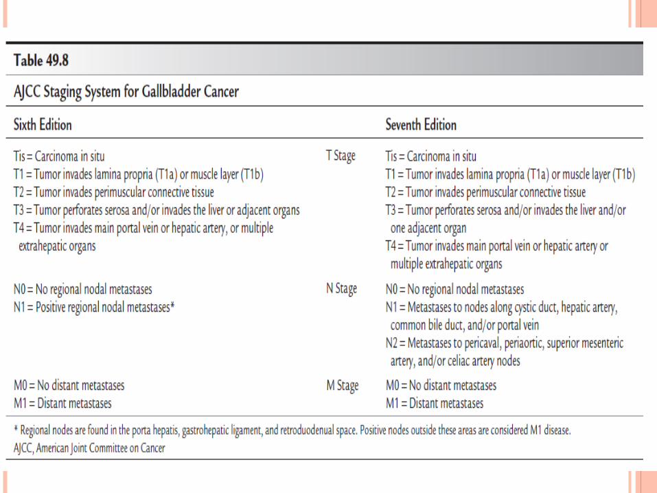
STAGING
SURGICAL MANAGEMENT
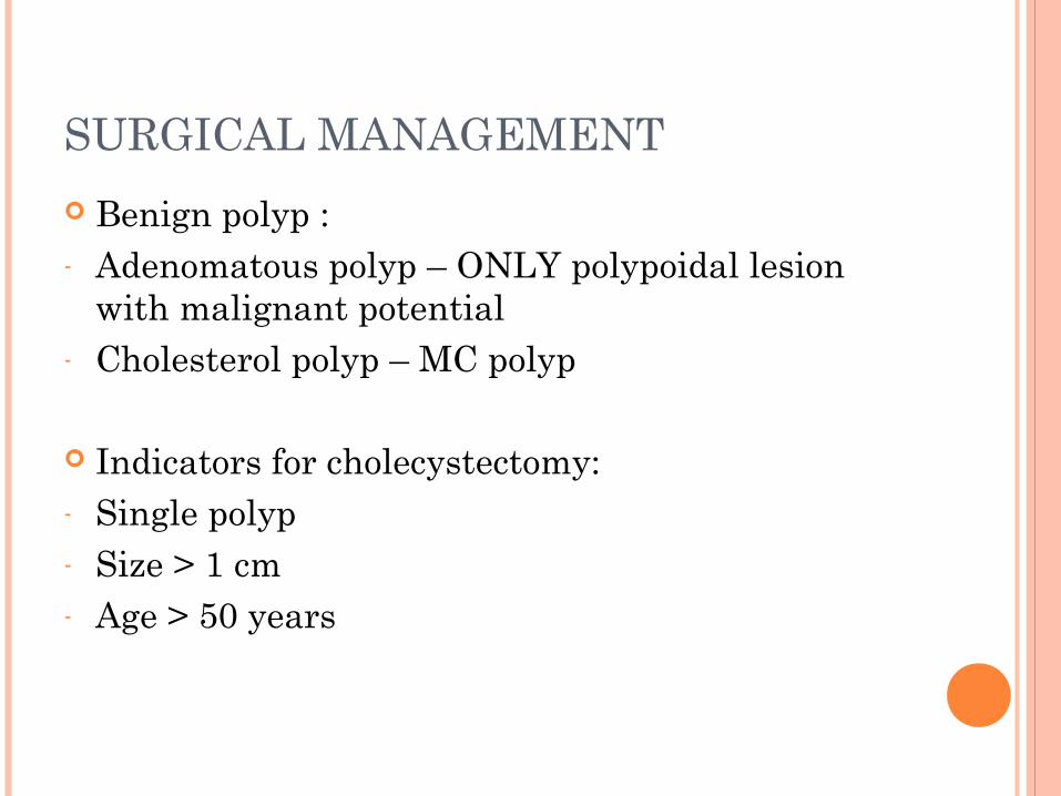
Benign polyp :- Adenomatous polyp – ONLY polypoidal lesion
with malignant potential- Cholesterol polyp – MC polyp
Indicators for cholecystectomy:- Single polyp- Size > 1 cm- Age > 50 years
Old concept – Offer OPEN cholecystectomy
Current concept – Offer Laparoscopic cholecystectomy + Frozen
Diagnosis – USG required If polyp presents with abdominal pain – rule out
other causes

INCIDENTALLY DETECTED GB CA
Incidence : 0.27 – 2.1% If diagnosis made by frozen – Prepare for
curative resection IF NOT COMFORTABLE – REFER NO EFFECT ON OUTCOME

T1a with margins negative : Standard cholecystectom cures 85 – 100%
T1b – controversial
T2 onwards – plan liver resection

NON CURATIVE CHOLECYSTECTOMY
Careful work up required which includes :
- Reviewing pre-cholecystectomy USG to localise extent
- Discuss case with operating surgeon
- Re-review T stage and margins pathologically

T1B LESIONS
If cystic duct stump / margins +ve – Bile duct resection and reconstruction OR Re-resection of cystic duct stump and frozen
proceed
EXTENT OF RESECTION BY STAGE
Rational approach to CA GB depends on :
- Stage of disease- Location of tumour- Margins status – if cholecystectomy has already
been performed.- Whether a prior noncurative cholecystectomy has
been performed

T1a – Simple cholecystectomy
T1b – Higher locoregional recurrence rates after simple cholecystectomy
T2,T3 – Complete enbloc resection with segment Ivb and V of liver

If invasion of hepatic inflow vascular structures is documented :
- Extended right hepatectomy + LN clearance of hepatoduodenal ligament + negative cystic duct/bile duct margins
- Abandon major resection IF:1. Nodal spread2. Metastases
LIVER RESECTION
Goal : To ensure a margin of 1-2 cm
Anatomic resection – better than wedge resection
If excision of segment IV b and V inadequate – DO extended right hepatectomy:
ESP in cases of large tumors invading portal pedicle
Tumors of lower end of GB encroaching onto porta
If isolated invasion of organ system present
EG: Stomach , duodenum, colon
In absence of distant metastases – DO local resection

LYMPH NODAL DISSECTION
Weigh risks vs benefits
Range of operations include : Excision of cystic duct node– Portal clearance– pancreaticoduodencetomy
1st manouvre : Mobilisation of duodenum – To assess aortocaval and retropancreatic nodes
Assess celiac node LN – If suspicious DO frozen and terminate procedure IF MALIGNANT

WHETHER ROUTINE BILE DUCT RESECTION IS NECESSARY FOR ADEQUATE LN CLEARANCE??
Excising extrahepatic bile duct – makes LN dissection easy
Increases morbidity of operation
No difference noted in the number of LN harvested with OR without bile duct resection
In general – bile duct resection NOT needed---- Unless suspicion of PORTA infiltration
Stage of disease and NOT extent of resection determines survival of patients


DID YOU KNOW? “Honeymoon and alcohol”
Roots trace back to Babylon Tradition for the soon to be father- in-law to
supply his daughter’s fiance with a month of mead
Time period referred to as the HONEYMONTH


DID YOU KNOW?
Adolf Hitler was one of the world’s best known abstainers from alcohol.


