Fluid and Electrolyte Metabolism/Renal and Urologic Disorders
Adam Weinstein, MDAssistant Professor Pediatric Nephrology
Children’s Hospital at Dartmouth Hitchcock
Disclosures• I have no relevant financial relationships with the manufacturers(s) of any commercial products(s) and/or provider of commercial services discussed in this CME activity.
• I do not intend to discuss an unapproved/investigative use of a commercial product/device in my presentation.
Objectives• Evaluate and plan treatment for various etiologies of
hyponatremia and their associated fluid and electrolyte abnormalities
• Distinguish between the causes of and initiate evaluation for glomerular and non‐glomerular hematuria
• Interpret which markers of severity in the setting of acute glomerulonephritis warrant evaluation and treatment
• Review the causes of and management approaches for nephrotic syndrome and asymptomatic proteinuria in children

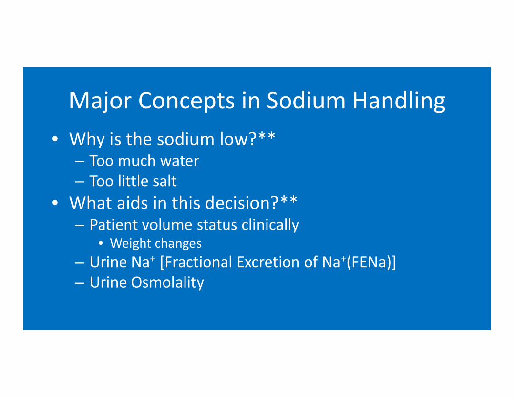
Major Concepts in Sodium Handling• Why is the sodium low?**
– Too much water– Too little salt
• What aids in this decision?**– Patient volume status clinically
• Weight changes– Urine Na+ [Fractional Excretion of Na+(FENa)]– Urine Osmolality
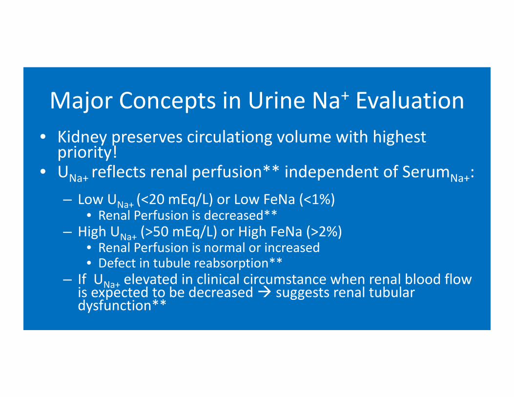
Major Concepts in Urine Na+ Evaluation• Kidney preserves circulating volume with highest priority!
• UNa+ reflects renal perfusion** independent of SerumNa+:– Low UNa+ (<20 mEq/L) or Low FeNa (<1%)
• Renal Perfusion is decreased**– High UNa+ (>50 mEq/L) or High FeNa (>2%)
• Renal Perfusion is normal or increased• Defect in tubule reabsorption**
– If UNa+ elevated in clinical circumstance when renal blood flow is expected to be decreased suggests renal tubular dysfunction**
Major Concepts in Urine Osm Evaluation
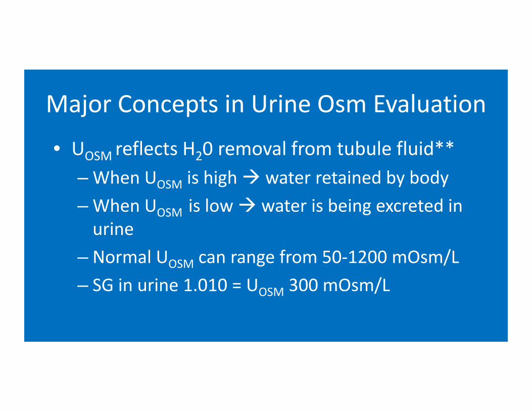
• UOSM reflects H20 removal from tubule fluid**– When UOSM is high water retained by body– When UOSM is low water is being excreted in urine
– Normal UOSM can range from 50‐1200 mOsm/L– SG in urine 1.010 = UOSM 300 mOsm/L

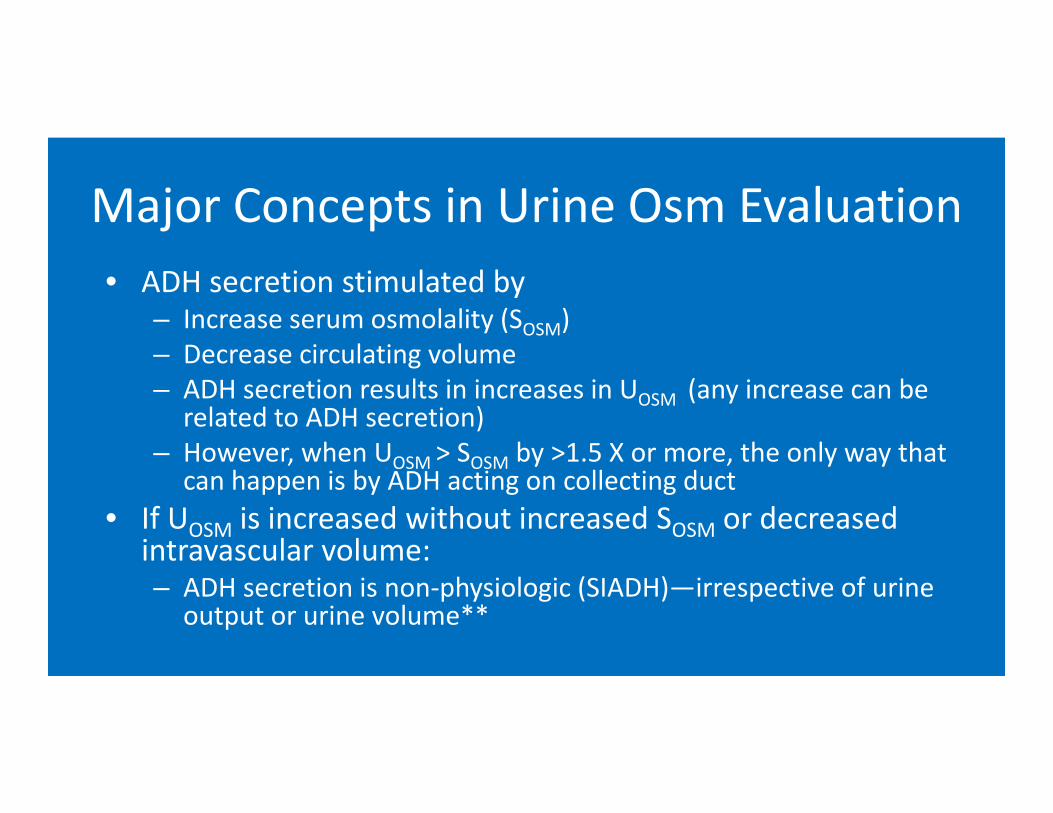
Major Concepts in Urine Osm Evaluation• ADH secretion stimulated by
– Increase serum osmolality (SOSM)– Decrease circulating volume– ADH secretion results in increases in UOSM (any increase can be
related to ADH secretion)– However, when UOSM > SOSM by >1.5 X or more, the only way that
can happen is by ADH acting on collecting duct• If UOSM is increased without increased SOSM or decreased
intravascular volume: – ADH secretion is non‐physiologic (SIADH)—irrespective of urine
output or urine volume**

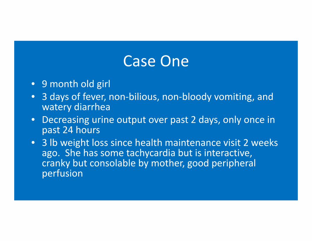
Hyponatremia Case One• 9 month old girl • 3 days of fever, non‐bilious, non‐bloody vomiting, and watery diarrhea
• Decreasing urine output over past 2 days, only once in past 24 hours
• 3 lb weight loss since health maintenance visit 2 weeks ago. She has some tachycardia but is interactive, cranky but consolable by mother, good peripheral perfusion

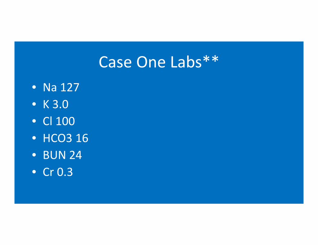
Case One Labs**• Na 127• K 3.0• Cl 100• HCO3 16• BUN 24• Cr 0.3

Questions Case One• What do you suspect the FENa and Urine Osmolality are?
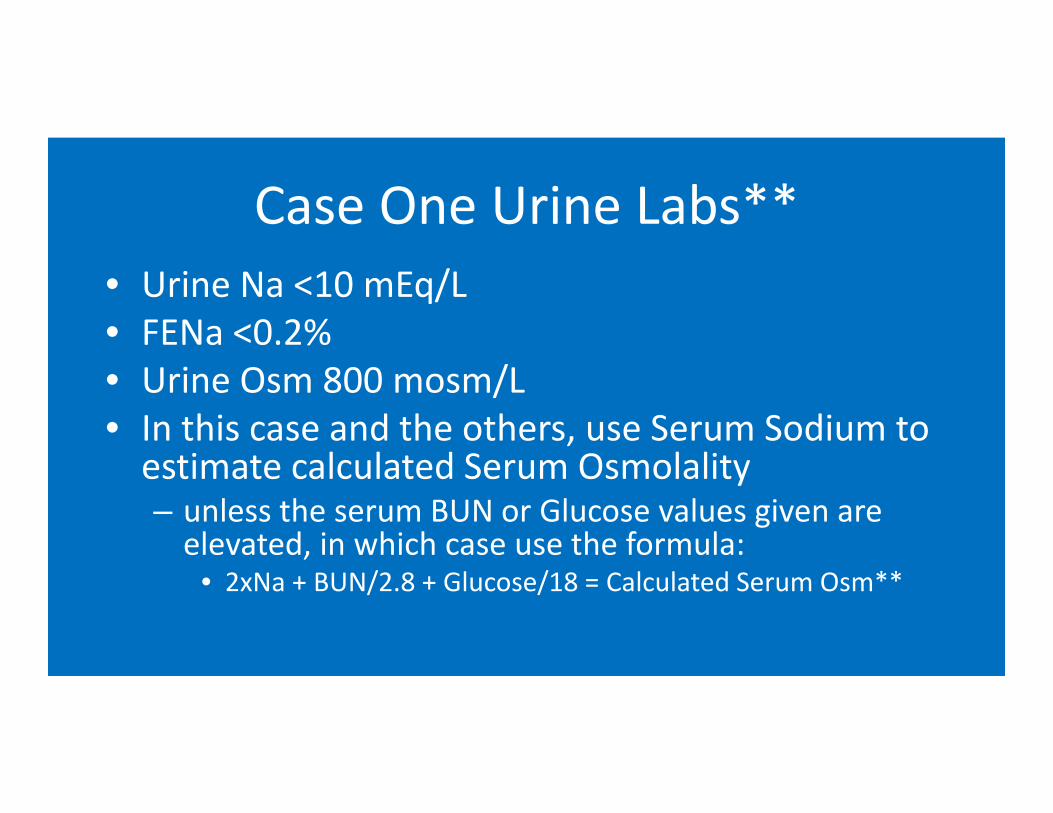
Case One Urine Labs**• Urine Na <10 mEq/L• FENa <0.2%• Urine Osm 800 mosm/L• In this case and the others, use Serum Sodium to estimate calculated Serum Osmolality– unless the serum BUN or Glucose values given are elevated, in which case use the formula:
• 2xNa + BUN/2.8 + Glucose/18 = Calculated Serum Osm**

Questions Case One• What is the diagnosis?**
• Does the patient seem to have more vomiting or diarrhea?**
• Explain the hypokalemia and how would you treat it?**
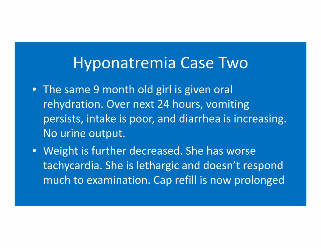
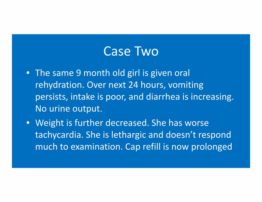
Hyponatremia Case Two• The same 9 month old girl is given oral rehydration. Over next 24 hours, vomitingpersists, intake is poor, and diarrhea is increasing. No urine output.
• Weight is further decreased. She has worse tachycardia. She is lethargic and doesn’t respond much to examination. Cap refill is now prolonged

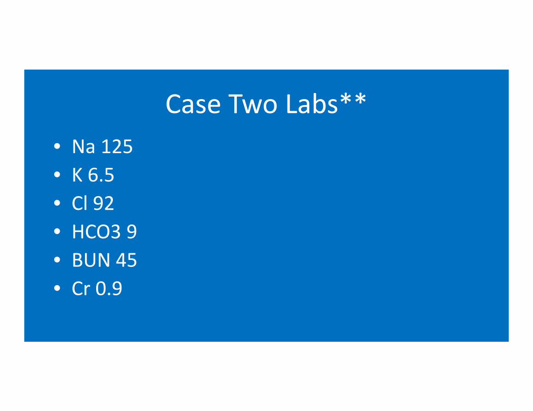
Case Two Labs**• Na 125• K 6.5• Cl 92• HCO3 9• BUN 45• Cr 0.9
Questions Case Two• What do you suspect the FENa and Urine Osmolality are?
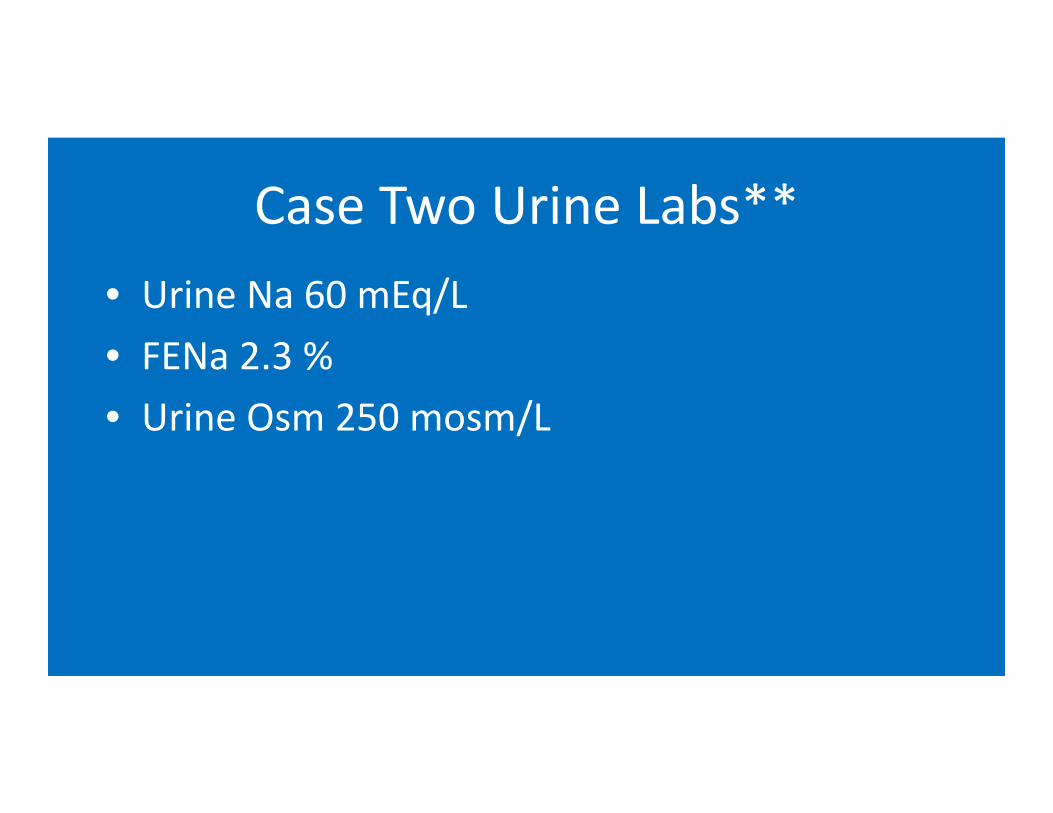
Case Two Urine Labs**• Urine Na 60 mEq/L• FENa 2.3 %• Urine Osm 250 mosm/L

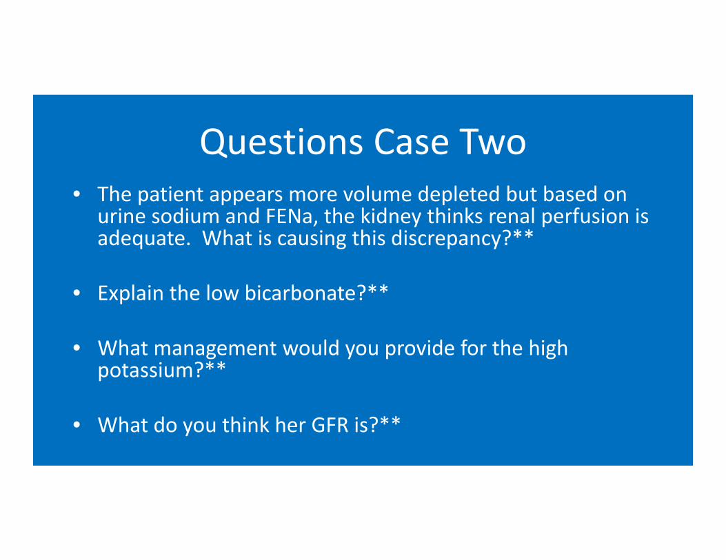
Questions Case Two• The patient appears more volume depleted but based on
urine sodium and FENa, the kidney thinks renal perfusion is adequate. What is causing this discrepancy?**
• Explain the low bicarbonate?**
• What management would you provide for the high potassium?**
• What do you think her GFR is?**
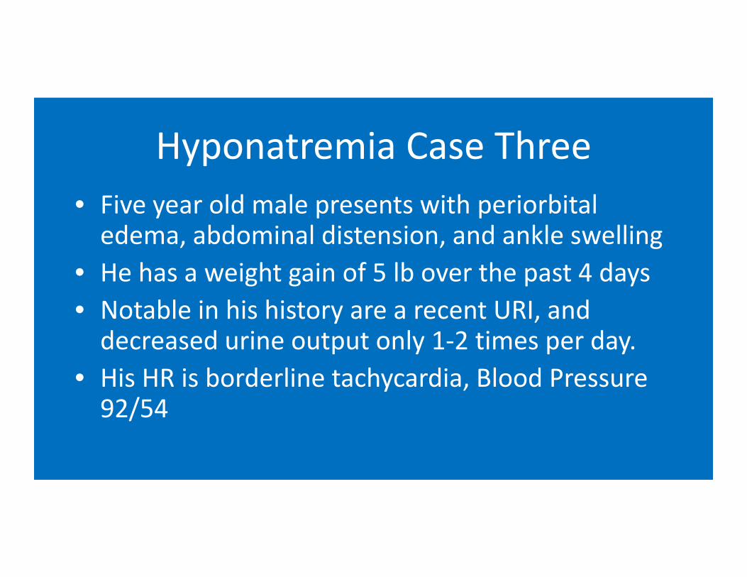
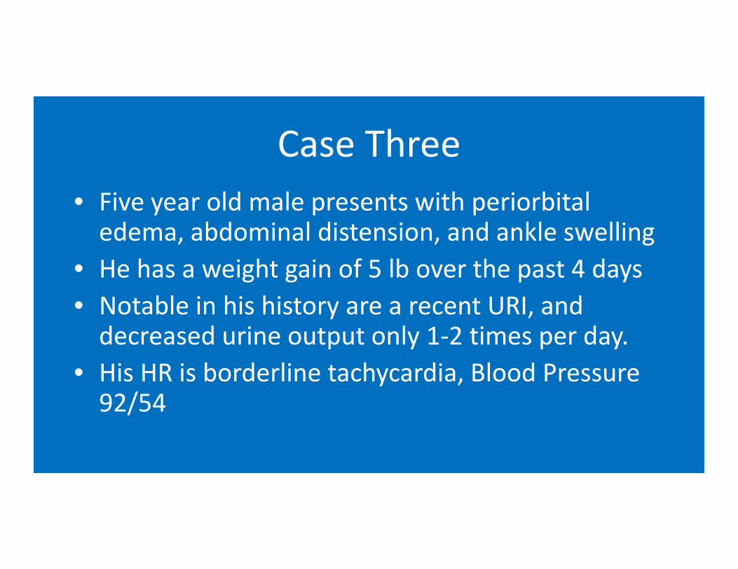
Hyponatremia Case Three• Five year old male presents with periorbital edema, abdominal distension, and ankle swelling
• He has a weight gain of 5 lb over the past 4 days• Notable in his history are a recent URI, and decreased urine output only 1‐2 times per day.
• His HR is borderline tachycardia, Blood Pressure 92/54

Case Three Labs**• Na 128• K 4.9• Cl 90• HCO3 26• BUN 12• Cr 0.5

Questions Case Three• What do you think the child’s diagnosis is?**• What do you suspect the FENa and Urine Osmolality are?**
• Why is hyponatremia associated with his condition?**
• How do you fix the hyponatremia?**
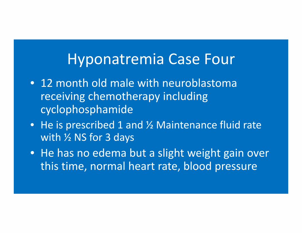
Hyponatremia Case Four • 12 month old male with neuroblastomareceiving chemotherapy including cyclophosphamide
• He is prescribed 1 and ½ Maintenance fluid rate with ½ NS for 3 days
• He has no edema but a slight weight gain over this time, normal heart rate, blood pressure

Case Four Labs**• Na 128• K 3.6• Cl 96• HCO3 22• BUN 5• Cr 0.3
Questions Case Four• What do you suspect the FENa and Urine Osmolality are?

Case Four Urine Labs**• Urine Na 95 mEq/L• FENa 2.1 %• Urine Osm 780 mosm/L

Questions Case Four• What is the child’s diagnosis is?**• What will your management be?**
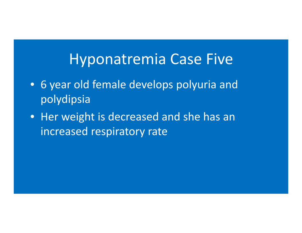
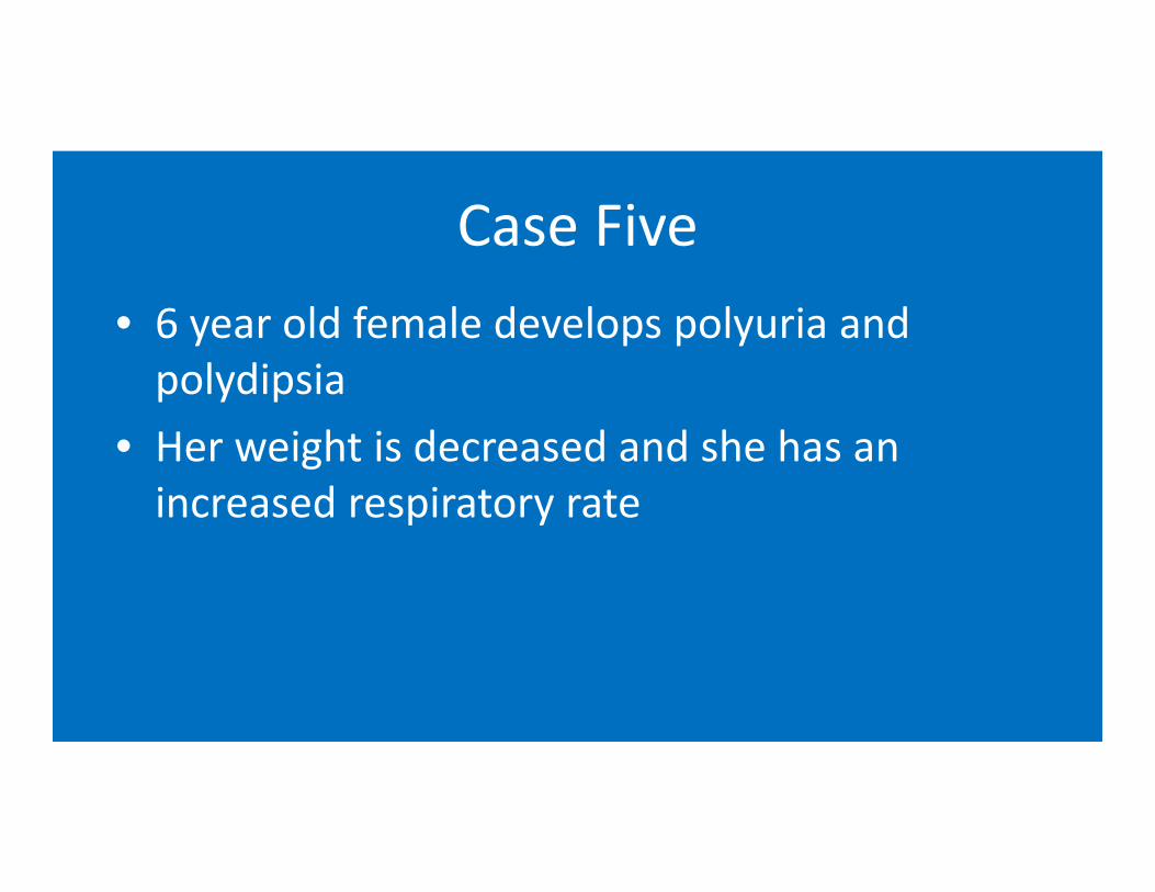
Hyponatremia Case Five• 6 year old female develops polyuria and polydipsia
• Her weight is decreased and she has an increased respiratory rate
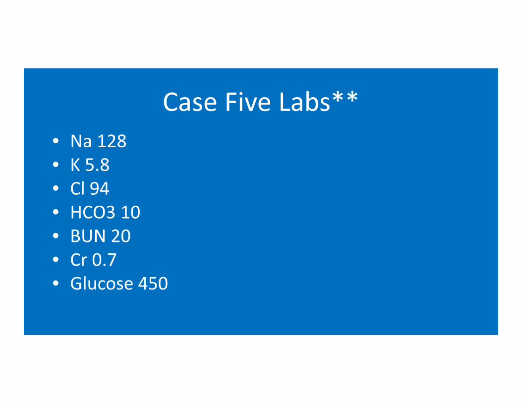
Case Five Labs**• Na 128• K 5.8• Cl 94• HCO3 10• BUN 20• Cr 0.7• Glucose 450

Questions Case Five• Why is the serum sodium low?**

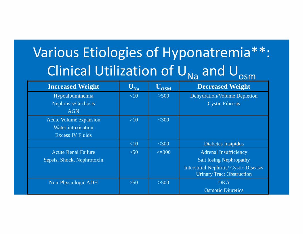
Various Etiologies of Hyponatremia**: Clinical Utilization of UNa and UosmIncreased Weight UNa UOSM Decreased Weight
HypoalbuminemiaNephrosis/Cirrhosis
AGN
<10 >500 Dehydration/Volume DepletionCystic Fibrosis
Acute Volume expansionWater intoxicationExcess IV Fluids
>10 <300
<10 <300 Diabetes InsipidusAcute Renal Failure
Sepsis, Shock, Nephrotoxin>50 <=300 Adrenal Insufficiency
Salt losing NephropathyInterstitial Nephritis/ Cystic Disease/
Urinary Tract ObstructionNon-Physiologic ADH >50 >500 DKA
Osmotic Diuretics

Take Home Points for Practice• The etiology of hyponatremia can be determined through
assessment of clinical volume status, urine sodium excretion (FENa), & urine osmolality
• If the renal response to a suggested clinical volume state appears inappropriate, consider renal tubular pathology—tubular injury, tubular dysfunction, or inappropriate hormone secretion (e.g. SIADH)
• Treatment of low volume states respond well to volume and electrolyte repletion; treatment of excess volume states respond to management of underlying disorder (e.g. nephroticsyndrome) and thoughtful supportive restrictions (e.g. SIADH)
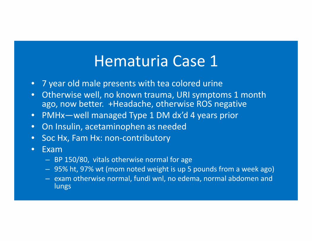
Hematuria Case 1• 7 year old male presents with tea colored urine• Otherwise well, no known trauma, URI symptoms 1 month
ago, now better. +Headache, otherwise ROS negative• PMHx—well managed Type 1 DM dx’d 4 years prior• On Insulin, acetaminophen as needed• Soc Hx, Fam Hx: non‐contributory• Exam
– BP 150/80, vitals otherwise normal for age– 95% ht, 97% wt (mom noted weight is up 5 pounds from a week ago)– exam otherwise normal, fundi wnl, no edema, normal abdomen and
lungs

What is the next step?

What is the next step?• First, confirm true hematuria**‐‐
– Is U/A + for blood? • If U/A neg blood—ingestion (beets, med)
• Next confirm RBC in urine**– Is Microscopy + for RBCs?– If Microscopy neg for RBCs then think:
• Myoglobinuria• Hemoglobinuria• Old specimen or other false positive
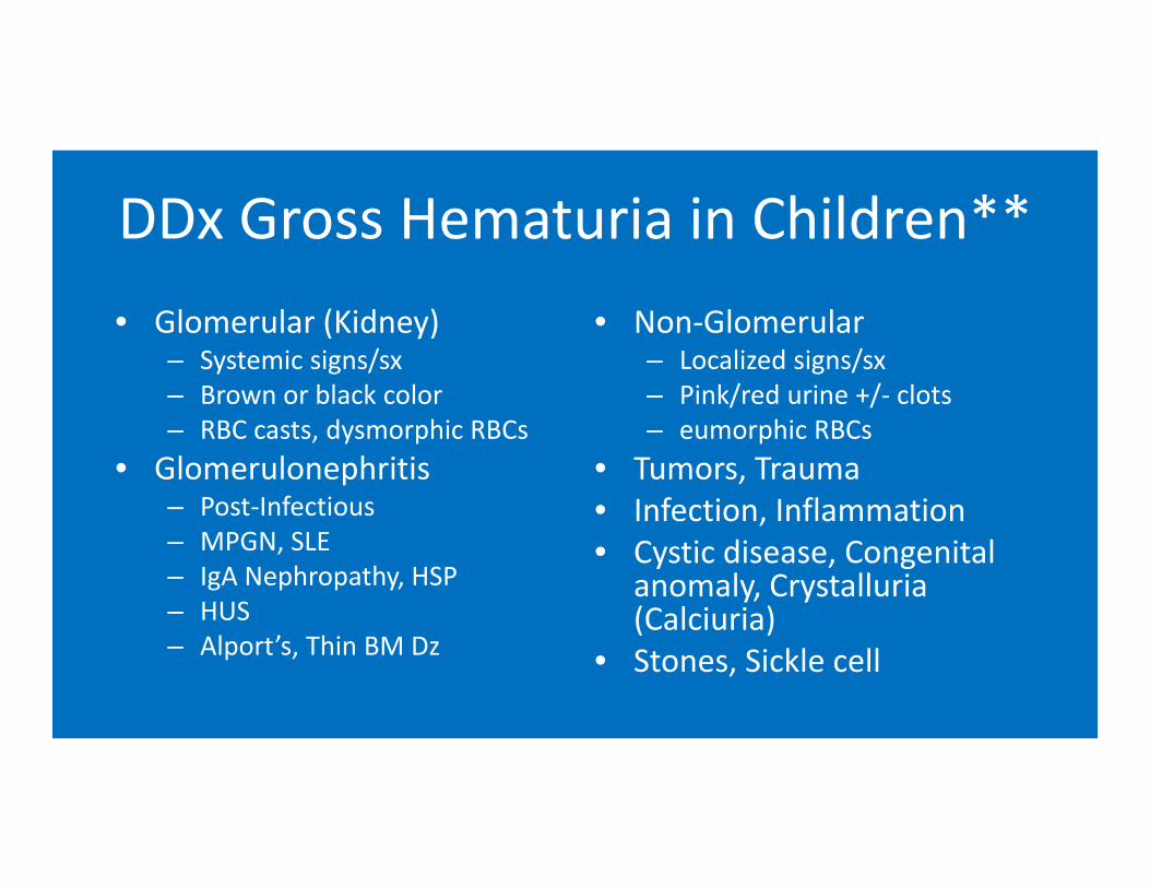
DDx Gross Hematuria in Children**• Glomerular (Kidney)
– Systemic signs/sx– Brown or black color– RBC casts, dysmorphic RBCs
• Glomerulonephritis– Post‐Infectious– MPGN, SLE– IgA Nephropathy, HSP– HUS– Alport’s, Thin BM Dz
• Non‐Glomerular– Localized signs/sx– Pink/red urine +/‐ clots– eumorphic RBCs
• Tumors, Trauma• Infection, Inflammation• Cystic disease, Congenital
anomaly, Crystalluria(Calciuria)
• Stones, Sickle cell
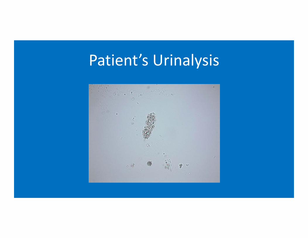
Patient’s Urinalysis

Acute Glomerulonephritis
• What next?

What are clinical indicators of a severe glomerulonephritis?
• Acute kidney injury/elevated creatinine• Hypertension• Severe proteinuria, nephrotic range proteinuria, in particular nephrotic syndrome
• Why and how can glomerulonephritis cause nephrotic syndrome?
Recap on Podocytes and Glomerular Basement Membrane
• Podocytes– Help create the negatively charged slit diaphragm that prevents many serum proteins (e.g. albumin) from filtering across the glomerulus
• Minimal Change Disease is a result of podocytes that are no longer functioning (they are “effaced”)– Negative charge slit diaphragm is gone– Serum proteins (e.g. albumin) filter into the Bowman’s Space
Severe Glomerulonephritis• When there is severe involvement of the basement membrane, then one might think of the podocytes as innocent bystanders
• Therefore, Nephrotic Syndrome is a manifestation of severe proteinuria and a marker for a severe glomerulonephritis (GN)
• If such a GN goes untreated, or does not resolve spontaneously (e.g. post‐infectious), then this results in a poor long‐term prognosis
Serology Interpretation• Let’s say you get C3 and C4 and ASLO titer• Results show a + ASLO titer, but C3 and C4 are normal
• What is your diagnostic interpretation?**
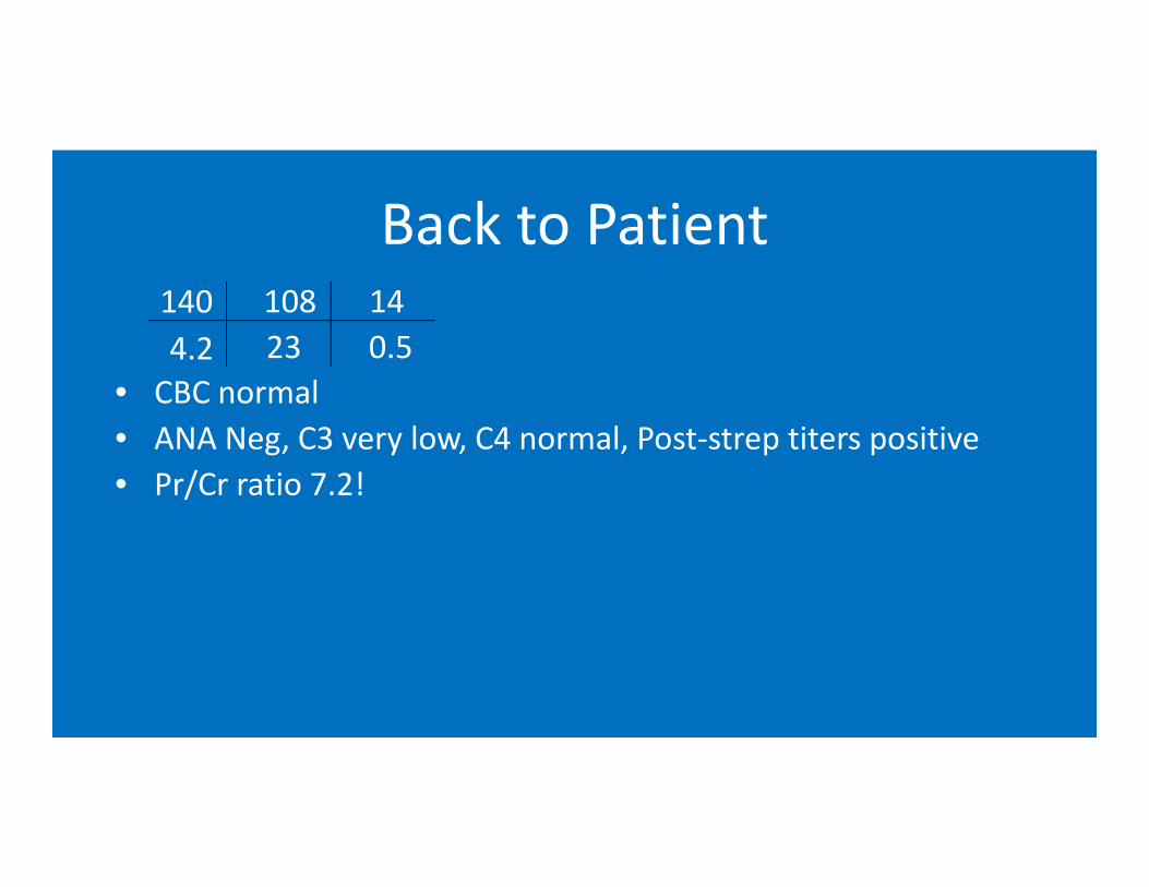
Back to Patient
• CBC normal• ANA Neg, C3 very low, C4 normal, Post‐strep titers positive• Pr/Cr ratio 7.2!
4.2 0.514
23108140
What is your presumptive diagnosis?**
A. IgA NephropathyB. Alport SyndromeC. Membranoproliferative GND. Post‐Infectious GN
How could you confirm this?**A. Monitoring for spontaneous resolution and
return of C3 to normal over next 2‐3 monthsB. Response to empiric treatment with
CorticosteroidsC. Renal Biopsy
But what about his Hypertension?**A. Provide reassurance. If this is post‐strep GN, it will get
better on its own so it does not require treatment
B. It is severely elevated for a 7 yo so warrants treatment with a low sodium diet and an ACE Inhibitor until the GN resolves
C. It is severely elevated for a 7 yo, so warrants treatment with a low sodium diet and thiazide diuretic until the GN resolves

Why did he gain weight?**A. With his acute GN and HTN he was more sedentary,
so this is increased body fatB. With his acute GN, his GFR may be decreased so this
is likely fluid weight from renal failureC. With his acute GN, renin is stimulated and his
nephrons are likely reabsorbing salt and water so this is fluid weight
D. With his acute GN and being sick, he probably actually lost weight, so this is measurement error
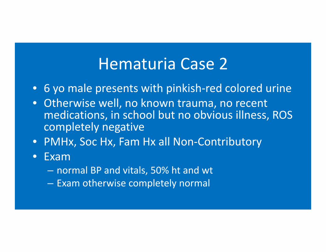
Hematuria Case 2• 6 yo male presents with pinkish‐red colored urine• Otherwise well, no known trauma, no recent medications, in school but no obvious illness, ROS completely negative
• PMHx, Soc Hx, Fam Hx all Non‐Contributory• Exam
– normal BP and vitals, 50% ht and wt– Exam otherwise completely normal
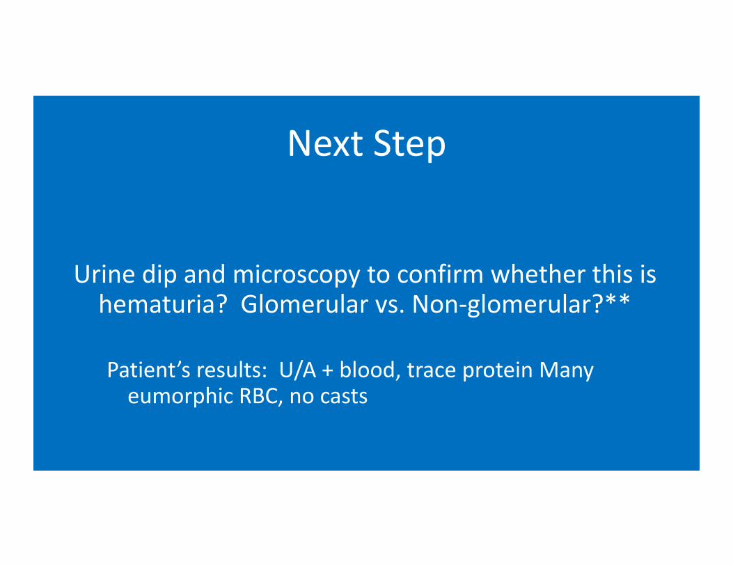
Next Step
Urine dip and microscopy to confirm whether this is hematuria? Glomerular vs. Non‐glomerular?**
Patient’s results: U/A + blood, trace protein Many eumorphic RBC, no casts

How would you evaluate at this point?

How would you evaluate at this point?**
• DDx of non‐glomerular hematuria**– Urine Ca/Cr and Renal U/S
• If either positive for stones, consider 24 hour urine for stone chemistries
– Any symptoms/signs of infection? If so, Urine Cx– Could consider Lytes BUN, Cr, CBC, Pr/Cr—if still need to be certain to exclude a GN etiology

Results• Ca/Cr 0.47 (Nml <0.2)• Renal U/S neg, other screens normal
• 3 months later—presents with flank pain and passes stone!

Hypercalciuria• Why care?
– Presence of hematuria worrisome to parents– Risk for stones**—VERY PAINFUL!!!– Can be associated with nephrocalcinosis**
• deposits of calcium within tubulointerstitial parenchyma• can cause progressive scarring and loss of function
• Who develops?– Idiopathic (probably multifactorial—genetics, diet, etc…)– Certain ‘tubulopathies’ (e.g. Dent’s disease, Bartter’s syndrome)
– Medication induced

• When Infants and children develop stones– Stone is not the disease. It’s a sign of the disease
• Every child with kidney stones or nephrocalcinosis should have a thorough evaluation as to the cause**– H&P into systemic disorders
• Growth and development, blood pressure, bone related signs/symptoms• Dietary history (attention to excess or deficiency), fluid intake, meds including vitamins and supplements
– Fam Hx of stones, hematuria, renal failure– Urinalysis, urine and serum chemistries, imaging– STONE analysis (send the stone for chemical analysis)
Childhood Kidney Stones**
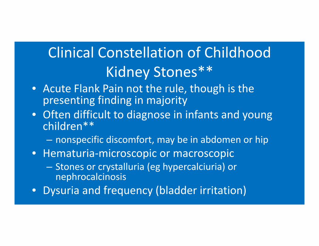
• Acute Flank Pain not the rule, though is the presenting finding in majority
• Often difficult to diagnose in infants and young children**– nonspecific discomfort, may be in abdomen or hip
• Hematuria‐microscopic or macroscopic– Stones or crystalluria (eg hypercalciuria) or nephrocalcinosis
• Dysuria and frequency (bladder irritation)
Clinical Constellation of Childhood Kidney Stones**
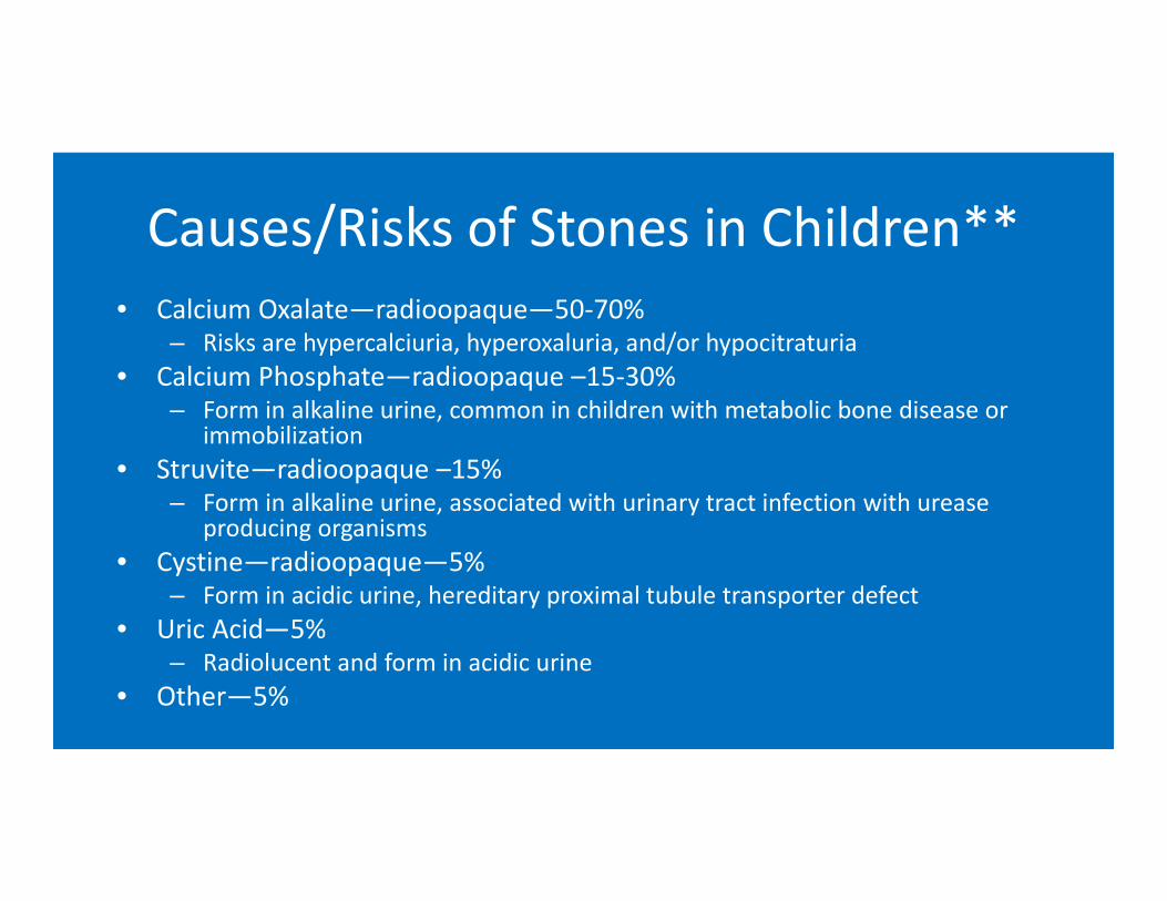
• Calcium Oxalate—radioopaque—50‐70%– Risks are hypercalciuria, hyperoxaluria, and/or hypocitraturia
• Calcium Phosphate—radioopaque –15‐30%– Form in alkaline urine, common in children with metabolic bone disease or
immobilization• Struvite—radioopaque –15%
– Form in alkaline urine, associated with urinary tract infection with urease producing organisms
• Cystine—radioopaque—5% – Form in acidic urine, hereditary proximal tubule transporter defect
• Uric Acid—5%– Radiolucent and form in acidic urine
• Other—5%
Causes/Risks of Stones in Children**
How do you treat idiopathic (most common form) hypercalciuria (and prevent nephrolithiasis)?**
A. Drink lots of fluids to make a dilute urineB. Low Sodium DietC. High Citrate DietD. Thiazide Diuretic
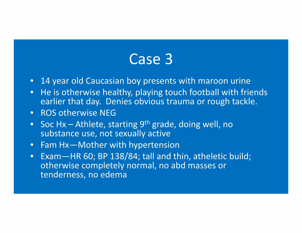
Hematuria Case 3• 14 year old Caucasian boy presents with maroon urine• He is otherwise healthy, playing touch football with friends
earlier that day. Denies obvious trauma or rough tackle.• ROS otherwise NEG• Soc Hx – Athlete, starting 9th grade, doing well, no
substance use, not sexually active• Fam Hx—Mother with hypertension• Exam—HR 60; BP 138/84; tall and thin, athletic build;
otherwise completely normal, no abd masses or tenderness, no edema

Next step‐‐Urinalysis• 1.025, pH 6, trace protein, large blood, many dysmorphic RBCs/hpf, no casts

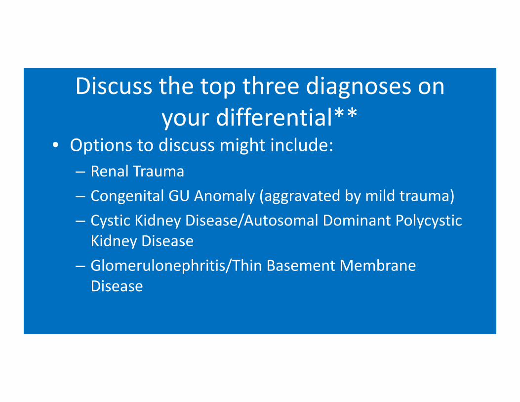
Discuss the top three diagnoses on your differential**

What would your evaluation be?**

Evaluation**• Urine Calcium/Cr normal• Basic Metabolic Panel normal• Renal Ultrasound reveals b/l enlarged kidneys with multiple cysts
• Diagnosis– Presumed Autosomal Dominant Polycystic Kidney Disease (ADPKD)
ADPKD in children**• A significant minority of cases of ADPKD present in childhood**
– Occasionally on prenatal imaging, especially with known family history– More commonly with acute onset gross hematuria (often in setting of
trauma or exertion)– Hypertension is a common presentation
• Asymptomatic screening of children of parents with ADPKD is not currently recommended**– Screening of these children’s Blood Pressure and a urinalysis for
protein at health maintenance is generally recommended– Presence of hypertension and/or proteinuria are associated with
progression of chronic kidney disease, and can be treated to delay progression**

Name three things you might do as far as clinical management**
ADPKD Management**• Strict Blood Pressure Control using RAAS inhibition—target
<120/80 has illustrated slower decline of renal function• Control of proteinuria with RAAS inhibition—similarly, this
delays progression of CKD and may prevent future development of ESRD
• If kidneys >15cm– may need to restrict from high impact sports/activities (risk of trauma and cyst rupture—massive bleeding)
• High Fluid intake‐‐ ADH is associated with cyst growth, so suppressing ADH with super‐hydration may slow progression
• Patient Education and connections with resources/networks
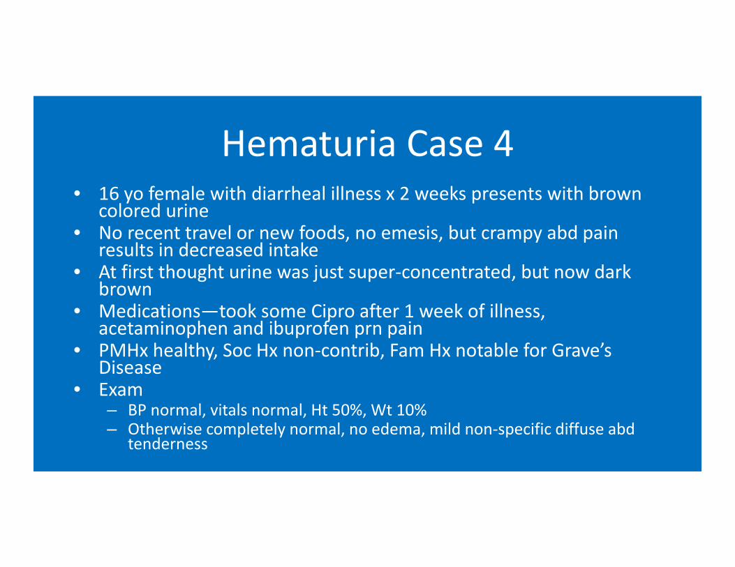
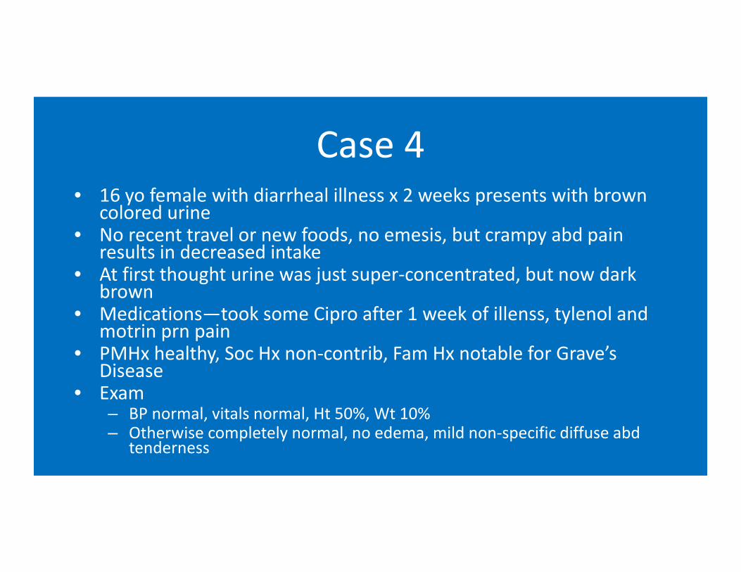
Hematuria Case 4• 16 yo female with diarrheal illness x 2 weeks presents with brown
colored urine• No recent travel or new foods, no emesis, but crampy abd pain
results in decreased intake• At first thought urine was just super‐concentrated, but now dark
brown• Medications—took some Cipro after 1 week of illness,
acetaminophen and ibuprofen prn pain• PMHx healthy, Soc Hx non‐contrib, Fam Hx notable for Grave’s
Disease• Exam
– BP normal, vitals normal, Ht 50%, Wt 10%– Otherwise completely normal, no edema, mild non‐specific diffuse abd
tenderness

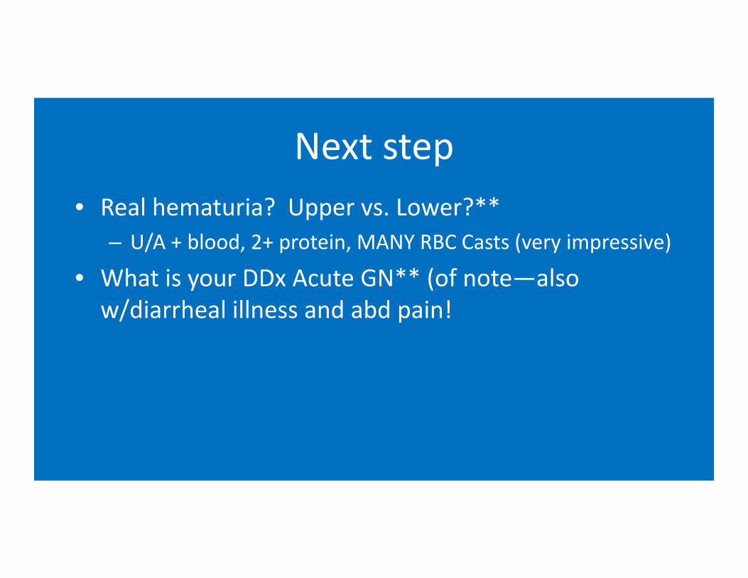
Next step• Real hematuria? Upper vs. Lower?**
– U/A + blood, 2+ protein, MANY RBC Casts (very impressive)
• What is your DDx Acute GN** (of note—also w/diarrheal illness and abd pain!
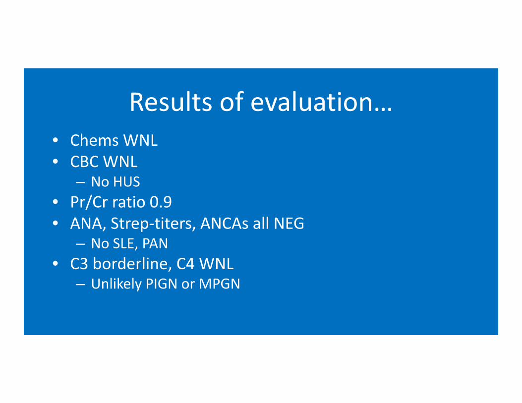
Results of evaluation…• Chems WNL• CBC WNL
– No HUS• Pr/Cr ratio 0.9• ANA, Strep‐titers, ANCAs all NEG
– No SLE, PAN• C3 borderline, C4 WNL
– Unlikely PIGN or MPGN

Hmm..• Diagnosed with C. Diff Colitis
– Treated with Flagyl and her diarrhea & abd pain resolved within 1‐2 weeks
– Never developed purpura or arthritis (no HSP)– Hematuria and proteinuria resolved – C3 completely normal 2 months later
• IgA Nephropathy? PIGN (can occur with chronic infections)– What to do? – What’s the treatment for IgA Nephropathy?

Answers• Watchful waiting—she’s in spontaneous remission
– If she relapses again with another GI illness or Resp illness, then likely IgA Nephropathy**
– If never happens again, then likely PIGN**• Only proven treatment for IgA Nephropathy is ACE‐Inhibitor to relax glomerular capillary bed/decrease filtration pressure and prevent scarring– Fish Oil has a promising study, but not confirmed– For severe clinical presentations, steroids +/‐ other immunosuppressants
• Some subjective experience, but no RCT yet to show benefit
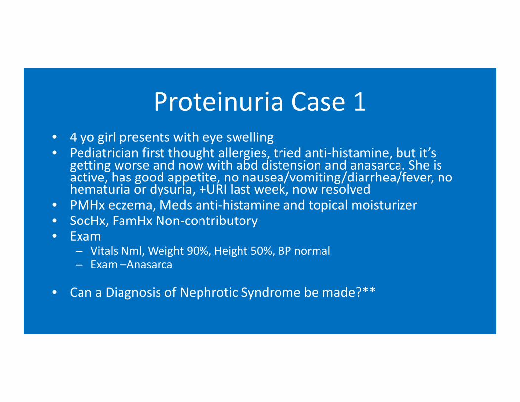
Proteinuria Case 1• 4 yo girl presents with eye swelling• Pediatrician first thought allergies, tried anti‐histamine, but it’s
getting worse and now with abd distension and anasarca. She is active, has good appetite, no nausea/vomiting/diarrhea/fever, no hematuria or dysuria, +URI last week, now resolved
• PMHx eczema, Meds anti‐histamine and topical moisturizer• SocHx, FamHx Non‐contributory• Exam
– Vitals Nml, Weight 90%, Height 50%, BP normal– Exam –Anasarca
• Can a Diagnosis of Nephrotic Syndrome be made?**
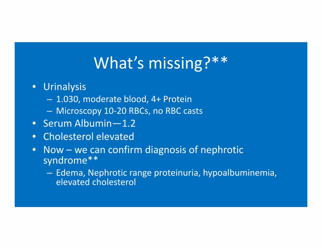
What’s missing?**• Urinalysis
– 1.030, moderate blood, 4+ Protein– Microscopy 10‐20 RBCs, no RBC casts
• Serum Albumin—1.2• Cholesterol elevated• Now – we can confirm diagnosis of nephroticsyndrome**– Edema, Nephrotic range proteinuria, hypoalbuminemia, elevated cholesterol

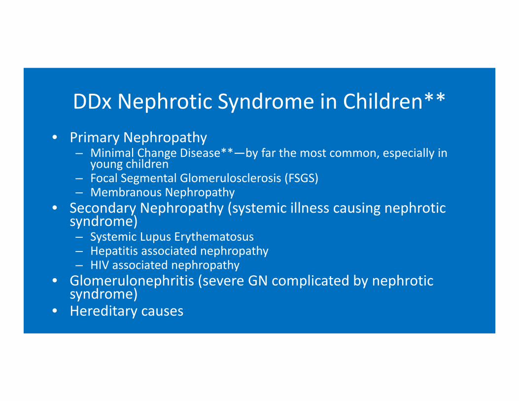
DDx Nephrotic Syndrome in Children**• Primary Nephropathy
– Minimal Change Disease**—by far the most common, especially in young children
– Focal Segmental Glomerulosclerosis (FSGS)– Membranous Nephropathy
• Secondary Nephropathy (systemic illness causing nephroticsyndrome)– Systemic Lupus Erythematosus– Hepatitis associated nephropathy – HIV associated nephropathy
• Glomerulonephritis (severe GN complicated by nephroticsyndrome)
• Hereditary causes
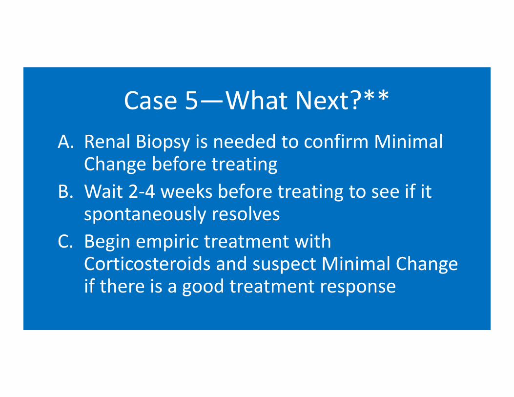
Case 1—What Next?**A. Renal Biopsy is needed to confirm Minimal
Change before treatingB. Wait 2‐4 weeks before treating to see if it
spontaneously resolvesC. Begin empiric treatment with
Corticosteroids and suspect Minimal Change if there is a good treatment response

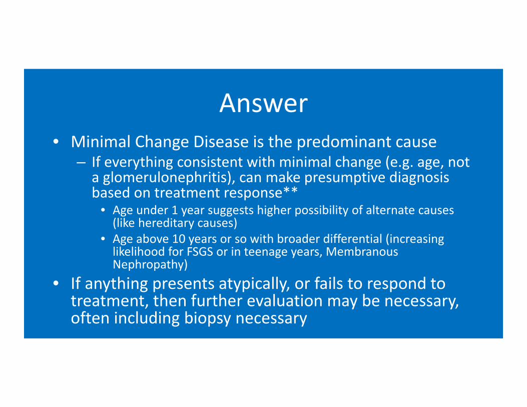
Answer• Minimal Change Disease is the predominant cause
– If everything consistent with minimal change (e.g. age, not a glomerulonephritis), can make presumptive diagnosis based on treatment response**
• Age under 1 year suggests higher possibility of alternate causes (like hereditary causes)
• Age above 10 years or so with broader differential (increasing likelihood for FSGS or in teenage years, Membranous Nephropathy)
• If anything presents atypically, or fails to respond to treatment, then further evaluation may be necessary, often including biopsy necessary
Minimal Change Disease• Presumptive treatment, 2mg/kg or 60mg Prednisone Daily**
– Usual response in 2‐4 weeks, small number may take longer– Treat intercurrent infections/triggers
• Majority recur**– Significant minority one and done (up to 40%)– Others may develop frequent relapsing or steroid dependent
nephrotic syndrome and require steroid‐sparing therapy (e.g. cyclophosphamide)
– If develops steroid resistance—reconsider other etiologies (e.g. FSGS)• Consistent treatment response suggests an ultimately good
prognosis, even if having recurrences**

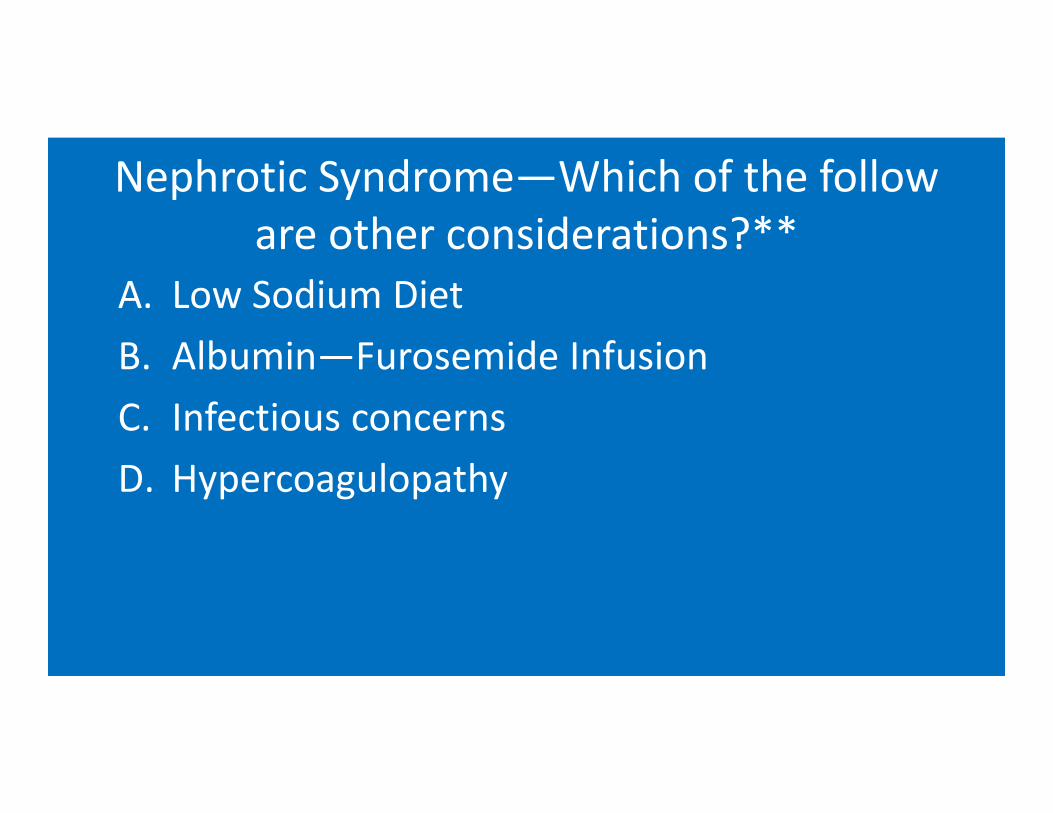
Nephrotic Syndrome—Which of the follow are other considerations?**
A. Low Sodium DietB. Albumin—Furosemide InfusionC. Infectious concernsD. Hypercoagulopathy
Answer• All of them!• Low Sodium Diet is helpful– high sodium intake results in
worsening edema (fills interstitial volume)**• When fluid overload/edema is causing symptoms (resp
distress) or complications, albumin‐furosemide infusions can help mobilize edema**– If this is needed, should be done at center with experience with
this treatment approach– Acute intravascular mobilization of this edema can lead to
pulmonary edema, and risk for respiratory failure or congestive heart failure

Answer• Nephrotic syndrome is associated with an increased risk for
infection** – Loss of IgG in urine– hypogammaglobulinemia– Fluid can get infected, in particular, cellulitis and Strep pneumo
peritonitis or pneumonia • Nephrotic syndrome is associated with a hypercoaguable
state and at increased risk for venous thrombosis** (especially with membranous nephropathy)– Loss of pro‐coagulants and anti‐coagulants in urine but balance
of these losses in a hypercoaguable state
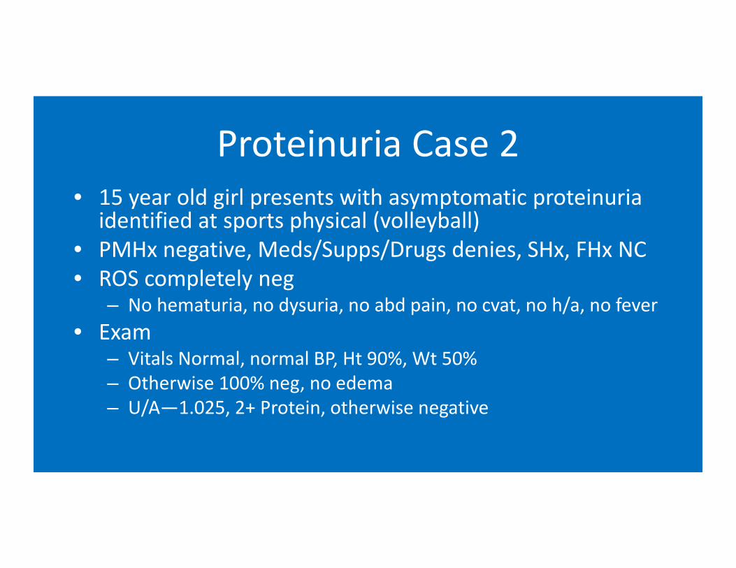
Proteinuria Case 2• 15 year old girl presents with asymptomatic proteinuria
identified at sports physical (volleyball)• PMHx negative, Meds/Supps/Drugs denies, SHx, FHx NC• ROS completely neg
– No hematuria, no dysuria, no abd pain, no cvat, no h/a, no fever• Exam
– Vitals Normal, normal BP, Ht 90%, Wt 50%– Otherwise 100% neg, no edema– U/A—1.025, 2+ Protein, otherwise negative

What to do next?
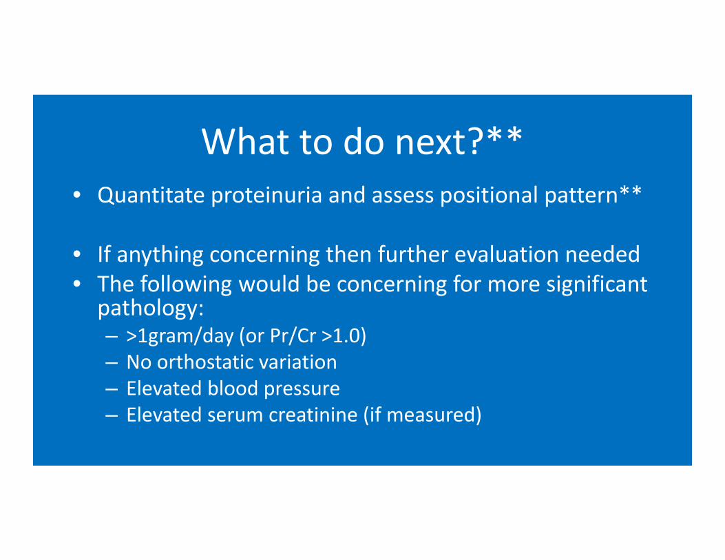
What to do next?**• Quantitate proteinuria and assess positional pattern**
• If anything concerning then further evaluation needed• The following would be concerning for more significant pathology:– >1gram/day (or Pr/Cr >1.0)– No orthostatic variation– Elevated blood pressure– Elevated serum creatinine (if measured)

DDx Proteinuria in Children**• Benign
– False positive (concentrated specimen)– Recent exercise, dehydration, physical stress– Orthostatic (benign positional) proteinuria
• Classic is tall adolescent or 20‐something
• Pathologic– FSGS, Membranous GN– SLE, Hepatitis Infection of HIV associated– Diabetic Nephropathy may start with asymptomatic proteinuria even in
teens– Glomerulonephritis – Hereditary
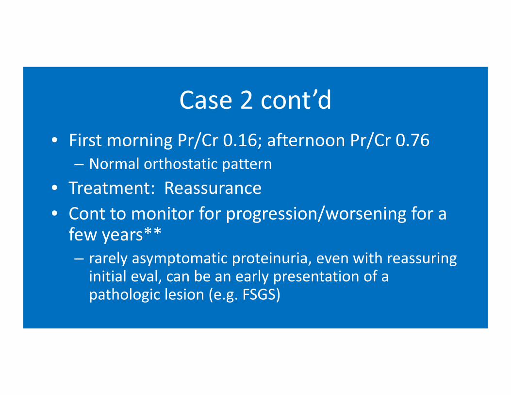
Case 2 cont’d• First morning Pr/Cr 0.16; afternoon Pr/Cr 0.76
– Normal orthostatic pattern• Treatment: Reassurance• Cont to monitor for progression/worsening for a few years** – rarely asymptomatic proteinuria, even with reassuring initial eval, can be an early presentation of a pathologic lesion (e.g. FSGS)

Take Home Points for Practice• Gross Hematuria may be a manifestation of
– Glomerulonephritis, presenting with RBC casts, hypertension, and proteinuria or
– Non‐glomerular causes such as infection, hypercalciuria or congenital or acquired structural anomalies (stones, cysts, urinary tract dilation), presenting with urinary and/or abdominal/flank symptoms
• Elevated serum creatinine, hypertension, and nephrotic syndrome are clinical indicators of severity for an acute glomerulonephritis and warrant evaluation and treatment
• Nephrotic syndrome due to minimal change disease is most often responsive to steroids and has an excellent long‐term prognosis, but is associated with serious complications requring vigilence and support

Suggested ReadingPan CG. Evaluation of Gross Hematuria. PediatrClin N Am 2006, 401‐412.
Gipson DS et al. Management of Childhood Onset Nephrotic Syndrome. Pediatrics 2009, 124: 747‐757. DOI: 10.1542/peds.2008‐1559
STOPNARRATIVES ARE PROVIDED AS A
REFERENCE FOR THE CASE PRESENTATION SESSIONS.
IN ORDER FOR YOU TO ACHIEVE THE MOST BENEFIT FROM THESE SESSIONS, WE ASK
THAT YOU DO NOT REFER TO THE NARRATIVES PRIOR TO ATTENDING THE CASE
PRESENTATION SESSIONS

Fluid and Electrolyte Metabolism/Renal and Urologic Disorders
Adam Weinstein, MDAssistant Professor Pediatric Nephrology
Children’s Hospital at Dartmouth Hitchcock

Disclosures• I have no relevant financial relationships with the manufacturers(s) of any commercial products(s) and/or provider of commercial services discussed in this CME activity.
• I do not intend to discuss an unapproved/investigative use of a commercial product/device in my presentation.

Objectives• Evaluate and plan treatment for various etiologies of
hyponatremia and their associated fluid and electrolyte abnormalities
• Distinguish between the causes of and initiate evaluation for glomerular and non‐glomerular hematuria
• Interpret which markers of severity in the setting of acute glomerulonephritis warrant evaluation and treatment
• Review the causes of and management approaches for nephrotic syndrome and asymptomatic proteinuria in children
Major Concepts in Sodium Handling• Why is the sodium low?**
– Too much water– Too little salt
• What aids in this decision?**– Patient volume status clinically
• Weight changes– Urine Na+ [Fractional Excretion of Na+(FENa)]– Urine Osmolality
Major Concepts in Urine Na+ Evaluation• Kidney preserves circulationg volume with highest priority!
• UNa+ reflects renal perfusion** independent of SerumNa+:– Low UNa+ (<20 mEq/L) or Low FeNa (<1%)
• Renal Perfusion is decreased**– High UNa+ (>50 mEq/L) or High FeNa (>2%)
• Renal Perfusion is normal or increased• Defect in tubule reabsorption**
– If UNa+ elevated in clinical circumstance when renal blood flow is expected to be decreased suggests renal tubular dysfunction**

Major Concepts in Urine Osm Evaluation
• UOSM reflects H20 removal from tubule fluid**– When UOSM is high water retained by body– When UOSM is low water is being excreted in urine
– Normal UOSM can range from 50‐1200 mOsm/L– SG in urine 1.010 = UOSM 300 mOsm/L
Major Concepts in Urine Osm Evaluation• ADH secretion stimulated by
– Increase serum osmolality (SOSM)– Decrease circulating volume– ADH secretion results in increases in UOSM (any increase can be
related to ADH secretion)– However, when UOSM > SOSM by >1.5 X or more, the only way that
can happen is by ADH acting on collecting duct• If UOSM is increased without increased SOSM or decreased
intravascular volume: – ADH secretion is non‐physiologic (SIADH)—irrespective of urine
output or urine volume**
Case One• 9 month old girl • 3 days of fever, non‐bilious, non‐bloody vomiting, and watery diarrhea
• Decreasing urine output over past 2 days, only once in past 24 hours
• 3 lb weight loss since health maintenance visit 2 weeks ago. She has some tachycardia but is interactive, cranky but consolable by mother, good peripheral perfusion
Case One Labs**• Na 127• K 3.0• Cl 100• HCO3 16• BUN 24• Cr 0.3

Questions Case One• What do you suspect the FENa and Urine Osmolality are?

Case One Urine Labs**• Urine Na <10 mEq/L• FENa <0.2%• Urine Osm 800 mosm/L• In this case and the others, use Serum Sodium to estimate calculated Serum Osmolality– unless the serum BUN or Glucose values given are elevated, in which case use the formula:
• 2xNa + BUN/2.8 + Glucose/18 = Calculated Serum Osm**
Questions Case One• What is the diagnosis?**
• Does the patient seem to have more vomiting or diarrhea?**
• Explain the hypokalemia and how would you treat it?**
Answers Case One• Hyponatremic Dehydration**
– the infant must have been getting hypotonic oral solution (juice, water, even pedialyte) as a treatment
• She has more diarrhea at this point, based on the normal anion gap (11) metabolic acidosis** – if vomiting were predominant, would expect a metabolic alkalosis**
Answers Case One• The hypokalemia is in part from GI losses, but even more so, in the setting of volume depletion aldosterone is stimulated and causes high urinary excretion**– As she is rehydrated, her potassium may get lower as renal perfusion increases and acidosis is corrected**
• High levels of H+ causes K to shift extracellular to allow H+ to move intracellular and reduce the acidemia
• So as acidosis is corrected, K will shift back intracellularly, and serum K will drop
Answers Case One• Repleting her potassium is particularly important to prevent symptoms such as weakness, paralysis, and arrhythmia.** – Most safe and effective would be IV rehydration with NS solution with KCl (20 to 40 mEq/L) added in continuously
– If she were having symptoms from hypokalemia, then can give as IV K bolus– but important to be judicious and avoid prescribing K bolus except when absolutely needed due to serious complications from inadvertent hyperkalemia
Case Two• The same 9 month old girl is given oral rehydration. Over next 24 hours, vomitingpersists, intake is poor, and diarrhea is increasing. No urine output.
• Weight is further decreased. She has worse tachycardia. She is lethargic and doesn’t respond much to examination. Cap refill is now prolonged
Case Two Labs**• Na 125• K 6.5• Cl 92• HCO3 9• BUN 45• Cr 0.9

Questions Case Two• What do you suspect the FENa and Urine Osmolality are?

Case Two Urine Labs**• Urine Na 60 mEq/L• FENa 2.3 %• Urine Osm 250 mosm/L
Questions Case Two• The patient appears more volume depleted but based on
urine sodium and FENa, the kidney thinks renal perfusion is adequate. What is causing this discrepancy?**
• Explain the low bicarbonate?**
• What management would you provide for the high potassium?**
• What do you think her GFR is?**
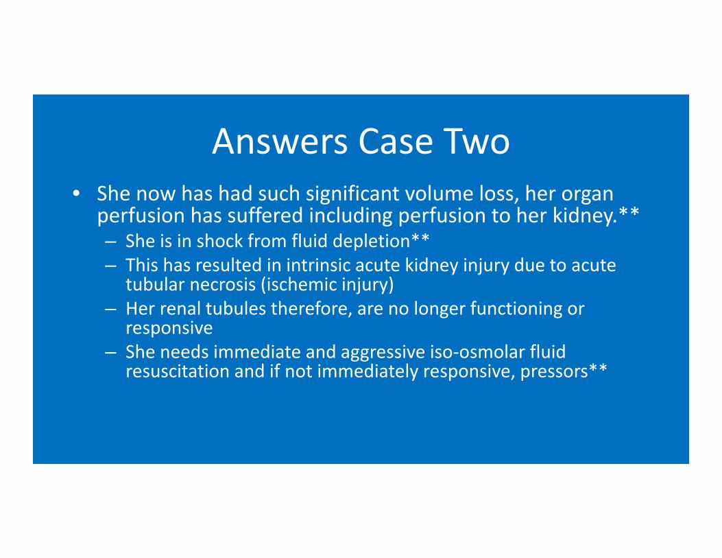
Answers Case Two• She now has had such significant volume loss, her organ
perfusion has suffered including perfusion to her kidney.** – She is in shock from fluid depletion**– This has resulted in intrinsic acute kidney injury due to acute
tubular necrosis (ischemic injury) – Her renal tubules therefore, are no longer functioning or
responsive– She needs immediate and aggressive iso‐osmolar fluid
resuscitation and if not immediately responsive, pressors**
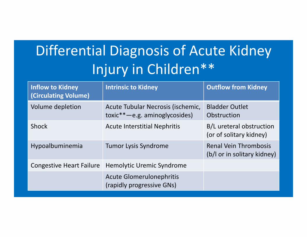
Differential Diagnosis of Acute Kidney Injury in Children**
Inflow to Kidney (Circulating Volume)
Intrinsic to Kidney Outflow from Kidney
Volume depletion Acute Tubular Necrosis (ischemic, toxic**—e.g. aminoglycosides)
Bladder Outlet Obstruction
Shock Acute Interstitial Nephritis B/L ureteral obstruction (or of solitary kidney)
Hypoalbuminemia Tumor Lysis Syndrome Renal Vein Thrombosis (b/l or in solitary kidney)
Congestive Heart Failure Hemolytic Uremic Syndrome
Acute Glomerulonephritis (rapidly progressive GNs)
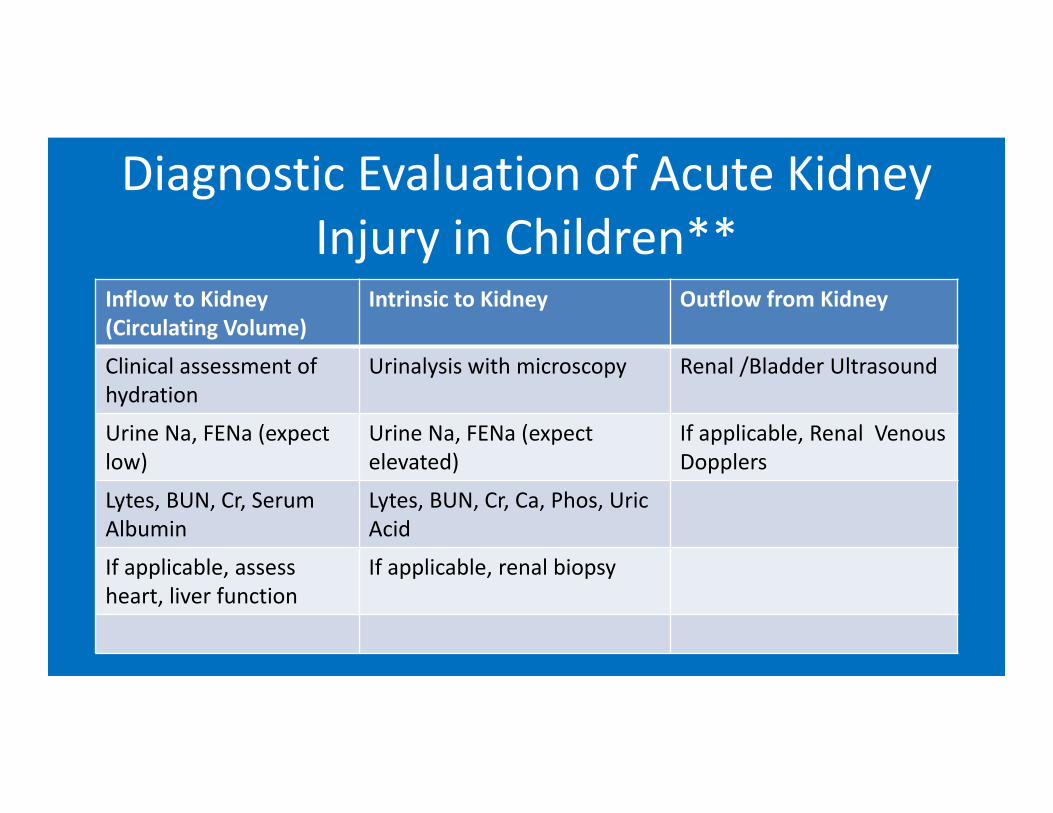
Diagnostic Evaluation of Acute Kidney Injury in Children**
Inflow to Kidney (Circulating Volume)
Intrinsic to Kidney Outflow from Kidney
Clinical assessment of hydration
Urinalysis with microscopy Renal /Bladder Ultrasound
Urine Na, FENa (expect low)
Urine Na, FENa (expect elevated)
If applicable, Renal Venous Dopplers
Lytes, BUN, Cr, Serum Albumin
Lytes, BUN, Cr, Ca, Phos, UricAcid
If applicable, assess heart, liver function
If applicable, renal biopsy
Answers Case Two• Her bicarbonate level is lower suggesting worsened metabolic acidosis. Note that the anion gap is now elevated (125 – (92 + 9) = 24**– Given her volume depletion and poor perfusion this now represents and anion gap metabolic acidosis, likely from lactic acidosis**
– With correction of her perfusion, then, this should improve
Answers Case Two• Her potassium is now elevated, likely because of impaired renal excretion from low perfusion and tubular injury**– Hopefully correction of her perfusion will correct some tubular function and improve potassium excretion
– If her kidney injury persists despite that, then additional measures are needed to prevent hyperkalemia complications (EKG changes fatal arrhythmia)**
• IV Calcium to stabilize the cardiac myocytes• Albuterol (Beta‐agonist), Insulin + Glucose, and/or Bicarbonate can all help K shift intracellularly
• Means for excretion– renal excretion (if responsive to improved perfusion), GI excretion through Na‐K resins, and/or dialysis
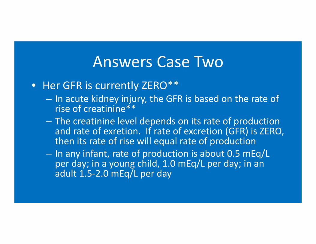
Answers Case Two• Her GFR is currently ZERO**
– In acute kidney injury, the GFR is based on the rate of rise of creatinine**
– The creatinine level depends on its rate of production and rate of exretion. If rate of excretion (GFR) is ZERO, then its rate of rise will equal rate of production
– In any infant, rate of production is about 0.5 mEq/L per day; in a young child, 1.0 mEq/L per day; in an adult 1.5‐2.0 mEq/L per day
Case Three• Five year old male presents with periorbital edema, abdominal distension, and ankle swelling
• He has a weight gain of 5 lb over the past 4 days• Notable in his history are a recent URI, and decreased urine output only 1‐2 times per day.
• His HR is borderline tachycardia, Blood Pressure 92/54

Case Three Labs**• Na 128• K 4.9• Cl 90• HCO3 26• BUN 12• Cr 0.5
Questions Case Three• What do you think the child’s diagnosis is?**• What do you suspect the FENa and Urine Osmolality are?**
• Why is hyponatremia associated with his condition?**
• How do you fix the hyponatremia?**
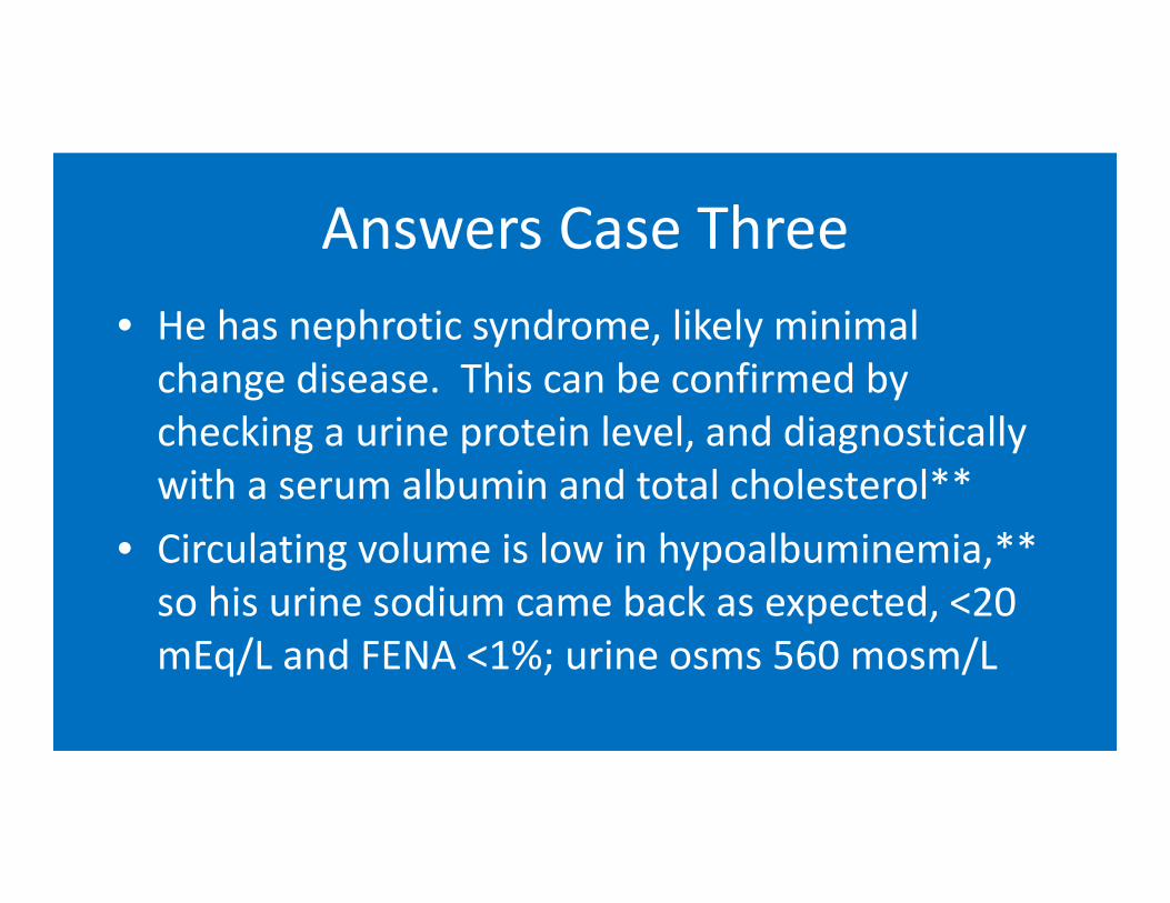
Answers Case Three• He has nephrotic syndrome, likely minimal change disease. This can be confirmed by checking a urine protein level, and diagnostically with a serum albumin and total cholesterol**
• Circulating volume is low in hypoalbuminemia,** so his urine sodium came back as expected, <20 mEq/L and FENA <1%; urine osms 560 mosm/L
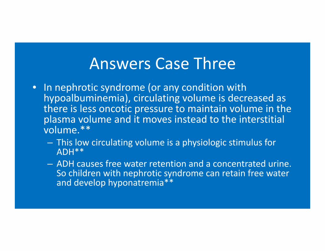
Answers Case Three• In nephrotic syndrome (or any condition with hypoalbuminemia), circulating volume is decreased as there is less oncotic pressure to maintain volume in the plasma volume and it moves instead to the interstitial volume.** – This low circulating volume is a physiologic stimulus for ADH**
– ADH causes free water retention and a concentrated urine. So children with nephrotic syndrome can retain free water and develop hyponatremia**
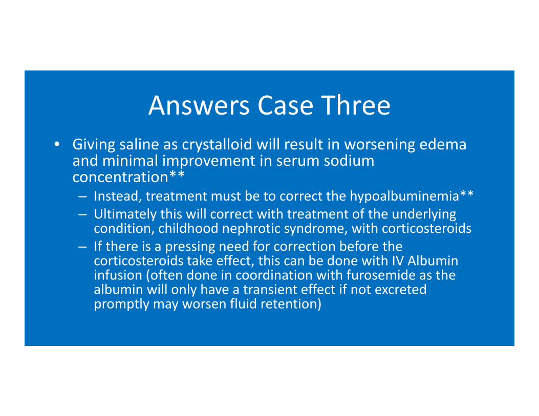
Answers Case Three• Giving saline as crystalloid will result in worsening edema
and minimal improvement in serum sodium concentration** – Instead, treatment must be to correct the hypoalbuminemia** – Ultimately this will correct with treatment of the underlying
condition, childhood nephrotic syndrome, with corticosteroids– If there is a pressing need for correction before the
corticosteroids take effect, this can be done with IV Albumin infusion (often done in coordination with furosemide as the albumin will only have a transient effect if not excreted promptly may worsen fluid retention)
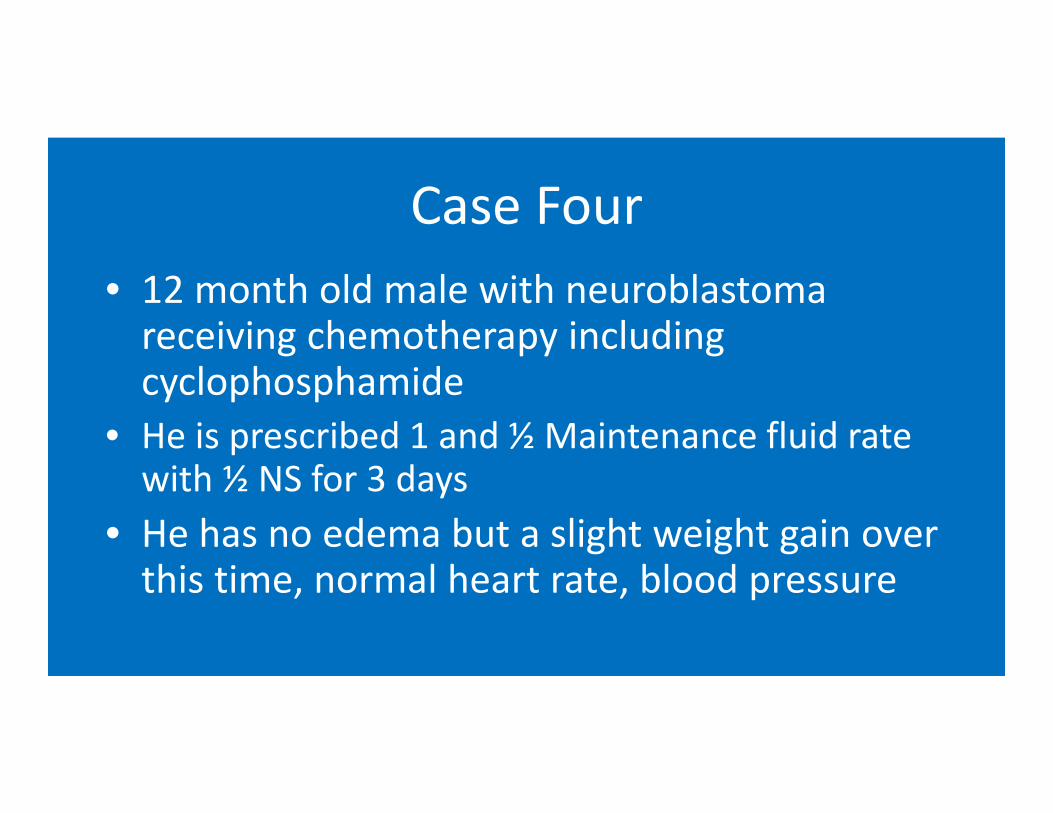
Case Four • 12 month old male with neuroblastomareceiving chemotherapy including cyclophosphamide
• He is prescribed 1 and ½ Maintenance fluid rate with ½ NS for 3 days
• He has no edema but a slight weight gain over this time, normal heart rate, blood pressure
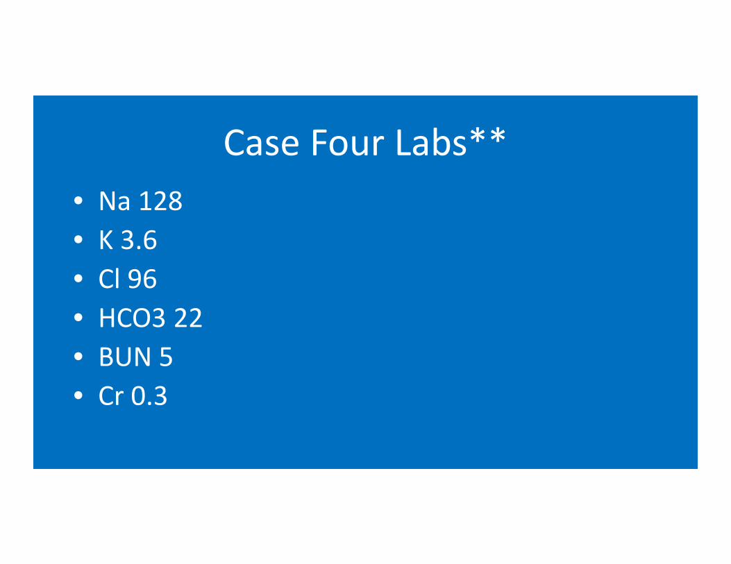
Case Four Labs**• Na 128• K 3.6• Cl 96• HCO3 22• BUN 5• Cr 0.3

Questions Case Four• What do you suspect the FENa and Urine Osmolality are?

Case Four Urine Labs**• Urine Na 95 mEq/L• FENa 2.1 %• Urine Osm 780 mosm/L
Questions Case Four• What is the child’s diagnosis is?**• What will your management be?**
Answers Case Four• This child has SIADH** • He has no physiologic stimuli for ADH secretion**
– He has normal circulating volume and no increase in serum osmolality
– Yet he has a concentrated urine, which means ADH is present and active
– With no physiologic stimuli, this indicates inappropriate ADH secretion.
Answers Case Four• Treatment is restriction of free water intake**• Giving extra isotonic sodium chloride may actually make matters worse– The kidney senses replete circulating volume so will excrete (not reabsorb) the sodium
– Because of the inappropriate ADH, the kidney will retain the fluid (water) in the saline even so
– Therefore giving fluids at a brisk rate (rather than a restricted rate), even isotonic fluids, will make matters worse
Conditions Associated with SIADH**• CNS Disease—stroke, hemorrhage, infection, trauma• Certain malignancies (mostly adult ones like lung cancer), but neuroblastoma can cause it
• Medications– carbamazepine, cyclophosphamide, SSRIs, and many others
• Surgery/Trauma• Pulmonary Disease, in particular infection• Pain, Vomiting, Stress Response• Pregnancy
Case Five• 6 year old female develops polyuria and polydipsia
• Her weight is decreased and she has an increased respiratory rate

Case Five Labs**• Na 128• K 5.8• Cl 94• HCO3 10• BUN 20• Cr 0.7• Glucose 450
Questions Case Five• Why is the serum sodium low?**
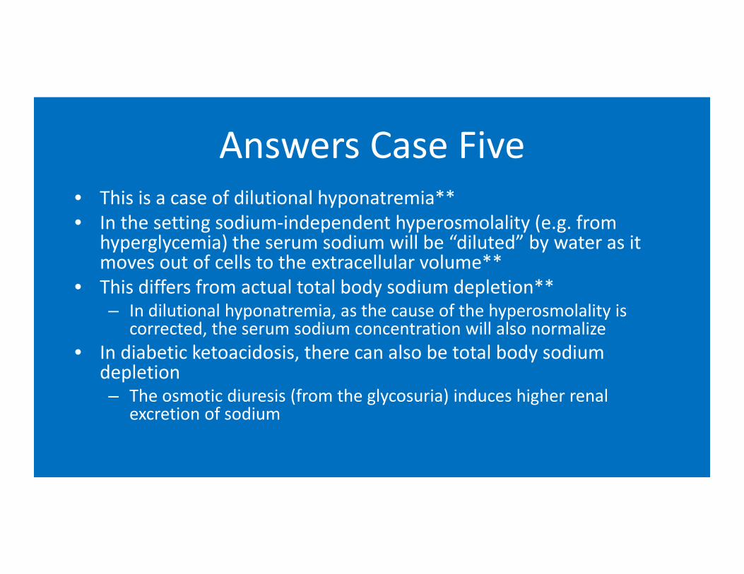
Answers Case Five• This is a case of dilutional hyponatremia**• In the setting sodium‐independent hyperosmolality (e.g. from
hyperglycemia) the serum sodium will be “diluted” by water as it moves out of cells to the extracellular volume**
• This differs from actual total body sodium depletion** – In dilutional hyponatremia, as the cause of the hyperosmolality is
corrected, the serum sodium concentration will also normalize• In diabetic ketoacidosis, there can also be total body sodium
depletion– The osmotic diuresis (from the glycosuria) induces higher renal
excretion of sodium
Various Etiologies of Hyponatremia**: Clinical Utilization of UNa and UosmIncreased Weight UNa UOSM Decreased Weight
HypoalbuminemiaNephrosis/Cirrhosis
AGN
<10 >500 Dehydration/Volume DepletionCystic Fibrosis
Acute Volume expansionWater intoxicationExcess IV Fluids
>10 <300
<10 <300 Diabetes InsipidusAcute Renal Failure
Sepsis, Shock, Nephrotoxin>50 <=300 Adrenal Insufficiency
Salt losing NephropathyInterstitial Nephritis/ Cystic Disease/
Urinary Tract ObstructionNon-Physiologic ADH >50 >500 DKA
Osmotic Diuretics
Take Home Points for Practice• The etiology of hyponatremia can be determined through
assessment of clinical volume status, urine sodium excretion (FENa), & urine osmolality
• If the renal response to a suggested clinical volume state appears inappropriate, consider renal tubular pathology—tubular injury, tubular dysfunction, or inappropriate hormone secretion (e.g. SIADH)
• Treatment of low volume states respond well to volume and electrolyte repletion; treatment of excess volume states respond to management of underlying disorder (e.g. nephroticsyndrome) and thoughtful supportive restrictions (e.g. SIADH)
Case 1• 7 year old male presents with tea colored urine• Otherwise well, no known trauma, URI symptoms 1 month
ago, now better. +Headache, otherwise ROS negative• PMHx—well managed Type 1 DM dx’d 4 years prior• On Insulin, acetaminophen as needed• Soc Hx, Fam Hx: non‐contributory• Exam
– BP 150/80, vitals otherwise normal for age– 95% ht, 97% wt (mom noted weight is up 5 pounds from a week ago)– exam otherwise normal, fundi wnl, no edema, normal abdomen and
lungs

What is the next step?
What is the next step?• First, confirm true hematuria**‐‐
– Is U/A + for blood? • If U/A neg blood—ingestion (beets, med)
• Next confirm RBC in urine**– Is Microscopy + for RBCs?– If Microscopy neg for RBCs then think:
• Myoglobinuria• Hemoglobinuria• Old specimen or other false positive

DDx Gross Hematuria in Children**• Glomerular (Kidney)
– Systemic signs/sx– Brown or black color– RBC casts, dysmorphic RBCs
• Glomerulonephritis– Post‐Infectious– MPGN, SLE– IgA Nephropathy, HSP– HUS– Alport’s, Thin BM Dz
• Non‐Glomerular– Localized signs/sx– Pink/red urine +/‐ clots– eumorphic RBCs
• Tumors, Trauma• Infection, Inflammation• Cystic disease, Congenital
anomaly, Crystalluria(Calciuria)
• Stones, Sickle cell

Patient’s Urinalysis
Acute Glomerulonephritis
• What next?
Answer• Should include evaluation of Severity**
– Lytes, BUN, Cr– Quantitate proteinuria– Blood Pressure
• Should include evaluation of Etiology**– CBC, Strep titers, ANA, C3, C4, ANCA, and/or anti‐GBM titers, etc…each should be considered based on clinical context
• In this case, perhaps CBC, Strep titers, C3, C4– Renal Ultrasound if not straightforward and you still need to consider non‐glomerular etiologies
What are clinical indicators of a severe glomerulonephritis?
• Acute kidney injury/elevated creatinine• Hypertension• Severe proteinuria, nephrotic range proteinuria, in particular nephrotic syndrome
• Why and how can glomerulonephritis cause nephrotic syndrome?

Recap on Podocytes and Glomerular Basement Membrane
• Podocytes– Help create the negatively charged slit diaphragm that prevents many serum proteins (e.g. albumin) from filtering across the glomerulus
• Minimal Change Disease is a result of podocytes that are no longer functioning (they are “effaced”)– Negative charge slit diaphragm is gone– Serum proteins (e.g. albumin) filter into the Bowman’s Space

Severe Glomerulonephritis• When there is severe involvement of the basement membrane, then one might think of the podocytes as innocent bystanders
• Therefore, Nephrotic Syndrome is a manifestation of severe proteinuria and a marker for a severe glomerulonephritis (GN)
• If such a GN goes untreated, or does not resolve spontaneously (e.g. post‐infectious), then this results in a poor long‐term prognosis

Serology Interpretation• Let’s say you get C3 and C4 and ASLO titer• Results show a + ASLO titer, but C3 and C4 are normal
• What is your diagnostic interpretation?**

Answer• Should state that the normal C3 and C4 makes this less likely to be post‐strep or post‐infectious GN.
• The positive strep titers likely represents past strep associated illness (anytime over prior 6‐12 months), but not necessarily resulting in post‐infectious GN.
• More likely another cause, like IgA Nephropathy, for example…

Back to Patient
• CBC normal• ANA Neg, C3 very low, C4 normal, Post‐strep titers positive• Pr/Cr ratio 7.2!
4.2 0.514
23108140

What is your presumptive diagnosis?**
A. IgA NephropathyB. Alport SyndromeC. Membranoproliferative GND. Post‐Infectious GN
What is your presumptive diagnosis?
D. Post‐Infectious GN (PIGN/PSGN)**• Low C3, + Post‐Strep titers, and acute presentation are all
consistent with PIGN**• As this is also a common etiology, based on the above, can make
a presumptive diagnosis of Post‐Infectious GN• IgA Nephropathy and Alport Syndrome are not associated with a
low C3**• IgA Nephropathy occurs concurrent with a resp infection, not
afterwards**• MPGN is associated with a low C3, but is significantly less
common than PIGN

How could you confirm this?**A. Monitoring for spontaneous resolution and
return of C3 to normal over next 2‐3 monthsB. Response to empiric treatment with
CorticosteroidsC. Renal Biopsy
How could you confirm this?A. Monitoring for spontaneous resolution and return of
C3 to normal over next 2‐3 months**• Should expect full resolution of PIGN over coming
months • Often gross hematuria resolves over 1‐2 weeks, and
hypertension and proteinuria gradually resolve over a few weeks to months as well
• If no resolution after 2‐3 months, then may need to consider alternate etiologies
How could you confirm this?• PIGN is treated with supportive measures**;
corticosteroids have not been shown to help it resolve faster or prevent complications
• Most PIGN resolves completely, but longitudinal follow‐up may illustrate higher risks for hypertension and proteinuria again as adults
– Not from recurrence, rather from chronic impacts on glomeruli number and filtration pressures
– Even so, these effects are uncommon, generally not during childhood, and prognosis is generally excellent
How could you confirm this?• If no improvement over 2‐3 months,** then may
have to consider alternative etiologies (like MPGN) and would confirm this via renal biopsy
• At present time, given likelihood of PIGN, a renal biopsy is not necessary
• However, in the setting of AKI, and risk for a rapidly progressive course, a renal biopsy would be done to confirm a PIGN etiology (rather than MPGN, which would require different treatment)

But what about his Hypertension?**A. Provide reassurance. If this is post‐strep GN, it will get
better on its own so it does not require treatment
B. It is severely elevated for a 7 yo so warrants treatment with a low sodium diet and an ACE Inhibitor until the GN resolves
C. It is severely elevated for a 7 yo, so warrants treatment with a low sodium diet and thiazide diuretic until the GN resolves
But what about his Hypertension?• His BP elevation is severe (Stage 2 hypertension) and his
symptoms of headache may suggest it is symptomatic• Even though PIGN is self‐limited, associated hypertension
can be severe and result in acute complications like hypertensive encephalopathy**
• Therefore BP elevation is typically treated**– Either an ACE‐Inhibitor or a diuretic may be good first line choice– However if there is an elevated creatinine an ACE‐Inhibitor would be
contra‐indicated– ACE‐Inhibitors through their effect on glomerular hemodynamics
may increase the creatinine even further
Why did he gain weight?**A. With his acute GN and HTN he was more sedentary,
so this is increased body fatB. With his acute GN, his GFR may be decreased so this
is likely fluid weight from renal failureC. With his acute GN, renin is stimulated and his
nephrons are likely reabsorbing salt and water so this is fluid weight
D. With his acute GN and being sick, he probably actually lost weight, so this is measurement error
Why did he gain weight?A. With his acute GN, renin is stimulated and
his nephrons are likely reabsorbing salt and water so this is fluid weight**
• Choice B is not correct in this case because his creatinine and GFR are normal
• However, this is another plausible option for someone with an impaired GFR
Case 2• 6 yo male presents with pinkish‐red colored urine• Otherwise well, no known trauma, no recent medications, in school but no obvious illness, ROS completely negative
• PMHx, Soc Hx, Fam Hx all Non‐Contributory• Exam
– normal BP and vitals, 50% ht and wt– Exam otherwise completely normal

Next Step
Urine dip and microscopy to confirm whether this is hematuria? Glomerular vs. Non‐glomerular?**
Patient’s results: U/A + blood, trace protein Many eumorphic RBC, no casts

How would you evaluate at this point?
How would you evaluate at this point?**
• DDx of non‐glomerular hematuria**– Urine Ca/Cr and Renal U/S
• If either positive for stones, consider 24 hour urine for stone chemistries
– Any symptoms/signs of infection? If so, Urine Cx– Could consider Lytes BUN, Cr, CBC, Pr/Cr—if still need to be certain to exclude a GN etiology
Results• Ca/Cr 0.47 (Nml <0.2)• Renal U/S neg, other screens normal
• 3 months later—presents with flank pain and passes stone!
Hypercalciuria• Why care?
– Presence of hematuria worrisome to parents– Risk for stones**—VERY PAINFUL!!!– Can be associated with nephrocalcinosis**
• deposits of calcium within tubulointerstitial parenchyma• can cause progressive scarring and loss of function
• Who develops?– Idiopathic (probably multifactorial—genetics, diet, etc…)– Certain ‘tubulopathies’ (e.g. Dent’s disease, Bartter’s syndrome)
– Medication induced
• When Infants and children develop stones– Stone is not the disease. It’s a sign of the disease
• Every child with kidney stones or nephrocalcinosis should have a thorough evaluation as to the cause**– H&P into systemic disorders
• Growth and development, blood pressure, bone related signs/symptoms• Dietary history (attention to excess or deficiency), fluid intake, meds including vitamins and supplements
– Fam Hx of stones, hematuria, renal failure– Urinalysis, urine and serum chemistries, imaging– STONE analysis (send the stone for chemical analysis)
Childhood Kidney Stones**

• Acute Flank Pain not the rule, though is the presenting finding in majority
• Often difficult to diagnose in infants and young children**– nonspecific discomfort, may be in abdomen or hip
• Hematuria‐microscopic or macroscopic– Stones or crystalluria (eg hypercalciuria) or nephrocalcinosis
• Dysuria and frequency (bladder irritation)
Clinical Constellation of Childhood Kidney Stones**

• Calcium Oxalate—radioopaque—50‐70%– Risks are hypercalciuria, hyperoxaluria, and/or hypocitraturia
• Calcium Phosphate—radioopaque –15‐30%– Form in alkaline urine, common in children with metabolic bone disease or
immobilization• Struvite—radioopaque –15%
– Form in alkaline urine, associated with urinary tract infection with urease producing organisms
• Cystine—radioopaque—5% – Form in acidic urine, hereditary proximal tubule transporter defect
• Uric Acid—5%– Radiolucent and form in acidic urine
• Other—5%
Causes/Risks of Stones in Children**

How do you treat idiopathic (most common form) hypercalciuria (and prevent nephrolithiasis)?**
A. Drink lots of fluids to make a dilute urineB. Low Sodium DietC. High Citrate DietD. Thiazide Diuretic
Answer• All of these can be effective measures to make
urine calcium more soluble (dilute urine, calcium citrate is soluble) or reduce urine calcium (low sodium diet, thiazide diuretic**
• Typical management is to discuss these options with the patient and family– Most asymptomatic (apart from the hematuria)
patients choose more conservative therapies (fluid intake, diet)
– Most patients with recurrent stones choose pharmacotherapy (e.g. thiazide)
Case 3• 14 year old Caucasian boy presents with maroon urine• He is otherwise healthy, playing touch football with friends
earlier that day. Denies obvious trauma or rough tackle.• ROS otherwise NEG• Soc Hx – Athlete, starting 9th grade, doing well, no
substance use, not sexually active• Fam Hx—Mother with hypertension• Exam—HR 60; BP 138/84; tall and thin, atheletic build;
otherwise completely normal, no abd masses or tenderness, no edema
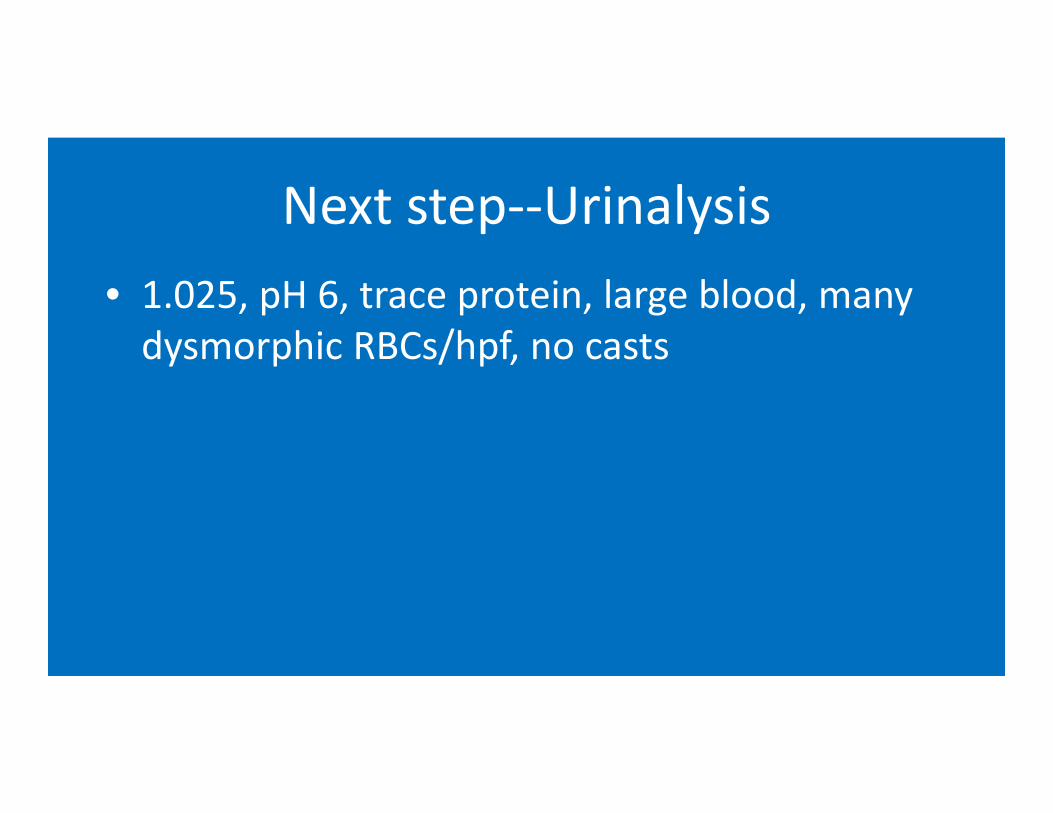
Next step‐‐Urinalysis• 1.025, pH 6, trace protein, large blood, many dysmorphic RBCs/hpf, no casts

Discuss the top three diagnoses on your differential**
Discuss the top three diagnoses on your differential**
• Options to discuss might include:– Renal Trauma– Congenital GU Anomaly (aggravated by mild trauma)– Cystic Kidney Disease/Autosomal Dominant Polycystic Kidney Disease
– Glomerulonephritis/Thin Basement Membrane Disease
What would your evaluation be?**
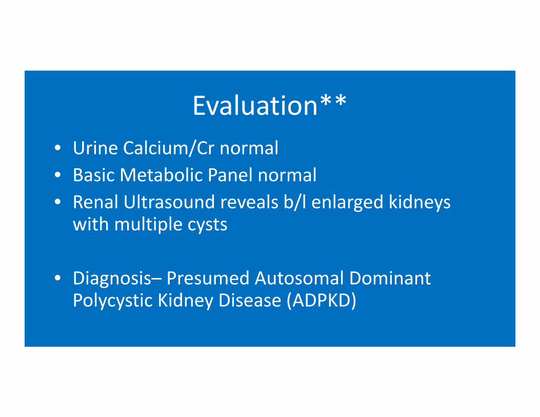
Evaluation**• Urine Calcium/Cr normal• Basic Metabolic Panel normal• Renal Ultrasound reveals b/l enlarged kidneys with multiple cysts
• Diagnosis– Presumed Autosomal Dominant Polycystic Kidney Disease (ADPKD)

ADPKD in children**• A significant minority of cases of ADPKD present in childhood**
– Occasionally on prenatal imaging, especially with known family history– More commonly with acute onset gross hematuria (often in setting of
trauma or exertion)– Hypertension is a common presentation
• Asymptomatic screening of children of parents with ADPKD is not currently recommended**– Screening of these children’s Blood Pressure and a urinalysis for
protein at health maintenance is generally recommended– Presence of hypertension and/or proteinuria are associated with
progression of chronic kidney disease, and can be treated to delay progression**

Name three things you might do as far as clinical management**

ADPKD Management**• Strict Blood Pressure Control using RAAS inhibition—target
<120/80 has illustrated slower decline of renal function• Control of proteinuria with RAAS inhibition—similarly, this
delays progression of CKD and may prevent future development of ESRD
• If kidneys >15cm– may need to restrict from high impact sports/activities (risk of trauma and cyst rupture—massive bleeding)
• High Fluid intake‐‐ ADH is associated with cyst growth, so suppressing ADH with super‐hydration may slow progression
• Patient Education and connections with resources/networks
Case 4• 16 yo female with diarrheal illness x 2 weeks presents with brown
colored urine• No recent travel or new foods, no emesis, but crampy abd pain
results in decreased intake• At first thought urine was just super‐concentrated, but now dark
brown• Medications—took some Cipro after 1 week of illenss, tylenol and
motrin prn pain• PMHx healthy, Soc Hx non‐contrib, Fam Hx notable for Grave’s
Disease• Exam
– BP normal, vitals normal, Ht 50%, Wt 10%– Otherwise completely normal, no edema, mild non‐specific diffuse abd
tenderness
Next step• Real hematuria? Upper vs. Lower?**
– U/A + blood, 2+ protein, MANY RBC Casts (very impressive)
• What is your DDx Acute GN** (of note—also w/diarrheal illness and abd pain!
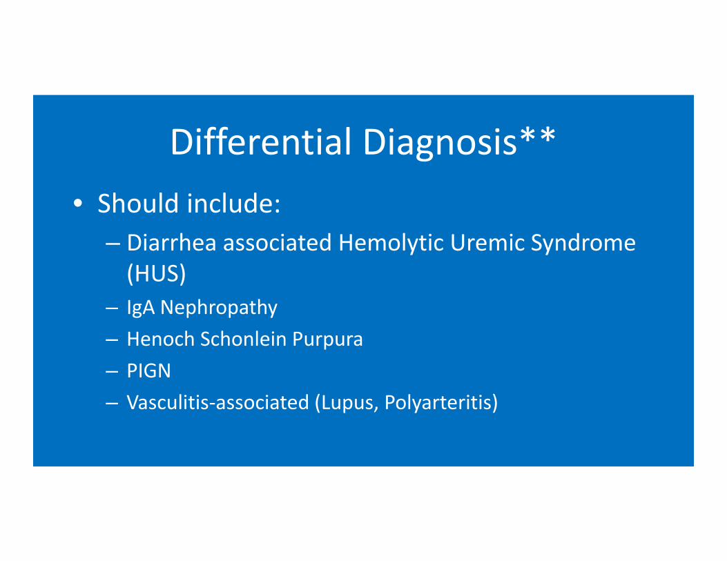
Differential Diagnosis**• Should include:
– Diarrhea associated Hemolytic Uremic Syndrome (HUS)
– IgA Nephropathy– Henoch Schonlein Purpura– PIGN– Vasculitis‐associated (Lupus, Polyarteritis)
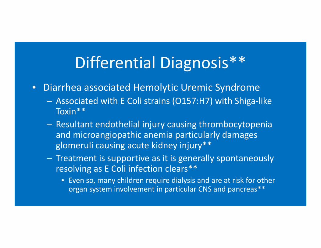
Differential Diagnosis**• Diarrhea associated Hemolytic Uremic Syndrome
– Associated with E Coli strains (O157:H7) with Shiga‐like Toxin**
– Resultant endothelial injury causing thrombocytopenia and microangiopathic anemia particularly damages glomeruli causing acute kidney injury**
– Treatment is supportive as it is generally spontaneously resolving as E Coli infection clears**
• Even so, many children require dialysis and are at risk for other organ system involvement in particular CNS and pancreas**
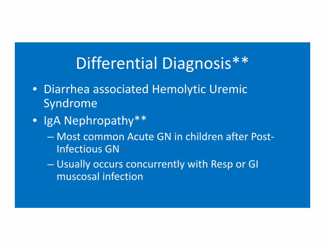
Differential Diagnosis**• Diarrhea associated Hemolytic Uremic Syndrome
• IgA Nephropathy**– Most common Acute GN in children after Post‐Infectious GN
– Usually occurs concurrently with Resp or GI muscosal infection
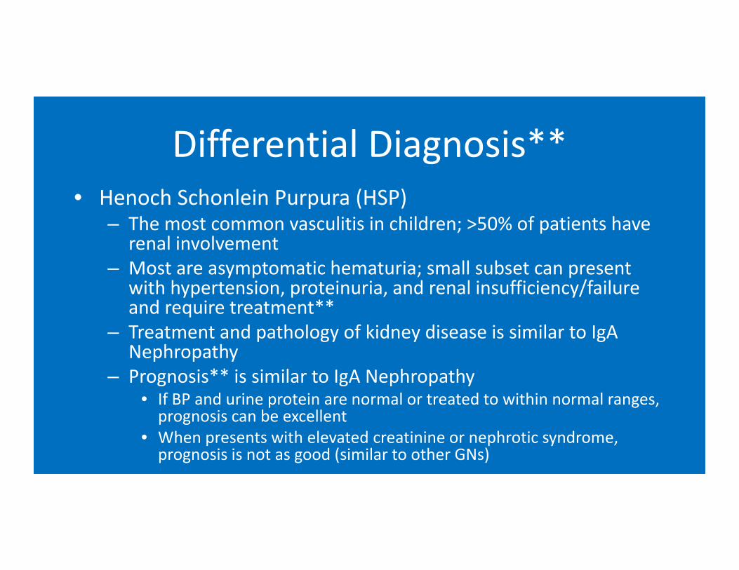
Differential Diagnosis**• Henoch Schonlein Purpura (HSP)
– The most common vasculitis in children; >50% of patients have renal involvement
– Most are asymptomatic hematuria; small subset can present with hypertension, proteinuria, and renal insufficiency/failure and require treatment**
– Treatment and pathology of kidney disease is similar to IgA Nephropathy
– Prognosis** is similar to IgA Nephropathy• If BP and urine protein are normal or treated to within normal ranges, prognosis can be excellent
• When presents with elevated creatinine or nephrotic syndrome, prognosis is not as good (similar to other GNs)

Results of evaluation…• Chems WNL• CBC WNL
– No HUS• Pr/Cr ratio 0.9• ANA, Strep‐titers, ANCAs all NEG
– No SLE, PAN• C3 borderline, C4 WNL
– Unlikely PIGN or MPGN
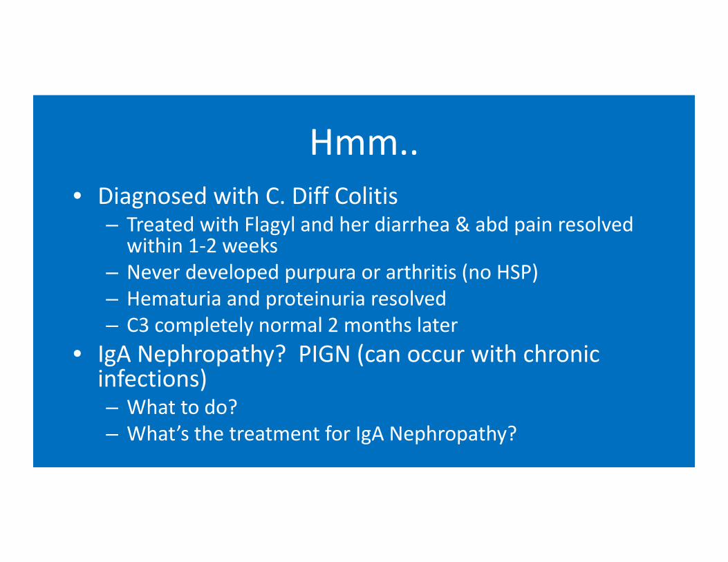
Hmm..• Diagnosed with C. Diff Colitis
– Treated with Flagyl and her diarrhea & abd pain resolved within 1‐2 weeks
– Never developed purpura or arthritis (no HSP)– Hematuria and proteinuria resolved – C3 completely normal 2 months later
• IgA Nephropathy? PIGN (can occur with chronic infections)– What to do? – What’s the treatment for IgA Nephropathy?
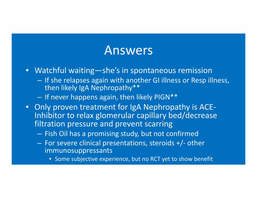
Answers• Watchful waiting—she’s in spontaneous remission
– If she relapses again with another GI illness or Resp illness, then likely IgA Nephropathy**
– If never happens again, then likely PIGN**• Only proven treatment for IgA Nephropathy is ACE‐Inhibitor to relax glomerular capillary bed/decrease filtration pressure and prevent scarring– Fish Oil has a promising study, but not confirmed– For severe clinical presentations, steroids +/‐ other immunosuppressants
• Some subjective experience, but no RCT yet to show benefit

Case 5• 4 yo girl presents with eye swelling• Pediatrician first thought allergies, tried anti‐histamine, but it’s
getting worse and now with abd distension and anasarca. She is active, has good appetite, no nausea/vomiting/diarrhea/fever, no hematuria or dysuria, +URI last week, now resolved
• PMHx eczema, Meds anti‐histamine and topical moisturizer• SocHx, FamHx Non‐contributory• Exam
– Vitals Nml, Weight 90%, Height 50%, BP normal– Exam –Anasarca
• Can a Diagnosis of Nephrotic Syndrome be made?**

What’s missing?**• Urinalysis
– 1.030, moderate blood, 4+ Protein– Microscopy 10‐20 RBCs, no RBC casts
• Serum Albumin—1.2• Cholesterol elevated• Now – we can confirm diagnosis of nephroticsyndrome**– Edema, Nephrotic range proteinuria, hypoalbuminemia, elevated cholesterol
DDx Nephrotic Syndrome in Children**• Primary Nephropathy
– Minimal Change Disease**—by far the most common, especially in young children
– Focal Segmental Glomerulosclerosis (FSGS)– Membranous Nephropathy
• Secondary Nephropathy (systemic illness causing nephroticsyndrome)– Systemic Lupus Erythematosus– Hepatitis associated nephropathy – HIV associated nephropathy
• Glomerulonephritis (severe GN complicated by nephroticsyndrome)
• Hereditary causes
Case 5—What Next?**A. Renal Biopsy is needed to confirm Minimal
Change before treatingB. Wait 2‐4 weeks before treating to see if it
spontaneously resolvesC. Begin empiric treatment with
Corticosteroids and suspect Minimal Change if there is a good treatment response
Answer• Minimal Change Disease is the predominant cause
– If everything consistent with minimal change (e.g. age, not a glomerulonephritis), can make presumptive diagnosis based on treatment response**
• Age under 1 year suggests higher possibility of alternate causes (like hereditary causes)
• Age above 10 years or so with broader differential (increasing likelihood for FSGS or in teenage years, Membranous Nephropathy)
• If anything presents atypically, or fails to respond to treatment, then further evaluation may be necessary, often including biopsy necessary

Minimal Change Disease• Presumptive treatment, 2mg/kg or 60mg Prednisone Daily**
– Usual response in 2‐4 weeks, small number may take longer– Treat intercurrent infections/triggers
• Majority recur**– Significant minority one and done (up to 40%)– Others may develop frequent relapsing or steroid dependent
nephrotic syndrome and require steroid‐sparing therapy (e.g. cyclophosphamide)
– If develops steroid resistance—reconsider other etiologies (e.g. FSGS)• Consistent treatment response suggests an ultimately good
prognosis, even if having recurrences**
Nephrotic Syndrome—Which of the follow are other considerations?**
A. Low Sodium DietB. Albumin—Furosemide InfusionC. Infectious concernsD. Hypercoagulopathy
Answer• All of them!• Low Sodium Diet is helpful– high sodium intake results in worsening edema (fills interstitial volume)**
• When fluid overload/edema is causing symptoms (resp distress) or complications, albumin‐furosemide infusions can help mobilize edema**
Answer• Nephrotic syndrome is associated with an increased risk for
infection** – Loss of IgG in urine– hypogammaglobulinemia– Fluid can get infected, in particular, cellulitis and Strep pneumo
peritonitis or pneumonia • Nephrotic syndrome is associated with a hypercoaguable
state and at increased risk for venous thrombosis** (especially with membranous nephropathy)– Loss of pro‐coagulants and anti‐coagulants in urine but balance
of these losses in a hypercoaguable state
Case 6• 15 year old girl presents with asymptomatic proteinuria
identified at sports physical (volleyball)• PMHx negative, Meds/Supps/Drugs denies, SHx, FHx NC• ROS completely neg
– No hematuria, no dysuria, no abd pain, no cvat, no h/a, no fever• Exam
– Vitals Normal, normal BP, Ht 90%, Wt 50%– Otherwise 100% neg, no edema– U/A—1.025, 2+ Protein, otherwise negative

What to do next?

What to do next?**• Quantitate proteinuria and assess positional pattern**
• If anything concerning then further evaluation needed• The following would be concerning for more significant pathology:– >1gram/day (or Pr/Cr >1.0)– No orthostatic variation– Elevated blood pressure– Elevated serum creatinine (if measured)
DDx Proteinuria in Children**• Benign
– False positive (concentrated specimen)– Recent exercise, dehydration, physical stress– Orthostatic (benign positional) proteinuria
• Classic is tall adolescent or 20‐something
• Pathologic– FSGS, Membranous GN– SLE, Hepatitis Infection of HIV associated– Diabetic Nephropathy may start with asymptomatic proteinuria even in
teens– Glomerulonephritis – Hereditary
Case 6 cont’d• First morning Pr/Cr 0.16; afternoon Pr/Cr 0.76
– Normal orthostatic pattern• Treatment: Reassurance• Cont to monitor for progression/worsening for a few years** – rarely asymptomatic proteinuria, even with reassuring initial eval, can be an early presentation of a pathologic lesion (e.g. FSGS)
Take Home Points for Practice• Gross Hematuria may be a manifestation of
– Glomerulonephritis, presenting with RBC casts, hypertension, and proteinuria or
– Non‐glomerular causes such as infection, hypercalciuria or congenital or acquired structural anomalies (stones, cysts, urinary tract dilation), presenting with urinary and/or abdominal/flank symptoms
• Elevated serum creatinine, hypertension, and nephrotic syndrome are clinical indicators of severity for an acute glomerulonephritis and warrant evaluation and treatment
• Nephrotic syndrome due to minimal change disease is most often responsive to steroids and has an excellent long‐term prognosis, but is associated with serious complications requring vigilence and support
Suggested ReadingPan CG. Evaluation of Gross Hematuria. PediatrClin N Am 2006, 401‐412.
Gipson DS et al. Management of Childhood Onset Nephrotic Syndrome. Pediatrics 2009, 124: 747‐757. DOI: 10.1542/peds.2008‐1559


