Endocrine DevelopmentHormone from hormon (Gk) to set in motion, to urge on (E.H. Starling 1866-1927)
1
Objectives
• Understanding of hormone types • Focus on hypothalamus and pituitary development• Understanding of endocrine gland development • Understanding of endocrine developmental functions• Brief understanding of molecular regulation and signalling
mechanisms
2
Citation: The Developing Human: clinically oriented embryology 9th ed. Keith L. Moore, T.V.N. Persaud, Mark G. Torchia. Philadelphia, PA: Saunders, 2011. Chapter 9 – Pharyngeal Apparatus, Face, and NeckChapter 12 ‐ Urogenital System
Citation: Larsen's human embryology 4th ed. Schoenwolf, Gary C; Larsen, William J, (William James). Philadelphia, PA : Elsevier/Churchill Livingstone, c2009. Chapter 16 ‐ Development of the Pharyngeal Apparatus and FaceChapter 15 ‐ Development of the Urogenital System
3
Nussey, S. and Whitehead, S. (2001). Endocrinology ‐ An Integrated Approach. UK Oxford: BIOS Scientific Publishers. ISBN‐10: 1‐85996‐252‐1. Detailed Table of Contents | Bookshelf Link•Chapter 1. Principles of endocrinology•Chapter 2. The endocrine pancreas•Chapter 3. The thyroid gland•Chapter 4. The adrenal gland•Chapter 5. The parathyroid glands and vitamin D•Chapter 6. The gonad•Chapter 7. The pituitary gland•Chapter 8. Cardiovascular and renal endocrinology
4
Hormones
Produced by endocrine glands – ductless glands that secrete hormones into the blood
• Amino acid derivatives – noradrenaline (norepinephrin), adrenalin (epinephrin), thyroid hormone
• Peptides and proteins – Thyroid stimulating hormone, leutenising hormone, follicle stimulating hormone (HPG axis)
• Steroids – androgens, glucocorticoids (regulate inflammation), mineralocorticoids (salt and water balance)
All hormones act upon cells in different tissues and can be classified by the "distance" of their action, the classical description is that hormones are delivered by the blood.
• Autocrine ‐ acts on self (extracellular fluid) – e.g. cytokines such as interleukin‐1 in monocytes• Paracrine ‐ acts locally (extracellular fluid or blood) – e.g. fgf, Wnt, shh, • Endocrine ‐ acts by secretion into blood stream (endocrine organs are richly vascularized).
Hormone Receptors• cell surface ‐modified amino acids, peptides, proteins – e.g. FSH receptor, G‐protein coupled
receptor• intracellular cytoplasmic/nuclear – steroids – androgen receptor, Nuclear receptor subfamily
3, C4 (NR3C4)
5
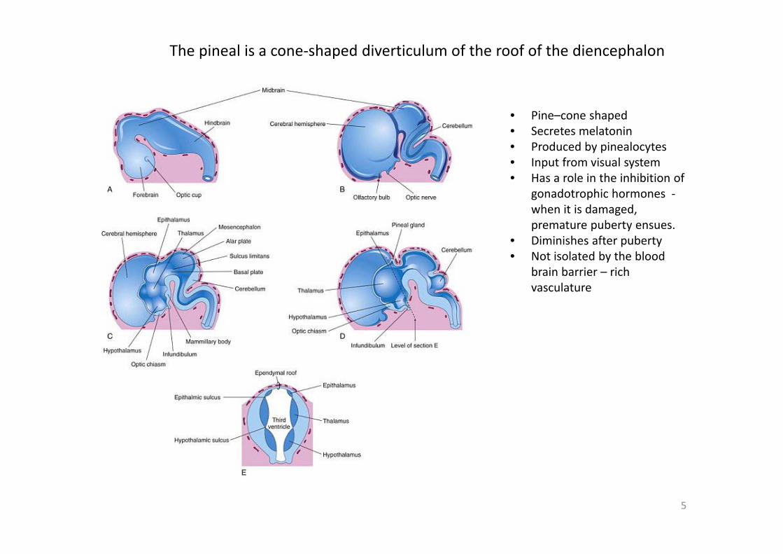
The pineal is a cone‐shaped diverticulum of the roof of the diencephalon
• Pine–cone shaped• Secretes melatonin• Produced by pinealocytes• Input from visual system• Has a role in the inhibition of
gonadotrophic hormones ‐when it is damaged, premature puberty ensues.
• Diminishes after puberty• Not isolated by the blood
brain barrier – rich vasculature
6
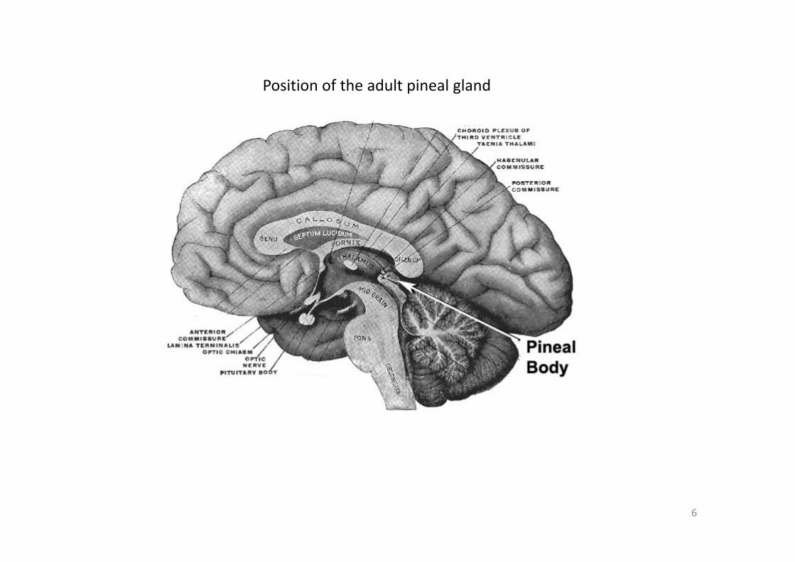
Position of the adult pineal gland
7
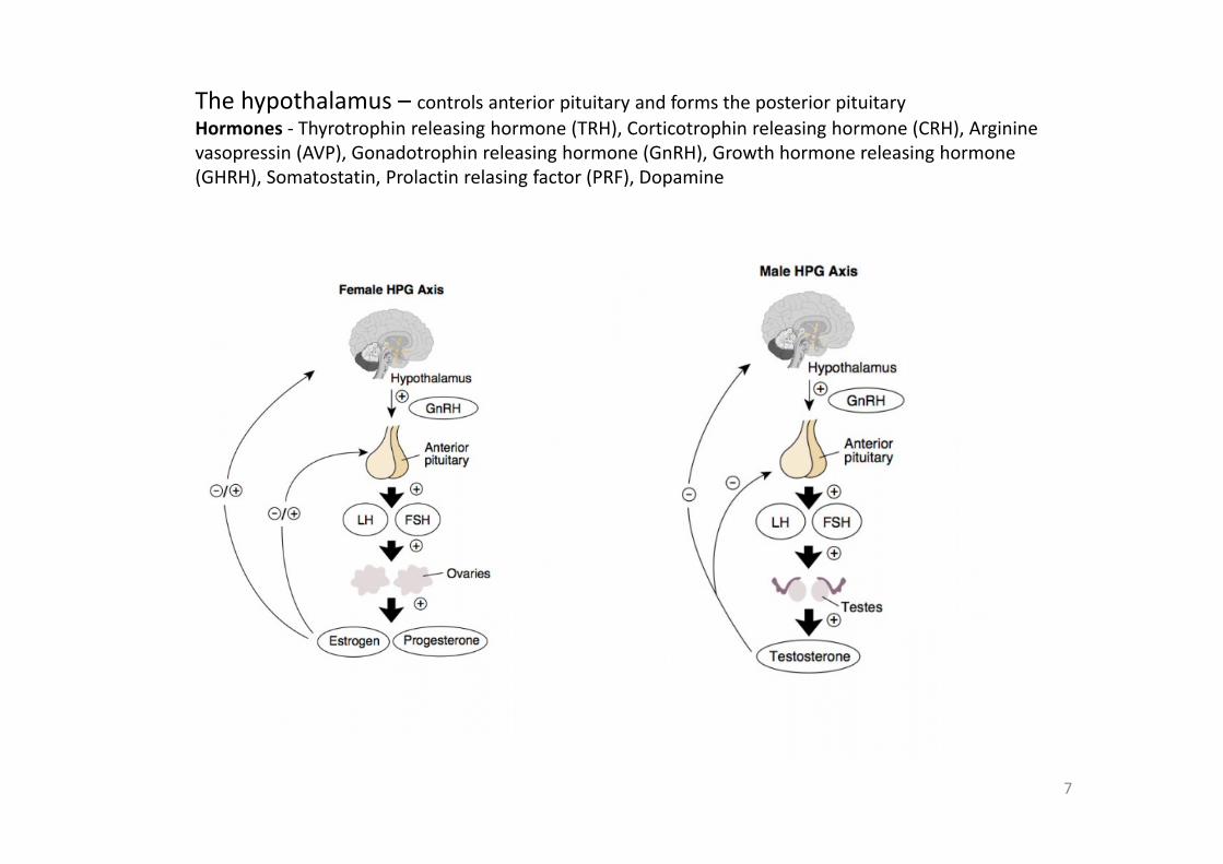
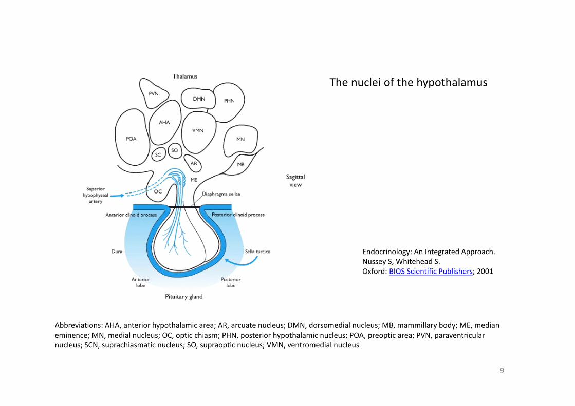
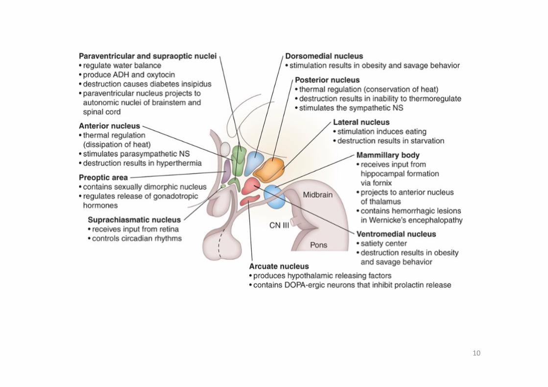
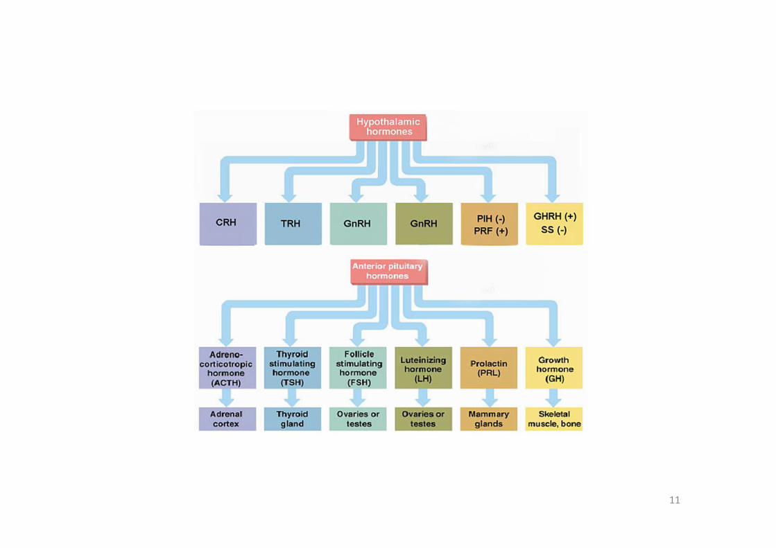
The hypothalamus – controls anterior pituitary and forms the posterior pituitaryHormones ‐ Thyrotrophin releasing hormone (TRH), Corticotrophin releasing hormone (CRH), Arginine vasopressin (AVP), Gonadotrophin releasing hormone (GnRH), Growth hormone releasing hormone (GHRH), Somatostatin, Prolactin relasing factor (PRF), Dopamine
8
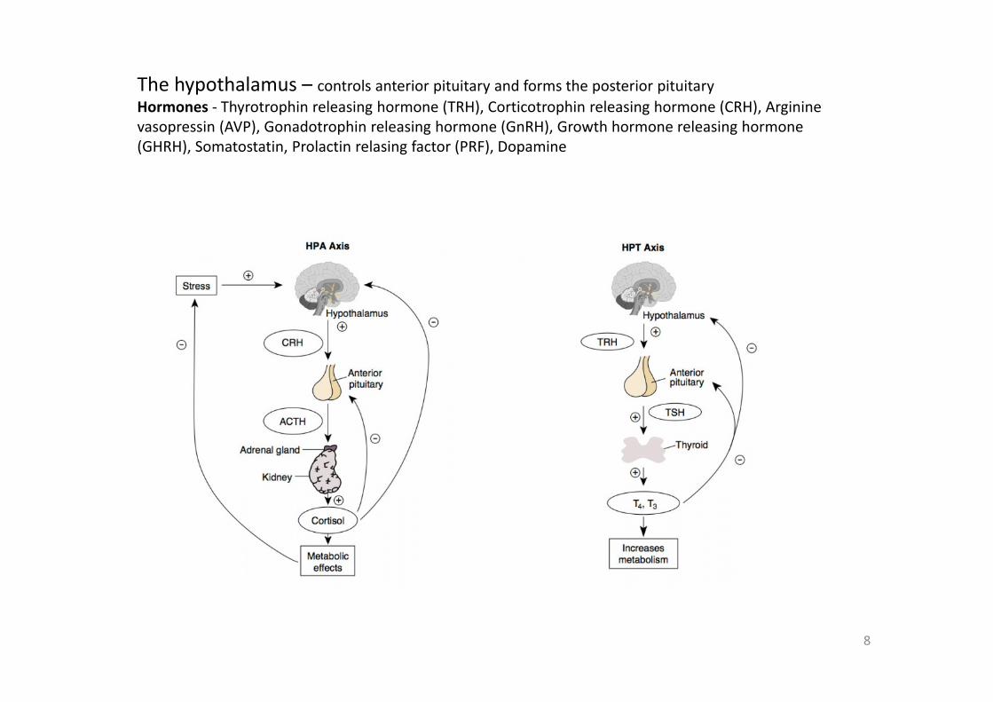
The hypothalamus – controls anterior pituitary and forms the posterior pituitaryHormones ‐ Thyrotrophin releasing hormone (TRH), Corticotrophin releasing hormone (CRH), Arginine vasopressin (AVP), Gonadotrophin releasing hormone (GnRH), Growth hormone releasing hormone (GHRH), Somatostatin, Prolactin relasing factor (PRF), Dopamine
9
Abbreviations: AHA, anterior hypothalamic area; AR, arcuate nucleus; DMN, dorsomedial nucleus; MB, mammillary body; ME, median eminence; MN, medial nucleus; OC, optic chiasm; PHN, posterior hypothalamic nucleus; POA, preoptic area; PVN, paraventricularnucleus; SCN, suprachiasmatic nucleus; SO, supraoptic nucleus; VMN, ventromedial nucleus
Endocrinology: An Integrated Approach.Nussey S, Whitehead S.Oxford: BIOS Scientific Publishers; 2001
The nuclei of the hypothalamus
10
11
12
Pituitary development movie – Interaction between the floor of the diencephalon and the ectodermal roof of the stomadeum
13
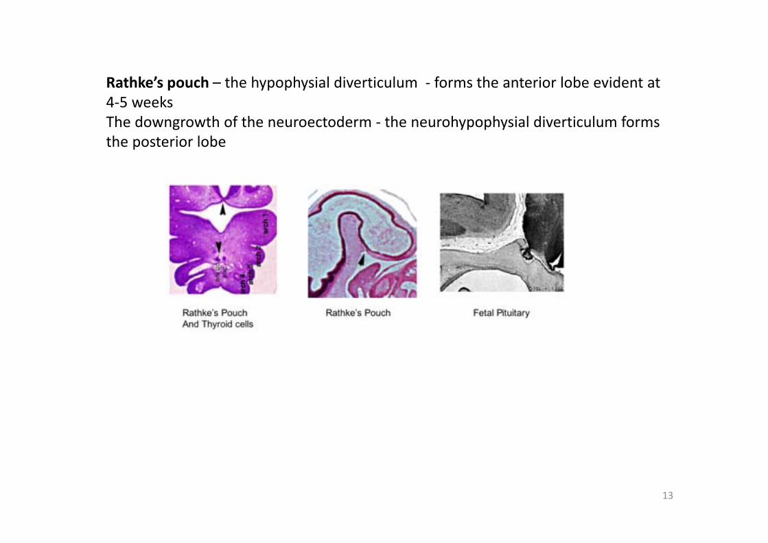
Rathke’s pouch – the hypophysial diverticulum ‐ forms the anterior lobe evident at 4‐5 weeksThe downgrowth of the neuroectoderm ‐ the neurohypophysial diverticulum forms the posterior lobe
14
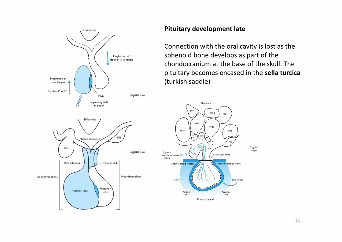
Pituitary development late
Connection with the oral cavity is lost as the sphenoid bone develops as part of the chondocranium at the base of the skull. The pituitary becomes encased in the sella turcica(turkish saddle)
15
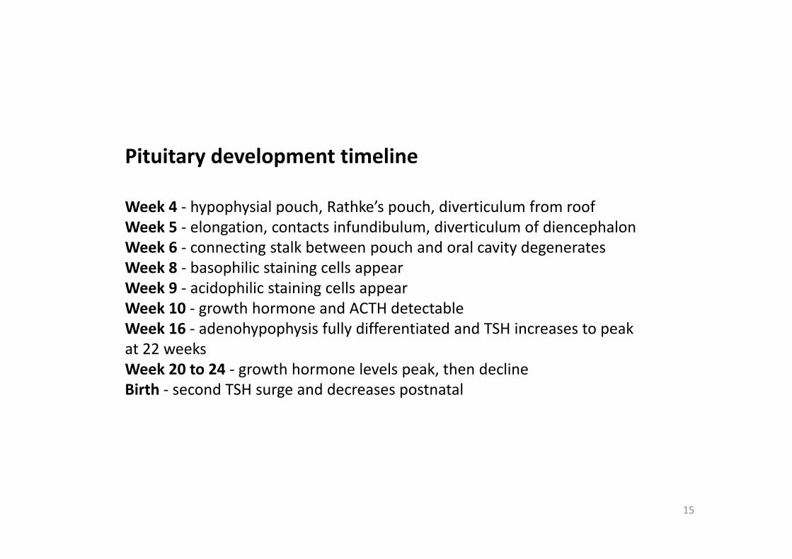
Pituitary development timeline
Week 4 ‐ hypophysial pouch, Rathke’s pouch, diverticulum from roof Week 5 ‐ elongation, contacts infundibulum, diverticulum of diencephalon Week 6 ‐ connecting stalk between pouch and oral cavity degenerates Week 8 ‐ basophilic staining cells appear Week 9 ‐ acidophilic staining cells appear Week 10 ‐ growth hormone and ACTH detectable Week 16 ‐ adenohypophysis fully differentiated and TSH increases to peak at 22 weeks Week 20 to 24 ‐ growth hormone levels peak, then decline Birth ‐ second TSH surge and decreases postnatal

16
The vascular system of the pituitary
The pituitary gland maintains connections with the brain but sits outside of the blood brain barrier.
The pituitary has one of the richest blood supplies of anywhere in the body.
The posterior lobe is supplied by the inferior hypophyseal artery
The anterior lobe is supplied by the superior hypophyseal artery
17
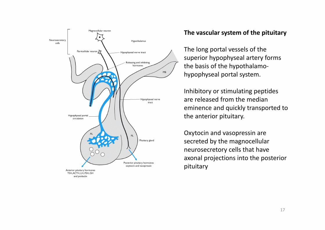
The vascular system of the pituitary
The long portal vessels of the superior hypophyseal artery forms the basis of the hypothalamo‐hypophyseal portal system.
Inhibitory or stimulating peptides are released from the median eminence and quickly transported to the anterior pituitary.
Oxytocin and vasopressin are secreted by the magnocellularneurosecretory cells that have axonal projections into the posterior pituitary
18
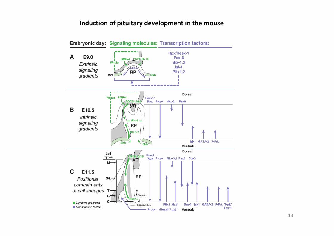
Induction of pituitary development in the mouse
19
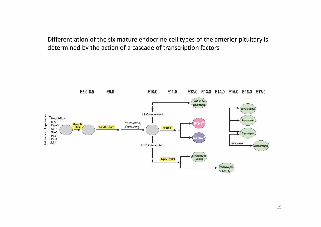
Differentiation of the six mature endocrine cell types of the anterior pituitary is determined by the action of a cascade of transcription factors
20
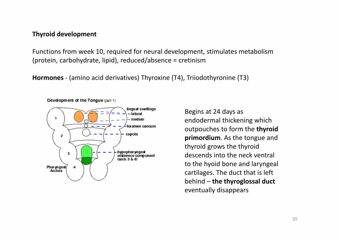
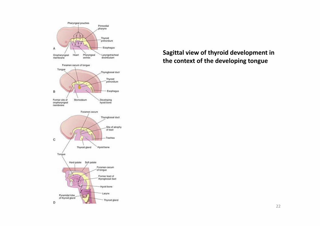
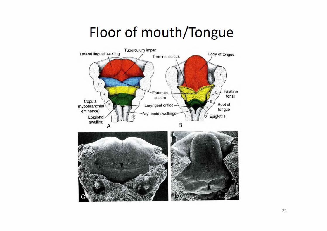
Thyroid development
Functions from week 10, required for neural development, stimulates metabolism (protein, carbohydrate, lipid), reduced/absence = cretinism
Hormones ‐ (amino acid derivatives) Thyroxine (T4), Triiodothyronine (T3)
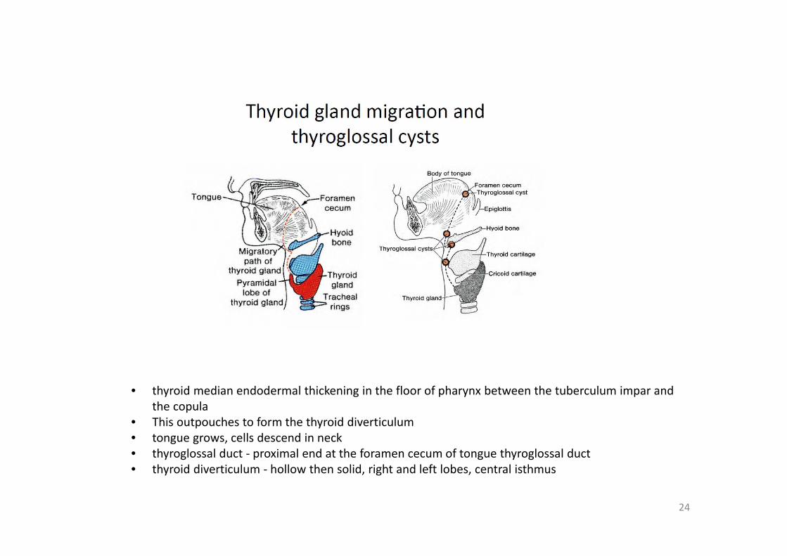
Begins at 24 days as endodermal thickening which outpouches to form the thyroid primordium. As the tongue and thyroid grows the thyroid descends into the neck ventral to the hyoid bone and laryngeal cartilages. The duct that is left behind – the thyroglossal duct eventually disappears
21
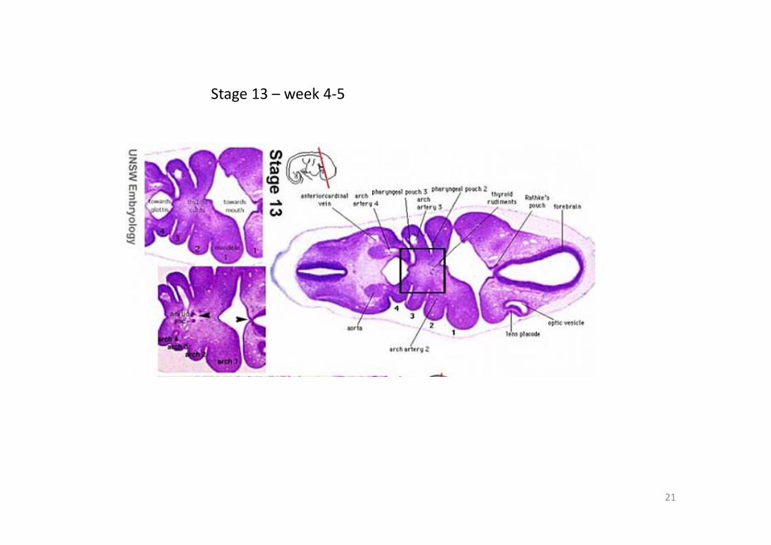
Stage 13 – week 4‐5
22
Sagittal view of thyroid development in the context of the developing tongue
23
24
• thyroid median endodermal thickening in the floor of pharynx between the tuberculum impar and the copula
• This outpouches to form the thyroid diverticulum • tongue grows, cells descend in neck • thyroglossal duct ‐ proximal end at the foramen cecum of tongue thyroglossal duct • thyroid diverticulum ‐ hollow then solid, right and left lobes, central isthmus
25
Parathyroid function
• 4 glands in the neck on the posterior surface of the thyroid about the size of a grain of rice
• Parathyroid Hormone ‐ Increase calcium ions [Ca2+] in the blood stimulates osteoclasts, increase Ca GIT absorption (opposite effect to calcitonin produced by the parafollicular cells of the thyroid formed from the ultimobranchial body)
• Adult Calcium and Phosphate ‐ Daily turnover in human with dietary intake of 1000 mg/day
• secreted by chief cells • Principal cells cords of cells
26
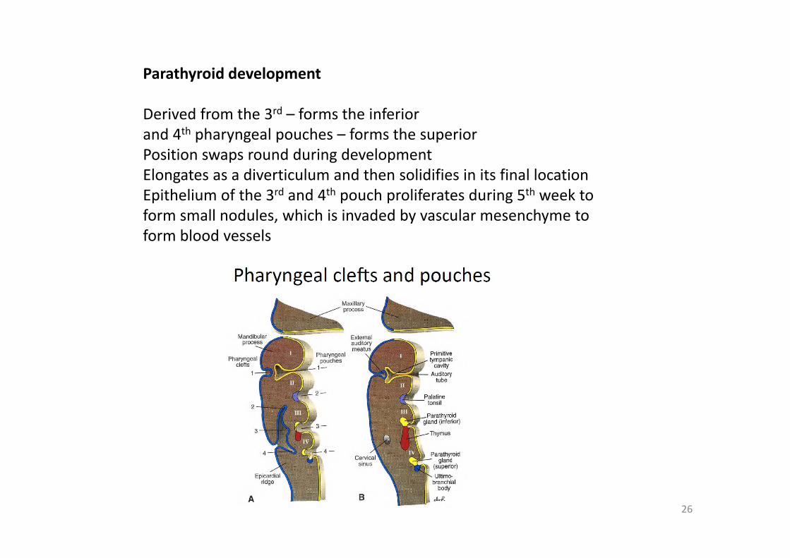
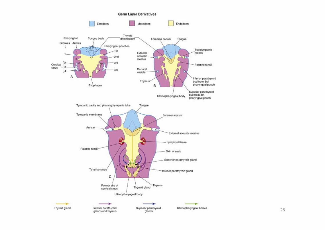
Parathyroid development
Derived from the 3rd – forms the inferiorand 4th pharyngeal pouches – forms the superiorPosition swaps round during developmentElongates as a diverticulum and then solidifies in its final locationEpithelium of the 3rd and 4th pouch proliferates during 5th week to form small nodules, which is invaded by vascular mesenchyme to form blood vessels
27
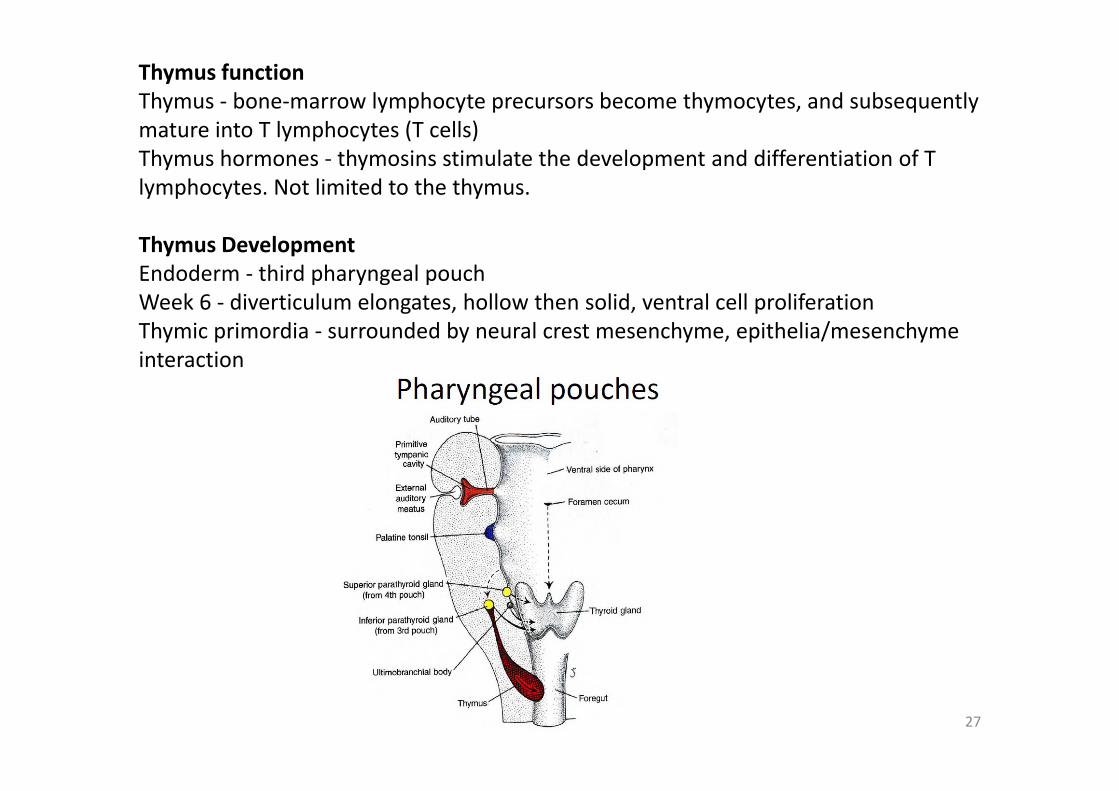
Thymus functionThymus ‐ bone‐marrow lymphocyte precursors become thymocytes, and subsequently mature into T lymphocytes (T cells) Thymus hormones ‐ thymosins stimulate the development and differentiation of T lymphocytes. Not limited to the thymus.
Thymus DevelopmentEndoderm ‐ third pharyngeal pouch Week 6 ‐ diverticulum elongates, hollow then solid, ventral cell proliferation Thymic primordia ‐ surrounded by neural crest mesenchyme, epithelia/mesenchyme interaction
28
29
Schoenwolf: Larsen's Human Embryology, 4th ed.Copyright © 2008
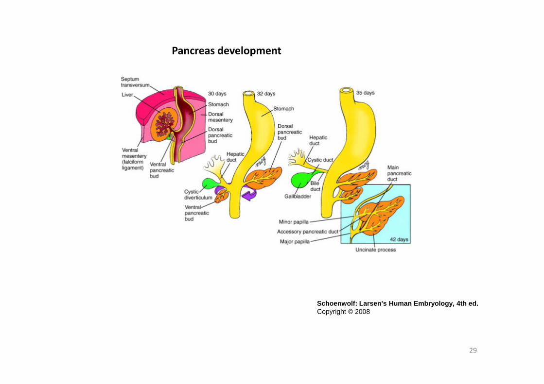
Pancreas development
30
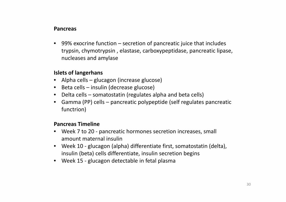
Pancreas
• 99% exocrine function – secretion of pancreatic juice that includes trypsin, chymotrypsin , elastase, carboxypeptidase, pancreatic lipase, nucleases and amylase
Islets of langerhans• Alpha cells – glucagon (increase glucose)• Beta cells – insulin (decrease glucose)• Delta cells – somatostatin (regulates alpha and beta cells)• Gamma (PP) cells – pancreatic polypeptide (self regulates pancreatic
functrion)
Pancreas Timeline• Week 7 to 20 ‐ pancreatic hormones secretion increases, small
amount maternal insulin • Week 10 ‐ glucagon (alpha) differentiate first, somatostatin (delta),
insulin (beta) cells differentiate, insulin secretion begins • Week 15 ‐ glucagon detectable in fetal plasma
31
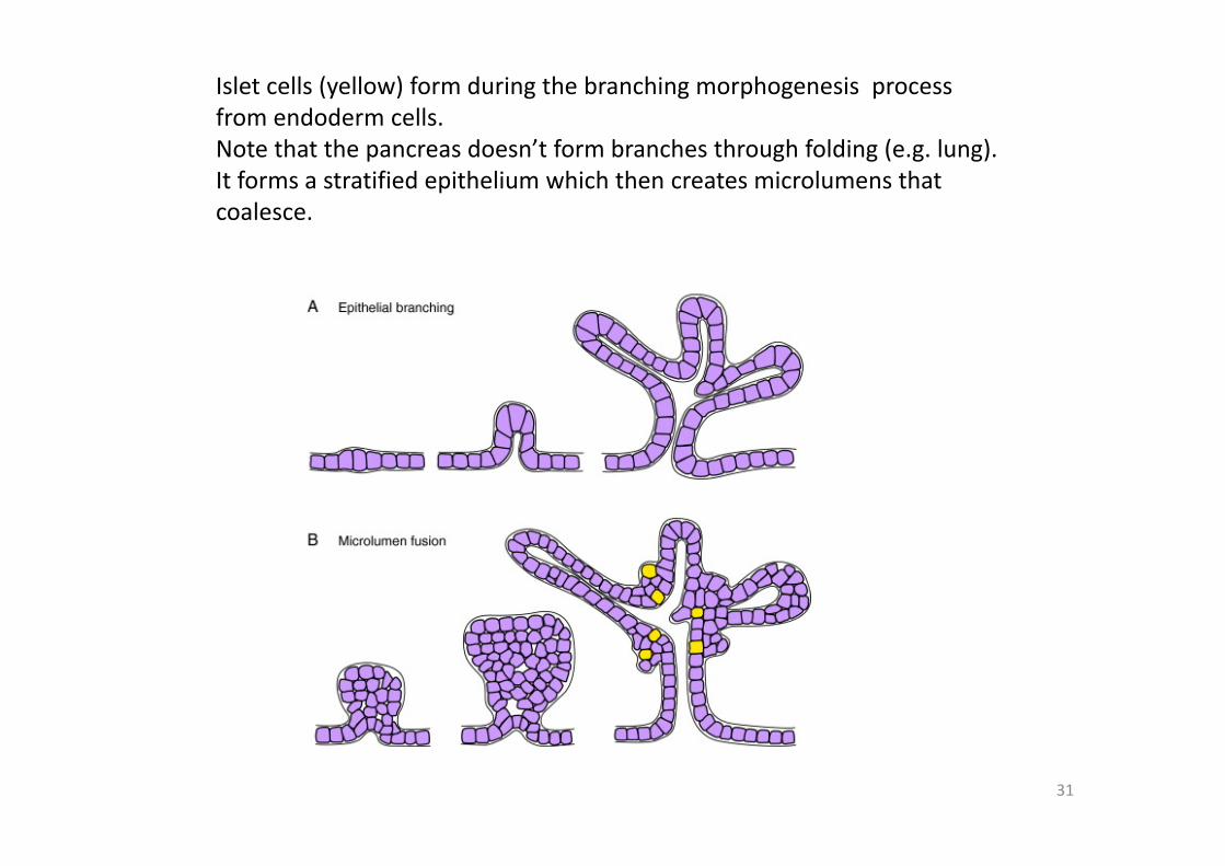
Islet cells (yellow) form during the branching morphogenesis process from endoderm cells. Note that the pancreas doesn’t form branches through folding (e.g. lung). It forms a stratified epithelium which then creates microlumens that coalesce.
32
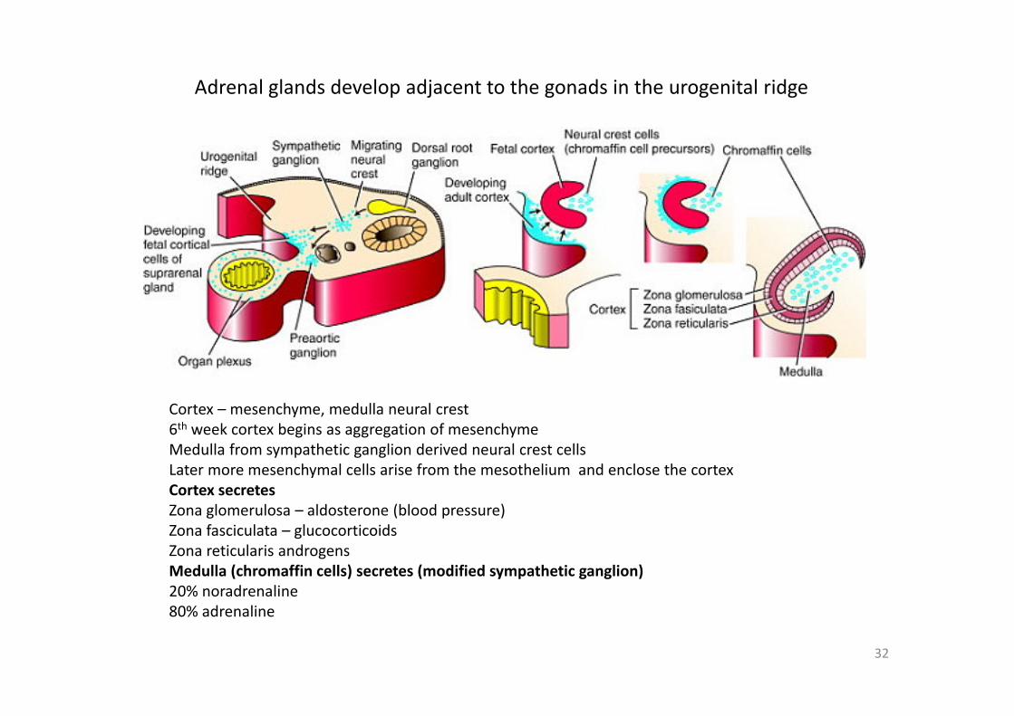
Adrenal glands develop adjacent to the gonads in the urogenital ridge
Cortex – mesenchyme, medulla neural crest6th week cortex begins as aggregation of mesenchyme Medulla from sympathetic ganglion derived neural crest cellsLater more mesenchymal cells arise from the mesothelium and enclose the cortexCortex secretes Zona glomerulosa – aldosterone (blood pressure)Zona fasciculata – glucocorticoidsZona reticularis androgensMedulla (chromaffin cells) secretes (modified sympathetic ganglion)20% noradrenaline80% adrenaline
33
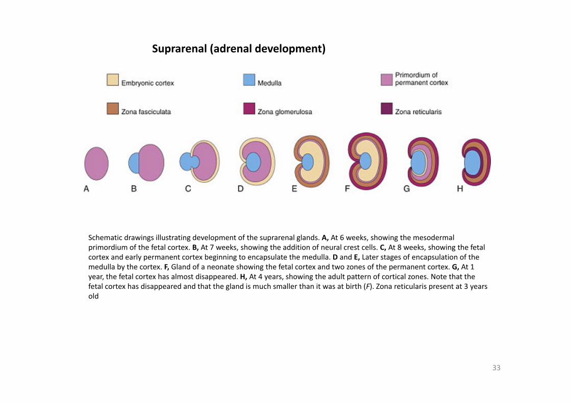
Suprarenal (adrenal development)
Schematic drawings illustrating development of the suprarenal glands. A, At 6 weeks, showing the mesodermal primordium of the fetal cortex. B, At 7 weeks, showing the addition of neural crest cells. C, At 8 weeks, showing the fetalcortex and early permanent cortex beginning to encapsulate the medulla. D and E, Later stages of encapsulation of the medulla by the cortex. F, Gland of a neonate showing the fetal cortex and two zones of the permanent cortex. G, At 1 year, the fetal cortex has almost disappeared. H, At 4 years, showing the adult pattern of cortical zones. Note that the fetal cortex has disappeared and that the gland is much smaller than it was at birth (F). Zona reticularis present at 3 years old
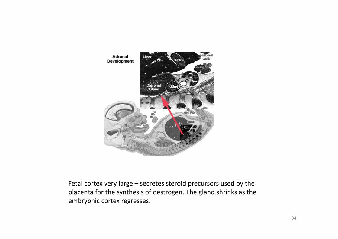
34
Fetal cortex very large – secretes steroid precursors used by the placenta for the synthesis of oestrogen. The gland shrinks as the embryonic cortex regresses.
35
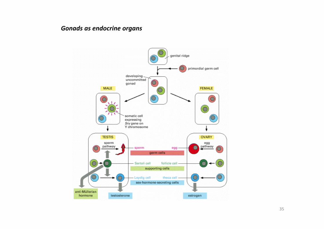
Gonads as endocrine organs
36
37
Placenta
• Human chorionic gonadotropin (hCG) ‐ like leutenizing hormone, supports corpus luteum in ovary, pregnant state rather than menstrual, maternal urine in some pregnancy testing
• Human chorionic somatommotropin (hCS) ‐ or human placental lactogen stimulate (maternal) mammary development (possible) – function similar to growth hormone
• Human chorionic thyrotropin (hCT) or TSH• Human chorionic corticotropin (hCACTH) or corticotropin‐releasing hormone• progesterone and oestrogens ‐ support maternal endometrium • Relaxin
38
Other
Endocrine HeartAtrial natriuretic peptide (ANP) ‐ Increase Filtration rate / decrease Na+ reabsorption Endothelins ‐ ET‐1, ET‐2, ET‐3, Vasoconstriction / Increase NO Nitric oxide (NO) ‐ Vasodilatation Endocrine KidneyRenin ‐ Increase Angiotensin‐aldosterone system Prostaglandins ‐ decrease Na+ reabsorption Erythropoietin ‐ Increase Erythrocyte (rbc) production 1,25 (OH)2 vitamin D ‐ calcium homeostasis Prekallikreins ‐ Increase Kinin production GIT EndocrineEnteric control of digestive function Gastrin ‐ Secreted from stomach (G cells), role in control of gastric acid secretion Cholecystokinin ‐ small intestine hormone, stimulates secretion of pancreatic enzymes and bile Secretin ‐ small intestine hormone (epithelial cells), stimulates secretion of bicarbonate‐rich fluids from pancreas and liver Adipose TissueLeptin ‐ polypeptide hormone produced in adipose and many other tissues with also many different roles Adiponectin ‐ regulation of energy homeostasis and glucose and lipid metabolism, as well as acting as an anti‐inflammatory on the cellular vascular wall Resistin ‐ (for resistance to insulin, RETN) a 108 amino acid polypeptide and the related resistin‐like protein‐beta (Resistin‐like molecule‐beta, RELMbeta) stimulate endogenous glucose production


