CentralBringing Excellence in Open Access
JSM Ophthalmology
Cite this article: Abib FC, Hida RY, dos Santos RM (2017) Corneal Endothelium: Histology, Physiology and In-vivo Examination with Specular Microscope. JSM Ophthalmol 5(4): 1063.
*Corresponding authorFernando Cesar Abib, Ophthalmologist, Federal University of Paraná, Av. João Gualberto, 1881 Sala 701, Curitiba, Brazil, Tel: 55-41-3252-2609; Email:
Submitted: 05 October 2017
Accepted: 29 November 2017
Published: 30 November 2017
ISSN: 2333-6447
Copyright© 2017 Abib et al.
OPEN ACCESS
Research Article
Corneal Endothelium: Histology, Physiology and In-vivo Examination with Specular MicroscopeFernando Cesar Abib1*, Richard Yudi Hida2, and Renata Martins dos Santos3
1Ophthalmologist, Clinica de Olhos Prof. Dr. Fernando Abib and Head of Ocular Oncology of Hospital Erasto Gaertner, Professor of Human Anatomy of Federal University of Paraná, Brazil2Phisician by Pontifical Catholic University of Parana, Brazil3Ophthalmologist, Hospital das Clínicas of University of São Paulo and Santa Casa de Sao Paulo, Brazil
INTRODUCTION The corneal endothelium may also correctly be called as the
posterior corneal epithelium due to its anatomical nature. In this study, the authors will be citing it as corneal endothelium, as it is classically known [1,2].
The corneal endothelium is an anatomic structure responsible for the maintenance of relative state of the corneal dehydration (deturgesence), one of the main factors that contributes to its transparency [1,3].
Endothelial cells theoretically do not multiply during life; they gradually die during lifetime. A minimum quantity of them
is necessary to maintain the cornea biologically useful to our organism, that is, transparent [3]. When its population reaches a critical level, the cornea loses its transparency permanently, and corneal transplantation maybe required to recover its transparency. This critical level is approximately from 700 to 400 cells per mm² of the corneal internal surface [4,5].
The Corneal Endothelium can be submitted to morphometric examination, in vivo, using several types or models of specular microscopes that capture images, so called samples of the corneal endothelium. There are many software that ` the endothelial images obtained by corneal specular microscopes. This type of equipment is used in ophthalmology, but it is also necessary in
Abstract
Purpose: To review histology and physiology of the corneal endothelium of the human eye.
Methods: Present the main parameters of the endothelial mosaic and its cells to assist the performance and interpretation of corneal specular microscopy examination in the clinic and ocular surgery based on classical literature.
The review: The corneal endothelium is a monolayer of cells distributed side by side and called endothelial mosaic. This cells present, in average, 18 to 20 μm width, 4 to 6 μm thickness and nucleus with 7 μm of diameter. As a consequence of the apoptosis of some of these cells during life, there is an increase of the remaining cells surface. Regarding the number of sides, the population of these cells presents hexagonal predominance, a smaller part of the cells shows less or more than six sides, which are approximate similar frequency. The endothelial cells population decreases with the age and the specular microscopy results will show a cellular density decrease, and both polymegethism and pleomorphism increase. A minimum quantity of these cells are necessary to maintain the cornea biologically useful to our organism, that is, transparency. When the endothelial cell population reaches a critical level, the cornea loses its transparency permanently, as corneal transplantation is the only way to recover vision for such cases. The specular microscopes perform in vivo examination of the corneal endothelium and it is used to specific finalities: endothelial triage, endothelial diagnosis, parameters setting for the corneal endothelial follow-up and scientific research. The results of the corneal specular microscopy: endothelial cell density, cellular area, coefficient of variation, hexagonal cell percentage and endothelial mosaic attached structures. Reliability Indexes for Corneal Specular Microscopy is used to demonstrate the sample error of the clinical corneal specular microscopy is reported with five different pieces equipment: Bio-Optics, Tomey EM-3000, Topcon SP3000P, Konan Cell Check XL, and Nidek CEM-530.). Corneal Endothelial Zones as a purpose of standardization with its respective dimensions and limits, to instruct medical doctors and SM manufacturers to best describe the areas where the endothelial images are acquired: Central, Mid-periphery, Periphery and Limbar. These zones have the form of a central disk and concentric rings.
Conclusion: The knowledge of the endothelial mosaic of the cornea and its physiology helps the routine of the examination and the use of the concepts presented in this review makes its results more accurate as a complementary method.

CentralBringing Excellence in Open Access
Abib et al. (2017)Email: [email protected]
JSM Ophthalmol 5(4): 1063 (2017) 2/8
the Eye Banking routine, where donated corneas are evaluated through an appropriate version of corneal specular microscope [6,7].
CORNEAL ENDOTHELIUM HISTOLOGYIn a 7-week old human embryo, the corneal endothelial cells
originate from the neural crest cells and then differentiate into mesenchymal cells. At the same stage, the corneal endothelium consists of a double layer of cuboidal or flat cells. In a 5-month old fetus, the corneal endothelium appears as a monolayer by the 18th week [8]. The decrease in corneal endothelium cell density is faster during the prenatal months. After birth, the natural eye growth including axial length and corneal diameter continues until approximately the fifth year of life. During the first 2 postnatal years, cell density decreases from approximately 4,000 cells/mm² to the adult cell density of approximately 2,500-3,000 cells/mm² [9].
Endothelial cell mitosis is rare and its biological control is unknown. As the death of these cells occurs continuously during life, a negative balance that gradually reduces cell density throughout life is well established. This loss was estimated to be 0.56% per year [7,9,10].
Corneal endothelial cell
Each endothelial cells presents approximately, in average, 18 to 20μm in width, 4 to 6 μm of thickness and its nucleus with 7μm of diameter (Figure 1). As a consequence of apoptosis, some of these cells during life, there is an increase of the remaining cell surface [3,6].
Regarding the number of sides, most of the endothelial cell population is predominantly hexagonal. A smaller quantity of cells shows less or more than six sides [9,11].
Corneal endothelial cell junctions
The junction between the lateral cellular borders is due to the tight junctions and gap junctions, contributing to the endothelial mosaic formation. Between the anterior surface of the endothelial cells and the Descemet’s membrane, there are junction complexes with less power of adherence [3].
Corneal endothelial mosaic
The corneal endothelial cells are displayed in one simple layer, side by side; covering the internal surface of the Descemet’s membrane, forming a real mosaic of cells (Figures 1 and 2).
CORNEAL ENDOTHELIUM PHYSIOLOGY The corneal endothelial barrier is due to great amount of
junction complexes, including tight and gap junctions, throughout the endothelial cells. This barrier decreases the corneal hydration [12].
The natural corneal hydration is due to hydrostatic pressure of the aqueous humor and to the oncotic pressure of the corneal stroma. The total sum of these forces overcomes the endothelial barrier effectiveness; and causes a continuous flow of corneal hydration. This flow is directly dependent of the magnitude of the aqueous humor hydrostatic pressure and the magnitude of the stromal oncotic pressure [12].
Figure 1 Scanning electron photomicrography of the corneal internal surface where endothelial cells which constitute the endothelial mosaic are seen. Márcia Reis Guimarães.
Figure 2 Corneal specular microscopy with normal endothelial morphology.
Endothelial pump is the mechanism through which the cornea removes the water that hydrated its stroma by the way described above. It results from the total amount of the enzymatic activity existing in the lateral borders of its cells. The enzymes involved in this process are the Na+/K+ATPase and the carbonic anhydrase. They are responsible for the active transportation of endothelium ions to the anterior chamber.
The endothelial cells have a large number of mitochondria, in charge of the production of the energy that supplies the endothelial pump. The largest fraction of the necessary O2 diffuses itself from the natural environment to limbal arterial capillaries.
When we are asleep, the palpebral occlusion decreases the cornea availability of O2 and increases the anaerobic metabolism to supply the corneal metabolic demand. An increase on the concentration of stromal lactic acid occurs and consequently, there is a tendency for the increase of corneal stromal oncotic pressure, supporting its hydration.
In the cornea with normal endothelium, this tendency to hydrate the corneal stroma is immediately compensated by the functional reserve of the endothelial pumping enzymatic activity.

CentralBringing Excellence in Open Access
Abib et al. (2017)Email: [email protected]
JSM Ophthalmol 5(4): 1063 (2017) 3/8
In the cornea with borderline endothelial pumping function due to any corneal disease or low cellularity, the metabolic alteration described above can cause matinal corneal edema which symptoms are blurred vision, dimmed vision or even vision with halos [6,7,12]. The estimated endothelial density threshold for such symptoms is believed to be approximately 700 cells/mm² in normal individuals with normal intraocular pressure. This symptomatic matinal edema is initially compensated after the patient awakes, sometime after some blinking, through cornea exposure to the environment and O2 exposure for appropriate corneal metabolism. With the endothelial pumping deterioration, and worsening of the endothelial status, the edema is compensated even later during the day, until a total corneal decompensation occurs, and its threshold is estimated in approximately 400 cells/mm² [2,12].
CORNEAL ENDOTHELIUM AND AGEThe corneal endothelial cells mitosis is rare and its biological
control is unknown. As the death of these cells occurs in a continuous way during life, a negative balance is established and it leads its population to a gradual reduction throughout life. This loss was estimated by MURPHY in 0.56% per year [9]. The behavior of the corneal endothelial density over a lifetime is shown on the Graphic 01. The normal values are presented inside the four different areas delimited by outer lines. The central line shows the average corneal endothelial density. The next two outer lines following the central line show the limits of the average ± 1 standard deviation, and the two further lines represent the limit of the average ± 2 standard deviation [10].
The other data of the corneal endothelium, such as the coefficient of variation (evaluates the polymegethism) and the percentage of hexagonal cells (evaluates the pleomorphism) are more complex for interpretation, so its analysis using specific software may be more objective [7,13,14].
As some of the endothelial cell population die, the surrounding cells increase its size to cover the exposed Descemet’s membrane. Thus, with age, there is an increase in size in a fraction of its cells population, in the same way; other areas remain with cells of the same size. The size variation of these cells is called polymegethism (Figures 3 and 4) [13,15].
In the process of Descemet’s membrane recovering, there is a necessity for some small exposed areas to be resurfaced. Only a section of a surrounding cell stretches toward the exposed Descemet’s membrane. For this cell stretching, they undergo through a shape re-adaptation, originating cells with less or more than six sides, characterizing as pleomorphism (Figures 5 and 6) [13,15].
So the authors summarizes that the endothelial cells population decreases with age in such a way that the specular microscopy data will show evidence of low cellular density, and increase in polymegethism and pleomorphism.
Graphic 1 – The behavior of the corneal endothelial density over a lifetime. The central line shows the average corneal endothelial density. The next two outer lines following the central line show the limits of the average ± 1 standard deviation, and the two further lines represent the limit of the average ± 2 standard deviation
Figure 3 Corneal specular microscopy of the endothelial mosaic with moderate polymegethism.
Figure 4 Corneal specular microscopy of the endothelial mosaic with severe polymegethism.
CORNEAL ENDOTHELIAL MOSAIC PROFILEThe corneal endothelial mosaic profile generated by specular
microscope data estimates its cell density and the presence of structures attached to it.
Endothelial cell density

CentralBringing Excellence in Open Access
Abib et al. (2017)Email: [email protected]
JSM Ophthalmol 5(4): 1063 (2017) 4/8
The endothelial cell density has the purpose of quantify numerically the existing cells in each square millimeters of the corneal endothelial mosaic area [6,7,13].
During life, there is a natural decline of the endothelial cell density that can be worsened by ocular trauma, glaucoma, uveitis, and procedures such as intraocular surgeries and even, among other causes, contact lenses. It is believed that when the population of these cells reaches a critical level, the cornea permanently loses its transparency, and corneal transplantation may be the only way to recover it. The referred critical level is known as the “corneal decompensation threshold” and it is approximately from 700 to 400 cells/mm².
The Figures 7 to 11 show the corneal endothelium and the estimated cell density measured with 5 different specular microscopes.
Endothelial mosaic attached structures
Endothelial mosaic attached structures are defined as cellular or non-cellular structures that can infiltrate the spaces between the corneal endothelial cells and/or the Descemet’s membrane, such as inflammatory, pigmentary or foreign body endothelial deposits, Descemet’s membrane folds, excrescences of the Descemet’s membrane so called guttae, excrescence form lesions or pseudo guttae, and vesicles and endothelial mosaic strip.
CORNEAL ENDOTHELIAL CELL DATAThe Figures 7 to 11 show the corneal endothelial cell data
to describe the cellular area, polymegethism and pleomorphism with 5 different specular microscopes.
Average endothelial cellular size or cellular area.
It represents the average cell size of a determined population of the studied cells (µm²). In the available specular microscopes it is described as the cellular area.
Coefficient of Variation
The endothelial cell population normally presents a determined cell size dispersion pattern around an average cell size. This cell size dispersion is called polymegethism, and it is also indicative of these cells functional reserve diminution or even stress or suffering. The data, which evaluates the polymegethism, is the coefficient of variation (CV).
The CV can be expressed in decimal or percentage terms, when the decimal is multiplied by 100.
Corneal Hexagonal Cells Percentage
The morphology of the normal corneal endothelial cells are predominantly hexagonal, a small part of the population of these cells will have a number of different sides, higher or lower than six. This variation of number of sides is denominated pleomorphism and it is the indicative of the diminution of these cells functional reserve or even stress or suffering. This cellular morphological aspect is evaluated by the percentage of cells with less than six sides, six sides and more than six sides.
All of this data which evaluates the corneal endothelium is influenced by age.
Figure 5 Corneal specular microscopy of the endothelial mosaic with moderate pleomorphism. To identify a pleomorphic cell - count how many cells surround it - each one helps in the constitution of one of its sides. One cell that has 7 neighbor cells, also has 7 sides.
Figure 6 Corneal specular microscopy of the endothelial mosaic with severe pleomorphism. To identify the pleomorphic cell - count how many cells surround it - each one helps in the constitution of one of its sides. There is a ten-side cell in this endothelial mosaic.
Figure 7 Illustration of the screen of a corneal specular microscopy (BioOptics) and its data analysis performed by Bambi 2000 Plus software. It shows the corneal endothelial mosaic with marked cells in the center (red). This is the equipment which generates the largest quantity of data.
CentralBringing Excellence in Open Access
Abib et al. (2017)Email: [email protected]
JSM Ophthalmol 5(4): 1063 (2017) 5/8
The Sampling Process of Corneal Specular Microscopy
The right eyes of 150 patients examined with corneal endothelial specular microscopy in five different ophthalmological clinics using 30 consecutive examinations with each specular microscope: Bio-Optics LSM 2000C and Bambi Plus 2000 analysis software (Bio-Optics, Portland, OR), Konan CellCheck XL (Konan, Nishinomiya, Japan), Tomey (Tomey, Nagoya, Japan), Topcon SP3000P (Paramus, NJ), and Nidek CEM-530 (Nidek, Aichi, Japan). Corneas with any corneal dystrophy, degeneration or iridocorneal endothelial (ICE) syndrome were excluded [16-19].
Only one endothelial image was acquired from the central corneal area for each right cornea, similar to our usual clinical routine. All cells from the endothelial image were marked by an examiner skilled in endothelial cell counting.
The counted endothelial cells in each image composed the endothelial sample. The results of the examinations were calculated by specific corneal specular microscope software to include the corneal endothelial density, the coefficient of variation, and the hexagonal cell percentage.
The endothelial samples were analyzed by Cells Analyzer software (Corneal Endothelial Statistical Lab; Technicall, São Paulo, Brazil).
The chosen statistical power for endothelial sample analysis was 95% reliability degree (RD) and 0.05 (5%) relative error (RE) also called sample error. With this software the sample error for each examination was calculated using the regular results of the specular microscopes. The endothelial examinations were classified based on the cutoff value of RE (0.05 or 5%): the counted cells were considered sufficient to represent the entire corneal endothelial mosaic if the RE was <0.05, but insufficient if the RE ≥0.05. The results of the studies that utilized eight different specular microscopes, to demonstrate the sample errors of the corneal specular microscopy examinations performed with all endothelial cells inside of only one central image are listed in the Figure 10, Figure 12.
Our conclusions of these studies were:
-Sample errors were present in a very high number of CSM examinations, and these examinations could not be compared with others in the same eye. CSM examinations need to add more cells through one or more images to achieve reliability and reproducibility. The Cells Analyzer tutorial routine is extremely useful for this purpose.
-It is recommended for all examinations to have a sample validation. It is also recommended that all studies involving endothelial semiology by specular microscopy or by confocal microscopy consist only of cases with samples that are statistically significant for the cornea from which the samples were collected. This can be easily obtained if the examinations are guided by reliability indices professed by the Cells Analyzer software.
-No. of Cells: the number of cells used to generate the data
-Area: the average cellular area used to generate the data
-Polymegethism: represented by the coefficient of variation (CV)
-Shape & Pleomorphism: the median/mean number of sides,
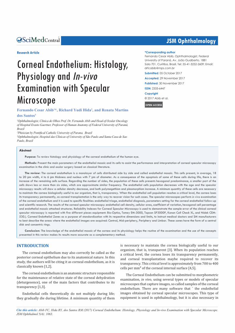
Figure 8 Illustration of the screen of a corneal specular microscopy (Konan model Cellcheck XL). The number of cells (NUM) used to calculate the endothelial results was 96. The largest cell (MAX) has 1038 µm2, the smaller cell (MIN) has 247 µm2, the average of the cellular size or the cellular area (AVE) was estimated in 515 µm2. The endothelial cell density (CD) was estimated in 1942 cells/mm2. The standard deviation of the average cellular area (SD) was 183 µm2. The coefficient of variation (CV) 0,36 or 36%. The hexagonal cells percentage (HEX) 51%. Corneal thickness (PACH) 541 μm.
percentage of cell less than 6 sides (<Hex), percentage with 6 sides (=Hex) and percentage with more than six sides (>Hex) 20.3%.
-Shape factor
Area counted: total area used for data
Bio-Optics
Corneal endothelial sample size characterization: the number of cells averaged 97 ± 22, and the calculated RE was 6.52 ± 0.86. Only 3 out of 30 examinations (10%) had a sufficient number of endothelial cells where the sampling error was smaller than the planned error (5%), whereas 27 out of 30 examinations (90%) had an insufficient number of endothelial cells; that is, the sampling error was larger than the planned error (5%).
Konan Cell Check XL
Corneal Endothelial Sample Size Characterization: the average of the counted cells was 139 ± 19 cells and the calculated RE was 6.00 ± 2.10. Only 7 out of 30 examinations (27%) had a sufficient number of endothelial cells where the sampling error was smaller than the planned error (5%); whereas 23 out of 30 examinations (73%) had an insufficient number of endothelial cells, that is, the sampling error was larger than the planned error (5%).
Nidek CEM-530
Corneal Endothelial Sample Size Characterization: the average of the counted cells was 140 ± 36 cells and the calculated RE was

CentralBringing Excellence in Open Access
Abib et al. (2017)Email: [email protected]
JSM Ophthalmol 5(4): 1063 (2017) 6/8
Figure 9 Illustration of the screen of a corneal specular microscopy (Tomey EM3000). The number of cells (Number) used to calculate the endothelial data was 122. The largest cell (Max) has 967 µm2, the smaller cell (Min) has 136 µm2, and the average of the cellular size or the cellular area (AVG) was estimated in 438 µm2. The endothelial cell density (CD) was estimated in 2284 cells/mm2. The standard deviation of the cellular areas (SD) was 154 µm2. The coefficient of variation (CV) 0.35 or 35%. The hexagonal cells percentage (6A) 38% is shown only in the pleomorfism histogram (Apex histogram).
Figure 10 Illustration of the screen of a corneal specular microscopy (Topcon SP3000P). The number of cells used to calculate the endothelial results was 20. The largest cell (MAX) has 565 µm2, the smaller cell (MIN) has 196 µm2, and the average of the cellular size or the cellular area (AVG) was estimated in 352 µm2. The endothelial cell density (CD) was estimated in 2838 cells/mm2. The standard deviation of the average cellular area (SD) was 100 µm2. The coefficient of variation (CV) 0.28 or 28%. The hexagonal cells percentage (HEX) 71%. Corneal thickness (T) 0,544 mm.
5.7 ± 1.5. Only 9 out of 30 examinations (30%) had a sufficient number of endothelial cells where the sampling error was smaller than the planned error (5%), whereas 21 out of 30 examinations (70%) with Topcon SP3000P, showed an insufficient number of endothelial cells.
Tomey EM-3000 Corneal endothelial sample size characterization: the number
of cells averaged 141± 40, and the calculated RE was 6.69 ± 1.95. Only 4 out of 30 examinations (13.33%) had a sufficient number of endothelial cells where the sampling error was smaller than
the planned error (5%), whereas 26 out of 30 examinations (86.67%) had an insufficient number of endothelial cells; that is, the sampling error was larger than the planned error (5%).
Topcon SP3000P
Corneal endothelial sample size characterization: the number of cells averaged 87 ± 18, and the calculated RE was 8.03 ± 1.8. Only 1 out of 30 examinations (3.33%) had a sufficient number of endothelial cells where the sampling error was smaller than the planned error (5%), whereas 29 out of 30 examinations (96.67%) with Topcon SP-3000P showed an insufficient number of endothelial cells.
Corneal endothelial zones
The front of the cornea appears elliptical, 11.7 mm in the horizontal meridian and 10.6 mm in the vertical. The posterior surface of the cornea appears circular, about 11.7 mm in diameter, posterior radius 5.85 mm. This difference is due to greater overlap of sclera and conjunctiva above and below than laterally.
The axial thickness of the cornea is 0.52 mm with peripheral thickness of 0.67 mm. In the central third, the optic zone, the radius of the curvature of the anterior surface is about 7.8 mm and that of the posterior 6.5 mm.
The Corneal Specular Microscope (SM) is the medical equipment that provides endothelial samples of the endothelial mosaic and uses the morphometric data of these cells to describe it. Many SMs use different areas of the endothelial mosaic to acquire endothelial images, however their manufacturers refer to determined corneal zones without the adequate morphologic accuracy, describing them on the most convenient way.
The purpose is to provide a brief review and standardization
CentralBringing Excellence in Open Access
Abib et al. (2017)Email: [email protected]
JSM Ophthalmol 5(4): 1063 (2017) 7/8
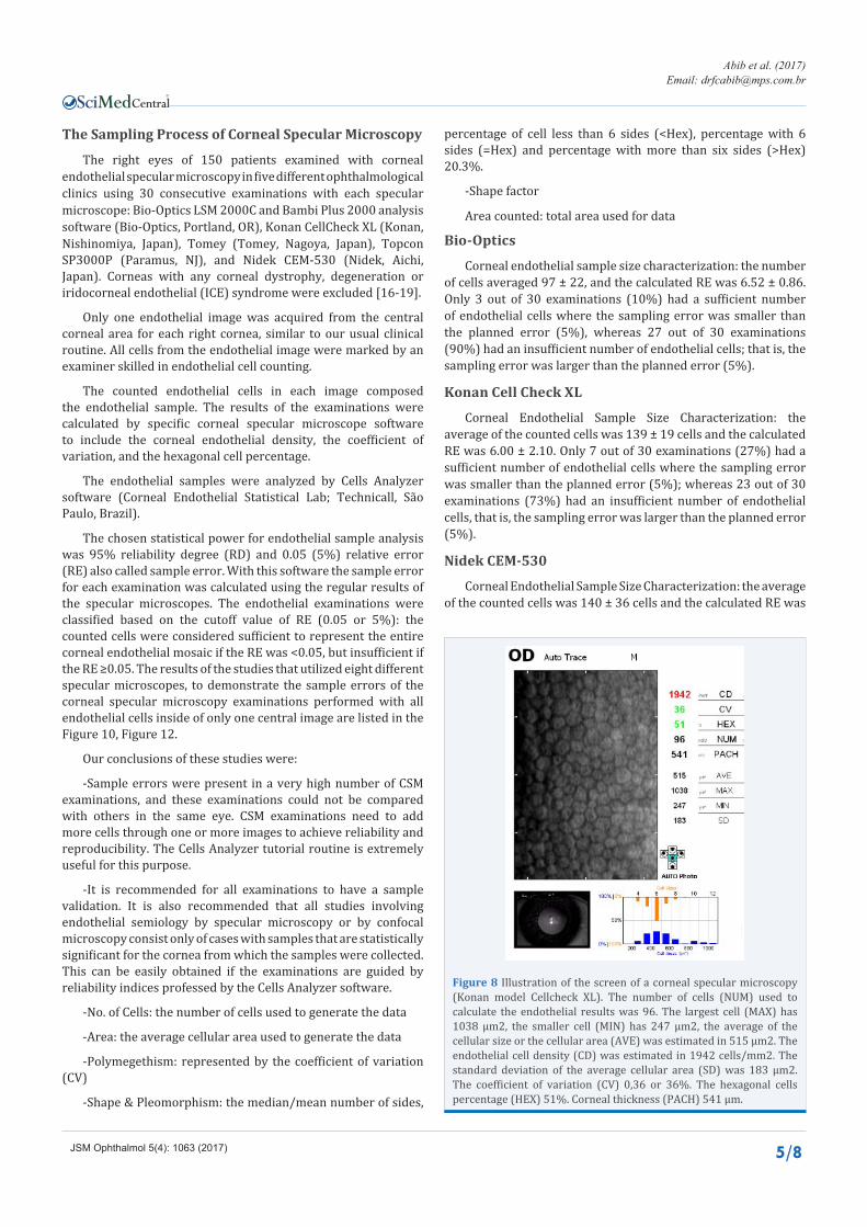
Figure 11 Illustration of the screen of a corneal specular microscopy (NIDEK CEM-530). The number of cells (NUM) used to calculate the endothelial results was 140. The largest cell (MAX) has 1145 µm2, the smaller cell (MIN) has 121 µm2, the average of the cellular size or the cellular area (AVG) was estimated in 437 µm2. The endothelial cell density (CD) was estimated in 2287 cells/mm2. The standard deviation of the average cellular area (SD) was 145 µm2. The coefficient of variation (CV) 0,35 or 35%. The hexagonal cells percentage (HEX) 73%. Corneal thickness (CT) 541 μm.
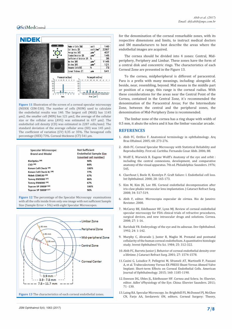
Figure 13 The characteristics of each corneal endothelial zones.
Figure 12 The percentage of the Specular Microscopy examinations with all the cells inside from only one image with not sufficient Sample Size (Sample Error > 5%) with eight Specular Microscopes.
for the denomination of the corneal remarkable zones, with its respective dimensions and limits, to instruct medical doctors and SM manufacturers to best describe the areas where the endothelial images are acquired.
The cornea should be divided into 4 zones: Central, Mid-periphery, Periphery and Limbar. These zones have the form of a central disk and concentric rings. The characteristics of each Corneal Zone are presented in the Figure 13.
To the cornea, midpheripheral is different of paracentral. Para is a prefix with many meanings, including: alongside of, beside, near, resembling, beyond. Mid means in the middle part or position of a range, this range is the corneal radius. With these considerations for the areas near the Central Point of the Cornea, contained in the Central Zone, it´s recommended the denomination of the Paracentral Areas; For the Intermediate Zone, between the central and the peripheral zones, the denomination of Mid-Periphery Zone is recommended.
The limbar zone of the cornea has a ring shape with width of 0.5 mm, it abuts the sclera and it has the limbar vascular arcade.
REFERENCES1. Abib FC, Oréfice F. Anatomical terminology in ophthalmology. Arq
Bras Oftalmol. 2005; 68: 273-276.
2. Abib FC. Corneal Specular Microscopy with Statistical Reliability and Reproducibility. First ed. Curitiba: Fernando Cesar Abib. 2006; 88.
3. Wolff E, Warwick R. Eugene Wolff’s Anatomy of the eye and orbit : including the central connexions, development, and comparative anatomy of the visual apparatus. 7th ed. Philadelphia: Saunders. 1976; 545.
4. Claerhout I, Beele H, Kestelyn P. Graft failure: I. Endothelial cell loss. Int Ophthalmol. 2008; 28: 165-173.
5. Kim M, Kim JK, Lee HK. Corneal endothelial decompensation after iris-claw phakic intraocular lens implantation. J Cataract Refract Surg. 2008; 34: 517-519.
6. Abib F, editor. Microscopia especular de córnea. Rio de Janeiro: Revinter. 2000.
7. McCarey BE, Edelhauser HF, Lynn MJ. Review of corneal endothelial specular microscopy for FDA clinical trials of refractive procedures, surgical devices, and new intraocular drugs and solutions. Cornea. 2008; 27: 1-16.
8. Barishak YR. Embryology of the eye and its adnexae. Dev Ophthalmol. 1992; 24: 1-142.
9. Murphy C, Alvarado J, Juster R, Maglio M. Prenatal and postnatal cellularity of the human corneal endothelium. A quantitative histologic study. Invest Ophthalmol Vis Sci. 1984; 25: 312-322.
10. Abib FC, Barreto Junior J. Behavior of corneal endothelial density over a lifetime. J Cataract Refract Surg. 2001; 27: 1574-1578.
11. Casini G, Loiudice P, Pellegrini M, Sframeli AT, Martinelli P, Passani A, et al. Trabeculectomy Versus EX-PRESS Shunt Versus Ahmed Valve Implant: Short-term Effects on Corneal Endothelial Cells. American Journal of Ophthalmology. 2015; 160: 1185-1190.
12. Dawson DG, Ubles JL, Edelhouser HF. Cornea and Sclera. In: Elsevier, editor. Adler´sPhysiology of the Eye. China: Elsevier Saunders. 2011; 71 -130.
13. Laing RA. Specular Microscopy. In: Brightbill FS, McDonnel PJ, McGhee CN, Farjo AA, Serdarevic ON, editors. Corneal Surgery: Theory,

CentralBringing Excellence in Open Access
Abib et al. (2017)Email: [email protected]
JSM Ophthalmol 5(4): 1063 (2017) 8/8
technique and tissue. Fourth ed. China: Mosby Elsevier. 2009; 105-116.
14. Laing RA, Oak SS, Leibowitz HM. Specialized Microscopy of the Cornea: Specular Microscopy. In: Leibowitz HM, Warring III GO, editors. Corneal Disorders, Clinical diagnosis and management. 2nd ed. Philadelphia: W.B. Saunders. 1998; 1172.
15. Abib FC, Abib DS. Behavior Of Endothelial Cellular Area, Coefficient Of Variation And Hexagonality Percentage Over A Lifetime. ARVO Meeting Abstracts. 2011; 52: 6443.
16. Abib FC, Holzchuh R, Schaefer A, Schaefer T, Godois R. The endothelial
sample size analysis in corneal specular microscopy clinical examinations. Cornea. 2012; 31: 546-550.
17. Abib FC, Costa AA, Haddad CP, Abib DS, Neto JL. The sampling error from specular microscopy examinations and their reliability indexes. Cornea. 2013; 32: 377-378.
18. Abib F, Schaefer A, Schaefer T, Abib D. The sampling error of the Konan Cell Check XL e Nidek CEM530 specular microscopes. In: International Congress of Sociedade Brasileira de Oftalmologia. Foz do Iguaçu, Brasil. 2013.
Abib FC, Hida RY, dos Santos RM (2017) Corneal Endothelium: Histology, Physiology and In-vivo Examination with Specular Microscope. JSM Ophthalmol 5(4): 1063.
Cite this article


