
CONGESTIVE
HEART FAILURE
Dr. Jamal Dabbas Interventional cardiologist & internist
1

CONGESTIVE HEART FAILURE

DEFINITION Congestive Heart Failure is a clinical syndrome in which the heart is unable to pump sufficient blood to meet the metabolic requirements of the body, or can do so only at an elevated filling pressure

EPIDEMIOLOGY • HF is a burgeoning problem worldwide, with more than 20 million people affected. • The overall prevalence of HF in the adult population in developed countries is 2%. • HF prevalence follows an exponential pattern, rising with age, and affects 6–10% of people over age 65. • Although the relative incidence of HF is lower in women than in men, women constitute at least one- half the cases of HF because of their longer life expectancy • Although HF once was thought to arise primarily in the setting of a depressed left ventricular (LV) ejection fraction (EF), epidemiologic studies have shown that approximately one-half of patients who develop HF have a normal or preserved EF (EF 40–50%). • Accordingly, HF patients are now broadly categorized into one of two groups: (1) HF with a depressed EF (commonly referred to as systolic failure) or (2) HF with a preserved EF (commonly referred to as diastolic failure).

CLASSIFICATION

• However, both the above classifications are outdated and not used clinically. They are used only academically for better understanding • The classification currently used clinically is that of systolic-failure versus diastolic-failure which was explained in epidemiology. • Apart from this, it is also classified as acute / chronic failure.

ETIOLOGY • Heart failure can result from any disorder that affects the ability of the heart to contract (systolic function) and/or relax (diastolic dysfunction) • Common causes are given in the table below


PATHOPHYSIOLOGY Normal Cardiac Performance • To understand the pathophysiologic processes in heart failure, a basic understanding of normal cardiac function is necessary. • Cardiac output (CO) is defined as the volume of blood ejected per unit time (L/min) and is the product of heart rate (HR) and stroke volume (SV): CO = HR × SV • Heart rate is controlled by the autonomic nervous system. • Stroke volume, or the volume of blood ejected during systole, depends on preload, afterload, and contractility.
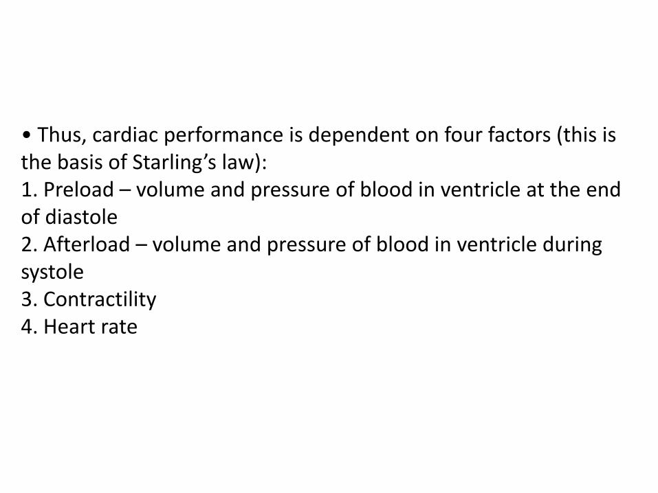
• Thus, cardiac performance is dependent on four factors (this is the basis of Starling’s law): 1. Preload – volume and pressure of blood in ventricle at the end of diastole 2. Afterload – volume and pressure of blood in ventricle during systole 3. Contractility 4. Heart rate

Compensatory mechanisms • Heart failure is a progressive disorder initiated by an event that impairs the ability of the heart to contract and/or relax. • The index event may have an acute onset, as with myocardial infarction, or the onset may be slow, as with long-standing hypertension. • Regardless of the index event, the decrease in the heart’s pumping capacity results in the heart having to rely on compensatory responses to maintain an adequate cardiac output.

• The compensatory mechanisms include: 1. Tachycardia and increased contractility through Sympathetic stimulation 2. Increased preload due to decreased sodium and water retention because of activation of RAAS, which is activated by decreased renal perfusion 3. Vasoconstriction and increased afterload- vasoconstriction occurs due to a number of neurohormones like NE, angiotensin 2, endothelin- 1 and vasopressin. Vasoconstriction increases peripheral vascular resistence and hence further decreases cardiac output 4. Ventricular hypertrophy and remodelling


CLINICAL PRESENTATION General • Patient presentation may range from asymptomatic to cardiogenic shock • The clinical picture depends on the nature of the underlying heart disease, the type of heart failure that it has evoked, and the neurohumoral changes that have developed

Symptoms • Dyspnea, particularly on exertion • Orthopnea • Paroxysmal nocturnal dyspnea • Exercise intolerance • Tachypnea • Cough • Fatigue • Nocturia • Hemoptysis • Abdominal pain • Anorexia • Nausea • Bloating • Poor appetite, early satiety • Ascites • Mental status changes

Signs • Pulmonary rales • Pulmonary edema • S3 gallop • Cool extremities • Pleural effusion • Cheyne-Stokes respiration • Tachycardia • Narrow pulse pressure • Cardiomegaly • Peripheral edema • Jugular venous distension • Hepatojugular reflux •Hepatomegaly

INVESTIGATIONS Blood tests • Blood gas analysis – to assess respiratory gas exchange • Serum creatinine and urea – to assess renal function • Serum alanine- and aspartate-aminotransferase plus other liver function tests – increased due to hepatic congestion • Complete blood count (CBC) – to investigate possibility of anaemia and if heart failure is due to it • Thyroid function tests to investigate possibility of thyrotoxicosis • Brain natriuretic peptide (BNP) – elevated in heart failure ( >100 pg/mL) and is a marker of risk; it is useful in the investigation of patients with breathlessness or peripheral oedema. • Neopterin levels increase and are biomarkers of cardiovascular remodelling

Electrocardiogram • A routine 12-lead ECG is recommended. The major importance of the ECG is to assess cardiac rhythm and determine the presence of LV hypertrophy or a prior MI (presence or absence of Q waves) as well as to determine QRS width to ascertain whether the patient may benefit from resynchronization therapy (see below). A normal ECG virtually excludes LV systolic dysfunction

Echocardiogram • Non-invasive cardiac imaging is essential for the diagnosis, evaluation, and management of HF. The most useful test is the two-dimensional (2-D) echocardiogram/Doppler, which can provide a semiquantitative assessment of LV size and function as well as the presence or absence of valvular and/or regional wall motion abnormalities (indicative of a prior MI).

• Echocardiogram assesses left ventricle size, valve function, pericardial effusion, wall motion abnormalities, and ejection fraction • Although the history, physical examination, and laboratory tests can provide important clues to the underlying cause of heart failure, the echocardiogram is the single most useful test in the evaluation of a patient with heart failure

Echocardiography is very useful and should be considered in all patients with heart failure in order to: * determine the aetiology * detect hitherto unsuspected valvular heart disease, * such as occult mitral stenosis, and other conditions that may be amenable to specific remedies * identify patients who will benefit from long-term therapy with drugs, such as ACE inhibitors (see below).

Chest radiography • A chest x-ray provides useful information about cardiac size and shape, as well as the state of the pulmonary vasculature (for edema), and may identify non-cardiac causes of the patient's symptoms


TREATMENT Goals of therapy • Relieve or reduce symptoms • Delay progression of the disease • Decrease hospitalization • Mainly decrease preload and afterload Although these goals are still important, identification of risk factors for heart failure development and recognition of its progressive nature have led to increased emphasis on preventing the development of this disorder.

• With this in mind, the American College of Cardiology/American Heart Association (ACC/AHA) guidelines for the evaluation and management of chronic heart failure use a staging system that recognizes not only the evolution and progression of the disorder, but also emphasizes risk factor modification and preventive treatment strategies.

• The New York Heart Association (NYHA) system is primarily intended to classify symptomatic heart failure according to the clinician’s subjective evaluation and does not recognize preventive measures or the progression of the disorder.


Heart Failure
The most common reason for hospitalization in adults >65 years old.

Mild Mild
Drugs
Diet
Fluid
Restriction
Heart Failure- (progression)
CDHF(Pulmonary Edema) Severe End Stage
Cardiogenic shock
Cardiomyopathy
Irreversible
Needs new ventricle
VAD
IABP VAD
IABP
Heart Transplant
Control With
Emergency-Upright, O2, morphine, etc

Definition-Heart Failure (HF)
Key Concepts
• CO = SV x HR-becomes insufficient to
meet metabolic needs of body
• SV- determined by preload, afterload
and myocardial contractility
• EF< 40% (need to understand)
• *Classifications HF
– Systolic failure- dec. contractility
– Diastolic failure- dec. filling
– Mixed

•Keys to understanding HF
• All organs (liver, lungs, legs, etc.) return blood to heart
•When heart begins to fail/ weaken> unable to pump blood forward-fluid backs up >
Inc. pressure within all organs.
•Organ response
•LUNGS: congested > “stiffer” , inc effort to breathe; fluid starts to escape into
alveoli; fluid interferes with O2 exchange, aggravates shortness of breath.
•Shortness of breath during exertion, may be early symptoms > progresses > later
require extra pillows at night to breathe > experience "P.N.D." or paroxysmal
nocturnal dyspnea .
•Pulmonary edema
•Legs, ankles, feet- blood from feet and legs > back-up of fluid and pressure in these
areas, heart unable to pump blood as promptly as received > inc. fluid within feet
and legs causes fluid to "seep" out of blood vessels ; inc. weight

Heart Failure (ADHF)Pneumonic (emergency mgt >recall for later!)
U
N
Upright Position
Nitrates
L
O
A
D
Lasix
Oxygen
ACE, ARBs, Amiodorone
Dig, Dobutamine
M
E
Morphine Sulfate
Extremities Down

Heart Failure Etiology and Pathophysiology
• Systolic failure- most common cause
– Hallmark finding: Dec. in *left ventricular ejection fraction (EF)
• Due to
– Impaired contractile function (e.g., MI)
– Increased afterload (e.g., hypertension)
– Cardiomyopathy
– Mechanical abnormalities (e.g., valve disease)

Heart Failure Etiology and Pathophysiology
• Diastolic failure
– Impaired ability of ventricles to relax and fill
during diastole > dec. stroke volume and CO
– Diagnosis based on presence of pulmonary
congestion, pulmonary hypertension, ventricular
hypertrophy
– *normal ejection fraction (EF)- Know why!

Heart Failure Etiology and Pathophysiology
• Mixed systolic and diastolic failure
– Seen in disease states such as dilated
cardiomyopathy (DCM)
– Poor EFs (<35%)
– High pulmonary pressures
• Biventricular failure (both ventricles may be
dilated and have poor filling and emptying
capacity)

Preload
• Volume of blood in ventricles at end diastole
• Depends on venous return
• Depends on compliance
Afterload
•Force needed to eject blood into circulation
•Arterial B/P, pulmonary artery pressure
•Valvular disease increases afterload
Factors effecting
heart pump
effectiveness

Cardiomegaly/ventricular remodeling occurs as heart overworked> changes in size, shape, and
function of heart after injury to left ventricle. Injury due to acute myocardial infarction or due to
causes that inc. pressure or volume overload as in Heart failure

Heart Failure
(AKA-congestive heart failure)
• Pathophysiology
• A. Cardiac compensatory mechanisms
– 1.tachycardia
– 2.ventricular dilation-Starling’s law
– 3.myocardial hypertrophy
• Hypoxia leads to dec. contractility

Pathophysiology-Summary •
•
B. Homeostatic Compensatory mechanisms
Sympathetic Nervous System-(beta blockers block this)
– 1. Vascular system- norepinephrine- vasoconstriction (What effect on afterload?)
– 2. Kidneys
• A. Dec. CO and B/P > renin angiotensin release. (ACE)
• B. Aldosterone release > Na and H2O retention
– 3. Liver- stores venous volume (ascites, +HJR, Hepatomegaly- can store 10 L. check enzymes
Counter-regulatory-
• Inc. Na > release of ADH (diuretics)
• *Release of atrial natriuretic factor > Na and H20 excretion, prevents severe cardiac decompensation
• What is BNP? What drug is synthetic form BNP?

Heart Failure Etiology and Pathophysiology
• Compensatory mechanisms- activated to
maintain adequate CO
– Neurohormonal responses: Endothelin -stimulated by
ADH, catecholamines, and angiotensin II >
• Arterial vasoconstriction
• Inc. in cardiac contractility
• Hypertrophy

Heart Failure Etiology and Pathophysiology
• **Counter regulatory processes
– Natriuretic peptides: atrial natriuretic peptide (ANP) and
b-type natriuretic peptide (BNP)- *also dx test for HF
• Released in response to inc. in atrial volume and ventricular
pressure
• Promote venous and arterial vasodilation, reduce preload
and afterload
• Prolonged HF > depletion of these factors

Heart Failure Etiology and Pathophysiology
• Counter regulatory processes
– Natriuretic peptides- endothelin and aldosterone
antagonists
• Enhance diuresis
• Block effects of the RAAS
– Natriuretic peptides- inhibit development of
cardiac hypertrophy; may have antiinflammatory
effects

Pathophysiology-
Structural Changes with HF
• Dec. contractility
• Inc. preload (volume)
• Inc. afterload (resistance)
• **Ventricular remodeling (ACE inhibitors
can prevent this)
– Ventricular hypertrophy
– Ventricular dilation

END RESULT
FLUID OVERLOAD > Acute Decompensated Heart
Failure (ADHF)/Pulmonary Edema
>Medical Emergency!

Heart Failure Classification
Systems • New York Heart Association Functional
Classification of HF
– Classes I to IV
• ACC/AHA Stages of HF (newer)
– Stages A to D


NY ASSN Funct Class
ACC/AHA
Stages

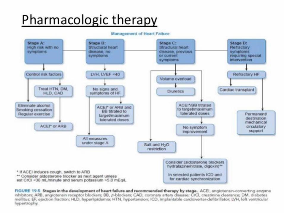
Stage A At high risk for developing heart failure. Includes people with:
Hypertension
Diabetes mellitus
CAD (including heart attack)
History of cardiotoxic drug therapy
History of alcohol abuse
History of rheumatic fever
Family history of CMP
Exercise regularly
Quit smoking
Treat hypertension
Treat lipid disorders
Discourage alcohol or illicit drug use
If previous heart attack/ current diabetes mellitus or HTN, use ACE-I
Stage B Those diagnosed with “systolic” heart failure- have never had symptoms of heart failure (usually by finding an ejection fraction of less than 40% on echocardiogram
Care measures in Stage A +
Should be on ACE-I
Add beta -blockers
Surgical consultation for coronary artery revascularization and valve repair/replacement (as appropriate
Stage C Patients with known heart failure with current or prior symptoms.
Symptoms include: SOB, fatigue
Reduced exercise intolerance
All care measures from Stage A apply, ACE-I and beta-blockers should be used + Diuretics, Digoxin,
Dietary sodium restriction
Weight monitoring, Fluid restriction Withdrawal drugs that worsen condition
Maybe Spironolactone therapy
Stage D Presence of advanced symptoms, after assuring optimized medical care
All therapies -Stages A, B and C + evaluation for:Cardiac transplantation, VADs, surgical options, research therapies, Continuous intravenous inotropic infusions/ End-of-life care
Therapies

Heart Failure Etiology and
Pathophysiology • Primary risk factors
– Coronary artery disease (CAD)
– Advancing age
• Contributing risk factors – Hypertension
– Diabetes
– Tobacco use
– Obesity
– High serum cholesterol
– African American descent
– Valvular heart disease
– Hypervolemia

CHF-due to – 1. Impaired cardiac function
• Coronary heart disease
• Cardiomyopathies
• Rheumatic fever
• Endocarditis
– 2. Increased cardiac workload
• Hypertension
• Valvular disorders
• Anemias
• Congenital heart defects
– 3.Acute non-cardiac conditions
• Volume overload
• Hyperthyroid, Fever,infection

Classifications- (how to describe)
•
•
•
•
Systolic versus diastolic
–Systolic- loss of contractility get dec. CO
–Diastolic- decreased filling or preload
Left-sided versus right –sided
– Left- lungs
– Right-peripheral
High output- hypermetabolic state
Acute versus chronic
– Acute- MI
– Chronic-cardiomyopathy

Symptoms

Left Ventricular Failure • Signs and symptoms
– dyspnea
– orthopnea PND
– Cheyne Stokes
– fatigue
– Anxiety
– rales
– NOTE L FOR LEFT AND L FOR LUNGS
– Why does this occur??

Heart Failure Clinical
Manifestations • Acute decompensated heart failure (ADHF)
– > Pulmonary edema, often life-threatening
• Early
– Increase in the respiratory rate
– Decrease in PaO2
• Later
– Tachypnea
– Respiratory acidemia

Heart Failure Clinical
Manifestations •
• Physical findings
• Orthopnea
• Dyspnea, tachypnea
• Use of accessory muscles
• Cyanosis
• Cool and clammy skin
Acute decompensated heart failure (ADHF)
•Physical findings
•*Cough with frothy,
blood-tinged sputum-
why??? > (see next slide)
•Breath sounds: Crackles,
wheezes, rhonchi
•Tachycardia
•Hypotension or
hypertension

ADHF/Pulmonary Edema (advanced L side HF)
• When PA WEDGE pressure is approx 30mmHg
– Signs and symptoms
• 1.wheezing
• 2.pallor, cyanosis
• 3.Inc. HR and BP
• 4.s3 gallop
The Auscultation Assistant - Rubs and Gallops
• 5.rales,copious pink, frothy sputum

Right Heart Failure
• Signs and Symptoms
– fatigue, weakness,
lethargy
– wt. gain, inc. abd. girth,
anorexia, RUQ pain
– elevated neck veins
– Hepatomegaly +HJR
– may not see signs of LVF

What does this
show?

What is present in this extremity, common to right sided HF?
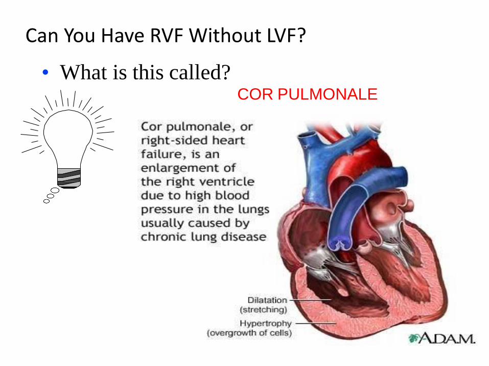
Can You Have RVF Without LVF?
• What is this called? COR PULMONALE

Heart Failure
Complications • Pleural effusion
• Atrial fibrillation (most common
dysrhythmia)
– Loss of atrial contraction (kick) -reduce CO by
10% to 20%
– Promotes thrombus/embolus formation inc. risk
for stroke
– Treatment may include cardioversion,
antidysrhythmics, and/or anticoagulants

Heart Failure
Complications • **High risk of fatal dysrhythmias (e.g., sudden
cardiac death, ventricular tachycardia) with HF and
an EF <35%
– HF lead to severe hepatomegaly, especially with
RV failure
• Fibrosis and cirrhosis - develop over time
– Renal insufficiency or failure

Heart Failure Diagnostic
Studies • Primary goal- determine underlying cause
– History and physical examination( dyspnea)
– Chest x-ray
– ECG
– Lab studies (e.g., cardiac enzymes, BNP- (beta
natriuretic peptide- normal value less than 100)
electrolytes
– EF

Heart Failure Diagnostic
Studies • Primary goal- determine underlying cause
– Hemodynamic assessment-Hemodynamic Monitoring-CVP- (right side) and Swan Ganz (left and right side)
– Echocardiogram-TEE best
– Stress testing- exercise or medicine
– Cardiac catheterization- determine heart pressures ( inc.PAW )
– Ejection fraction (EF)

Nursing Assessment
• Vital signs
• PA readings
• Urine output
• -What else!!

Decreased cardiac output
• Plan frequent rest periods
• Monitor VS and O2 sat at rest and during activity
• Take apical pulse
• Review lab results and hemodynamic monitoring
results
• Fluid restriction- keep accurate I and O
• Elevate legs when sitting
• Teach relaxation and ROM exercises

Knowledge deficit
• Low Na diet
• Fluid restriction
• Daily weight
• When to call Dr.
• Medications
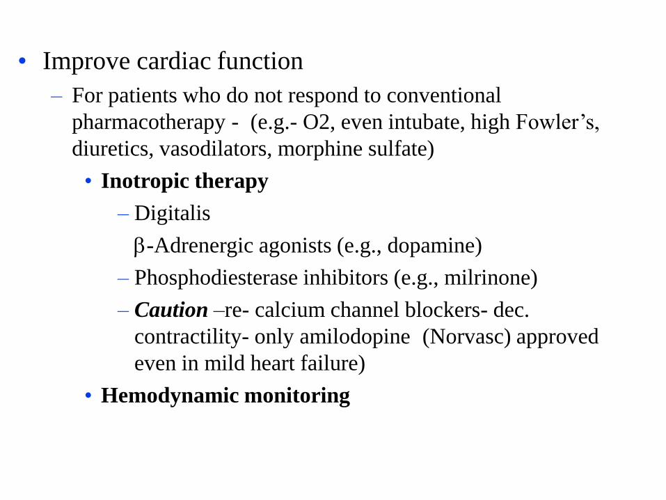
• Improve cardiac function
– For patients who do not respond to conventional
pharmacotherapy - (e.g.- O2, even intubate, high Fowler’s,
diuretics, vasodilators, morphine sulfate)
• Inotropic therapy
– Digitalis
-Adrenergic agonists (e.g., dopamine)
– Phosphodiesterase inhibitors (e.g., milrinone)
– Caution –re- calcium channel blockers- dec.
contractility- only amilodopine (Norvasc) approved
even in mild heart failure)
• Hemodynamic monitoring
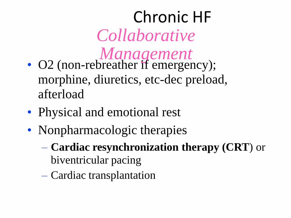
Chronic HF Collaborative Management
• O2 (non-rebreather if emergency);
morphine, diuretics, etc-dec preload,
afterload
• Physical and emotional rest
• Nonpharmacologic therapies
– Cardiac resynchronization therapy (CRT) or
biventricular pacing
– Cardiac transplantation

CRT-Cardiac Resynchronization Therapy
HOW IT WORKS:
Standard implanted pacemakers -
equipped with two wires (or "leads")
conduct pacing signals to specific regions
of heart (usually at positions A and C).
Biventricular pacing devices have added
a third lead (to position B) that is
designed to conduct signals directly into
the left ventricle. Combination of all
three lead > synchronized pumping of
ventricles, inc. efficiency of each beat
and pumping more blood on the whole.

Chronic HF-Collaborative Management
Drug therapy
– Diuretics
• Thiazide
• Loop
• Spironolactone
– Vasodilators
• ACE inhibitors- pril or
ril *first line heart
failure
• Angiotensin II receptor
blockers
• Nitrates
-Adrenergic blockers-
al or ol
• Nesiritide- Natrecor
(BNP)

Chronic HF Collaborative Management
• Drug therapy (cont’d)
– Positive inotropic agents
• Digitalis
• Calcium sensitizers- (Levosimendan) new under
research; cardioprotective, inc. cardiac contractility
– BiDil (combination drug containing isosorbide
dinitrate and hydralazine) approved only for the
treatment of HF in African Americans

Chronic HF Collaborative Management
• Nutritional therapy
– Diet/weight reduction recommendations- individualized and culturally sensitive
– Dietary Approaches to Stop Hypertension (DASH) diet recommended
– Sodium- usually restricted to 2.5 g per day
– Potassium encouraged unless on K sparing diuretics (Aldactone)

Chronic HF Collaborative Management
• Nutritional therapy
– Fluid restriction may or may not be required
– Daily weights important
• Same time, same clothing each day
– *Weight gain of 3 lb (1.4 kg) over 2 days or a 3-
to 5-lb (2.3 kg) gain over a week-report to
health care provider

Intraaortic Balloon Pump (IABP)
• Provides temporary circulatory
assistance
– ↓ Afterload
– Augments aortic diastolic pressure
• Outcomes
– Improved coronary blood flow
– Improved perfusion of vital organs

Intraaortic balloon pump
IABP Machine

10 Commandments of Heart Failure Treatment
1. Maintain patient on 2- to 3-g sodium diet. Follow daily weight. Monitor
standing blood pressures in the office, as these patients are prone to
orthostasis. Determine target/ideal weight, which is not the dry weight. In
order to prevent worsening azotemia, some patients will need to have
some edema. Achieving target weight should mean no orthopnea or
paroxysmal nocturnal dyspnea. Consider home health teaching.
2. Avoid all nonsteroidal anti-inflammatory drugs because they block the
effect of ACE inhibitors and diuretics. The only proven safe calcium
channel blocker in heart failure is amlodipine (Lotrel /Norvasc).
3. Use ACE inhibitors in all heart failure patients unless they have an
absolute contraindication or intolerance. Use doses proven to improve
survival and back off if they are orthostatic. In those patients who cannot
take an ACE inhibitor, use an angiotensin receptor blocker like
irbesartan (Avapro).
4. Use loop diuretics (like furosemide [Lasix]) in most NYHA class II
through IV patients in dosages adequate to relieve pulmonary congestive
symptoms. Double the dosage (instead of giving twice daily) if there is
no response or if the serum creatinine level is > 2.0 mg per dL (180 µmol
per L).
5. For patients who respond poorly to large dosages of loop diuretics,
consider adding 5 to 10 mg of metolazone (Zaroxolyn) one hour before
the dose of furosemide once or twice a week as tolerated.

The 10 Commandments of Heart Failure Treatment
6. Consider adding 25 mg spironolactone in most class III or IV
patients. Do not start if the serum creatinine level is > 2.5 mg
per dL (220 µmol per L).
7. Use metoprolol (Lopressor), carvedilol (Coreg) or bisoprolol
(Zebeta) (beta blockers) in all class II and III heart failure
patients unless there is a contraindication. Start with low
doses and work up. Do not start if the patient is
decompensated.
8. Use digoxin in most symptomatic heart failure patients.
9. Encourage a graded exercise program.
10. Consider a cardiology consultation in patients who fail to
improve.
ACE = angiotensin-converting enzyme.

Medical Treatment-Drug Therapy (typical)
• Cardiac Glycoside-Digoxin
• Positive inotropes-dobutamine, Primacor. Natrecor
• Antihypertensives- WHY
• ACE inhibitors- stops remodeling (pril or ril)
– Catopril,enalapril,cozar,lisinopril
• Preload reduction *MSO4- important,
– Vasodilators-nitrates
– Diuretics-lasix, HCTZ, (Aldactone and Inspra)
– Beta blockers- dec. effects of SNS (Coreg)
– *Caution with CALCIUM CHANNEL BLOCKERS- dec cardiac contractility

Meds!
Angiotensin-converting enzyme inhibitors , such as captopril and enalapril,
block conversion of angiotensin I to angiotensin II, a vasoconstrictor that can
raise BP. These drugs alleviate heart failure symptoms by causing vasodilation
and decreasing myocardial workload.
Beta-adrenergic blockers , such as bisoprolol, metoprolol, and carvedilol,
reduce heart rate, peripheral vasoconstriction, and myocardial ischemia.
Diuretics prompt kidneys to excrete sodium, chloride, and water, reducing fluid
volume. Loop diuretics such as furosemide, bumetanide, and torsemide are
preferred first-line diuretics because of efficacy in patients with and without
renal impairment. Low-dose spironolactone may be added to a patient's
regimen if he has recent or recurrent symptoms at rest despite therapy with
ACE inhibitors, beta-blockers, digoxin, and diuretics.
Digoxin increases the heart's ability to contract and improves heart failure
symptoms and exercise tolerance in patients with mild to moderate heart failure

Other drug options include nesiritide (Natrecor), a preparation
of human BNP that mimics the action of endogenous BNP,
causing diuresis and vasodilation, reducing BP, and improving
cardiac output.
Intravenous (I.V.) positive inotropes such as dobutamine,
dopamine, and milrinone, as well as vasodilators such as
nitroglycerin or nitroprusside, are used for patients who
continue to have heart failure symptoms despite oral
medications. Although these drugs act in different ways, all are
given to try to improve cardiac function and promote diuresis
and clinical stability.

#14
• The nurse is caring for a hospitalized client with heart failure who is receiving captopril (Capoten) and spironolactone (aldactone). Which lab value will be most important to monitor?
•
•
•
•
A. Sodium
B. Blood urea nitrogen (BUN)
C. Potassium
D. Alkaline phosphatase (ALP)
•C. Potassium

CONGESTIVE HEART
FAILURE
Heart (or cardiac) failure:
It is defined as the inefficiency of the heart to pump sufficient amount of oxygenated blood to the organs to meet the metabolic demands and to collect the blood from the organs.
Congestive heart failure (CHF):
It is complex clinical syndrome characterized by abnormalities of left ventricular function and neurohormonal regulation, which are accompanied by effort intolerance, fluid retention, and reduced longevity
2

Based on amount of cardiac output
90
Low-cardiac output failure
• It is most common congestive heart failure. • The metabolic demands of the body organs are normal with in limits but the heart fails
to pump sufficient amount of oxygenated blood to the organs of the body.
• The primary cause of LCOF is the ventricular systolic dysfunction and ventricular diastolic dysfunction.
Ventricular systolic dysfunction
• Myocardial infarction weakens the muscles of ventricles and make them inefficient to pump the required volume of blood.
• Thus results in low cardiac output and low ejection fraction.
Ventricular diastolic dysfunction
• Hypertrophy is responsible for the stiffening of heart muscle
• The stiffened muscle of the ventricles fails to relax during diastolic and thus cannot collect sufficent amount of blood.
• This ultimately results in low cardiac output.
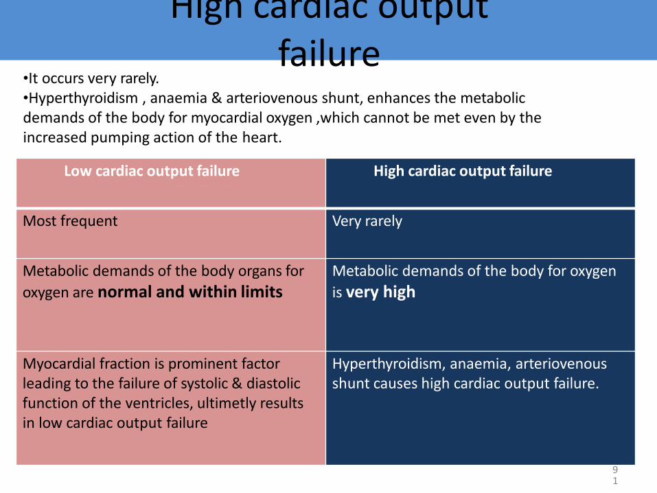
High cardiac output failure
91
Low cardiac output failure High cardiac output failure
Most frequent Very rarely
Metabolic demands of the body organs for
oxygen are normal and within limits Metabolic demands of the body for oxygen
is very high
Myocardial fraction is prominent factor leading to the failure of systolic & diastolic function of the ventricles, ultimetly results in low cardiac output failure
Hyperthyroidism, anaemia, arteriovenous shunt causes high cardiac output failure.
•It occurs very rarely. •Hyperthyroidism , anaemia & arteriovenous shunt, enhances the metabolic demands of the body for myocardial oxygen ,which cannot be met even by the increased pumping action of the heart.

92
Right side cardiac failure
• The failure of right ventricle to pump the entire blood present in it during systole results in retention of some amount of blood after every systole.
• Thus blood is accumulated in right ventricle after few systoles.
• The left ventricle fails to accept the blood from peripheral organs and ultimately results in generalized systemic oedema or peripheral oedema.
Left side cardiac failure Right side cardiac failure
Is the result of right side cardiac failure Is the result of left side cardiac failure
Inefficent pumping action of left ventricle is responsible for the accumulation of blood in the ventricles
Inefficient pumping action of right ventricle is responsible for the accumulation of blood in right ventricle
Left ventricle fails to accept/collect the
blood from lungs due to back pressure Right ventricle fails to accept/collect the
blood from peripheral organs.
Pulmonary congestion/oedema is the final result
Peripheral generalized oedema is the final result
Pathophysiology
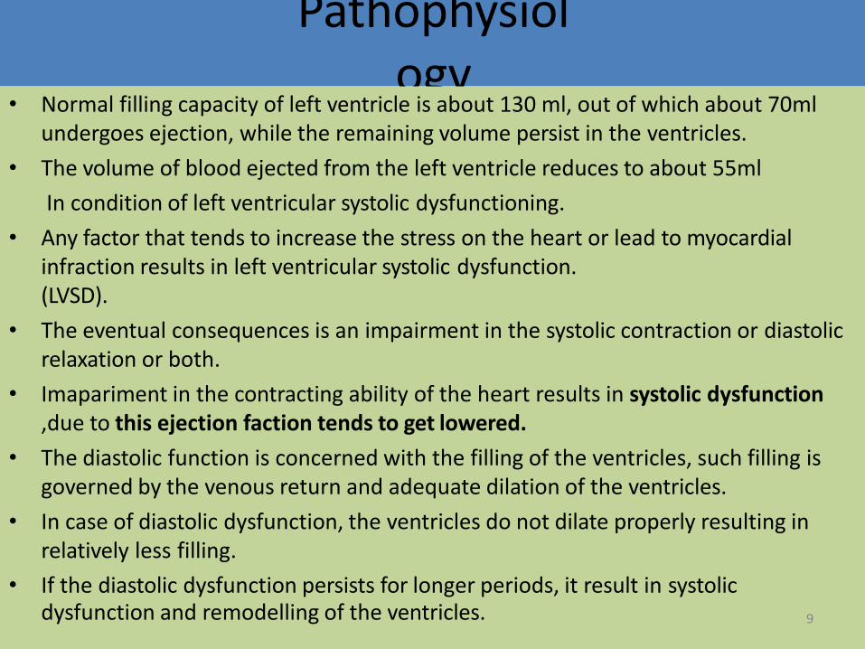
• Normal filling capacity of left ventricle is about 130 ml, out of which about 70ml undergoes ejection, while the remaining volume persist in the ventricles.
• The volume of blood ejected from the left ventricle reduces to about 55ml
In condition of left ventricular systolic dysfunctioning.
• Any factor that tends to increase the stress on the heart or lead to myocardial infraction results in left ventricular systolic dysfunction. (LVSD).
• The eventual consequences is an impairment in the systolic contraction or diastolic relaxation or both.
• Imapariment in the contracting ability of the heart results in systolic dysfunction ,due to this ejection faction tends to get lowered.
• The diastolic function is concerned with the filling of the ventricles, such filling is governed by the venous return and adequate dilation of the ventricles.
• In case of diastolic dysfunction, the ventricles do not dilate properly resulting in relatively less filling.
• If the diastolic dysfunction persists for longer periods, it result in systolic dysfunction and remodelling of the ventricles. 9

Compensatory mechanisms of congestive heart
failure
94
• To enhances the cardiac output, body compensates for the intrinsic cardiac effects in the following manner.
1. Increased sympathetic discharge
• To compensate for the decreased B.P, baroreceptors located in the arch of aorta carotid sinuses and walls of the heart get stimulated and causes activation of beta-adrenergic receptors leading to an increase in rate and force of contraction of heart.
• An increase in venous return (preload) is also seen due to the activation of alpha- adrenergic receptors.
• Increased rate and force of contraction together with the increased preload results in an initial increase in the cardiac output.
• Vasoconstriction of the arteries due to alpha stimulation also causes an increase in after load, leading to fall in ejection fraction.
• As a result the cardiac output decreases.

Activation of Renin- Angiotensin
Aldosterone (RAA)
95
• Fall in the cardiac output decreases the renal perfusion rate, as a result the RAA system gets activated .
• Angiotensin 2 causes vasoconstriction and an increase in the peripheral vascular
resistance(PVR).
• while aldosterone leads to increased retention of sodium and water, there by
increasing the blood volume.
• PVR effects the after load during which the heart is unable to pump the extra
blood volume.
• This leads to the development of back up pressure causing pulmonary congestion
and peripheral oedema.

Clinical manifestations/signs and symptoms
96
• Fluid retention
• Pulmonary congestion
• Dyspnoea & orthopnoea
CVS MANIFESTATIONS
• Resting tachycardia
• Ventricular arrhythmias
• Enlargement of heart
RENAL MANIFESTATIONS
• Nocturia
• Oliguria
OTHER MANIFESTATIONS
• Reduced cardiac output lead to poor perfusion of skeletal muscle resulting in fatigue.
• Reduced perfusion to brain results in altered mental states & confusion.
• Reduced perfusion may also causes the patient to appear pale with cold and sweaty hands.

TREATMENT
Non-drug Treatment/ Non-pharmacological Approach:
Physical exercise
salt intake
fluid intake
Alcohol consumption
Liquorice
97

TREATMENT OF CHF • There are two distinct goals of drug therapy
in CHF:
Relief of congestion/low cardiac output symptoms & restoration of cardiac performance:
o Inotropic drugs-digoxin, dobutamine,amrinone/milrinone.
o Diuretics: furosemide, thiazides.
o Vasodilators: ACE inhibitors/AT1 antagonist, hydralazine, nitrate.
o BETA blocker: metoprolol,bisprolol,carvedilol
Arrest/reversal of disease progression & prolongation of survival
ACE inhibitors/AT1antagonist (ARBs).
Beta-blockers
Aldosterone antagonist-spironolactone.. 98

Loop Diuretics Furosemide, Bumetanide , Torsemid
• The Na+, K+, 2Cl- symporter, a carrier-mediated process,. • It is the major reabsorptive mechanism in the thick ascending limb (TAL). • All four ions are transported by secondary active transport into the TAL epithelial cells, at their apical surface,
using the energy derived from the Na+/K+-ATPase co-transporter, also a carrier mediated mechanism.
Mechanism of Action of Loop Diuretics: • Loop diuretics act on the Na-K-2Cl symporter in the thick ascending limb of the loop of Henle to inhibit sodium
and chloride reabsorption. Because magnesium and calcium reabsorption in the thick ascending limb is dependent on sodium and chloride concentrations.
• loop diuretics also inhibit their reabsorption. By disrupting the reabsorption of these ions, loop diuretics prevent the urine from becoming dilute and disrupt the generation of a hypertonic renal medulla. Without such a concentrated medulla, water has less of an osmotic driving force to leave the collecting duct system, ultimately resulting in increased urine production. This diuresis leaves less water to be reabsorbed into the blood, resulting in a decrease in blood volume.
• Loop diuretics cause vasodilation of the veins and of the kidney's blood vessels, mechanically causing a decrease in blood pressure. The collective effects of decreased blood volume and vasodilation decrease blood pressure.
Adverse reaction: •pre-renal azotemia •Hypokalemia •Skin rash •ototoxicity
99

Potassium-Sparing Diuretics
100
• The K-sparing diuretics are weak diuretics alone. • They are primarily used as adjuncts to thiazides and loop diuretics or for potassium and
magnesium spacing. Instead of using thiazides alone for hypertension ,triamterene is also used by combination.
• Amiloride can be used for magnesium deficiency because it increases renal reabsorption. • If a patient who has hypomagnesemia, and you can't give them enough magnesium
orally, because of laxative action, give amiloride. • Also, amiloride is useful for patients taking lithium who have polyuria and complain of
having to get up three or four times at night. At a dose of 5 mg bid, amiloride reduces urine volume by 30%.
• "Don't use any K-sparing diuretics with angiotensin-converting enzyme inhibitors, angiotensin II receptor blockers [or] nonsteroidals.
• Be cautioned against using them when serum creatinine levels are above 2 mg/dL. Specific side effects seen with K-sparing diuretics include • Hyperchloremic acidosis; • Hyperkalemia, especially if administered with an ACE inhibitor, angiotensin II receptor
blocker or in patients with diabetes; • Gynecomastia, impotence in men or irregular menstrual cycles in women (only with use
of spironolactone); • Folic acid deficiency (with chronic use of triamterene); or acute renal failure (with
triamterene when used with indomethacin [Indocin]).

K+ Sparing Agents • Triamterene & amiloride – acts on
distal tubules to ↓ K secretion
• Spironolactone (Aldosterone antagonist)
it improve survival in CHF patients due to the effect on renin-angiotensin- aldosterone system with subsequent effect on myocardial remodeling and fibrosis
• Aldosterone inhibition minimize
potassium loss, prevent sodium and water retention, endothelial dysfunction and myocardial fibrosis.
101

Renin–Angiotensin- System • The renin-angiotensin system (RAS) or the renin-angiotensin-aldosterone
system (RAAS) is a hormone system that regulates blood pressure and water (fluid) balance.
• When blood volume is low, juxtaglomerular cellsin the kidneys secrete renin directly into circulation.
• Plasma renin then carries out the conversion of angiotensinogen released by the liver to angiotensin I.
• Angiotensin I is subsequently converted to angiotensin II by the enzyme angiotensin converting enzyme found in the lungs.
• Angiotensin II is a potent vaso-active peptide that causes blood vessels to constrict, resulting in increased blood pressure.
• Angiotensin II also stimulates the secretion of the hormone aldosterone from the adrenal cortex
• Aldosterone causes the tubules of the kidneys to increase the reabsorption of sodium and water into the blood. This increases the volume of fluid in the body, which also increases blood pressure.
• If the renin-angiotensin-aldosterone system is too active, blood pressure will be too high.
• There are many drugs that interrupt different steps in this system to lower blood pressure. These drugs are one of the main ways to control high blood pressure (hypertension),hear failure,kidney failure, and harmful effects of diabetes. 23

Inhibitors of Renin- Angiotensin- Aldosterone System
103
Angiotensin converting enzyme inhibitors
Angiotensin receptors blockers
Spironolactone (Aldosterone antagonist)

Angiotensin Converting Enzyme (ACE) Inhibitors
• Captopril, Lisinopril, Enalapril, Ramipril, Quinapril.
Mode of action:
• Angiotensin 1 Angiotensin 2
Hences, they inhibit the generation of angiotensin 2,a potent vasoconstrictor.
They also inhibit the release of aldosterone & vasopressin, thereby inhibiting fluid and slat retention thus decreasing the preload.
Elevate the levels of bradykinin, vasodilator thus enhancing renal & cardiac perfusion.
ACE Inhibitors
104

Angiotensin Receptor AT-1 blockers (ARB)
Losartan ,candesartan,valsartan
• Angiotensin 2 ,a vasocontrictor is concerned with ventricular remodelling and fluid retention.
• These drugs inhibit the binding of angiotensin 2 to its AT₁ receptor.
• Thus they preclude the a bove mentioned effects of angiotensin 2.
• These agents do not exert any action on bradykinin and thus do not produce cough.
• Has comparable effect to ACE I
• Can be used in certain conditions when ACE I are contraindicated
Adverse drug reactions •Hypotension •Impariment of renal functioning Dose Candesartan •Initial: 4-8mg •Targeted dose -32mg Valsartan •Initial:40mg •Targeted dose -160mg 10
5

Cardiac glycosides : Digoxin (DIGITALIS)
It inhibits the inhibit Na +,K + ATPase , pump which
Functions in the exchange of Na⁺ for k⁺ ions.
Such blockage results in intracellular accumulation of Na⁺ ions .
These ions are then exchanged with Ca₂⁺ ions through Na⁺ - Ca₂⁺ exchange carries.
These ca₂⁺ ions increase the contractility of the myocardium which is beneficial to the failing heart.
Digoxin enhances the cholinergic activity which reduces the HR and AV conduction .
Due to this the time required for diastolic filling gets enhanced while the myocardial o2 consumption is retarted.
The sympathetic outflow comprising renin, aldosterone is also decreased by dioxin
106

Drug reaction
• Bradycardia
• Nausea
• Vomiting
• Visual disturbances
• Non paroxysomal junctional tachycardia
• Supraventricular tachycardia
• Sexual dysfunction
• Neuralgic pain
USES: • For tachyarrhythmias
• For ventricular arrhythmias
107

Dopamine • Dopamine acts at a variety of receptors (dose dependant) • Rapid elimination- can only be administered as a continuous infusion
•Stimulates beta-adrenergic receptors and produces a positive inotropic response.
•Unlike the vasoconstriction seen with high doses of dopamine, dobutamine produces a mild vasodilatation
β -Adrenergic Agonists
Dobutamine
108

BIPYRIDINES phosphodiesterase inhibitors
• Targets PDE -3 (found in cardiac and smooth muscle)
• Ex. Inamrinone , milrinone
alter the intracellular movements of calcium by
influencing the sarcoplasmic reticulum
increasing inward calcium flux
in the heart during the
action potential
increase myocardial contractility
Inhibition of PDE3
Increase in cAMP
the conversion of inactive protein kinase to active form
Protein kinases are responsible for phosphorylation of Ca channels
increased Ca entry into the cell
↑ Vascular Permeability leads to ↓ in intravascular fluid Volume
increase in contractility
vasodilation
35

Vasodilators
• Isosorbide dinitrate, isosorbide mononitrate, and hydralazine also used specially in patients who cannot tolerate ACE inhibitors.
110

Vasodilator(Hydralazine)
111
• It directly relaxes the arterioles & arteries reducing the peripheral vascular reesistances & preload.
• It also help to reduce after load.
Adverse drug reaction:
• Nausea
• Palpitation
• Tachycardia
• Salt & water retention on prolong therapy.

NITRATES & NITRITES
Nitroglycerin is denitrated by
glutathione S -transferase in smooth muscle
Free nitrite ion is released, which is then converted to Nitric Oxide
activation of guanylyl cyclase enzyme
increase in cGMP
dephosphorylation of myosin light chain , preventing the interaction of myosin with actin(Myosin light chain kinase essential for smooth muscle contraction).
Results in vasodilation
112

NISIRITIDE(BNP)
113
• Brain (B-type) natriuretic peptide (BNP) is secreted constitutively by ventricular myocytes in response to stretch
• Niseritide = recombinant human BNP
• Naturally occurring atrial natriuretic peptide may vascular
permeability may reduce intravascular volume)
Main Side Effect: • hypotension

Human BNP binds to the particulate guanylate cyclase receptor of vascular smooth muscle and endothelial
intracellular concentrations (cGMP) ↑
smooth muscle cell relaxation
dilate veins and arteries
systemic and pulmonary vascular resistances ↑
Indirect ↑ in cardiac output and diuresis.
Effective in HF because preload and afterload↓
114

B-type natriuretic peptide (BNP) is a hormone produced by your heart. BNP is released in response to changes in pressure inside the heart. These changes can be related to heart failure and other cardiac problems. Levels goes up when heart failure develops or gets worse, and levels goes down when heart failure is stable. In most cases, BNP levels are higher in patients with heart failure than people who have normal heart function.

It’s measurement is a simple blood test to help diagnose or monitor heart failure.

Recombinant BNP (nesiritide) has been evaluated and approved for adjunctive therapy for acute CHF, although subsequent evidence of harm dramatically diminished its use for this indication.

An implantable cardioverter-defibrillator (ICD) is a specialized device designed to directly treat many dysrhythmias, and it is specifically designed to address ventricular tachyarrhythmias(V-tach) specially in patients with low ejection fraction post MI.

A permanent pacemaker is an implanted device that provides electrical stimuli, thereby causing cardiac contraction when intrinsic myocardial electrical activity is inappropriately slow or absent. All modern ICDs also function as pacemakers

Renin-Angiotensin System Inhibition With Angiotensin-Converting Enzyme Inhibitor or Angiotensin Receptor Blocker or ARNI: The introduction of an angiotensin receptor–neprilysin inhibitor (ARNI) (valsartan/sacubitril) and a sinoatrial node modulator (ivabradine), complements established pharmacological and device-based therapies and represents a milestone in the evolution of care for patients with heart failure (HF). Accordingly, the writing committees of the “2016 ACC/AHA/HFSA Focused Update on New Pharmacological Therapy for Heart Failure” and the “2016 ESC Guideline on the Diagnosis and Treatment of Acute and Chronic Heart Failure” concurrently developed recommendations for the incorporation of these therapies into clinical practice.


