Case Report/Clinical Techniques
Complex Apical Intraradicular Infection and ExtraradicularMineralized Biofilms as the Cause of Wet Canals andTreatment Failure: Report of 2 CasesDomenico Ricucci, MD, DDS,* George T.M. Candeiro, DDS, MSc, PhD,
†‡Calogero Bugea, DDS,
§
and Jos�e F. Siqueira, Jr, DDS, MSc, PhDjj
Abstract
This article describes 2 cases that showed persistentintracanal exudation (wet canal) even after severalvisits of antimicrobial endodontic treatment. Histologicand histobacteriologic investigation was conducted fordetermination of the cause. The 2 cases involved teethwith apical periodontitis lesions, which presentedpersistent exudation refractory to treatment afterseveral visits. In case 1, it was not possible to achievea dry canal, and surgery had to be performed. In case2, attempts to dry the canal succeeded and the canalwas filled, but follow-up examination showed anenlarged apical periodontitis lesion and extractionwas performed. Biopsy specimens consisting of theroot apex and apical periodontitis lesion for case 1and the whole root for case 2 were subjected to histo-logic and histobacteriologic analyses. Both casesshowed complex bacterial infection in the apical root,affecting both the intraradicular space and the outerroot surface. Case 1 showed bacterial biofilms in ram-ifications, on untouched walls, and extending to theexternal root surface to form a thick and partiallymineralized structure with high bacterial density.Different bacterial morphotypes were evidenced. Case2 had a ledge on the apical canal wall created duringinstrumentation, which was filled with necrotic debris,filling material, and bacteria. The walls of the apicalportion of the canal were covered by a bacterial bio-film, which was continuous with a thick extraradicularbiofilm covering the cementum and dentin in resorp-tive defects. The extraradicular biofilm showed areasof mineralization and was dominated by filamentousbacteria. The 2 cases with wet canals and treatmentfailure were associated with complex persistent infec-tion in the apical part of the root canal system extend-ing to form thick and partially mineralized biofilmstructures (calculus) on the outer apical root surface.(J Endod 2016;42:509–515)From the *Private Practice, Cetraro, Italy; †Faculty of Dentistry,Cear�a, Brazil; §Private Practice, Lecce, Italy; and jjDepartment of En
Address requests for reprints to Dr Domenico Ricucci, Piazza Ca0099-2399/$ - see front matter
Copyright ª 2016 American Association of Endodontists.http://dx.doi.org/10.1016/j.joen.2015.12.014
JOE — Volume 42, Number 3, March 2016
Key WordsExtraradicular infection, post-treatment apical periodontitis, treatment outcome,wet canal
Apical periodontitis is a disease caused by bacterial infection of the root canal system.The infection is usually restricted to the intraradicular space, but occasionally it can
spread to the extraradicular space (1). When formed, the extraradicular infection isusually an extension of the intraradicular infection. In some cases, an extraradicularbiofilm can be formed on the outer root surface area surrounding the apical foramina(2–4).
Extraradicular biofilms have been associated with post-treatment apical periodon-titis (4) and are usually associated with symptoms (1, 3). Some reports indicate that theextraradicular biofilm can become mineralized. Harn et al (5) reported a case of post-treatment apical periodontitis associated with a sinus tract in which a calculus-like de-posit was observed on the root surface during surgery. Ricucci et al (2) described 2cases in which calculus (mineralized biofilms) occurred on the outer apical root sur-face of teeth with post-treatment apical periodontitis. In their study on the prevalence ofbiofilms in treated and untreated teeth, Ricucci and Siqueira (1) also reported 6 casesof extraradicular biofilms (4 from untreated teeth and 2 from treated teeth). Two casesshowed areas of mineralization with a calculus-like appearance. An untreated tooth withan extensive calculus-like deposit on the external apical root surface was shown in areview article on endodontic biofilms (6). None of these reported cases had deep peri-odontal pockets reaching the apex, and most of them were associated with sinus tracts.
The purpose of this article was to contribute to the knowledge of the causes ofpersistent apical periodontitis by describing 2 cases that showed continuous intracanalexudation refractory to orthograde root canal treatment. Histologic and histobacterio-logic analysis of specimens obtained by apical surgery and extraction showed the occur-rence of apical intraradicular infection associated with extraradicular mineralizedbiofilm structures as the cause of persistent symptoms and disease.
Case ReportsCase 1
A 29-year-old man attended a general dental office complaining of several epi-sodes of swelling in the right anterior maxilla and the taste of purulent exudate. He re-ported that he was self-medicating with antibiotics and had no history of trauma. Hismedical history was noncontributory.
University of S~ao Paulo, S~ao Paulo, S~ao Paulo, Brazil; ‡Universitary Center Unichistus, Fortaleza,dodontics, Faculty of Dentistry, Est�acio de S�a University, Rio de Janeiro, Rio de Janeiro, Brazil.lvario, 7, 87022 Cetraro (CS), Italy. E-mail address: [email protected]
Intra- and Extraradicular Infection and Wet Canal 509
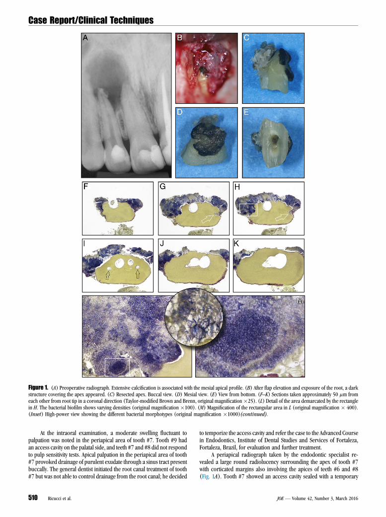
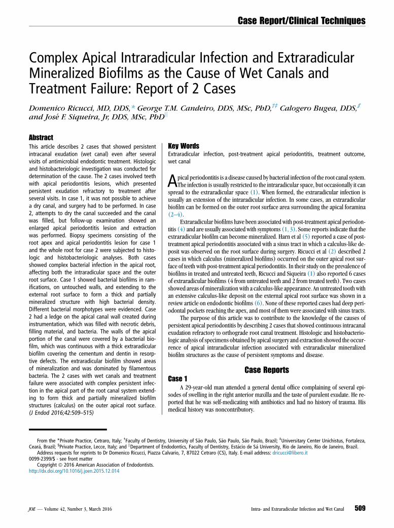
Figure 1. (A) Preoperative radiograph. Extensive calcification is associated with the mesial apical profile. (B) After flap elevation and exposure of the root, a darkstructure covering the apex appeared. (C) Resected apex. Buccal view. (D) Mesial view. (E) View from bottom. (F–K) Sections taken approximately 50 mm fromeach other from root tip in a coronal direction (Taylor-modified Brown and Brenn, original magnification�25). (L) Detail of the area demarcated by the rectanglein H. The bacterial biofilm shows varying densities (original magnification�100). (M) Magnification of the rectangular area in L (original magnification � 400).(Inset) High-power view showing the different bacterial morphotypes (original magnification �1000)(continued).
Case Report/Clinical Techniques
At the intraoral examination, a moderate swelling fluctuant topalpation was noted in the periapical area of tooth #7. Tooth #9 hadan access cavity on the palatal side, and teeth #7 and #8 did not respondto pulp sensitivity tests. Apical palpation in the periapical area of tooth#7 provoked drainage of purulent exudate through a sinus tract presentbuccally. The general dentist initiated the root canal treatment of tooth#7 but was not able to control drainage from the root canal; he decided
510 Ricucci et al.
to temporize the access cavity and refer the case to the Advanced Coursein Endodontics, Institute of Dental Studies and Services of Fortaleza,Fortaleza, Brazil, for evaluation and further treatment.
A periapical radiograph taken by the endodontic specialist re-vealed a large round radiolucency surrounding the apex of tooth #7with corticated margins also involving the apices of teeth #6 and #8(Fig. 1A). Tooth #7 showed an access cavity sealed with a temporary
JOE — Volume 42, Number 3, March 2016
Figure 1. (Continued). (N) High-power view of the ramification indicated by the left arrow in I. Necrotic debris and bacterial biofilm (originalmagnification�400). (O) High-power view of the ramification indicated by the right arrow in I. Necrotic debris in the center. Bacterial biofilm layering the walls.The empty space between the bacterial biofilm and the wall on the right is a shrinkage artifact (original magnification �400).
Case Report/Clinical Techniques
material, whereas extensive composite coronal restorations were pre-sent in tooth #8. A radiopaque area could be clearly distinguished onthe mesial apical profile of the lateral incisor, suggesting a calculus-like deposit (Fig. 1A). Periodontal probing did not reveal pocketsexceeding 2 mm deep around the roots of all the maxillary anteriorteeth. Thermal and electric pulp tests gave no response for teeth #7and #8, whereas tooth #6 responded normally.
Orthograde root canal treatment was scheduled for the maxillaryright incisors. Root canal treatment of the lateral incisor was initiatedfirst. After anesthesia and rubber dam isolation, the canal was reac-cessed and gently flushed with 20 mL 2.5% sodium hypochlorite(NaOCl) solution. The working length was established with an elec-tronic apex locator (RomiApex A-15; Romidan Ltd, Kiryat Ono, Israel).The root canal was instrumented with the Reciproc R50 file (VDW,Munich, Germany) and copious irrigation with 2.5% NaOCl. Duringall procedures, apical patency was maintained with a #10 K-file. Atcompletion of canal preparation, an intracanal exudate was observed,and the canal was filled with calcium hydroxide mixed with sterile sa-line and the access cavity temporized. The canal was reaccessed after15 days, and after canal revision, exudate was still present in the canal.Calcium hydroxide medication was placed once again. After 45 moredays, exudate was still present in the canal, and at this point a mixtureof iodoform and calcium hydroxide was packed into the canal. Duringeach visit, the root canal was instrumented and copiously irrigatedwith 2.5% NaOCl. Passive ultrasonic irrigation with the Irrisonic tip(Helse Dental Technology, Santa Rosa de Viterbo, SP, Brazil) wasused in all appointments for 1 minute. At the next visit, after another30 days, drainage of purulent exudate was still observed, making itimpossible to obtain dry conditions for root canal obturation. In themeantime, treatment of tooth #8 was completed uneventfully in 2 visits.Periradicular surgery for the lateral incisor was scheduled at thispoint.
The region was anesthetized with 2% mepivacaine with epineph-rine (1:100.000). A mucoperiosteal flap was raised followed by osteot-omy with a round bur under copious irrigation with saline solution toexpose the periapical pathologic tissue. The lesion seemed not to befirmly adhered to the root tip and was gently removed from the bonecrypt with curettes. A black, apparently mineralized, amorphous struc-ture was observed covering the buccal and mesial apical surfaces(Fig. 1B). Root end resection was accomplished with an ultrasonictip (W1; CVDentus, S~ao Jos�e dos Campos, SP, Brazil) at a 90� angle co-ronally to the calcified structure. A 4-mm-long fragment of the apicalroot was removed (Fig. 1C–E). The root tip and the fragments of softpathologic tissue were immersed in fixative for histopathologic and his-tobacteriologic analyses. The root canal preparation was then recapit-
JOE — Volume 42, Number 3, March 2016
ulated with hand files (Dentsply Maillefer, Ballaigues, Switzerland) andcopious irrigation with saline. Next, 17% EDTA was used for 3 minutes,and the root canal was dried with sterile paper points and filled withcold lateral condensation and sealer (EndoFill, Dentsply Maillefer).The excess apical gutta-percha point was removed and burnishedand the flap sutured. A postoperative radiograph was taken. Amoxicillin500 mg/8 h for 7 days, ibuprofen 600 mg/8 h for 3 days, and oral rinseswith 0.12% chlorhexidine digluconate 3 times a day for 7 days were pre-scribed. The postoperative period was uneventful.
After 6 months, the patient reported no discomfort. Vertical andlateral percussion as well as buccal and palatal palpation of the periap-ical area gave normal responses. Periodontal probing was withinnormal limits. A periapical radiograph revealed that healing was inprogress, with bone formation from the margin of the original radiolu-cency, in a centripetal direction.
Case 2A 70-year-oldman presented to the dental office of 1 of the authors
(C.B.) because ‘‘a bridge in the right mandible was decemented.’’ Thepatient declared that the bridge had been prepared several years beforeand that recently he felt it was loose. He did not experience any pain.
At inspection, the abutment of tooth #29 appeared completely de-stroyed by caries, and an explorer could be inserted into the exposedpulp chamber without provoking any pain or bleeding. Percussionand palpation gave negative responses. No sinus tract could be seenbuccally or lingually. Tooth #27 did not show any caries lesion and re-sponded normally to thermal and electric pulp tests. A periapical radio-graph showed a radiolucent area located on the mesial apical profile oftooth #29 (Fig. 2A). The diagnosis of pulp necrosis with asymptomaticapical periodontitis was made, and root canal treatment was scheduledfor tooth #29.
After anesthesia and rubber dam isolation, the carious tissue wasremoved, and the access cavity was prepared. The working length wasestablished with a #15 K-file and an electronic apex locator (DentaPortZX; Morita, Tokyo, Japan). A radiograph was taken to confirm the work-ing length, which showed the apical foramen ending at the mesial apicalprofile (Fig. 2B). Instrumentation was initiated with the ProTaper S1 in-strument (Dentsply Maillefer), but it became evident that successive in-struments (ProTaper S2 and F1) could not reach the establishedworking length because of ledge formation. Irrigation was performedwith 5% NaOCl (Niclor; Ogna, Muggi�o, MB, Italy) followed by 10%EDTA (Tubuliclean, Ogna). A sterile cotton pellet was placed in theorifice, and the access was sealed with Cavit (3M ESPE, Seefeld,Germany).
Intra- and Extraradicular Infection and Wet Canal 511
Figure 2. (A) Preoperative radiograph. (B) Working length radiograph. (C) Checking the working length after an attempt to pass an apical obstacle showed thatthe instruments were taken beyond the root canal limits. (D) Postobturation radiograph. (E) Radiograph taken after cementation of the new bridge. Note that thepost space was prepared at the expenses of the distal wall, but perforation did not occur. (F) Fourteen-month follow-up. A large osteolytic lesion is present on themesial profile of the root. (G) Radiograph of the extracted tooth. (H) Photograph of the mesial aspect of the root. A calculus-like structure can be appreciated in theapical third. (I) Close-up of the apical area (continued).
Case Report/Clinical Techniques
At the next visit 1 week later, an attempt was made to bypass theledge with GT hand files (Dentsply Tulsa Dental Specialties, Tulsa,OK); this resulted in root canal instruments taken beyond the limitsof the canal as confirmed by radiography after attempts to flatten theledge and maintain apical patency (Fig. 2C). It was not possible toobtain a dry canal after instrumentation because of continuous seepageof exudate from the periapical inflammatory tissue; therefore, a sterilecotton pellet was placed and the access temporized as describedpreviously.
The canal was revised 2 more times at intervals of 1 week, but thesituation remained unchanged, with fluid flooding the canal from theapex. Irrigation was repeated with the EndoVac system (Discus Dental,Culver City, CA).
512 Ricucci et al.
At the fifth visit, the canal was finally dry and obturated using Ther-mafil (Dentsply Maillefer) and Pulp Canal Sealer EWT (Kerr Corp, Or-ange, CA) (Fig. 2D). The access cavity was temporized, and the patientwas referred to the prosthodontist for placement of a new bridge(Fig. 2E).
The patient presented after 14months for follow-up, declaring thatthere were no symptoms over the whole postoperative period. A radio-graph showed that the radiolucency had enormously increased, de-stroying the alveolar bone along the entire mesial radicular profile(Fig. 2F). No sinus tracts could be observed, but a periodontal probepenetrated over 10 mm from the mesial side with purulent exudationand bleeding. Root canal deviation as a result of post preparationwas also evident.
JOE — Volume 42, Number 3, March 2016
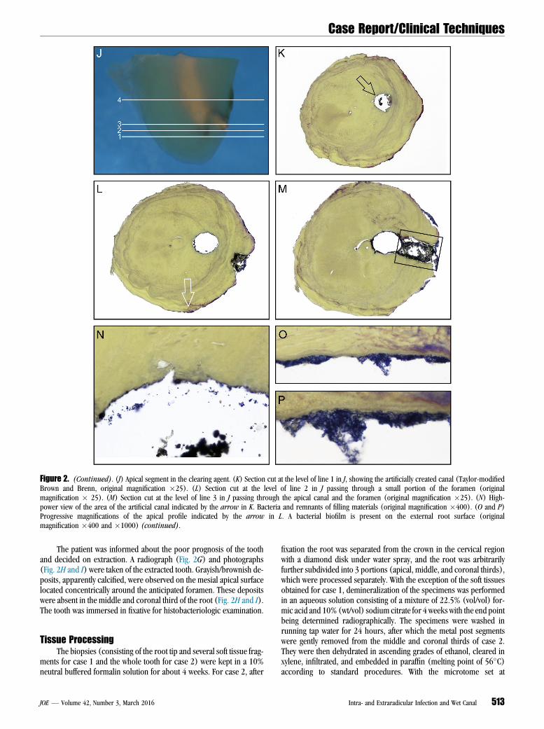
Figure 2. (Continued). (J) Apical segment in the clearing agent. (K) Section cut at the level of line 1 in J, showing the artificially created canal (Taylor-modifiedBrown and Brenn, original magnification �25). (L) Section cut at the level of line 2 in J passing through a small portion of the foramen (originalmagnification � 25). (M) Section cut at the level of line 3 in J passing through the apical canal and the foramen (original magnification �25). (N) High-power view of the area of the artificial canal indicated by the arrow in K. Bacteria and remnants of filling materials (original magnification �400). (O and P)Progressive magnifications of the apical profile indicated by the arrow in L. A bacterial biofilm is present on the external root surface (originalmagnification �400 and �1000) (continued).
Case Report/Clinical Techniques
The patient was informed about the poor prognosis of the toothand decided on extraction. A radiograph (Fig. 2G) and photographs(Fig. 2H and I) were taken of the extracted tooth. Grayish/brownish de-posits, apparently calcified, were observed on the mesial apical surfacelocated concentrically around the anticipated foramen. These depositswere absent in the middle and coronal third of the root (Fig. 2H and I).The tooth was immersed in fixative for histobacteriologic examination.
Tissue ProcessingThe biopsies (consisting of the root tip and several soft tissue frag-
ments for case 1 and the whole tooth for case 2) were kept in a 10%neutral buffered formalin solution for about 4 weeks. For case 2, after
JOE — Volume 42, Number 3, March 2016
fixation the root was separated from the crown in the cervical regionwith a diamond disk under water spray, and the root was arbitrarilyfurther subdivided into 3 portions (apical, middle, and coronal thirds),which were processed separately. With the exception of the soft tissuesobtained for case 1, demineralization of the specimens was performedin an aqueous solution consisting of a mixture of 22.5% (vol/vol) for-mic acid and 10% (wt/vol) sodium citrate for 4 weeks with the end pointbeing determined radiographically. The specimens were washed inrunning tap water for 24 hours, after which the metal post segmentswere gently removed from the middle and coronal thirds of case 2.They were then dehydrated in ascending grades of ethanol, cleared inxylene, infiltrated, and embedded in paraffin (melting point of 56�C)according to standard procedures. With the microtome set at
Intra- and Extraradicular Infection and Wet Canal 513
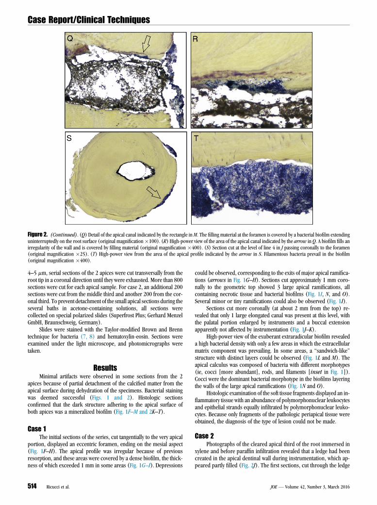
Figure 2. (Continued). (Q) Detail of the apical canal indicated by the rectangle in M. The filling material at the foramen is covered by a bacterial biofilm extendinguninterruptedly on the root surface (original magnification�100). (R) High-power view of the area of the apical canal indicated by the arrow in Q. A biofilm fills anirregularity of the wall and is covered by filling material (original magnification �400). (S) Section cut at the level of line 4 in J passing coronally to the foramen(original magnification �25). (T) High-power view from the area of the apical profile indicated by the arrow in S. Filamentous bacteria prevail in the biofilm(original magnification �400).
Case Report/Clinical Techniques
4–5 mm, serial sections of the 2 apices were cut transversally from theroot tip in a coronal direction until they were exhausted. More than 800sections were cut for each apical sample. For case 2, an additional 200sections were cut from the middle third and another 200 from the cor-onal third. To prevent detachment of the small apical sections during theseveral baths in acetone-containing solutions, all sections werecollected on special polarized slides (Superfrost Plus; Gerhard MenzelGmbH, Braunschweig, Germany).
Slides were stained with the Taylor-modified Brown and Brenntechnique for bacteria (7, 8) and hematoxylin-eosin. Sections wereexamined under the light microscope, and photomicrographs weretaken.
ResultsMinimal artifacts were observed in some sections from the 2
apices because of partial detachment of the calcified matter from theapical surface during dehydration of the specimens. Bacterial stainingwas deemed successful (Figs. 1 and 2). Histologic sectionsconfirmed that the dark structure adhering to the apical surface ofboth apices was a mineralized biofilm (Fig. 1F–M and 2K–T).
Case 1The initial sections of the series, cut tangentially to the very apical
portion, displayed an eccentric foramen, ending on the mesial aspect(Fig. 1F–H). The apical profile was irregular because of previousresorption, and these areas were covered by a dense biofilm, the thick-ness of which exceeded 1 mm in some areas (Fig. 1G–I). Depressions
514 Ricucci et al.
could be observed, corresponding to the exits of major apical ramifica-tions (arrows in Fig. 1G–H). Sections cut approximately 1 mm coro-nally to the geometric top showed 3 large apical ramifications, allcontaining necrotic tissue and bacterial biofilms (Fig. 1I, N, and O).Several minor or tiny ramifications could also be observed (Fig. 1I).
Sections cut more coronally (at about 2 mm from the top) re-vealed that only 1 large elongated canal was present at this level, withthe palatal portion enlarged by instruments and a buccal extensionapparently not affected by instrumentation (Fig. 1J–K).
High-power view of the exuberant extraradicular biofilm revealeda high bacterial density with only a few areas in which the extracellularmatrix component was prevailing. In some areas, a ‘‘sandwich-like’’structure with distinct layers could be observed (Fig. 1L and M). Theapical calculus was composed of bacteria with different morphotypes(ie, cocci [more abundant], rods, and filaments [inset in Fig. 1]).Cocci were the dominant bacterial morphotype in the biofilms layeringthe walls of the large apical ramifications (Fig. 1N and O).
Histologic examination of the soft tissue fragments displayed an in-flammatory tissue with an abundance of polymorphonuclear leukocytesand epithelial strands equally infiltrated by polymorphonuclear leuko-cytes. Because only fragments of the pathologic periapical tissue wereobtained, the diagnosis of the type of lesion could not be made.
Case 2Photographs of the cleared apical third of the root immersed in
xylene and before paraffin infiltration revealed that a ledge had beencreated in the apical dentinal wall during instrumentation, which ap-peared partly filled (Fig. 2J). The first sections, cut through the ledge
JOE — Volume 42, Number 3, March 2016
Case Report/Clinical Techniques
and not encompassing the foramen (located more coronally), re-vealed necrotic debris, filling material, and bacteria in this iatrogen-ically created space (Fig. 2K and N). Sections cut more coronallyand incorporating the foramen showed that the walls of the apicalportion of the canal were covered by a bacterial biofilm in some areas,and this was lined by filling materials toward the lumen (Fig. 2Q andR). A bacterial biofilm also lined the filling material at the foraminalopening. This was continuous with the biofilm covering the externalapical root (Fig. 2M and Q). In some sections, this biofilm extendedfor approximately half of the root circumference and exhibited varyingthickness, covering the cementum or the dentin in resorptive defectsdenuded from the cementum (Fig. 2S). Filamentous bacteria were theprevailing morphotype in the apical calculus (Fig. 2T). Bacterial bio-films were observed on the apical external surface up to a distance ofapproximately 3 mm from the root tip and were absent in the middleand coronal thirds.DiscussionThe clinical condition referred to as ‘‘wet canals’’ is characterized
by continuous seepage of inflammatory exudate into the canal persistingdespite treatment. The cause is arguably persistent infection, and the 2cases reported in this article significantly contribute to the understand-ing of the etiology of this clinical condition. Both teeth had persistentintracanal exudation, which was associated with apical intraradicularinfection continuous with a thick extraradicular biofilm as determinedby histobacteriology. In case 1, root canal filling had to be placed duringthe surgical procedures. In case 2, exudation was controlled afterseveral visits, but the lesion increased in size as revealed at the firstfollow-up examination.
The extraradicular biofilms associated with the 2 cases were thickand presented areas of mineralization that gave them a calculus-likeappearance similar to previous reports (1, 2). Different bacterialmorphotypes were found in the biofilms, indicating the occurrenceof a multispecies bacterial community. These extraradicular biofilmswere contiguous to intraradicular biofilms, which were unaffected bytreatment procedures. These observations give support to theassumption that extraradicular biofilms are usually an extension ofthe intraradicular infection (9). Whether or not these complex extrara-dicular bacterial structures exhibiting areas of mineralization becomeindependent of the intraradicular infectious process remains to bedetermined. Given the apparently high degree of organization of thesemature bacterial structures, it is reasonable to believe that they maynot be significantly influenced by intracanal procedures that succeededin eradicating the intraradicular infection. Consequently, these matureand mineralized extraradicular biofilms could become an independentinfectious entity. It is important to point out that this assumption is notsupported by the present findings because there was an intraradicularinfectious component in both cases. However, a previous article showed1 case of extraradicular biofilm in a tooth with no detectable intraradic-ular infection (3). Other studies should elaborate on this issue.
The extraradicular biofilm is a relatively rare occurrence (1)because bacteria leaving the apical foramen are directly combatedand eliminated by the host defense mechanisms. When developed, itis likely to be a consequence of massive infection of the root canal sys-tem associated with prolonged exposure of the canal space to the oralenvironment (10). The fact that most cases of extraradicular biofilmsare associated with sinus tracts may indicate that these cases are result-ing from chronicization of a previous abscess, a condition known to becaused by egression of pathogenic bacteria from the canal to the peri-radicular tissues (11). Moreover, the extraradicular occurrence of bac-teria may also be precipitated by overinstrumentation (3); this may have
JOE — Volume 42, Number 3, March 2016
occurred in case 2. This reinforces the need for controlled apical lengthof instrumentation in order to avoid postoperative complications.
Calculus is a bacterial biofilm that underwent mineralization. Insupra- or subgingival calculus, separate foci of mineralization increasein size and coalesce to form solid mineralized masses (12). Crystalsform by precipitation of mineral salts initially in the intercellular matrixof the biofilm, then on the bacterial cell surface, and eventually withinthe bacterial cells (13). Whether or not the same mechanism happensin extraradicular calculus cannot be inferred, but the presence ofmineralization foci in these apical structures may suggest likewise.There are some possible ways by which mineralization can developon the extraradicular biofilm. A possible source may be the inflamma-tory exudate and periradicular tissue fluids saturated with mineralsfrom bone solubilization. In addition, long-standing sinus tracts mayfunction as a route of communication between the periradicular areaand the external environment, permitting the passage of minerals andsalts from the oral fluids into the apical periodontitis lesion. Althoughit is possible that bacteria may play some role in mineralization of thebiofilm, the most common opinion on periodontal calculus is that bac-teria are only passively involved, being included in the mineralizationprocess along with the biofilm intercellular matrix (14). Communica-tion of the apical periodontitis lesion with a secondary periodontaldefect may also have been a source of mineralization in case 2.
In conclusion, complex persistent infection in the apical part of theroot canal system extending to form thick and partially mineralized bio-film structures on the outer apical root surface was the cause of wet ca-nals and endodontic treatment failure in the cases reported. Severalattempts to control infection and persistent exudation were performed,and the treatment was significantly arrested.
AcknowledgmentsThe authors deny any conflicts of interest related to this study.
References1. Ricucci D, Siqueira JF Jr. Biofilms and apical periodontitis: study of prevalence and
association with clinical and histopathologic findings. J Endod 2010;36:1277–88.2. Ricucci D, Martorano M, Bate AL, Pascon EA. Calculus-like deposit on the apical
external root surface of teeth with post-treatment apical periodontitis: report oftwo cases. Int Endod J 2005;38:262–71.
3. Ricucci D, Siqueira JF Jr, Lopes WS, et al. Extraradicular infection as the cause ofpersistent symptoms: a case series. J Endod 2015;41:265–73.
4. Tronstad L, Barnett F, Cervone F. Periapical bacterial plaque in teeth refractory toendodontic treatment. Endod Dent Traumatol 1990;6:73–7.
5. Harn WM, Chen YH, Yuan K, et al. Calculus-like deposit at apex of tooth with refrac-tory apical periodontitis. Endod Dent Traumatol 1998;14:237–40.
6. Siqueira JF, Rocas IN, Ricucci D. Biofilms in endodontic infection. Endod Topics2010;22:33–49.
7. Taylor RD. Modification of the Brown and Brenn Gram stain for the differentialstaining of gram-positive and gram-negative bacteria in tissue sections. Am J ClinPathol 1966;46:472–6.
8. Ricucci D, Bergenholtz G. Bacterial status in root-filled teeth exposed to the oralenvironment by loss of restoration and fracture or caries—a histobacteriologicalstudy of treated cases. Int Endod J 2003;36:787–802.
9. Siqueira JF, Rocas IN. Present status and future directions in endodontic microbi-ology. Endod Topics 2014;30:3–22.
10. Ricucci D, Siqueira JF Jr. Endodontology. An Integrated Biological and ClinicalView. London: Quintessence Publishing; 2013.
11. Siqueira JF Jr, Rocas IN. Microbiology and treatment of acute apical abscesses. ClinMicrobiol Rev 2013;26:255–73.
12. Jin Y, Yip HK. Supragingival calculus: formation and control. Crit Rev Oral Biol Med2002;13:426–41.
13. Zander HA, Hazen SP, Scott DB. Mineralization of dental calculus. Proc Soc Exp BiolMed 1960;103:257–60.
14. Hinrichs JE, Math VT. The role of dental calculus and other local predispos-ing factors. In: Newman MG, Takei HH, Klokkevold PR, Carranza FA, eds.Carranza’s Clinical Periodontology, 12th ed. St Louis: Elsevier/Saunders;2015:116–31.
Intra- and Extraradicular Infection and Wet Canal 515


