Download - Cognitive Neuroscience
Chapter Five Cognitive Neuroscience
In the 1930’s Wilder Penfield developed groundbreaking surgical techniques for the treatment of epilepsy. He created a preoperative procedure that involved direct electrical stimulation of brain tissue that is still in use today. Adapted from earlier animal studies, Penfield discovered that application of direct electrical stimulation of the convoluted surface of the brain, referred to as the cerebral cortex, allowed mapping the areas of the cortex responsible for important functions such as perception, motor control, and language, thus allowing the surgeon to remove the abnormal tissue associated with epileptic seizures while sparing as much critical functioning brain tissue as possible. Penfield’s work also documented the fact that electrical stimulation of the cortical surface of the human brain was associated with changes in mental experience.
“Wilder Penfield’s Brain Stimulation”
Because the brain itself does not possess pain receptors, patients were operated on under local anesthesia and were therefore awake and able to report their mental experience when told their brain was being electrically stimulated. Patients would respond with reports of changes in visual perception, such as “falling stars” when visual areas were stimulated, and they would report perception of auditory events (e.g., hearing music and insisting that a radio was playing in the room when it was not) when auditory areas were stimulated. Penfield mapped the cortical areas responsible for voluntary motor control of the body and demonstrated that the neurons in this region were organized like a map of the human body. He also discovered supplementary motor areas that seemed to be involved in motor planning as well as motor control. He reported on a patient whose hand moved when the portion of the motor control area representing the hand was stimulated, and the patient reported the mental experience that the doctor had moved the hand. However, when the nearby supplementary motor area, associated with planning of volitional movements of the same hand, was stimulated the patient’s hand moved and now the patient claimed to have moved it voluntarily. These examples raise questions regarding the association of physical changes in the brain and mental experience, as well as questions regarding how
119
different regions of cortex may implement different cognitive processes, questions that are at the heart of a new and rapidly changing area of research called cognitive neuroscience. Pinky and the Brain. Before getting too serious about the brain you can visit YouTube to view Pinky and the Brain singing the brain song. It is entertaining as well as educational.
“Parts of the Brain Song”
This chapter:
• Defines the relatively new scientific discipline of cognitive neuroscience. • Explains the 2 major influences leading to the rapid emergence of cognitive
neuroscience. • Discusses 2 questions of debate during the development of neuroscience. • Provides a brief introduction to the basic principles of neural communication in
the brain. • Provides a brief introduction to the basic functions of the major neuroanatomical
structures of interest to cognitive scientists. • Provides a focused review of selected cognitive neuroscience methodologies
from 3 broad classes of methods.
Outline of Topics I. Definition and Development of Cognitive Neuroscience II. Major Historical Questions about the Brain
A. The Mind/Brain Question B. Localization of Cortical Function
III. Neural signaling A. Structures of the Prototypical Neuron B. Electrical Signaling within Neurons C. Chemical Signaling between Neurons D. Signaling versus Computation
IV. Functional Neuroanatomy for Cognitive Scientists A. Introduction to the Cerebral Cortex B. 4 Cortical Lobes Plus 1 Important Additional Area C. 2 General Principles of Cortical Function D. Cortical Processing E. Subcortical Structures of Importance to Cognitive Scientists F. Cerebellum & Brainstem
120
V. Methods of Cognitive Neuroscience A. Studying Electrophysiological Processes
1. Direct Electrical Recording 2. Electroencephalography (EEG) 3. Event-related Potentials (ERP)
B. Imaging the structure and function of the brain 1. MRI 2. PET 3. Functional MRI (fMRI)
C. Lesion analysis 1. Single & Double Dissociations 2. Transcranial magnetic stimulation (TMS)
References Glossary
I. Definition and Development of Cognitive Neuroscience Cognitive neuroscience is a relatively new scientific discipline that combines the study of cognitive and biological processes in the brain, primarily in humans. Cognitive neuroscience is a scientific discipline that is the result of a fairly recent coming together of researchers in cognitive psychology and neuroscience to better understand how cognitive processes such as thoughts, emotions, perceptions, memory, and goals are implemented in the brain. Cognitive neuroscience has emerged as a distinct discipline only recently in response to 2 major influences. First, for many years cognitive psychologists studied cognitive processing in humans with only minimal attention to how such processes might be physically implemented due to a strong belief that that processing steps used to complete a cognitive task were not directly dependent on the underlying physical substrate, in this case the brain, performing the computations. Similarly, neuroscientists focused primarily on the physical functioning of the neurons and brains. One major impetus for the emergence of cognitive neuroscience was that cognitive psychologists and neuroscientists started serious discussions of how their respective areas of inquiry could complement each other and lead to a fuller understanding of how the human brain implements human cognition. On this view, neuroscientists could develop a better understanding of physical brain processes by considering the cognitive theories developed by psychologists, and cognitive psychologists could benefit from the knowledge of neuroscientists regarding how the brain worked to implement cognitive processes. A second force leading to the emergence of cognitive neuroscience was the development of techniques for observing the operation of the brain in a living human being (i.e., in vivo) from outside of the scalp and skull (i.e., noninvasive). Moreover, a critical technical development in 1990 (Ogawa et al., 1990) led to the realization that a widely available medical scanning technology that had been used for making images of the structure of the tissues of the body, magnetic resonance imaging (MRI), could be easily modified to produce functional images of the pattern of neural activity in the brain during performance of a cognitive task. Due to the fact that a large number of MRI
121
scanners were already in widespread use in medical centers and medical schools, this new functional MRI (fMRI) technique for imaging the function of the human brain became accessible to a large number of researchers in a short amount of time, leading to an explosion in the number of functional brain imaging studies and a rapid increase in the number of scientists wanting to focus on the new and exciting discipline of cognitive neuroscience.
“How the MRI Works”
II. Major Historical Questions about the Brain
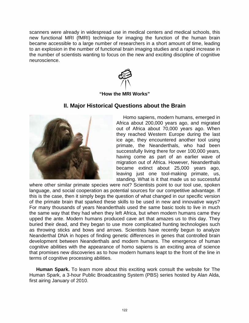
Homo sapiens, modern humans, emerged in Africa about 200,000 years ago, and migrated out of Africa about 70,000 years ago. When they reached Western Europe during the last ice age, they encountered another tool using primate, the Neanderthals, who had been successfully living there for over 100,000 years, having come as part of an earlier wave of migration out of Africa. However, Neanderthals became extinct about 25,000 years ago, leaving just one tool-making primate, us, standing. What is it that made us so successful
where other similar primate species were not? Scientists point to our tool use, spoken language, and social cooperation as potential sources for our competitive advantage. If this is the case, then it simply begs the question of what changed in our specific version of the primate brain that sparked these skills to be used in new and innovative ways? For many thousands of years Neanderthals used the same basic tools to live in much the same way that they had when they left Africa, but when modern humans came they upped the ante. Modern humans produced cave art that amazes us to this day. They buried their dead, and they began to use more complicated hunting technologies such as throwing sticks and bows and arrows. Scientists have recently begun to analyze Neanderthal DNA in hopes of finding genetic differences in genes that controlled brain development between Neanderthals and modern humans. The emergence of human cognitive abilities with the appearance of homo sapiens is an exciting area of science that promises new discoveries as to how modern humans leapt to the front of the line in terms of cognitive processing abilities. Human Spark. To learn more about this exciting work consult the website for The Human Spark, a 3-hour Public Broadcasting System (PBS) series hosted by Alan Alda, first airing January of 2010.
122
In her poem, The Brain – is wider than the Sky, Emily Dickenson (1860’s) wonders at the amazing ability of the human brain to contain (or represent?) the immense universe that we find ourselves a part of. This poem points to the complex relationship between mind and brain. How can it be that an organ that fits in the human skull can contain such immenseness as the sky itself? Dickinson suggests that it is because the brain can think about the sky, and not only that but the brain can also, with disarming effortlessness, think about the person who is thinking about the sky. The Brain – is wider than the Sky – For – put them side by side – The one the other will contain With ease – and You – beside –
A. The Mind/Brain Question A fundamental question regarding the brain is how physical matter leads to the sensations, feelings, thoughts, and emotions experienced by humans, that is, the mind. While the debate regarding the mind/brain relationship has not been resolved, and many subtle variations of argument have been proposed over the debate’s long history, two basic positions, called dualism and monism, are clearly at the heart of the debate. The monist (also called physicalist) position is that the mind, like the brain, is physical-- composed of the same substance as the brain. By contrast, the dualist position is that the brain is physical but the mind is nonphysical, and therefore that mind and brain have a dual nature. Because of its reliance on a nonphysical mind, most cognitive scientists find it difficult to reconcile dualist positions on the mind/brain problem with the scientific method which is based on observations of physical events and their causes (Ward, 2006). In fact, a reductionist monist position, where it is argued that mental events will someday be found to be understandable as physical brain states, appears to be the dominant view amongst cognitive neuroscientists (see Churchland, 2002; Crick, 1994; Edelman, 2004 for further reading, the later 2 authors are noted cognitive neuroscientists and Nobel laureates). The monist part of this position views the mind as caused by physical processes in the brain, and the reductionist part of this position emphasizes that just as chemical reactions have been effectively explained by reducing them to physical processes in the constituent atoms in the scientific discipline of chemistry, the human mind may also someday be explainable as interactions of the fundamental building blocks of the brain, neurons. While this reductionist monist position certainly has many supporters amongst cognitive neuroscientists, there are a variety of nonreductionist monists who believe that the brain may cause the mind, but that there may be aspects of the mind that preclude a fully reductionist explanation, that is, by referring to interactions of neurons. Nonreductionists often argue that the mind has emergent properties not understandable by breaking it down into its constituent parts. For example, an emergent property of water is that it changes into something
123
qualitatively different when it freezes and transforms to ice. Nonreductionist monists typically argue that the mind is the result of emergent properties of many neurons in the brain physically interacting in complex ways that cannot be reduced down to the level of individual neurons. This is an active area of debate, and students wishing to learn more about basic arguments regarding the mind/brain problem should consult Ravenscroft (2005).
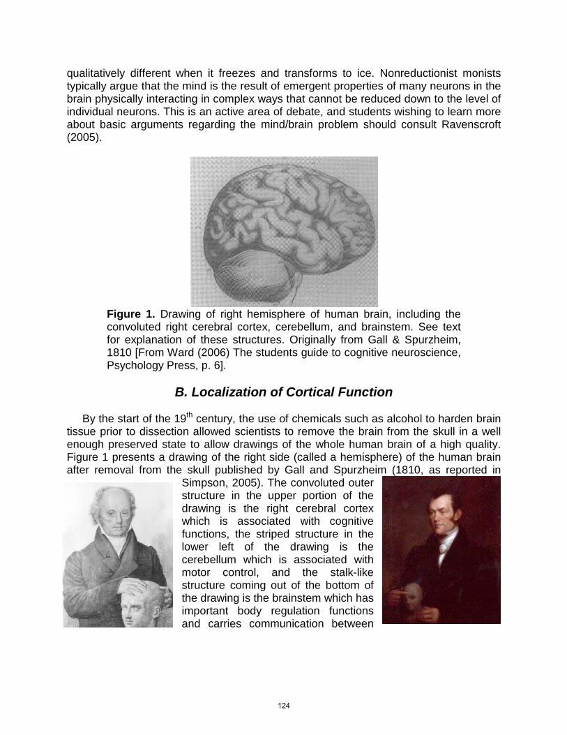
Figure 1. Drawing of right hemisphere of human brain, including the convoluted right cerebral cortex, cerebellum, and brainstem. See text for explanation of these structures. Originally from Gall & Spurzheim, 1810 [From Ward (2006) The students guide to cognitive neuroscience, Psychology Press, p. 6].
B. Localization of Cortical Function
By the start of the 19th century, the use of chemicals such as alcohol to harden brain tissue prior to dissection allowed scientists to remove the brain from the skull in a well enough preserved state to allow drawings of the whole human brain of a high quality. Figure 1 presents a drawing of the right side (called a hemisphere) of the human brain after removal from the skull published by Gall and Spurzheim (1810, as reported in
Simpson, 2005). The convoluted outer structure in the upper portion of the drawing is the right cerebral cortex which is associated with cognitive functions, the striped structure in the lower left of the drawing is the cerebellum which is associated with motor control, and the stalk-like structure coming out of the bottom of the drawing is the brainstem which has important body regulation functions and carries communication between
124
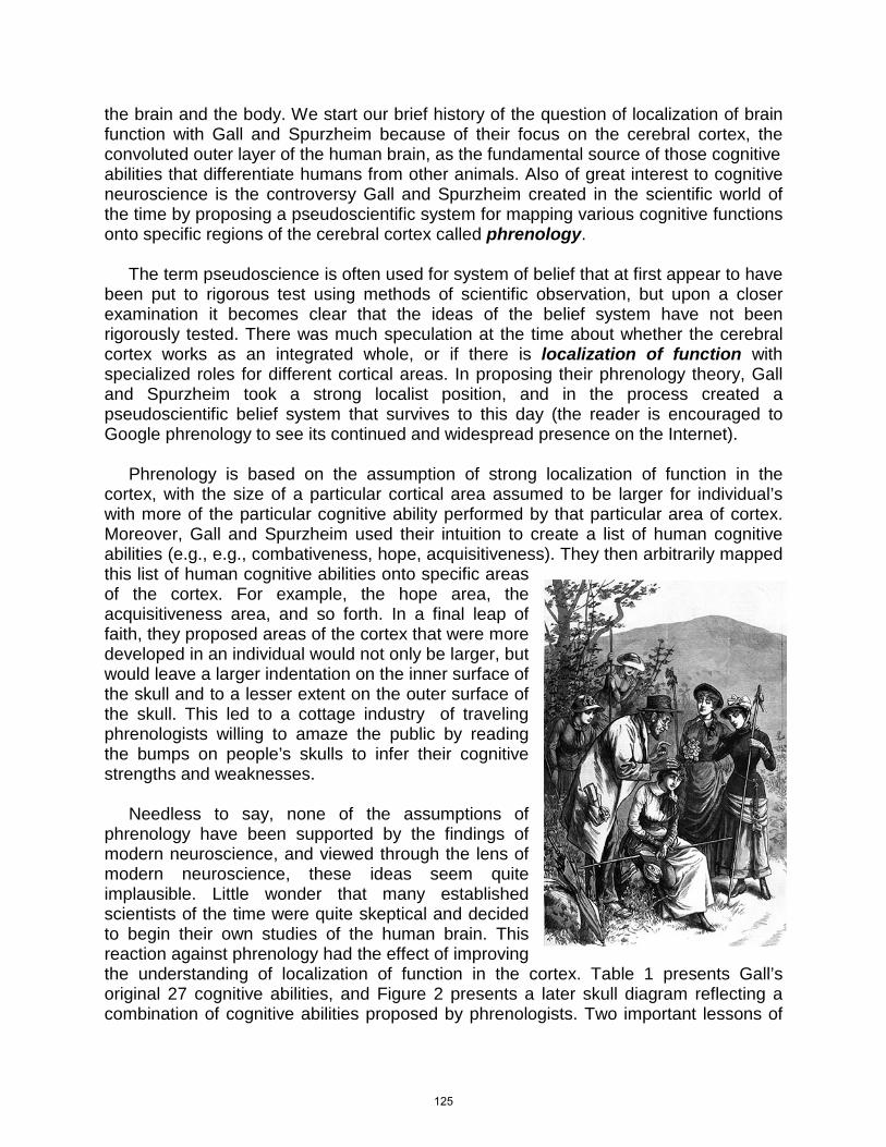
the brain and the body. We start our brief history of the question of localization of brain function with Gall and Spurzheim because of their focus on the cerebral cortex, the convoluted outer layer of the human brain, as the fundamental source of those cognitive abilities that differentiate humans from other animals. Also of great interest to cognitive neuroscience is the controversy Gall and Spurzheim created in the scientific world of the time by proposing a pseudoscientific system for mapping various cognitive functions onto specific regions of the cerebral cortex called phrenology. The term pseudoscience is often used for system of belief that at first appear to have been put to rigorous test using methods of scientific observation, but upon a closer examination it becomes clear that the ideas of the belief system have not been rigorously tested. There was much speculation at the time about whether the cerebral cortex works as an integrated whole, or if there is localization of function with specialized roles for different cortical areas. In proposing their phrenology theory, Gall and Spurzheim took a strong localist position, and in the process created a pseudoscientific belief system that survives to this day (the reader is encouraged to Google phrenology to see its continued and widespread presence on the Internet). Phrenology is based on the assumption of strong localization of function in the cortex, with the size of a particular cortical area assumed to be larger for individual’s with more of the particular cognitive ability performed by that particular area of cortex. Moreover, Gall and Spurzheim used their intuition to create a list of human cognitive abilities (e.g., e.g., combativeness, hope, acquisitiveness). They then arbitrarily mapped this list of human cognitive abilities onto specific areas of the cortex. For example, the hope area, the acquisitiveness area, and so forth. In a final leap of faith, they proposed areas of the cortex that were more developed in an individual would not only be larger, but would leave a larger indentation on the inner surface of the skull and to a lesser extent on the outer surface of the skull. This led to a cottage industry of traveling phrenologists willing to amaze the public by reading the bumps on people’s skulls to infer their cognitive strengths and weaknesses. Needless to say, none of the assumptions of phrenology have been supported by the findings of modern neuroscience, and viewed through the lens of modern neuroscience, these ideas seem quite implausible. Little wonder that many established scientists of the time were quite skeptical and decided to begin their own studies of the human brain. This reaction against phrenology had the effect of improving the understanding of localization of function in the cortex. Table 1 presents Gall’s original 27 cognitive abilities, and Figure 2 presents a later skull diagram reflecting a combination of cognitive abilities proposed by phrenologists. Two important lessons of
125
this false start in the study of localization of cortical function are that it is critical to have good theories of the cognitive processes of the mind, and it is also critical to have rigorous methods for associating cognitive functions with brain areas. Both of these critical ingredients were lacking in the phrenology of the 19th century.
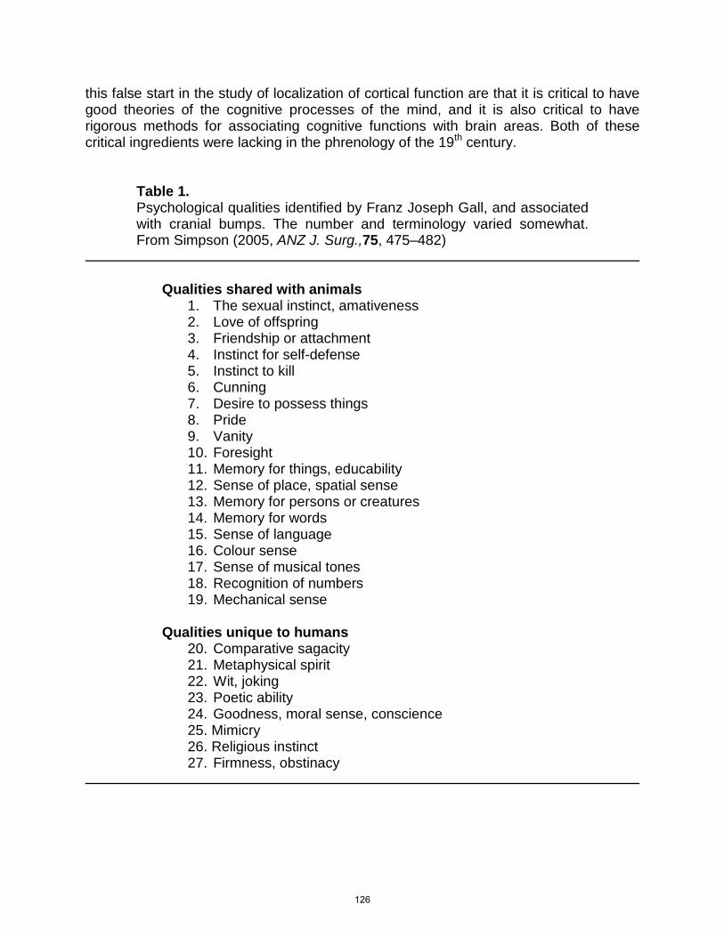
Table 1. Psychological qualities identified by Franz Joseph Gall, and associated with cranial bumps. The number and terminology varied somewhat. From Simpson (2005, ANZ J. Surg.,75, 475–482)
Qualities shared with animals 1. The sexual instinct, amativeness 2. Love of offspring 3. Friendship or attachment 4. Instinct for self-defense 5. Instinct to kill 6. Cunning 7. Desire to possess things 8. Pride 9. Vanity 10. Foresight 11. Memory for things, educability 12. Sense of place, spatial sense 13. Memory for persons or creatures 14. Memory for words 15. Sense of language 16. Colour sense 17. Sense of musical tones 18. Recognition of numbers 19. Mechanical sense
Qualities unique to humans
20. Comparative sagacity 21. Metaphysical spirit 22. Wit, joking 23. Poetic ability 24. Goodness, moral sense, conscience 25. Mimicry 26. Religious instinct 27. Firmness, obstinacy
126
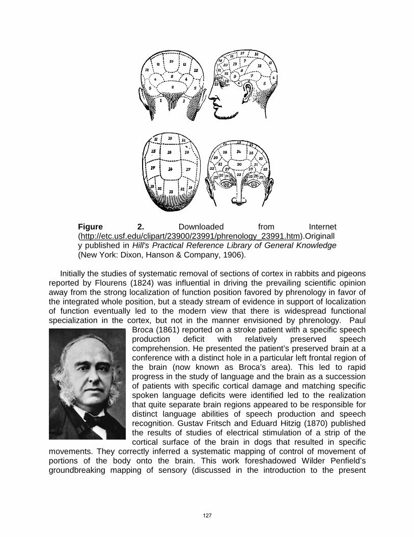
Figure 2. Downloaded from Internet (http://etc.usf.edu/clipart/23900/23991/phrenology_23991.htm).Originally published in Hill's Practical Reference Library of General Knowledge (New York: Dixon, Hanson & Company, 1906).
Initially the studies of systematic removal of sections of cortex in rabbits and pigeons reported by Flourens (1824) was influential in driving the prevailing scientific opinion away from the strong localization of function position favored by phrenology in favor of the integrated whole position, but a steady stream of evidence in support of localization of function eventually led to the modern view that there is widespread functional specialization in the cortex, but not in the manner envisioned by phrenology. Paul
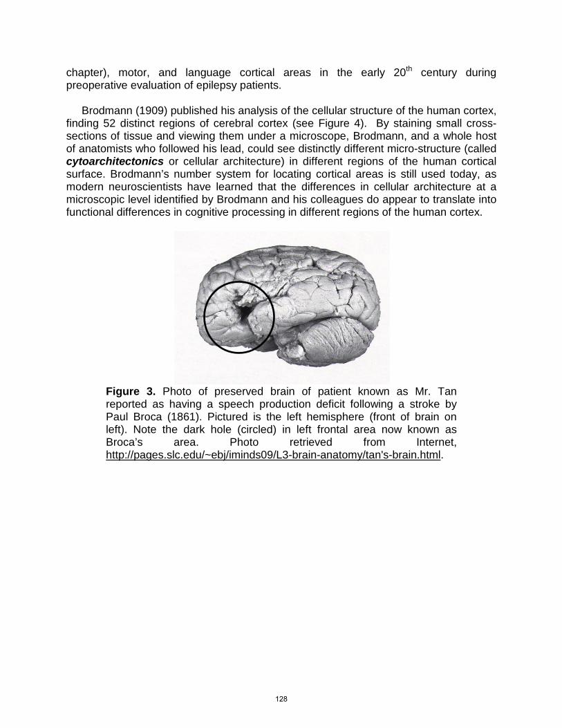
Broca (1861) reported on a stroke patient with a specific speech production deficit with relatively preserved speech comprehension. He presented the patient’s preserved brain at a conference with a distinct hole in a particular left frontal region of the brain (now known as Broca’s area). This led to rapid progress in the study of language and the brain as a succession of patients with specific cortical damage and matching specific spoken language deficits were identified led to the realization that quite separate brain regions appeared to be responsible for distinct language abilities of speech production and speech recognition. Gustav Fritsch and Eduard Hitzig (1870) published the results of studies of electrical stimulation of a strip of the cortical surface of the brain in dogs that resulted in specific
movements. They correctly inferred a systematic mapping of control of movement of portions of the body onto the brain. This work foreshadowed Wilder Penfield’s groundbreaking mapping of sensory (discussed in the introduction to the present
127
chapter), motor, and language cortical areas in the early 20th century during preoperative evaluation of epilepsy patients. Brodmann (1909) published his analysis of the cellular structure of the human cortex, finding 52 distinct regions of cerebral cortex (see Figure 4). By staining small cross-sections of tissue and viewing them under a microscope, Brodmann, and a whole host of anatomists who followed his lead, could see distinctly different micro-structure (called cytoarchitectonics or cellular architecture) in different regions of the human cortical surface. Brodmann’s number system for locating cortical areas is still used today, as modern neuroscientists have learned that the differences in cellular architecture at a microscopic level identified by Brodmann and his colleagues do appear to translate into functional differences in cognitive processing in different regions of the human cortex.
Figure 3. Photo of preserved brain of patient known as Mr. Tan reported as having a speech production deficit following a stroke by Paul Broca (1861). Pictured is the left hemisphere (front of brain on left). Note the dark hole (circled) in left frontal area now known as Broca’s area. Photo retrieved from Internet, http://pages.slc.edu/~ebj/iminds09/L3-brain-anatomy/tan's-brain.html.
128
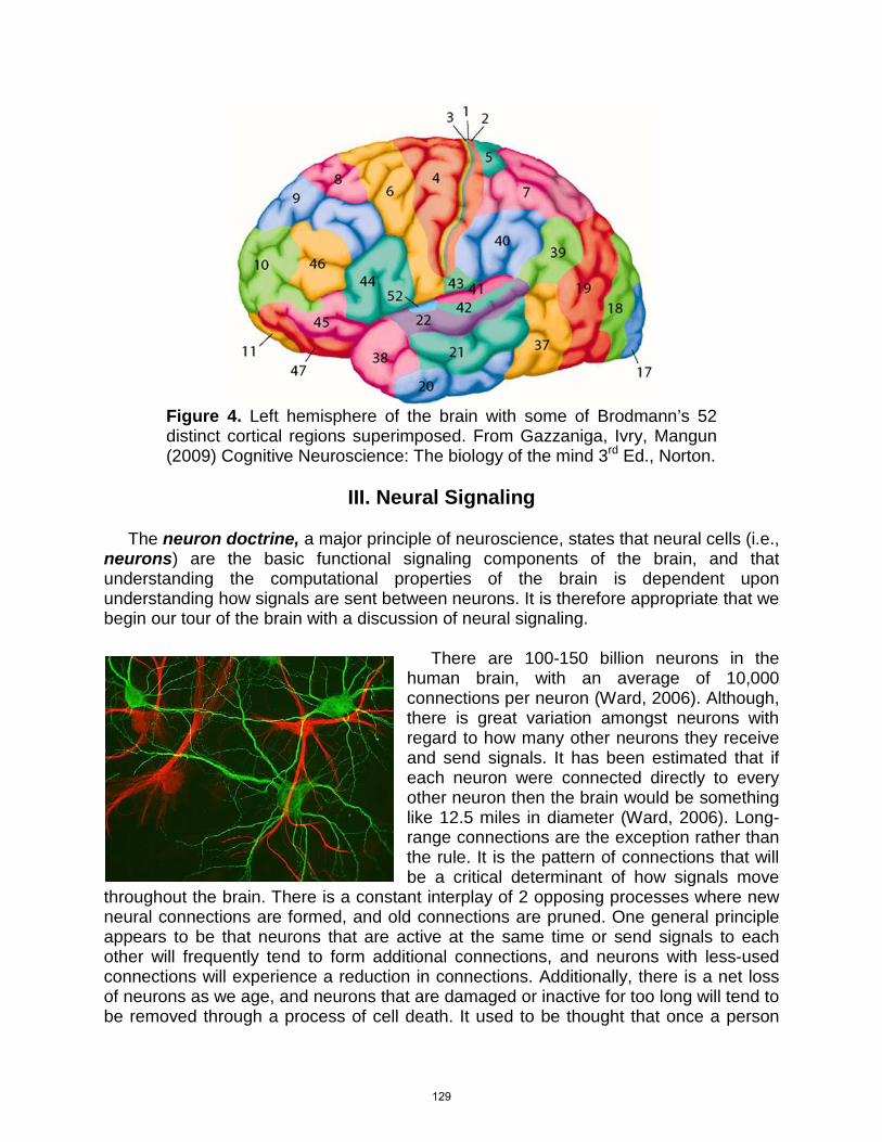
Figure 4. Left hemisphere of the brain with some of Brodmann’s 52 distinct cortical regions superimposed. From Gazzaniga, Ivry, Mangun (2009) Cognitive Neuroscience: The biology of the mind 3rd Ed., Norton.
III. Neural Signaling
The neuron doctrine, a major principle of neuroscience, states that neural cells (i.e., neurons) are the basic functional signaling components of the brain, and that understanding the computational properties of the brain is dependent upon understanding how signals are sent between neurons. It is therefore appropriate that we begin our tour of the brain with a discussion of neural signaling.
There are 100-150 billion neurons in the human brain, with an average of 10,000 connections per neuron (Ward, 2006). Although, there is great variation amongst neurons with regard to how many other neurons they receive and send signals. It has been estimated that if each neuron were connected directly to every other neuron then the brain would be something like 12.5 miles in diameter (Ward, 2006). Long-range connections are the exception rather than the rule. It is the pattern of connections that will be a critical determinant of how signals move
throughout the brain. There is a constant interplay of 2 opposing processes where new neural connections are formed, and old connections are pruned. One general principle appears to be that neurons that are active at the same time or send signals to each other will frequently tend to form additional connections, and neurons with less-used connections will experience a reduction in connections. Additionally, there is a net loss of neurons as we age, and neurons that are damaged or inactive for too long will tend to be removed through a process of cell death. It used to be thought that once a person
129
reaches adulthood no new neurons were created, but we now know this to be false. The extent to which adults can form new neurons, and in what brain areas, is still being actively researched. Neurons make up about 10% of the cells in our brain. The other major class of brain cells are the glia, which, carry out critical structural and metabolic supportive functions. Glial cells provide structural support for neurons, help guide neurons into place during the development of the brain, provide critical metabolites to neurons, and help shield them from harmful substances in the blood. The blood-brain barrier refers to the glial shield that screens many substances in the blood from entering the brain. Some glial cells (i.e., myelin) form fatty insulating sheaths around the axons of neurons, and some glial cells work to clean up debris and foreign substances.
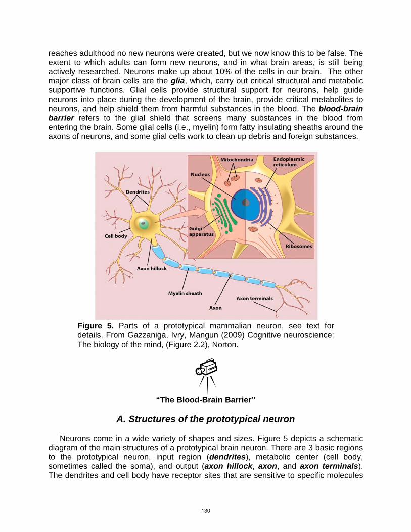
Figure 5. Parts of a prototypical mammalian neuron, see text for details. From Gazzaniga, Ivry, Mangun (2009) Cognitive neuroscience: The biology of the mind, (Figure 2.2), Norton.
“The Blood-Brain Barrier”
A. Structures of the prototypical neuron
Neurons come in a wide variety of shapes and sizes. Figure 5 depicts a schematic diagram of the main structures of a prototypical brain neuron. There are 3 basic regions to the prototypical neuron, input region (dendrites), metabolic center (cell body, sometimes called the soma), and output (axon hillock, axon, and axon terminals). The dendrites and cell body have receptor sites that are sensitive to specific molecules
130
of chemical messengers from other neurons, called neurotransmitters. A discrete active electrical signal, an action potential (AP), will originate at the axon hillock if conditions are right and be sent down the axon to the axon terminals. Myelin is a fatty covering of a particular type of glial cell that facilitates the transmission of the AP. Axons of neurons continuously sprout and grow toward dendrites of nearby neurons. Glial cells help provide a scaffolding structure and chemical signals to aid this process. Axons will position terminals next to a dendritic area that is appropriate for receiving signals from that neuron. The gap between the axon terminals and dendrites is called the synapse. Chemical signals in the form of neurotransmitter molecules are sent from the axon terminal across the synaptic cleft (or gap) and are received on the surface of the dendrite. Synapses that do not result in effective communication between neurons will eventually be pruned out. Sprouting of new synapses and pruning of ineffective synapses is a continuing process. These processes drive neural plasticity; the synaptic connections of the brain are continuously changing in response to the organism’s experience with the world. New synapses are formed, and old less-useful synapses are pruned.
“Neural Communication”
B. Electrical signaling within neurons
There are 6 basic steps of electrical signaling within the neuron to understand (see Figure 6). The process of internal electrical signaling is initiated by a chemical neurotransmitter molecule being sent across the synaptic cleft to the dendrite of the post-synaptic (i.e., receiving) neuron, resulting in a brief electrical post-synaptic potential (PSP). PSPs are discrete (i.e., short-lived) electrical events that are passively conducted down to the axon hillock, just like electricity in a wire. Excitatory PSPs involve positive current flow into the neuron and increase the probability the axon hillock will fire an AP, and inhibitory PSPs involve negative current flow (typically, positively charged particles move out of the neuron) and decrease the probability the axon hillock will fire an AP. The excitatory and inhibitory PSPs are passively conducted to the axon hillock where they are summed. If the combined total is above a set threshold, an AP will be fired. The AP is an all-or-none electrical wave that is actively propagated down the axon by a well-timed opening and closing of channels in the axons that permit the flow of electrically charged particles (called ions) of sodium and potassium. Due to the controlled opening and closing of ion channels, an AP has a constant size. Once the AP reaches the axon terminal it triggers chemical transmission of neurotransmitter across the synaptic cleft to the next neuron down the line. There are 3 aspects of internal electrical signaling that critically affect the overall signaling properties of the neuron. First, the AP is all-or-none and is of a constant size,
131
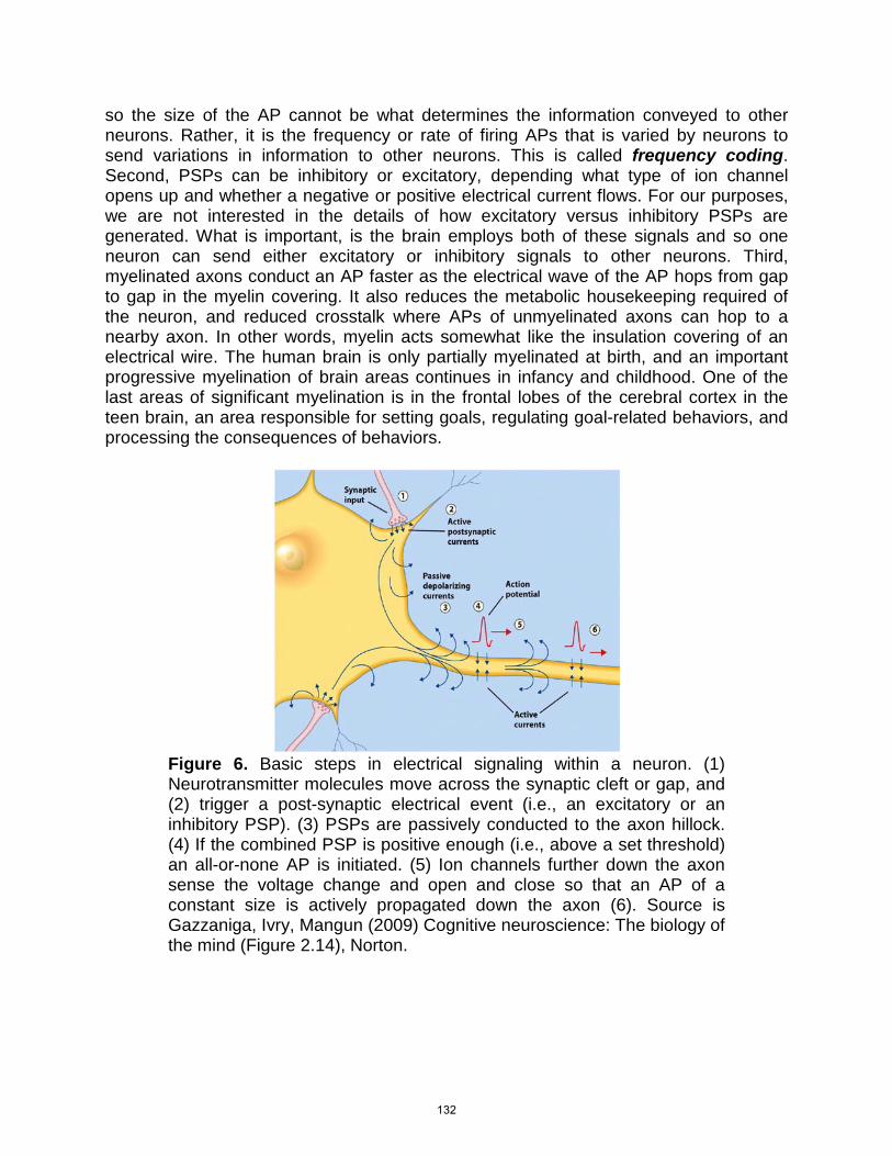
so the size of the AP cannot be what determines the information conveyed to other neurons. Rather, it is the frequency or rate of firing APs that is varied by neurons to send variations in information to other neurons. This is called frequency coding. Second, PSPs can be inhibitory or excitatory, depending what type of ion channel opens up and whether a negative or positive electrical current flows. For our purposes, we are not interested in the details of how excitatory versus inhibitory PSPs are generated. What is important, is the brain employs both of these signals and so one neuron can send either excitatory or inhibitory signals to other neurons. Third, myelinated axons conduct an AP faster as the electrical wave of the AP hops from gap to gap in the myelin covering. It also reduces the metabolic housekeeping required of the neuron, and reduced crosstalk where APs of unmyelinated axons can hop to a nearby axon. In other words, myelin acts somewhat like the insulation covering of an electrical wire. The human brain is only partially myelinated at birth, and an important progressive myelination of brain areas continues in infancy and childhood. One of the last areas of significant myelination is in the frontal lobes of the cerebral cortex in the teen brain, an area responsible for setting goals, regulating goal-related behaviors, and processing the consequences of behaviors.
Figure 6. Basic steps in electrical signaling within a neuron. (1) Neurotransmitter molecules move across the synaptic cleft or gap, and (2) trigger a post-synaptic electrical event (i.e., an excitatory or an inhibitory PSP). (3) PSPs are passively conducted to the axon hillock. (4) If the combined PSP is positive enough (i.e., above a set threshold) an all-or-none AP is initiated. (5) Ion channels further down the axon sense the voltage change and open and close so that an AP of a constant size is actively propagated down the axon (6). Source is Gazzaniga, Ivry, Mangun (2009) Cognitive neuroscience: The biology of the mind (Figure 2.14), Norton.
132
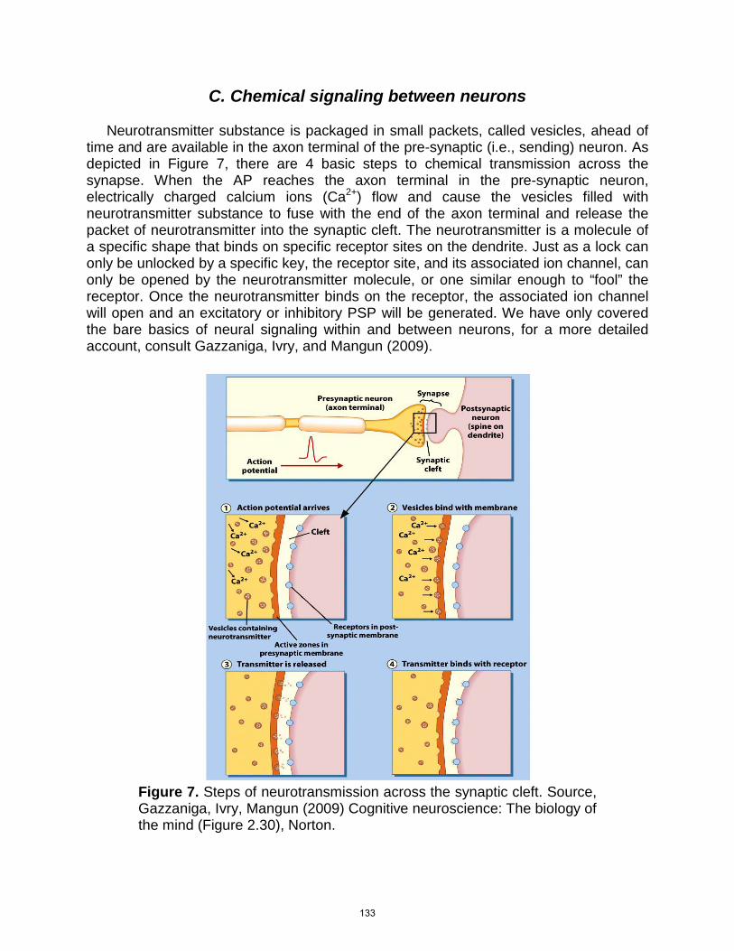
C. Chemical signaling between neurons Neurotransmitter substance is packaged in small packets, called vesicles, ahead of time and are available in the axon terminal of the pre-synaptic (i.e., sending) neuron. As depicted in Figure 7, there are 4 basic steps to chemical transmission across the synapse. When the AP reaches the axon terminal in the pre-synaptic neuron, electrically charged calcium ions (Ca2+) flow and cause the vesicles filled with neurotransmitter substance to fuse with the end of the axon terminal and release the packet of neurotransmitter into the synaptic cleft. The neurotransmitter is a molecule of a specific shape that binds on specific receptor sites on the dendrite. Just as a lock can only be unlocked by a specific key, the receptor site, and its associated ion channel, can only be opened by the neurotransmitter molecule, or one similar enough to “fool” the receptor. Once the neurotransmitter binds on the receptor, the associated ion channel will open and an excitatory or inhibitory PSP will be generated. We have only covered the bare basics of neural signaling within and between neurons, for a more detailed account, consult Gazzaniga, Ivry, and Mangun (2009).
Figure 7. Steps of neurotransmission across the synaptic cleft. Source, Gazzaniga, Ivry, Mangun (2009) Cognitive neuroscience: The biology of the mind (Figure 2.30), Norton.
133
D. Signaling versus Computation Cognitive science has traditionally been based on the idea that cognitive processes can be viewed as formal operations or computations performed on representations, the same kind of computations done by computers. However, this computational/representational understanding of the mind (also referred to as CRUM, Thagard, 2005) appears at first to be inconsistent with the notion of the brain as a massively complex network that processes a large number of signals simultaneously. Some have argued that, at a minimum, the distributed complex signal processing of the brain uses a form of computation that is a different kind than what is typically used to motivate traditional cognitive science (CRUM). However, others have argued that analysis of the computational properties of simulated neural networks yields a promising correspondence between simulated neural computation and CRUM-style computation. They suggest it is likely that once we know more of the details of neural computation in the brain, we will find it compatible with the CRUM formulation of cognitive science. Regardless of whether this claim is borne in future research, it seems clear that the more we know about how the brain implements cognitive processes, the closer we come to knowing how a sophisticated cognitive system such as the human mind can be physically implemented. Neural Communication Animations. The Mind Project (Markram, 2006), associated with Indiana University, has a website that contains animations of interest to students of cognitive science. The following link will take students to a page that presents a series of 4 animations about neural communication. Three of these, the action potential, synapses, and “classical” chemical neurotransmission, are directly related to the material on neural communication just covered in the preceding section. The final animation, electrical neurotransmission, covers neural communication across a specialized electrical connection between some neurons called a gap junction that is not covered in the present chapter. Electrical signaling across gap junctions is likely to be an area of active research activity over the next few years; students interested in this lesser known form of neural communication are encouraged to also view this animation.
“Mind Project Animations”
134
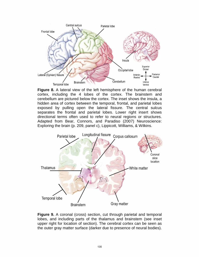
Figure 8. A lateral view of the left hemisphere of the human cerebral cortex, including the 4 lobes of the cortex. The brainstem and cerebellum are pictured below the cortex. The inset shows the insula, a hidden area of cortex between the temporal, frontal, and parietal lobes exposed by pulling open the lateral fissure. The central sulcus separates the frontal and parietal lobes. Lower right insert shows directional terms often used to refer to neural regions or structures. Adapted from Bear, Connors, and Paradiso (2007) Neuroscience: Exploring the brain (p. 209, panel c), Lippicott, Williams, & Wilkins.
Figure 9. A coronal (cross) section, cut through parietal and temporal lobes, and including parts of the thalamus and brainstem (see inset upper right for location of section). The cerebral cortex can be seen as the outer gray matter surface (darker due to presence of neural bodies).
135
The white matter containing myelinated axon tracts is depicted in deeper lighter areas. The corpus callosum, comprising the vast majority of the interhemispheric white matter tracts is visible in the center of the diagram. Parts of the thalamus (sits on top of the brainstem) and brainstem are also included in the section. Adapted from Bear, Connors, and Paradiso (2007) Neuroscience: Exploring the brain (p. 223, panel b), Lippicott, Williams, & Wilkins.
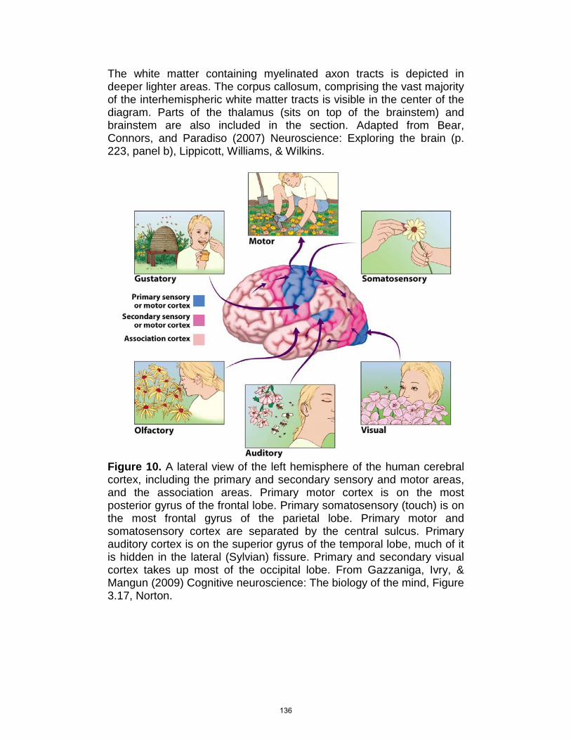
Figure 10. A lateral view of the left hemisphere of the human cerebral cortex, including the primary and secondary sensory and motor areas, and the association areas. Primary motor cortex is on the most posterior gyrus of the frontal lobe. Primary somatosensory (touch) is on the most frontal gyrus of the parietal lobe. Primary motor and somatosensory cortex are separated by the central sulcus. Primary auditory cortex is on the superior gyrus of the temporal lobe, much of it is hidden in the lateral (Sylvian) fissure. Primary and secondary visual cortex takes up most of the occipital lobe. From Gazzaniga, Ivry, & Mangun (2009) Cognitive neuroscience: The biology of the mind, Figure 3.17, Norton.
136
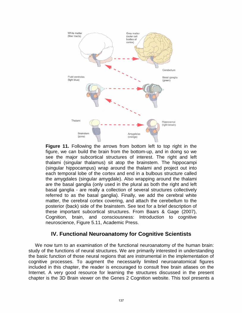
Figure 11. Following the arrows from bottom left to top right in the figure, we can build the brain from the bottom-up, and in doing so we see the major subcortical structures of interest. The right and left thalami (singular thalamus) sit atop the brainstem. The hippocampi (singular hippocampus) wrap around the thalami and project out into each temporal lobe of the cortex and end in a bulbous structure called the amygdales (singular amygdale). Also wrapping around the thalami are the basal ganglia (only used in the plural as both the right and left basal ganglia - are really a collection of several structures collectively referred to as the basal ganglia). Finally, we add the cerebral white matter, the cerebral cortex covering, and attach the cerebellum to the posterior (back) side of the brainstem. See text for a brief description of these important subcortical structures. From Baars & Gage (2007), Cognition, brain, and consciousness: Introduction to cognitive neuroscience, Figure 5.11, Academic Press.
IV. Functional Neuroanatomy for Cognitive Scientists
We now turn to an examination of the functional neuroanatomy of the human brain: study of the functions of neural structures. We are primarily interested in understanding the basic function of those neural regions that are instrumental in the implementation of cognitive processes. To augment the necessarily limited neuroanatomical figures included in this chapter, the reader is encouraged to consult free brain atlases on the Internet. A very good resource for learning the structures discussed in the present chapter is the 3D Brain viewer on the Genes 2 Cognition website. This tool presents a
137
user rotatable 3D brain model with important structures colorized for easy identification. An important feature is that once a structure is selected for viewing, occluding features are made translucent as the 3D brain model is rotated by the user, allowing for clear view of the shape and location of the structure in question. Another useful resource is the Whole Brain Atlas on the Harvard Medical School website. This tool includes a full structural brain scan (MRI) of a normal brain that can be viewed using three 2D viewers that allow viewing of 2D sections from 3 canonical angles (side, cross-section, above). This tool also has an option to select structures from a drop-down list to access a view where the structure in question is marked.
A. Introduction to the Cerebral Cortex We will begin with a lateral (side) view of the left side of the human brain as depicted in Figure 8. There are three major structures visible in a lateral view of the human brain: the cerebrum, brainstem, and cerebellum. The cerebrum comprises a major portion of the human brain. It is separated longitudinally (i.e., from front to back) into right and left hemispheres. The 2 cerebral hemispheres are, broadly speaking, mirror images of each other, consisting of the same basic structures, but there are some functional and anatomical variation between the 2, and the topic of hemispheric differences in structure and function is an active topic of research. Each cerebral hemisphere is covered by a convoluted surface structure, the cerebral cortex, of bumps (called gyri, singular gyrus), and grooves or infoldings (called sulci, singular sulcus, larger sulci also called fissures). The lateral cortex is made up of 4 lobes (subsections) visible in the lateral view of Figure 8: Frontal, parietal, temporal, and occipital. The frontal and parietal lobe are clearly separated by the central sulcus (see Figure 8) running laterally in both hemispheres, and the temporal lobe is clearly separated from the frontal lobe by the lateral (also called Sylvian) fissure. The boundaries between the temporal, parietal, and occipital lobes are not so well demarcated and will not be covered here. Two other important cortical areas are hidden from view. The insula, pictured in the upper right inset, may be viewed by pulling the temporal lobe out of the way at the lateral fissure. The cortex takes a lead role in cognitive processing and representation, and each of the 4 lateral lobes (frontal, parietal, temporal, and occipital) as well as the additional areas (insula) depicted in Figures 8 and 9, take unique roles in implementing cognition. Figure 9 presents a coronal section (cross-sectional slice) at the level of the parietal and temporal lobes (see inset in Figure 9 for slice location). The cortex is the outer, darker layer of neural cell bodies, referred to as gray matter, and measuring 0.1 - 0.2 inches in thickness. Below the cortex are the axonal tracts (white matter); communication superhighways containing large numbers of axons that carry information from one cortical region to another, as well as between the cortex and the rest of the brain and body. White matter tracts are of 4 basic types, but all types almost always carry bidirectional communication between neural areas. Arcuate tracts carry information for relatively short distances between nearby cortical regions within a hemisphere. Longitudinal tracts carry information between anterior and posterior
138
cortical regions within a hemisphere. Interhemispheric tracts allow communication between hemispheres, with nearly all of the interhemispheric tracts passing through the corpus callosum (see Figure 9). Finally, there are widespread tracts connecting the cortex with the brainstem and subcortical structures (important subcortical structures will be covered in the next subsection). Many regions of the cortex are involved in processing sensory and motor information. For example, the occipital lobe is almost exclusively devoted to processing visual information. The first cortical areas to receive sensory information within a sensory modality (e.g., vision, audition) from subcortical structures are referred to as the primary sensory cortex (see Figure 11). With the exception of olfaction (smell), all primary cortical areas get their input from the sensorimotor relay station of the brain, a subcortical structure called the thalamus, which sits on top of the end of the brainstem (see Figures 9 & 11). Secondary sensory areas get their inputs from primary sensory areas and continue the perceptual processing in the former. Primary and secondary motor areas work similarly, but in reverse. Much of the rest of the cortex visible in the lateral view of the brain (see Figure 11) is categorized as association cortex. Association areas also contain neurons that do a lot of sensory and motor processing, but also contain many neurons that are not so easily categorized and appear to support integration of sensory and motor information as well as a wide range of cognitive functions. Figure 11 depicts the major primary and secondary sensorimotor areas of the cortex for vision, audition, touch, and motor movement. Primary and secondary olfactory areas are located out of view on the ventral (underside) of the frontal lobe and the medial temporal lobe. The gustatory cortex has a visible portion in the primary somatosensory cortex, but also a hidden portion in the insula and related areas hidden in the lateral fissure between the temporal and frontal lobes.
B. 4 Cortical Lobes Plus 1 Important Additional Area
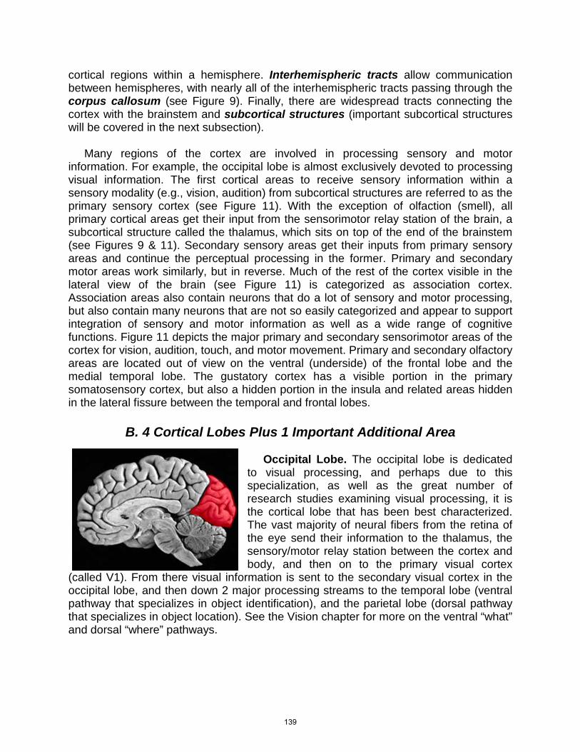
Occipital Lobe. The occipital lobe is dedicated to visual processing, and perhaps due to this specialization, as well as the great number of research studies examining visual processing, it is the cortical lobe that has been best characterized. The vast majority of neural fibers from the retina of the eye send their information to the thalamus, the sensory/motor relay station between the cortex and body, and then on to the primary visual cortex
(called V1). From there visual information is sent to the secondary visual cortex in the occipital lobe, and then down 2 major processing streams to the temporal lobe (ventral pathway that specializes in object identification), and the parietal lobe (dorsal pathway that specializes in object location). See the Vision chapter for more on the ventral “what” and dorsal “where” pathways.
139

Complete destruction of V1 in the occipital lobe of one hemisphere (e.g., right occipital V1) results in a total loss of conscious visual experience from the opposite visual hemifield (e.g., left visual hemifield), resulting in cortical blindness in that visual hemifield. In fact, patients with cortical blindness in part of the visual field can often correctly orient their eyes to the location in their blind spot where an object appears, even though they claim to have no conscious experience of seeing a visual event at that location. This blindsight phenomenon of patients orienting without conscious awareness is thought to be due to a second primitive visual system that takes a small portion of the output from the retina of the eye and sends it to brainstem areas that control the movement of the head and eyes. This is an example of unconscious processing of visual information (see the Vision chapter).
“Blindsight”
Parietal Lobe. The parietal lobe is the final portion of one of the 2 major visual processing pathways from the occipital lobe, the where pathway, which stresses processing of the spatial location of objects. Patients with damage to parietal areas will often have trouble judging the relative spatial location of objects (e.g., which of 2 objects is closer on a table), but have relatively preserved ability to identify the objects. The parietal lobes are also involved in how we move the focus of visual attention around the visual field, and so it should be of no great surprise that patients with parietal lesions often experience deficits in spatial awareness. Gazzaniga, Ivry, and Mangun (2009) report on an interesting case from the literature. Patients with parietal damage to one hemisphere will often neglect visual objects on the opposite side of the visual space (i.e., the opposite visual hemifield), due to the crossing of visual information as it moves from the retina to the occipital lobe. A study of such patients in Milan, Italy, had patients imagine standing at one end of a plaza (the Piazza del Duomo) that they, as long-time residents of Milan, were quite familiar with. The patients described the buildings on the same side as their parietal lesion (e.g., buildings on the right side of the plaza for a person with a right parietal lesion), while failing to mention, or neglecting, those buildings on the opposite side to their parietal lesion. This is simply the expected pattern of neglect, and not that interesting. Amazingly, however, when asked to imagine moving to the opposite end of the plaza and turn around and report the view from the other end of the plaza, these patients would now report the buildings they had neglected earlier, but were now on their non-neglected side due to the imagined move to the other end of the plaza. Moreover, they now neglected the buildings they had reported earlier.
140
This is a striking demonstration of a loss of spatial awareness to both perceived visual events, and in visual memory. The primary somatosensory cortex is located on the most frontal gyrus of the parietal lobe, just posterior to (i.e., in back of) the central sulcus (see Figures 8 & 10).The parietal lobe is important for mathematical and language processing, perhaps via spatial representations used in these domains. The parietal lobe is also involved in spatial localization in touch and audition.
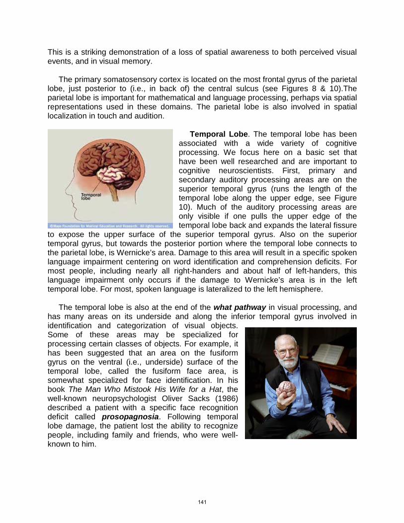
Temporal Lobe. The temporal lobe has been associated with a wide variety of cognitive processing. We focus here on a basic set that have been well researched and are important to cognitive neuroscientists. First, primary and secondary auditory processing areas are on the superior temporal gyrus (runs the length of the temporal lobe along the upper edge, see Figure 10). Much of the auditory processing areas are only visible if one pulls the upper edge of the temporal lobe back and expands the lateral fissure
to expose the upper surface of the superior temporal gyrus. Also on the superior temporal gyrus, but towards the posterior portion where the temporal lobe connects to the parietal lobe, is Wernicke’s area. Damage to this area will result in a specific spoken language impairment centering on word identification and comprehension deficits. For most people, including nearly all right-handers and about half of left-handers, this language impairment only occurs if the damage to Wernicke’s area is in the left temporal lobe. For most, spoken language is lateralized to the left hemisphere. The temporal lobe is also at the end of the what pathway in visual processing, and has many areas on its underside and along the inferior temporal gyrus involved in identification and categorization of visual objects. Some of these areas may be specialized for processing certain classes of objects. For example, it has been suggested that an area on the fusiform gyrus on the ventral (i.e., underside) surface of the temporal lobe, called the fusiform face area, is somewhat specialized for face identification. In his book The Man Who Mistook His Wife for a Hat, the well-known neuropsychologist Oliver Sacks (1986) described a patient with a specific face recognition deficit called prosopagnosia. Following temporal lobe damage, the patient lost the ability to recognize people, including family and friends, who were well-known to him.
141
Finally, the hippocampus is a subcortical structure that runs the length of the inside of the temporal lobe. Bilateral removal of the hippocampus results in a stark memory impairment that involves an inability to learn new facts or store life events in memory. At the end of the hippocampus is a bulbous structure called the amygdala, which is involved in emotional processing. Due to its direct connections to the hippocampus, it is connected to emotional effects in the memory.
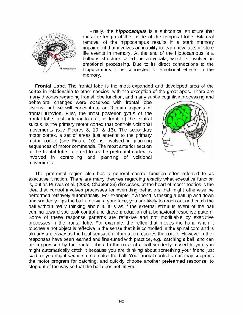
Frontal Lobe. The frontal lobe is the most expanded and developed area of the cortex in relationship to other species, with the exception of the great apes. There are many theories regarding frontal lobe function, and many subtle cognitive processing and behavioral changes were observed with frontal lobe lesions, but we will concentrate on 3 main aspects of frontal function. First, the most posterior gyrus of the frontal lobe, just anterior to (i.e., in front of) the central sulcus, is the primary motor cortex that controls volitional movements (see Figures 8, 10, & 13). The secondary motor cortex, a set of areas just anterior to the primary motor cortex (see Figure 10), is involved in planning sequences of motor commands. The most anterior section of the frontal lobe, referred to as the prefrontal cortex, is involved in controlling and planning of volitional movements. The prefrontal region also has a general control function often referred to as executive function. There are many theories regarding exactly what executive function is, but as Purves et al. (2008, Chapter 23) discusses, at the heart of most theories is the idea that control involves processes for overriding behaviors that might otherwise be performed relatively automatically. For example, if a friend is tossing a ball up and down and suddenly flips the ball up toward your face, you are likely to reach out and catch the ball without really thinking about it. It is as if the external stimulus event of the ball coming toward you took control and drove production of a behavioral response pattern. Some of these response patterns are reflexive and not modifiable by executive processes in the frontal lobe. For example, the reflex that moves the hand when it touches a hot object is reflexive in the sense that it is controlled in the spinal cord and is already underway as the heat sensation information reaches the cortex. However, other responses have been learned and fine-tuned with practice, e.g., catching a ball, and can be suppressed by the frontal lobes. In the case of a ball suddenly tossed to you, you might automatically catch it because you are thinking about something your friend just said, or you might choose to not catch the ball. Your frontal control areas may suppress the motor program for catching, and quickly choose another prelearned response, to step out of the way so that the ball does not hit you.
142

“Spiderman Reflexes”
Another way of thinking about this situation is to realize that the brain is adept at learning and storing behavior patterns (see procedural memory in the Memory chapter), and it can operate in what has been termed default mode, where prelearned behavior patterns are initiated in direct response to external events as opposed to internal goals and plans. Control over such automatic, stimulus-driven, default mode behavior patterns can happen in several ways: suppression of the automatic behavior pattern, switching to initiate an alternate behavioral pattern, and evaluation and simulation of possible consequences of different behavioral options. Patients with prefrontal damage often experience subtle changes in executive function that are varied and difficult to document. However, it seems clear that damage to prefrontal areas can result in (a) increased susceptibility to distraction from environmental events, (b) a decreased ability to suppress default mode behavioral patterns, and (c) may produce repetitions of unsuccessful behaviors in the face of negative feedback. Other patterns of prefrontal damage may lead to a reduced ability to suppress socially inappropriate behavior (e.g., laughing at inappropriate times), or to process the potential consequences of actions. Also of some note is the fact that the olfactory bulb, an area that processes the sense of smell, is located on the ventral surface (underside) of the prefrontal cortex. A major portion of the primary sensory cortex for olfaction is also on the ventral surface of the frontal lobe. Damage to the frontal lobes is often accompanied by damage to the olfactory processing pathway. As a result, a sudden decline in performance on a simple smell test can be diagnostic of frontal lobe damage, and indicate the need for more detailed cognitive testing to detect subtle changes in frontal lobe function.
Insula. The insular (meaning island) cortex is an area hidden in the infold of the lateral fissure (see Figure 8). This area includes the primary gustatory (taste) sensory cortex as well as areas that process the perception of pain. The insula is thought to represent the bodily state. Information from all parts of the body, including organ systems, is integrated into a sense of how you are feeling. As you sit in a
143
chair, you feel the pressure of the chair against your bottom, you may feel a tension in your neck from sitting still for too long, or you may begin to think about your blind date later in the day, and feel your heart rate and breathing quicken. Finally, the insula may be involved in processing aversive emotional feelings such as disgust and injustice. In fact, recent studies indicate the insula will become active when people choose risky options in a decision task or are presented with a patently unfair option that someone else has offered, e.g., receiving an offer to share only a small portion of money that you and another person both found at the same time, while the other person proposes keeping most of it.
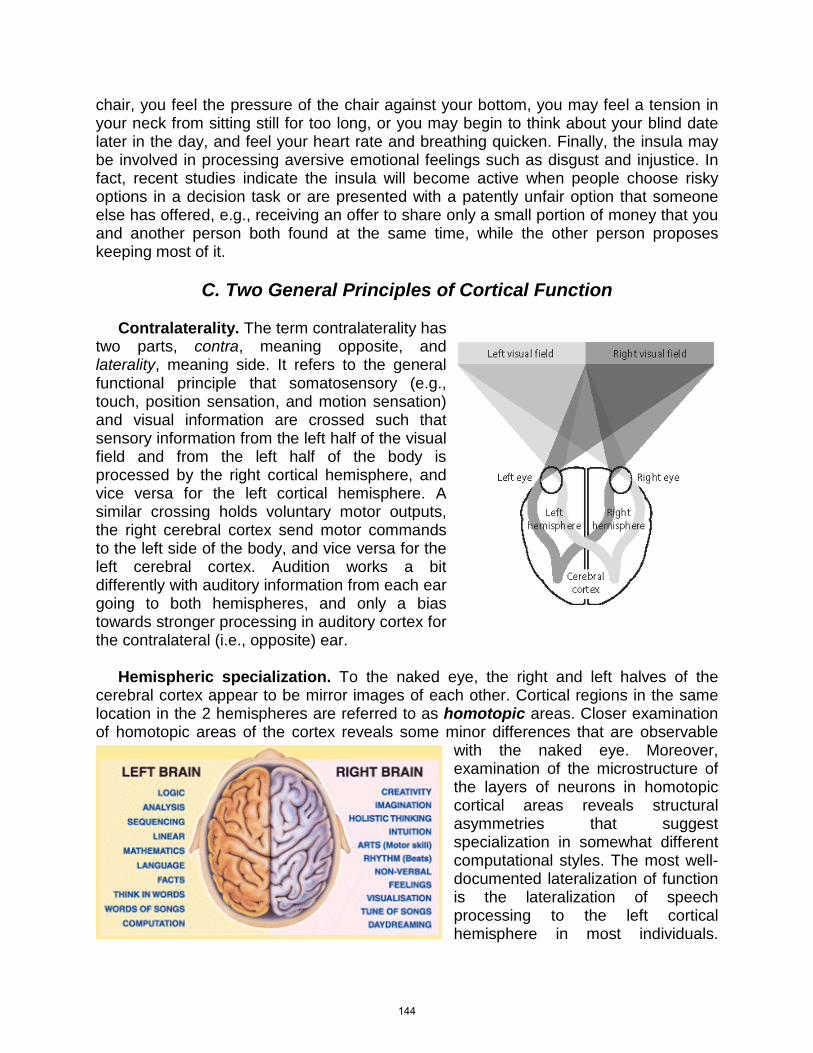
C. Two General Principles of Cortical Function Contralaterality. The term contralaterality has two parts, contra, meaning opposite, and laterality, meaning side. It refers to the general functional principle that somatosensory (e.g., touch, position sensation, and motion sensation) and visual information are crossed such that sensory information from the left half of the visual field and from the left half of the body is processed by the right cortical hemisphere, and vice versa for the left cortical hemisphere. A similar crossing holds voluntary motor outputs, the right cerebral cortex send motor commands to the left side of the body, and vice versa for the left cerebral cortex. Audition works a bit differently with auditory information from each ear going to both hemispheres, and only a bias towards stronger processing in auditory cortex for the contralateral (i.e., opposite) ear. Hemispheric specialization. To the naked eye, the right and left halves of the cerebral cortex appear to be mirror images of each other. Cortical regions in the same location in the 2 hemispheres are referred to as homotopic areas. Closer examination of homotopic areas of the cortex reveals some minor differences that are observable
with the naked eye. Moreover, examination of the microstructure of the layers of neurons in homotopic cortical areas reveals structural asymmetries that suggest specialization in somewhat different computational styles. The most well-documented lateralization of function is the lateralization of speech processing to the left cortical hemisphere in most individuals.
144
Nearly all right-handers (about 96%) have speech represented strongly in the left hemisphere, as well as 60% of left-handers, and over 95% of all humans have language represented strongly in the left hemisphere (Gazzaniga, Ivry, Mangun, 2009). There is also evidence that the processing of music, and of visual and spatial information is more pronounced in the right hemisphere for most people. This does not mean the right hemisphere is not involved in language processing, or the left in visuospatial processing. One must be careful about over-interpreting hemispheric differences in processing to mean that both sides do not work together in a given processing domain such as language, music, or vision. What can be said is that homotopic areas of the cortex appear to have somewhat diverged during evolutionary history to yield specializations in different computational styles for domains such as language or music. Just how the hemispheres work together to support cognitive function is an active area of inquiry.
“The Right Brain vs Left Brain test – Optical Illusion”
D. Cortical Processing
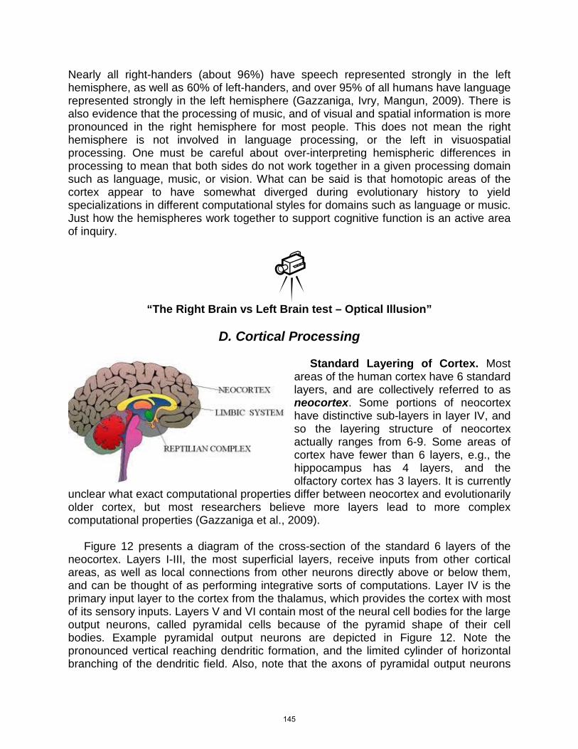
Standard Layering of Cortex. Most areas of the human cortex have 6 standard layers, and are collectively referred to as neocortex. Some portions of neocortex have distinctive sub-layers in layer IV, and so the layering structure of neocortex actually ranges from 6-9. Some areas of cortex have fewer than 6 layers, e.g., the hippocampus has 4 layers, and the olfactory cortex has 3 layers. It is currently
unclear what exact computational properties differ between neocortex and evolutionarily older cortex, but most researchers believe more layers lead to more complex computational properties (Gazzaniga et al., 2009). Figure 12 presents a diagram of the cross-section of the standard 6 layers of the neocortex. Layers I-III, the most superficial layers, receive inputs from other cortical areas, as well as local connections from other neurons directly above or below them, and can be thought of as performing integrative sorts of computations. Layer IV is the primary input layer to the cortex from the thalamus, which provides the cortex with most of its sensory inputs. Layers V and VI contain most of the neural cell bodies for the large output neurons, called pyramidal cells because of the pyramid shape of their cell bodies. Example pyramidal output neurons are depicted in Figure 12. Note the pronounced vertical reaching dendritic formation, and the limited cylinder of horizontal branching of the dendritic field. Also, note that the axons of pyramidal output neurons
145
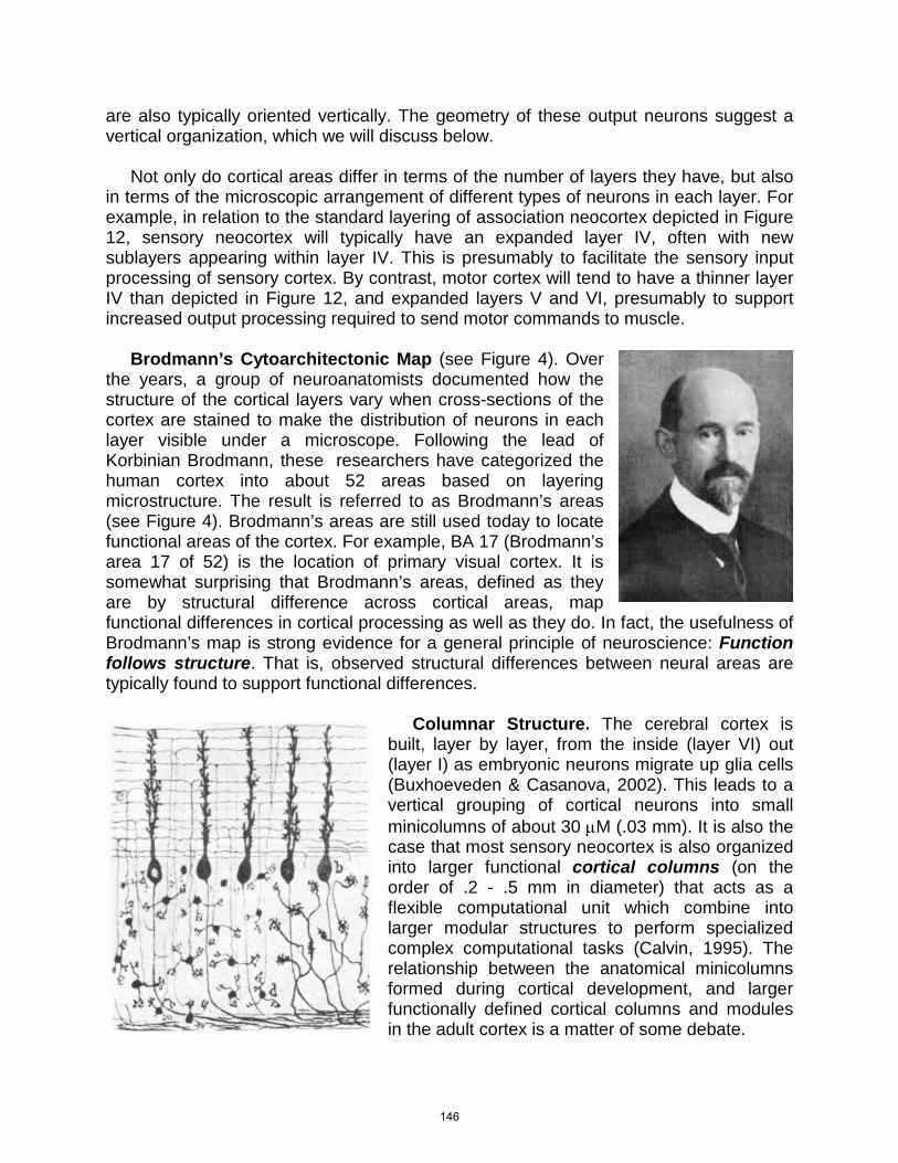
are also typically oriented vertically. The geometry of these output neurons suggest a vertical organization, which we will discuss below. Not only do cortical areas differ in terms of the number of layers they have, but also in terms of the microscopic arrangement of different types of neurons in each layer. For example, in relation to the standard layering of association neocortex depicted in Figure 12, sensory neocortex will typically have an expanded layer IV, often with new sublayers appearing within layer IV. This is presumably to facilitate the sensory input processing of sensory cortex. By contrast, motor cortex will tend to have a thinner layer IV than depicted in Figure 12, and expanded layers V and VI, presumably to support increased output processing required to send motor commands to muscle. Brodmann’s Cytoarchitectonic Map (see Figure 4). Over the years, a group of neuroanatomists documented how the structure of the cortical layers vary when cross-sections of the cortex are stained to make the distribution of neurons in each layer visible under a microscope. Following the lead of Korbinian Brodmann, these researchers have categorized the human cortex into about 52 areas based on layering microstructure. The result is referred to as Brodmann’s areas (see Figure 4). Brodmann’s areas are still used today to locate functional areas of the cortex. For example, BA 17 (Brodmann’s area 17 of 52) is the location of primary visual cortex. It is somewhat surprising that Brodmann’s areas, defined as they are by structural difference across cortical areas, map functional differences in cortical processing as well as they do. In fact, the usefulness of Brodmann’s map is strong evidence for a general principle of neuroscience: Function follows structure. That is, observed structural differences between neural areas are typically found to support functional differences.
Columnar Structure. The cerebral cortex is built, layer by layer, from the inside (layer VI) out (layer I) as embryonic neurons migrate up glia cells (Buxhoeveden & Casanova, 2002). This leads to a vertical grouping of cortical neurons into small minicolumns of about 30 µM (.03 mm). It is also the case that most sensory neocortex is also organized into larger functional cortical columns (on the order of .2 - .5 mm in diameter) that acts as a flexible computational unit which combine into larger modular structures to perform specialized complex computational tasks (Calvin, 1995). The relationship between the anatomical minicolumns formed during cortical development, and larger functionally defined cortical columns and modules in the adult cortex is a matter of some debate.
146

We can better understand the idea of a cortical column by considering a common finding when a microelectrode is inserted vertically into a column of cortical tissue and recording from sensory neurons from deeper neurons along the vertical insertion path. What is often found is that the sensory neurons all have the same receptive field, that is, visual neurons within a column all tend to be responsive to visual events in the same restricted region in the visual field and somatosensory neurons within a column all tend to be responsive to touch events in the same restricted location on the body. Not only do sensory neurons within a column tend to have the same receptive field, but they also tend to share many functional properties. This sharing of function for neurons within columns of cortex has led to the proposal of functionally defined cortical columns as a model for cortical computation. Blue Brain Project: Simulating Cortical Columns. The Blue Brain Project (Markram, 2006) is an ambitious research effort led by Henry Markram at École Polytechnique Fédérale de Lausanne in Switzerland. The goal of the project is to collect enough data on the microarchitecture of the neural connections in cortical columns (~.5 mm diameter and ~1.5 mm thick) in the rodent cortex to produce a large-scale biologically realistic simulation of the cortex. The very large numbers of neurons and neural connections in even a very small portion of the cortex make this a difficult computational task necessitating use of a powerful supercomputer, IBM’s Blue Gene supercomputer. To learn more, go to the Blue Brain Project website or watch some of the videos online about this ambitious and exciting project or reverse engineer the brain. Blue Brain Project videos:
“Henry Markram: Supercomputing the Brain’s Secrets”
“Bluebrain / Year One”
“Realtime Electrocorticographic Brain Mapping”
Cortical Maps. Sensory cortex typically organizes into larger-scale structures called maps (on the order of multiple cm2 of cortex). By far the most well-studied are the cortical maps of the sensory cortex, called topographic maps, because the arrangement of receptive fields of neurons in the sensory cortex is like a map of the
147
sensory world. For example, the primary visual cortex, V1, holds a map of the visual field. Actually, each V1 in each hemisphere of the brain contains a map of the opposite half of the visual field (i.e., right V1 contains a mapping of the left visual hemifield). Figure 13 presents a diagram of the mapping of neurons in primary motor cortex (on the pre-central gyrus, the most posterior gyrus of the frontal lobe). Note that neurons that initiate firing of muscle fibers for a body part will be right next to each other. A distorted map is formed where the representation of the size of each body part has to do with the number of neurons devoted to initiation of fine-tuned movements of that body part. Minicolumns, Functional Columns and Modules, and Maps. One simple idea that was proposed early in the progression of knowledge about the cortex was to assume that the small anatomical minicolumns formed during cortical development tend to morph into the larger functional cortical columns and modules observed in the adult cortex, and that these are in turn combined to form maps. This story is seductive in its simplicity, but is not yet supported by strong evidence. Much work remains to be done to connect these disparate spatial levels of scale into a coherent story of cortical computation. Ambitious studies like the Blue Brain project discussed above hold the promise of developing a new set of techniques for understanding how information is integrated as patterns of electrical activity within a network of neurons, and in doing so, may provide the evidence needed for an integrated story about cortical computation.
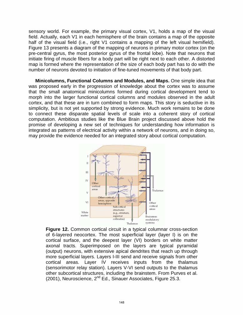
Figure 12. Common cortical circuit in a typical columnar cross-section of 6-layered neocortex. The most superficial layer (layer I) is on the cortical surface, and the deepest layer (VI) borders on white matter axonal tracts. Superimposed on the layers are typical pyramidal (output) neurons, with extensive apical dendrites that reach up through more superficial layers. Layers I-III send and receive signals from other cortical areas. Layer IV receives inputs from the thalamus (sensorimotor relay station). Layers V-VI send outputs to the thalamus other subcortical structures, including the brainstem. From Purves et al. (2001), Neuroscience, 2nd Ed., Sinauer Associates, Figure 25.3.
148
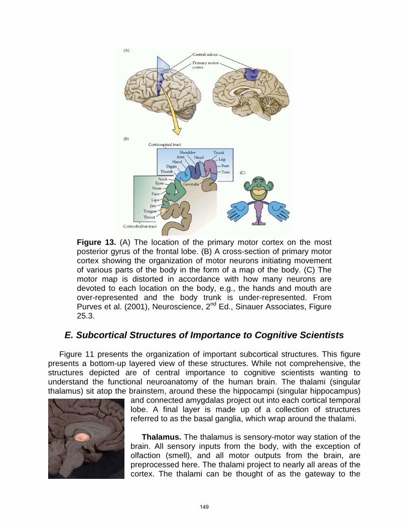
Figure 13. (A) The location of the primary motor cortex on the most posterior gyrus of the frontal lobe. (B) A cross-section of primary motor cortex showing the organization of motor neurons initiating movement of various parts of the body in the form of a map of the body. (C) The motor map is distorted in accordance with how many neurons are devoted to each location on the body, e.g., the hands and mouth are over-represented and the body trunk is under-represented. From Purves et al. (2001), Neuroscience, 2nd Ed., Sinauer Associates, Figure 25.3.
E. Subcortical Structures of Importance to Cognitive Scientists
Figure 11 presents the organization of important subcortical structures. This figure presents a bottom-up layered view of these structures. While not comprehensive, the structures depicted are of central importance to cognitive scientists wanting to understand the functional neuroanatomy of the human brain. The thalami (singular thalamus) sit atop the brainstem, around these the hippocampi (singular hippocampus)
and connected amygdalas project out into each cortical temporal lobe. A final layer is made up of a collection of structures referred to as the basal ganglia, which wrap around the thalami. Thalamus. The thalamus is sensory-motor way station of the brain. All sensory inputs from the body, with the exception of olfaction (smell), and all motor outputs from the brain, are preprocessed here. The thalami project to nearly all areas of the cortex. The thalami can be thought of as the gateway to the
149
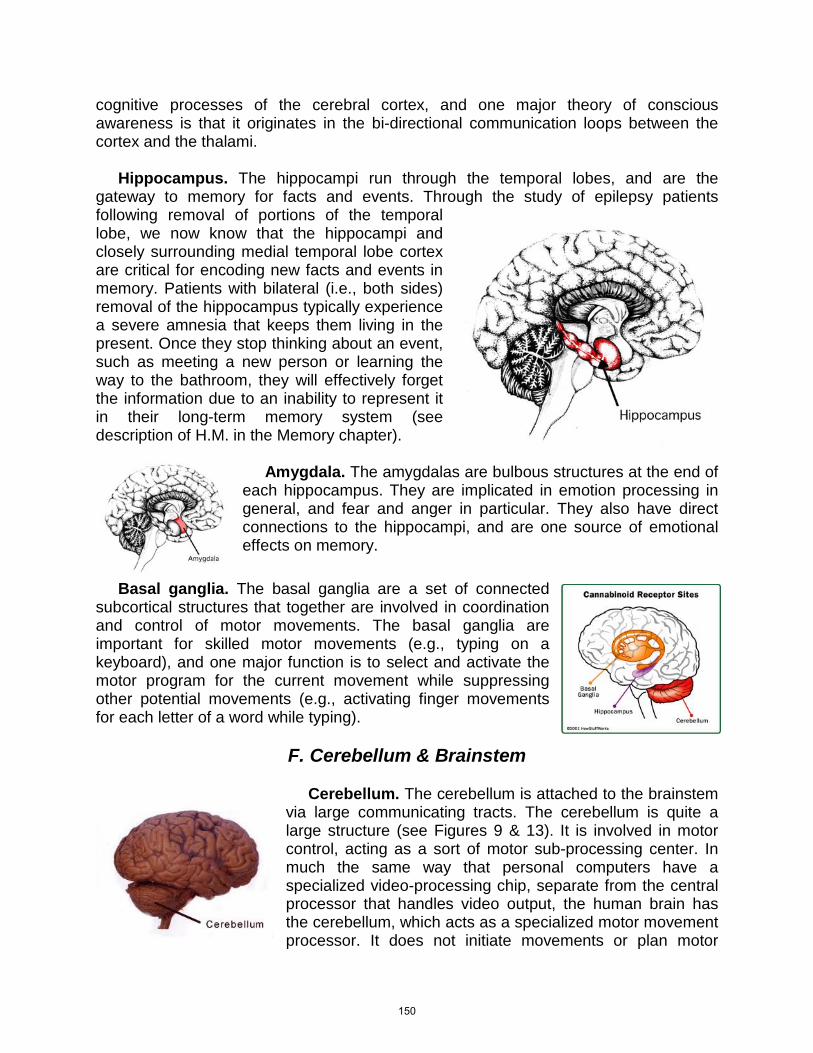
cognitive processes of the cerebral cortex, and one major theory of conscious awareness is that it originates in the bi-directional communication loops between the cortex and the thalami. Hippocampus. The hippocampi run through the temporal lobes, and are the gateway to memory for facts and events. Through the study of epilepsy patients following removal of portions of the temporal lobe, we now know that the hippocampi and closely surrounding medial temporal lobe cortex are critical for encoding new facts and events in memory. Patients with bilateral (i.e., both sides) removal of the hippocampus typically experience a severe amnesia that keeps them living in the present. Once they stop thinking about an event, such as meeting a new person or learning the way to the bathroom, they will effectively forget the information due to an inability to represent it in their long-term memory system (see description of H.M. in the Memory chapter).
Amygdala. The amygdalas are bulbous structures at the end of each hippocampus. They are implicated in emotion processing in general, and fear and anger in particular. They also have direct connections to the hippocampi, and are one source of emotional effects on memory.
Basal ganglia. The basal ganglia are a set of connected subcortical structures that together are involved in coordination and control of motor movements. The basal ganglia are important for skilled motor movements (e.g., typing on a keyboard), and one major function is to select and activate the motor program for the current movement while suppressing other potential movements (e.g., activating finger movements for each letter of a word while typing).
F. Cerebellum & Brainstem
Cerebellum. The cerebellum is attached to the brainstem via large communicating tracts. The cerebellum is quite a large structure (see Figures 9 & 13). It is involved in motor control, acting as a sort of motor sub-processing center. In much the same way that personal computers have a specialized video-processing chip, separate from the central processor that handles video output, the human brain has the cerebellum, which acts as a specialized motor movement processor. It does not initiate movements or plan motor
150
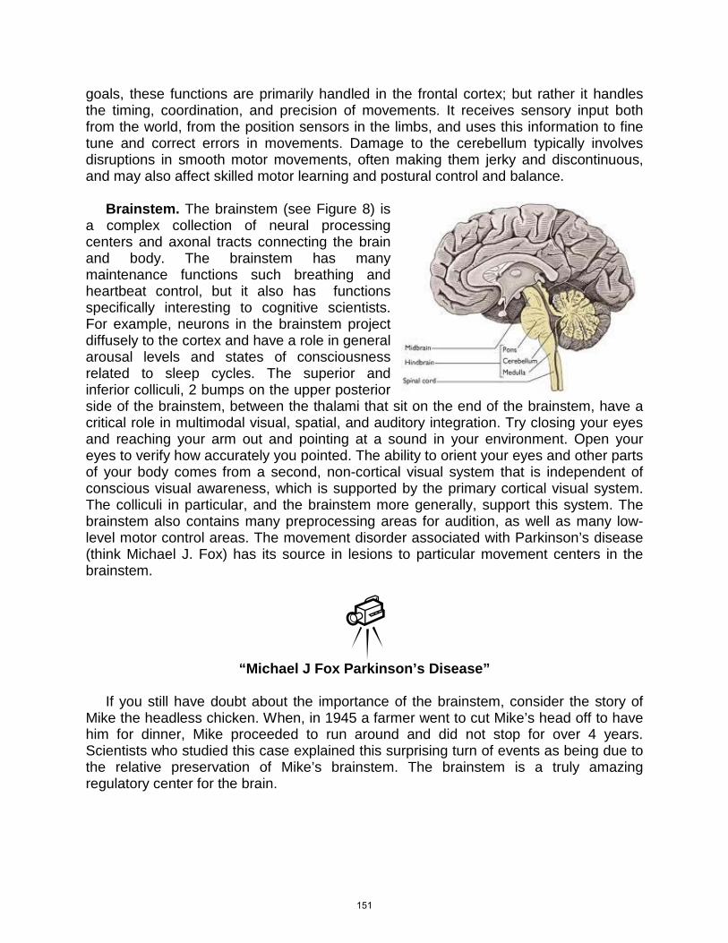
goals, these functions are primarily handled in the frontal cortex; but rather it handles the timing, coordination, and precision of movements. It receives sensory input both from the world, from the position sensors in the limbs, and uses this information to fine tune and correct errors in movements. Damage to the cerebellum typically involves disruptions in smooth motor movements, often making them jerky and discontinuous, and may also affect skilled motor learning and postural control and balance. Brainstem. The brainstem (see Figure 8) is a complex collection of neural processing centers and axonal tracts connecting the brain and body. The brainstem has many maintenance functions such breathing and heartbeat control, but it also has functions specifically interesting to cognitive scientists. For example, neurons in the brainstem project diffusely to the cortex and have a role in general arousal levels and states of consciousness related to sleep cycles. The superior and inferior colliculi, 2 bumps on the upper posterior side of the brainstem, between the thalami that sit on the end of the brainstem, have a critical role in multimodal visual, spatial, and auditory integration. Try closing your eyes and reaching your arm out and pointing at a sound in your environment. Open your eyes to verify how accurately you pointed. The ability to orient your eyes and other parts of your body comes from a second, non-cortical visual system that is independent of conscious visual awareness, which is supported by the primary cortical visual system. The colliculi in particular, and the brainstem more generally, support this system. The brainstem also contains many preprocessing areas for audition, as well as many low-level motor control areas. The movement disorder associated with Parkinson’s disease (think Michael J. Fox) has its source in lesions to particular movement centers in the brainstem.
“Michael J Fox Parkinson’s Disease”
If you still have doubt about the importance of the brainstem, consider the story of Mike the headless chicken. When, in 1945 a farmer went to cut Mike’s head off to have him for dinner, Mike proceeded to run around and did not stop for over 4 years. Scientists who studied this case explained this surprising turn of events as being due to the relative preservation of Mike’s brainstem. The brainstem is a truly amazing regulatory center for the brain.
151
“Headless Chicken Lives”
V. Methods of Cognitive Neuroscience
There are a large number of technologies and procedures that allow for the study of brain structure and function. While this vast array of methods has contributed greatly to neuroscience, cognitive neuroscience uses methodologies from 3 basic approaches. One approach attempts to record the electrical activity of individual neurons or collections of neurons during performance of a cognitive task. Another approach is to record the metabolic activity in small patches of neural tissue during performance of a cognitive task. This approach is often used in conjunction with techniques for creating a 3D image of the structures of the brain, with the result being a 3D image of a pattern of functional brain activation superimposed on a 3D image of brain structures. A third approach attempts to ascertain differences in cognitive function following lesions or damage to neural tissue. There are many specific methodologies used for each of these approaches, we will focus on the examples that are most frequently used in cognitive neuroscience research. Each of the 3 basic approaches to studying cognition in the brain has strengths and weaknesses. However, it is important to note that scientists often view results that are confirmed by multiple methodologies as being more certain. This is called the principle of converging operations, an idea that is quite powerful in science.
A. Studying Electrophysiological Processes Direct electrical recording. Because signals within a neuron are electrical in nature, measure of the electrical PSPs in the dendrites and APs in the axon of a neuron are a direct window to neural information processing. The direct electrical recording approach involves bringing an electrode into contact with the surface of the brain, or inserting a small electrode into the brain (remember, the brain itself does not have pain or touch receptors) and then recording the electrical activity of a single, or a group, of neurons. For the most part, this type of recording is done in animals. While it is relatively rare for researchers to have the opportunity to record from electrodes inserted into human brains, there are limited opportunities for direct recording of awake patients undergoing brain surgery. Reese et al. (2002) reported on a study where single cell recordings were obtained from several neurons on the ventromedial surface (ventral refers to the underside, and medial refers to the midline where the temporal lobe is next to the brainstem) of the temporal lobe of a patient (See Figure 14). Because the brain has no pain receptors, direct recordings can be made while the patient is awake. Panels A and B of Figure 14 depict the electrode site of the particular neuron that generated the spike (neural action potentials) recordings of
152
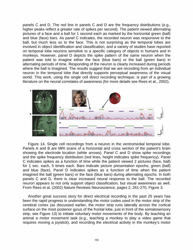
panels C and D. The red line in panels C and D are the frequency distributions (e.g., higher peaks reflect a greater rate of spikes per second). The patient viewed alternating pictures of a face and a ball for 1 second each as marked by the horizontal green (ball) and blue (face) bars. As panel C indicates, the recorded neuron was responsive to the ball, but much less so to the face. This is not surprising as the temporal lobes are involved in object identification and classification, and a variety of studies have reported on temporal lobe neurons sensitive to a specific category of objects in humans and in monkeys. However, panel D depicts the spike pattern of the same neuron when the patient was told to imagine either the face (blue bars) or the ball (green bars) in alternating periods of time. Responding of the neuron is clearly increased during periods where the ball is imagined. The results suggest that we are recording from an individual neuron in the temporal lobe that directly supports perceptual awareness of the visual world. This work, using the single cell direct recording technique, is part of a growing literature on the neural correlates of awareness (for more details see Rees et al., 2002).
Figure 14. Single cell recordings from a neuron in the ventromedial temporal lobe. Panels A and B are MRI scans of a horizontal and cross section of the patient’s brain showing the electrode location (white arrows). Panel C and D show spike recordings and the spike frequency distribution (red lines, height indicates spike frequency). Panel C indicates spikes as a function of time while the patient viewed 2 pictures (face, ball) for 1 sec. each, 5 times each. Bars indicate picture presentation epochs, green (ball) and blue (face). Panel D indicates spikes as a function of time when the patient imagined the ball (green bars) or the face (blue bars) during alternating epochs. In both panels C and D, there is clear increased neural response to the ball. The recorded neuron appears to not only support object classification, but visual awareness as well. From Rees et al. (2002) Nature Reviews Neuroscience, pages 2, 261-270, Figure 3. Another great success story for direct electrical recording in the past 25 years has been the rapid progress in understanding the motor codes used in the motor strip of the cerebral cortex (as discussed earlier, the motor strip runs laterally across the cortical surface on the most posterior gyrus of the frontal lobe, just in front of the somatosensory strip, see Figure 13) to initiate voluntary motor movements of the body. By teaching an animal a motor movement task (e.g., teaching a monkey to play a video game that requires moving a joystick), and recording the electrical activity in the monkey’s motor
153
strip, researchers have been able to “decode” some of the commands used by the motor strip. From such studies, it became clear that knowing how the brain controls motor movements of the body could form the basis of developing technologies that would allow the mind to control the movement of artificial limbs, or develop what has come to be called a brain-machine interface (BMI, also referred to as neural interface systems, NIS). In a now classic study, researchers at Duke University (Carmena et al., 2003) were able to show strong proof of concept. In this study a monkey was trained to play a videogame by manipulation of a joystick, while electrodes implanted in the monkey’s motor cortex directly recorded motor signals associated with movements of the monkey’s hand and arm as they were controlling the joystick (see Figure 16). Eventually, the computer that processed the cortical motor commands took over controlling the video game, that is, the monkey was playing the game through thought alone, the joystick was disconnected and non-functional. In recent years, much progress has been made. Researchers have developed small chips with arrays of recording electrodes for implantation in the human brain. Additionally, there are several tests currently underway of the use of this type of BMI chip in humans to overcome paralysis by allowing them to control a computer, robotic arms, or robotic legs directly with their minds (see Figure 17). Many interesting videos showing the operation of BMIs in monkeys and humans have been posted online. Brain Machine Interface Videos Online:
“Robo-Monkey Uses Brain Power to Feed Itself”
Brain Gate is a company developing a chip that can be implanted in humans who are paralyzed due to injury. This video shows a patient playing video games using his mind to imagine moving the game controller, and the computer decodes the motor program signals of the cortex and executes the movements for him.
“BrainGate Lets Your Brain Control the Computer”
Some researchers are attempting to use EEG electrode caps to record motor program signals from motor cortex noninvasively (i.e., without implanting a chip in the brain). Here is a video of a wheel chair that is controlled by thought alone using an EEG cap.
154
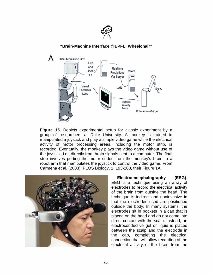
“Brain-Machine Interface @EPFL: Wheelchair”
Figure 15. Depicts experimental setup for classic experiment by a group of researchers at Duke University. A monkey is trained to manipulated a joystick and play a simple video game while the electrical activity of motor processing areas, including the motor strip, is recorded. Eventually, the monkey plays the video game without use of the joystick, i.e., directly from brain signals sent to a computer. The final step involves porting the motor codes from the monkey’s brain to a robot arm that manipulates the joystick to control the video game. From Carmena et al. (2003), PLOS Biology, 1, 193-208, their Figure 1A.
Electroencephalography (EEG). EEG is a technique using an array of electrodes to record the electrical activity of the brain from outside the head. The technique is indirect and noninvasive in that the electrodes used are positioned outside the body. In many systems, the electrodes sit in pockets in a cap that is placed on the head and do not come into direct contact with the scalp. Instead, an electroconductive gel or liquid is placed between the scalp and the electrode in the cap, completing the electrical connection that will allow recording of the electrical activity of the brain from the
155
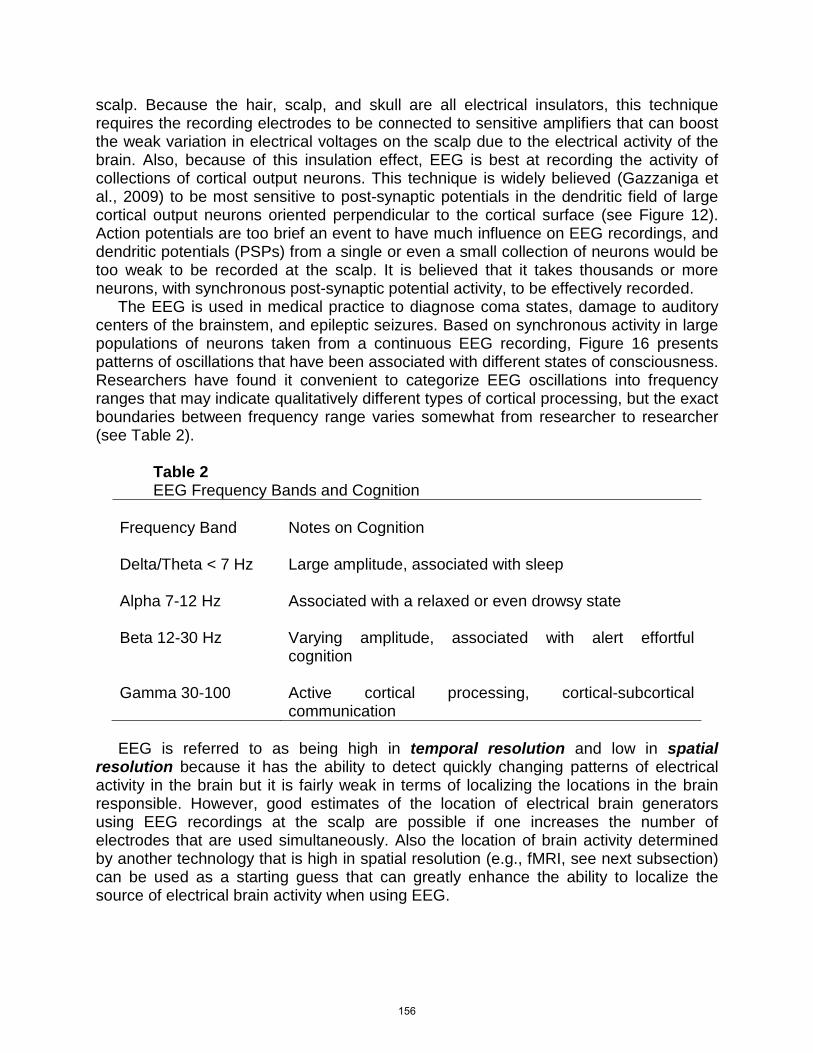
scalp. Because the hair, scalp, and skull are all electrical insulators, this technique requires the recording electrodes to be connected to sensitive amplifiers that can boost the weak variation in electrical voltages on the scalp due to the electrical activity of the brain. Also, because of this insulation effect, EEG is best at recording the activity of collections of cortical output neurons. This technique is widely believed (Gazzaniga et al., 2009) to be most sensitive to post-synaptic potentials in the dendritic field of large cortical output neurons oriented perpendicular to the cortical surface (see Figure 12). Action potentials are too brief an event to have much influence on EEG recordings, and dendritic potentials (PSPs) from a single or even a small collection of neurons would be too weak to be recorded at the scalp. It is believed that it takes thousands or more neurons, with synchronous post-synaptic potential activity, to be effectively recorded. The EEG is used in medical practice to diagnose coma states, damage to auditory centers of the brainstem, and epileptic seizures. Based on synchronous activity in large populations of neurons taken from a continuous EEG recording, Figure 16 presents patterns of oscillations that have been associated with different states of consciousness. Researchers have found it convenient to categorize EEG oscillations into frequency ranges that may indicate qualitatively different types of cortical processing, but the exact boundaries between frequency range varies somewhat from researcher to researcher (see Table 2).
Table 2 EEG Frequency Bands and Cognition
Frequency Band
Notes on Cognition
Delta/Theta < 7 Hz
Large amplitude, associated with sleep
Alpha 7-12 Hz
Associated with a relaxed or even drowsy state
Beta 12-30 Hz
Varying amplitude, associated with alert effortful cognition
Gamma 30-100
Active cortical processing, cortical-subcortical communication
EEG is referred to as being high in temporal resolution and low in spatial resolution because it has the ability to detect quickly changing patterns of electrical activity in the brain but it is fairly weak in terms of localizing the locations in the brain responsible. However, good estimates of the location of electrical brain generators using EEG recordings at the scalp are possible if one increases the number of electrodes that are used simultaneously. Also the location of brain activity determined by another technology that is high in spatial resolution (e.g., fMRI, see next subsection) can be used as a starting guess that can greatly enhance the ability to localize the source of electrical brain activity when using EEG.
156
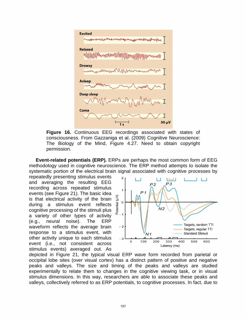
Figure 16. Continuous EEG recordings associated with states of consciousness. From Gazzaniga et al. (2009) Cognitive Neuroscience: The Biology of the Mind, Figure 4.27. Need to obtain copyright permission.
Event-related potentials (ERP). ERPs are perhaps the most common form of EEG methodology used in cognitive neuroscience. The ERP method attempts to isolate the systematic portion of the electrical brain signal associated with cognitive processes by repeatedly presenting stimulus events and averaging the resulting EEG recording across repeated stimulus events (see Figure 21). The basic idea is that electrical activity of the brain during a stimulus event reflects cognitive processing of the stimuli plus a variety of other types of activity (e.g., neural noise). The ERP waveform reflects the average brain response to a stimulus event, with other activity unique to each stimulus event (i.e., not consistent across stimulus events) averaged out. As depicted in Figure 21, the typical visual ERP wave form recorded from parietal or occipital lobe sites (over visual cortex) has a distinct pattern of positive and negative peaks and valleys. The size and timing of the peaks and valleys are studied experimentally to relate them to changes in the cognitive viewing task, or in visual stimulus dimensions. In this way, researchers are able to associate these peaks and valleys, collectively referred to as ERP potentials, to cognitive processes. In fact, due to
157
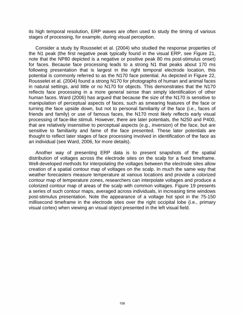
its high temporal resolution, ERP waves are often used to study the timing of various stages of processing, for example, during visual perception. Consider a study by Rousselet et al. (2004) who studied the response properties of the N1 peak (the first negative peak typically found in the visual ERP, see Figure 21, note that the NP80 depicted is a negative or positive peak 80 ms post-stimulus onset) for faces. Because face processing leads to a strong N1 that peaks about 170 ms following presentation that is largest in the right temporal electrode location, this potential is commonly referred to as the N170 face potential. As depicted in Figure 22, Rousselet et al. (2004) found a strong N170 for photographs of human and animal faces in natural settings, and little or no N170 for objects. This demonstrates that the N170 reflects face processing in a more general sense than simply identification of other human faces. Ward (2006) has argued that because the size of the N170 is sensitive to manipulation of perceptual aspects of faces, such as smearing features of the face or turning the face upside down, but not to personal familiarity of the face (i.e., faces of friends and family) or use of famous faces, the N170 most likely reflects early visual processing of face-like stimuli. However, there are later potentials, the N250 and P400, that are relatively insensitive to perceptual aspects (e.g., inversion) of the face, but are sensitive to familiarity and fame of the face presented. These later potentials are thought to reflect later stages of face processing involved in identification of the face as an individual (see Ward, 2006, for more details). Another way of presenting ERP data is to present snapshots of the spatial distribution of voltages across the electrode sites on the scalp for a fixed timeframe. Well-developed methods for interpolating the voltages between the electrode sites allow creation of a spatial contour map of voltages on the scalp. In much the same way that weather forecasters measure temperature at various locations and provide a colorized contour map of temperature zones, researchers can interpolate voltages and produce a colorized contour map of areas of the scalp with common voltages. Figure 19 presents a series of such contour maps, averaged across individuals, in increasing time windows post-stimulus presentation. Note the appearance of a voltage hot spot in the 75-150 millisecond timeframe in the electrode sites over the right occipital lobe (i.e., primary visual cortex) when viewing an visual object presented in the left visual field.
158
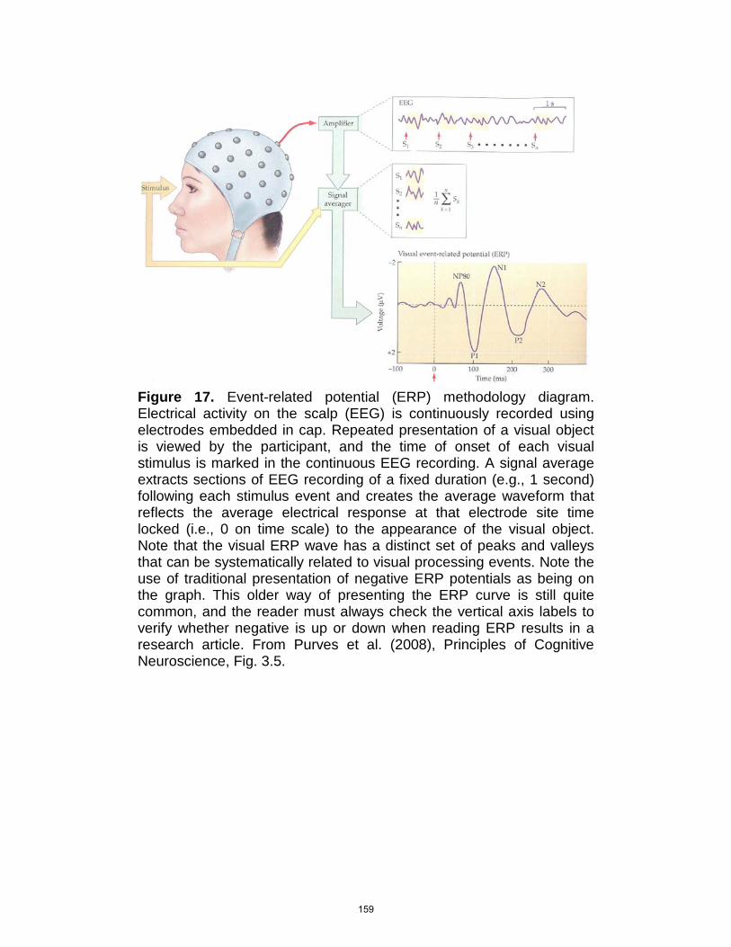
Figure 17. Event-related potential (ERP) methodology diagram. Electrical activity on the scalp (EEG) is continuously recorded using electrodes embedded in cap. Repeated presentation of a visual object is viewed by the participant, and the time of onset of each visual stimulus is marked in the continuous EEG recording. A signal average extracts sections of EEG recording of a fixed duration (e.g., 1 second) following each stimulus event and creates the average waveform that reflects the average electrical response at that electrode site time locked (i.e., 0 on time scale) to the appearance of the visual object. Note that the visual ERP wave has a distinct set of peaks and valleys that can be systematically related to visual processing events. Note the use of traditional presentation of negative ERP potentials as being on the graph. This older way of presenting the ERP curve is still quite common, and the reader must always check the vertical axis labels to verify whether negative is up or down when reading ERP results in a research article. From Purves et al. (2008), Principles of Cognitive Neuroscience, Fig. 3.5.
159
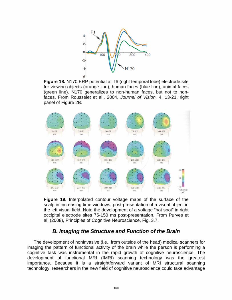
Figure 18. N170 ERP potential at T6 (right temporal lobe) electrode site for viewing objects (orange line), human faces (blue line), animal faces (green line). N170 generalizes to non-human faces, but not to non-faces. From Rousselet et al., 2004, Journal of Vision. 4, 13-21, right panel of Figure 2B.
Figure 19. Interpolated contour voltage maps of the surface of the scalp in increasing time windows, post-presentation of a visual object in the left visual field. Note the development of a voltage “hot spot” in right occipital electrode sites 75-150 ms post-presentation. From Purves et al. (2008), Principles of Cognitive Neuroscience, Fig. 3.7.
B. Imaging the Structure and Function of the Brain
The development of noninvasive (i.e., from outside of the head) medical scanners for imaging the pattern of functional activity of the brain while the person is performing a cognitive task was instrumental in the rapid growth of cognitive neuroscience. The development of functional MRI (fMRI) scanning technology was the greatest importance. Because it is a straightforward variant of MRI structural scanning technology, researchers in the new field of cognitive neuroscience could take advantage
160
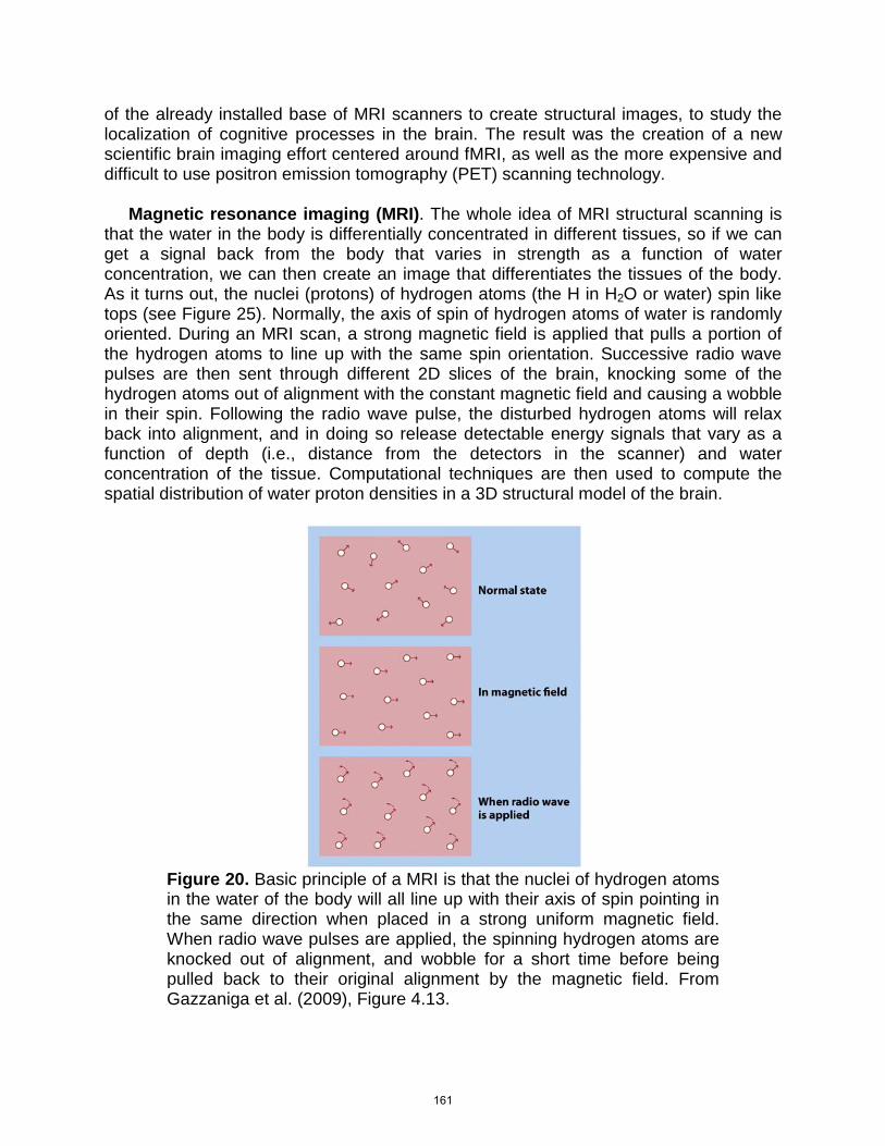
of the already installed base of MRI scanners to create structural images, to study the localization of cognitive processes in the brain. The result was the creation of a new scientific brain imaging effort centered around fMRI, as well as the more expensive and difficult to use positron emission tomography (PET) scanning technology. Magnetic resonance imaging (MRI). The whole idea of MRI structural scanning is that the water in the body is differentially concentrated in different tissues, so if we can get a signal back from the body that varies in strength as a function of water concentration, we can then create an image that differentiates the tissues of the body. As it turns out, the nuclei (protons) of hydrogen atoms (the H in H2O or water) spin like tops (see Figure 25). Normally, the axis of spin of hydrogen atoms of water is randomly oriented. During an MRI scan, a strong magnetic field is applied that pulls a portion of the hydrogen atoms to line up with the same spin orientation. Successive radio wave pulses are then sent through different 2D slices of the brain, knocking some of the hydrogen atoms out of alignment with the constant magnetic field and causing a wobble in their spin. Following the radio wave pulse, the disturbed hydrogen atoms will relax back into alignment, and in doing so release detectable energy signals that vary as a function of depth (i.e., distance from the detectors in the scanner) and water concentration of the tissue. Computational techniques are then used to compute the spatial distribution of water proton densities in a 3D structural model of the brain.
Figure 20. Basic principle of a MRI is that the nuclei of hydrogen atoms in the water of the body will all line up with their axis of spin pointing in the same direction when placed in a strong uniform magnetic field. When radio wave pulses are applied, the spinning hydrogen atoms are knocked out of alignment, and wobble for a short time before being pulled back to their original alignment by the magnetic field. From Gazzaniga et al. (2009), Figure 4.13.
161
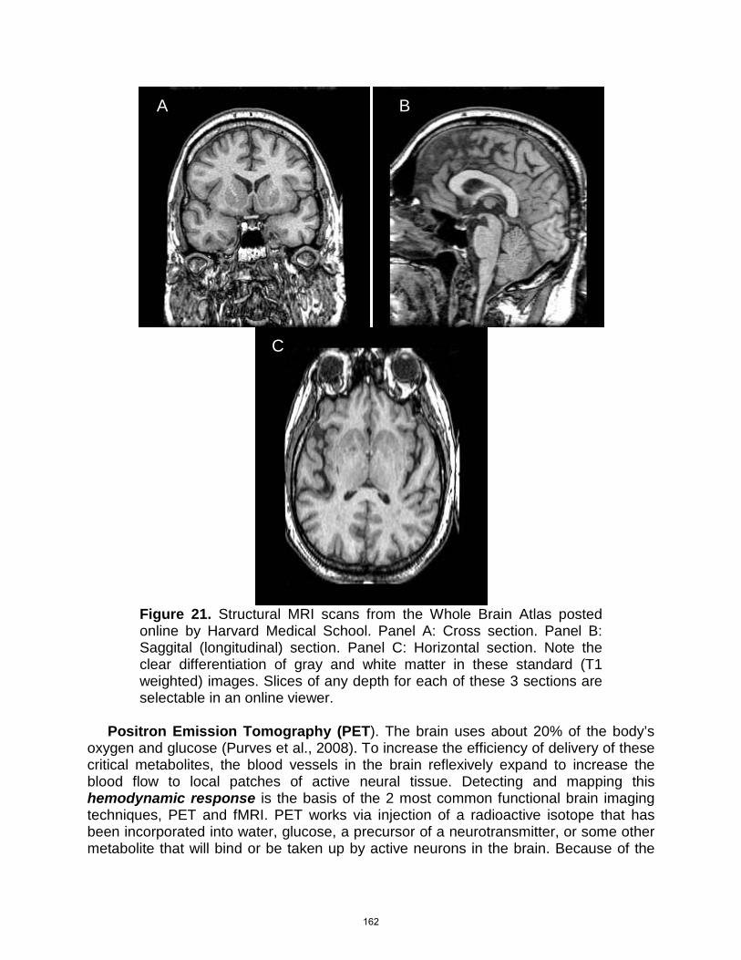
Figure 21. Structural MRI scans from the Whole Brain Atlas posted online by Harvard Medical School. Panel A: Cross section. Panel B: Saggital (longitudinal) section. Panel C: Horizontal section. Note the clear differentiation of gray and white matter in these standard (T1 weighted) images. Slices of any depth for each of these 3 sections are selectable in an online viewer.
Positron Emission Tomography (PET). The brain uses about 20% of the body’s oxygen and glucose (Purves et al., 2008). To increase the efficiency of delivery of these critical metabolites, the blood vessels in the brain reflexively expand to increase the blood flow to local patches of active neural tissue. Detecting and mapping this hemodynamic response is the basis of the 2 most common functional brain imaging techniques, PET and fMRI. PET works via injection of a radioactive isotope that has been incorporated into water, glucose, a precursor of a neurotransmitter, or some other metabolite that will bind or be taken up by active neurons in the brain. Because of the
A B
C
162
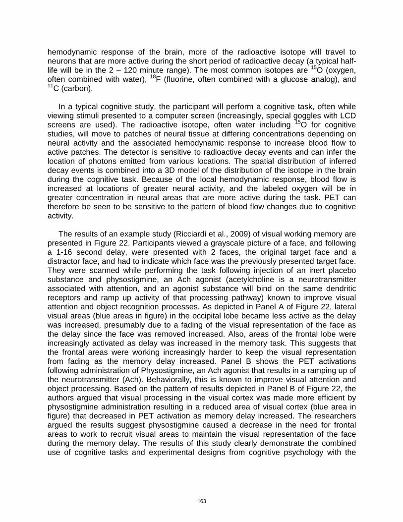
hemodynamic response of the brain, more of the radioactive isotope will travel to neurons that are more active during the short period of radioactive decay (a typical half-life will be in the 2 – 120 minute range). The most common isotopes are 15O (oxygen, often combined with water), 18F (fluorine, often combined with a glucose analog), and 11C (carbon). In a typical cognitive study, the participant will perform a cognitive task, often while viewing stimuli presented to a computer screen (increasingly, special goggles with LCD screens are used). The radioactive isotope, often water including 15O for cognitive studies, will move to patches of neural tissue at differing concentrations depending on neural activity and the associated hemodynamic response to increase blood flow to active patches. The detector is sensitive to radioactive decay events and can infer the location of photons emitted from various locations. The spatial distribution of inferred decay events is combined into a 3D model of the distribution of the isotope in the brain during the cognitive task. Because of the local hemodynamic response, blood flow is increased at locations of greater neural activity, and the labeled oxygen will be in greater concentration in neural areas that are more active during the task. PET can therefore be seen to be sensitive to the pattern of blood flow changes due to cognitive activity. The results of an example study (Ricciardi et al., 2009) of visual working memory are presented in Figure 22. Participants viewed a grayscale picture of a face, and following a 1-16 second delay, were presented with 2 faces, the original target face and a distractor face, and had to indicate which face was the previously presented target face. They were scanned while performing the task following injection of an inert placebo substance and physostigmine, an Ach agonist (acetylcholine is a neurotransmitter associated with attention, and an agonist substance will bind on the same dendritic receptors and ramp up activity of that processing pathway) known to improve visual attention and object recognition processes. As depicted in Panel A of Figure 22, lateral visual areas (blue areas in figure) in the occipital lobe became less active as the delay was increased, presumably due to a fading of the visual representation of the face as the delay since the face was removed increased. Also, areas of the frontal lobe were increasingly activated as delay was increased in the memory task. This suggests that the frontal areas were working increasingly harder to keep the visual representation from fading as the memory delay increased. Panel B shows the PET activations following administration of Physostigmine, an Ach agonist that results in a ramping up of the neurotransmitter (Ach). Behaviorally, this is known to improve visual attention and object processing. Based on the pattern of results depicted in Panel B of Figure 22, the authors argued that visual processing in the visual cortex was made more efficient by physostigmine administration resulting in a reduced area of visual cortex (blue area in figure) that decreased in PET activation as memory delay increased. The researchers argued the results suggest physostigmine caused a decrease in the need for frontal areas to work to recruit visual areas to maintain the visual representation of the face during the memory delay. The results of this study clearly demonstrate the combined use of cognitive tasks and experimental designs from cognitive psychology with the
163
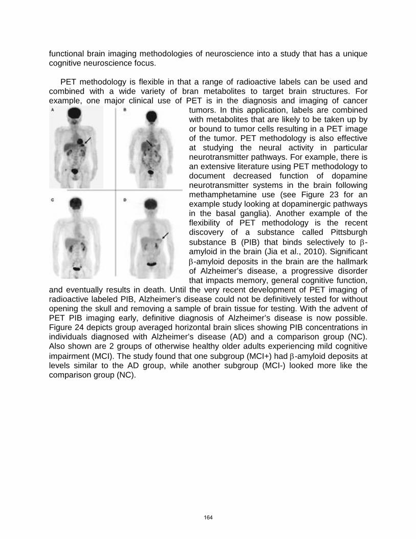
functional brain imaging methodologies of neuroscience into a study that has a unique cognitive neuroscience focus. PET methodology is flexible in that a range of radioactive labels can be used and combined with a wide variety of bran metabolites to target brain structures. For example, one major clinical use of PET is in the diagnosis and imaging of cancer
tumors. In this application, labels are combined with metabolites that are likely to be taken up by or bound to tumor cells resulting in a PET image of the tumor. PET methodology is also effective at studying the neural activity in particular neurotransmitter pathways. For example, there is an extensive literature using PET methodology to document decreased function of dopamine neurotransmitter systems in the brain following methamphetamine use (see Figure 23 for an example study looking at dopaminergic pathways in the basal ganglia). Another example of the flexibility of PET methodology is the recent discovery of a substance called Pittsburgh substance B (PIB) that binds selectively to β-amyloid in the brain (Jia et al., 2010). Significant β-amyloid deposits in the brain are the hallmark of Alzheimer’s disease, a progressive disorder that impacts memory, general cognitive function,
and eventually results in death. Until the very recent development of PET imaging of radioactive labeled PIB, Alzheimer’s disease could not be definitively tested for without opening the skull and removing a sample of brain tissue for testing. With the advent of PET PIB imaging early, definitive diagnosis of Alzheimer’s disease is now possible. Figure 24 depicts group averaged horizontal brain slices showing PIB concentrations in individuals diagnosed with Alzheimer’s disease (AD) and a comparison group (NC). Also shown are 2 groups of otherwise healthy older adults experiencing mild cognitive impairment (MCI). The study found that one subgroup (MCI+) had β-amyloid deposits at levels similar to the AD group, while another subgroup (MCI-) looked more like the comparison group (NC).
164
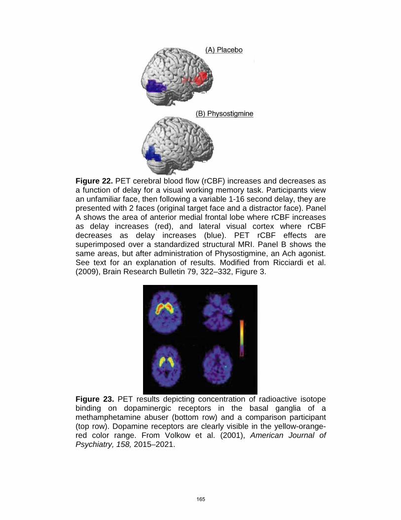
Figure 22. PET cerebral blood flow (rCBF) increases and decreases as a function of delay for a visual working memory task. Participants view an unfamiliar face, then following a variable 1-16 second delay, they are presented with 2 faces (original target face and a distractor face). Panel A shows the area of anterior medial frontal lobe where rCBF increases as delay increases (red), and lateral visual cortex where rCBF decreases as delay increases (blue). PET rCBF effects are superimposed over a standardized structural MRI. Panel B shows the same areas, but after administration of Physostigmine, an Ach agonist. See text for an explanation of results. Modified from Ricciardi et al. (2009), Brain Research Bulletin 79, 322–332, Figure 3.
Figure 23. PET results depicting concentration of radioactive isotope binding on dopaminergic receptors in the basal ganglia of a methamphetamine abuser (bottom row) and a comparison participant (top row). Dopamine receptors are clearly visible in the yellow-orange-red color range. From Volkow et al. (2001), American Journal of Psychiatry, 158, 2015–2021.
165
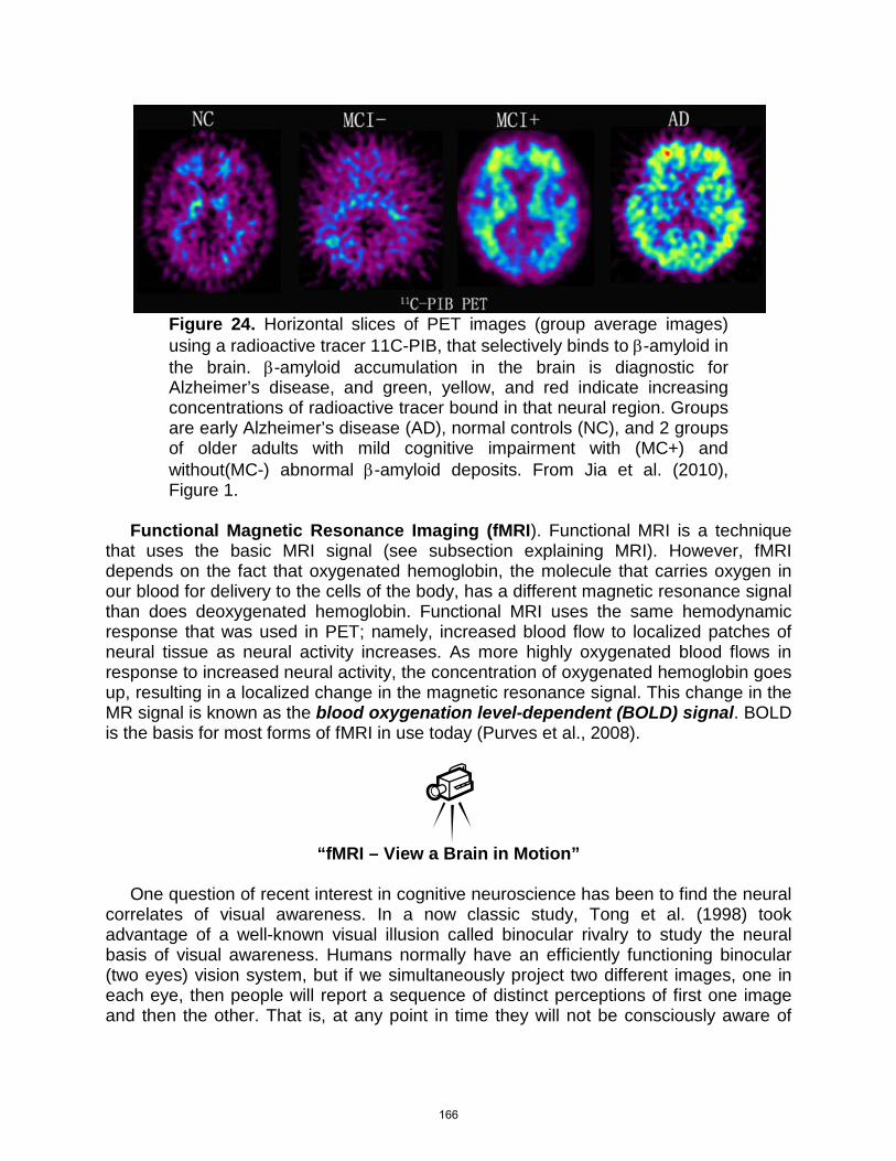
Figure 24. Horizontal slices of PET images (group average images) using a radioactive tracer 11C-PIB, that selectively binds to β-amyloid in the brain. β-amyloid accumulation in the brain is diagnostic for Alzheimer’s disease, and green, yellow, and red indicate increasing concentrations of radioactive tracer bound in that neural region. Groups are early Alzheimer’s disease (AD), normal controls (NC), and 2 groups of older adults with mild cognitive impairment with (MC+) and without(MC-) abnormal β-amyloid deposits. From Jia et al. (2010), Figure 1.
Functional Magnetic Resonance Imaging (fMRI). Functional MRI is a technique that uses the basic MRI signal (see subsection explaining MRI). However, fMRI depends on the fact that oxygenated hemoglobin, the molecule that carries oxygen in our blood for delivery to the cells of the body, has a different magnetic resonance signal than does deoxygenated hemoglobin. Functional MRI uses the same hemodynamic response that was used in PET; namely, increased blood flow to localized patches of neural tissue as neural activity increases. As more highly oxygenated blood flows in response to increased neural activity, the concentration of oxygenated hemoglobin goes up, resulting in a localized change in the magnetic resonance signal. This change in the MR signal is known as the blood oxygenation level-dependent (BOLD) signal. BOLD is the basis for most forms of fMRI in use today (Purves et al., 2008).
“fMRI – View a Brain in Motion”
One question of recent interest in cognitive neuroscience has been to find the neural correlates of visual awareness. In a now classic study, Tong et al. (1998) took advantage of a well-known visual illusion called binocular rivalry to study the neural basis of visual awareness. Humans normally have an efficiently functioning binocular (two eyes) vision system, but if we simultaneously project two different images, one in each eye, then people will report a sequence of distinct perceptions of first one image and then the other. That is, at any point in time they will not be consciously aware of
166
one of the pictures even though it is perfectly available to the visual system as it is being projected into one eye. The basic idea of the study was to examine perceptual processing areas in the temporal lobe “what” visual pathway (see Vision chapter) that is specialized for identification of objects. They chose the fusiform face area (FFA), and perahippocampal place area (PPA) on the ventral (underside) of the temporal lobe because they knew they could drive the activation of these areas with pictures of faces and places, and they wanted to know if these visual processing areas were far enough along the object identification pathway in the temporal lobe to be associated with visual awareness. If they used binocular rivalry presentation (i.e., a face in one eye and a place in the other) and participants reported only consciously seeing one picture at a time but both the FFA and PPA were strongly activated, then this would indicate these areas were being driven by the pictures on both retinas, rather than the single picture in visual awareness. If, on the other hand, the FFA was only active during periods when the face was in visual awareness, and the PPA was only active during periods when the place was in visual awareness, then this would be strong evidence that visual processing at this mid-level in the system was supporting visual awareness. Tong et al. had participants simultaneously view a picture of a house in one eye and a picture of a face in the other eye while being scanned. They had participants report when they had a perceptual reversal, and which picture they were currently perceiving. Figure 25 presents example FFA (faces results in more activation than houses), and PPA (houses results in more activation than faces) areas identified in one participant. Figure 26 presents the average activation in FFA and PPA as a function of time since a perceptual switch between perceived pictures simultaneously presented to the left and right eyes. Time zero indicates a perceptual switch. The left panel presents the results for a switch from the house to the face, and the right panel a switch from the face to the house. Note that in both panels there is a switch in the relative activation of the PPA and FFA directly following the time 0 point in each panel of Figure 26. In both cases, the area activated the most after time 0 matches the picture perceptually switched to. The results are consistent with the idea that the PPA and FFA are supporting visual awareness as their activation levels seem to be driven by the stimulus in visual awareness, not the stimuli as presented on the retina.
167

PET, fMRI & EEG/ERP Compared. Both PET and fMRI have relatively low temporal resolution, and relatively high spatial resolution (Gazzaniga et al., 2009). Comparatively, EEG/ERP has high temporal resolution, but poor spatial resolution. PET is limited by the speed of radioactive decay of the isotopes used, and even a fast isotope will require a minimum of 40 seconds of scanning to effectively count radioactive events. Functional MRI can operate faster, being limited by the BOLD response, which takes several seconds as opposed to tens of seconds in PET. EEG/ERP can detect changes in cognitive processes that occur in a thousandth of a second or less. PET can localize signals to a cube of neural tissue (called a voxel) on the order of a centimeter, and fMRI can now localize to the level of a millimeter voxel. EEG/ERP is quite limited in its ability to localize the area of brain that produces a recorded electrical event, but with large electrode array systems (e.g., 128 or 256 electrodes) it is possible to localize electrical brain generators at the level of a voxel of several cm. PET has limitations due to the use of radioactive isotopes, the much larger financial cost, and the fact that there are far fewer PET scanner in operation than MRI scanners. MRI scanners have become quite common at medical clinics, hospitals, medical schools, and research universities. MRI scanners also pose no known health risks other than magnetic metals mistakenly being brought into the strong magnetic field when the scanner is on. Accidents reported have been due to flying metal objects rather than a magnetic field negatively affecting the body. EEG/ERP setups are quite reasonably priced in relationship to fMRI.
“Why Not to Wear Metal During MRI Video”
Figure 25. Differential fMRI activations of a single participant superimposed on a structural MRI scan for the same individual. To determine the extent of the fusiform face area (FFA) and perahippocampal place area (PPA) in each individual, the differential activation to binocular presentation (to both eyes at the same time) of a picture of a house versus a face was computed. The view is of a horizontal section at the level of the ventral surface of the temporal lobe. By radiological convention the left hemisphere is presented on the right in each scan. Left Panel: The additional FFA response to faces (in
168
comparison to houses) in this individual is primarily in the right temporal lobe. Right Panel: The additional PPA response to houses (in comparison to faces) in this individual was bilateral, but stronger on the right side. From Tong et al. (1998), Neuron, 21, 753–759, Figure 2.
Figure 26. The signal strength in the FFA (blue) and PPA (red) as a function of time during the binocular rivalry viewing condition. There is a reversal in activations in the FFA and PPA as the participant reports (time 0) a perceptual switch from house to face (left panel), and from face to house (right panel). From Tong et al. (1998), Neuron, 21, 753–759, Figure 3.
C. Lesion Analysis
A brain lesion refers to any structural change or abnormality in neurons of the brain. Lesions are the result of disease processes or a physical injury event. The area of behavioral neuroscience has made important strides in our knowledge of how the brain works by using a methodology where animals are trained to produce a certain behavior, a portion of the brain is lesioned
or damaged, and changes in behavior on the learned task are evaluated in light of the pattern of brain injury. While this methodology continues to provide important insights into how the brain works, experimentally produced lesions are limited to animal research.
169
A more common approach has been to identify patients with brain lesions due to disease or injury that occurs as a normal part of life, and to study the changes in cognitive processing abilities as a function of different patterns of brain lesion (Gazzaniga et al., 2009). Neurologists and neuropsychologists have well-established roles as the medical doctors responsible for treating brain injury and/or disease, and the psychologists specialize in assessing changes in cognitive processes due to injury or disease. Two common sources of brain lesions are strokes and trauma. Strokes, or cerebrovascular accidents, refer to a disruption of the blood supply to the brain, and can be due to a blockage in a blood vessel or a bursting of a blood vessel resulting in internal bleeding. Either way, the blood supply to particular neural areas will be disrupted and damage to or loss of neural tissue will soon result. Brain lesions from strokes are not random. Because we all share the same basic structured system of arteries that supply blood to the brain, and because some locations in this system are more likely to lead to a stroke, certain patterns of stroke damage are more likely than others. Traumatic brain injury (TBI) can result from many types of accidents, such as car accidents, sports-related heat trauma, or gunshot injuries. TBI can result in a closed or open headed injury. In a closed head injury the brain is injured due to rapid acceleration/deceleration of the head and brain. The brain is supported by a clear fluid called cerebrospinal fluid (CSF), and has a series of CSF filled chambers (called ventricles) that act to dampen shocks. In a closed head injury the skull may be either accelerated or decelerated (i.e., stopped) suddenly, but the brain, suspended in CSF will continue on and bounce against the skull, resulting in focal bruising and tearing where the brain contacts the skull, and widespread shearing of axons as the brain is deformed when it contacts the skull. A common closed-head TBI due to an auto accident results from the victim hitting the windshield, and resultant frontal lobe damage (and damage to the anterior portions of the temporal lobe), but there is often an opposing brain injury as the brain bounces back against the rear of the skull, resulting in occipital lobe damage. This type of action can also result in brainstem injuries as the brain moves back and forth supported on the brainstem stalk. Open head injuries result when a fall cracks open the skull, or a mechanical blow to the head results in an object penetrating the skull. Before the advent of structural brain imaging technologies, open headed wounds, often incurred by soldiers in battle, were a leading way to localize a brain lesion while the patient was still alive. With the advent of MRI and computerized tomography (CT, uses x-rays to reconstruct a 3D model of the internal tissues of the body, typically with less spatial resolution than MRI), we can assess the extent of the brain lesion with much greater accuracy. Many diseases and degenerative disorders can also damage the brain, e.g., Parkinson’s disease or Alzheimer’s disease.
170
“Cerebrospinal Fluid Circulation”
Analysis of cognitive deficits due to a brain lesion is a difficult endeavor. Naturally occurring patterns of damage are rarely localized to one small area, and the pattern of damage will vary quite a lot across individuals, even with the same type of injury. Following a stroke or TBI there will be a period of continued brain degeneration as damaged neurons continue to die, followed by an extended period where there will be some recovery and reorganization of function. Finding a group of patients with similar patterns of brain lesions that have all reached a relatively stable stage in their recovery can be quite challenging for researchers wishing to assess the cognitive impact of damage to particular brain structures. Another challenge is finding uninjured individuals who are similar to the patients in other respects, e.g., age, gender, education, socioeconomic status, who can be part of an appropriate comparison group. Because of these difficulties, some researchers have argued for detailed testing and reporting of the results of an individual patient rather than studying groups of patients with potentially disparate patterns of brain lesions. However, it must be recognized that it is often difficult to generalize the results of such single-case studies. If we find a particular cognitive deficit following brain injury in a single patient, we typically have no way of knowing if this deficit was present prior to the injury. The result is that both group comparison and single-case study designs are in common usage in neurology and neuropsychology. Single and Double Dissociations. Neurologists and neuropsychologists wishing to establish evidence for localization of cognitive function to particular brain regions often look for 2 characteristic patterns of evidence from a lesion analysis study. For example, if we believe that brain area A is critical for the ability to recognize emotional expressions of faces, but brain area B is critical for the ability to recognize faces. We could test this theory by finding patient group A that includes patients with damage to brain area A but not to brain area B. We test patient group A, and some comparison individuals without brain injury, on a test of facial expressions and a test of facial recognition. Imagine that we find that the performance of the 2 groups is relatively equivalent on the facial recognition test, but that patient group A yields impaired performance on the facial expressions test. This result is called a single-dissociation (see Table 3) where a single patient group is compared to another group on 2 tasks, and found to be impaired in one area but not the other. Certainly, this single-dissociation result pattern is consistent with what we would predict if brain area A is responsible for emotional expression processing and not for facial identification. However, single-dissociations are problematic because we do not know if our results are simply due to a difference in difficulty or sensitivity to brain injury in general. Patients with significant brain injuries are often somewhat impaired on a wide range of cognitive tests, and will tend to be more impaired on more difficult tests. It may be the lack of impairment for
171
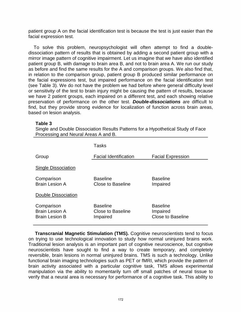
patient group A on the facial identification test is because the test is just easier than the facial expression test. To solve this problem, neuropsychologist will often attempt to find a double-dissociation pattern of results that is obtained by adding a second patient group with a mirror image pattern of cognitive impairment. Let us imagine that we have also identified patient group B, with damage to brain area B, and not to brain area A. We run our study as before and find the same results for the A and comparison groups. We also find that, in relation to the comparison group, patient group B produced similar performance on the facial expressions test, but impaired performance on the facial identification test (see Table 3). We do not have the problem we had before where general difficulty level or sensitivity of the test to brain injury might be causing the pattern of results, because we have 2 patient groups, each impaired on a different test, and each showing relative preservation of performance on the other test. Double-dissociations are difficult to find, but they provide strong evidence for localization of function across brain areas, based on lesion analysis.
Table 3 Single and Double Dissociation Results Patterns for a Hypothetical Study of Face Processing and Neural Areas A and B.
Tasks Group
Facial Identification
Facial Expression
Single Dissociation Comparison
Baseline
Baseline
Brain Lesion A Close to Baseline Impaired Double Dissociation Comparison
Baseline
Baseline
Brain Lesion A Close to Baseline Impaired Brain Lesion B Impaired Close to Baseline
Transcranial Magnetic Stimulation (TMS). Cognitive neuroscientists tend to focus on trying to use technological innovation to study how normal uninjured brains work. Traditional lesion analysis is an important part of cognitive neuroscience, but cognitive neuroscientists have sought to find a way to create temporary, and completely reversible, brain lesions in normal uninjured brains. TMS is such a technology. Unlike functional brain imaging technologies such as PET or fMRI, which provide the pattern of brain activity associated with a particular cognitive task, TMS allows experimental manipulation via the ability to momentarily turn off small patches of neural tissue to verify that a neural area is necessary for performance of a cognitive task. This ability to
172
safely perform experimental manipulation with TMS is a very important strength of the TMS technology as it allows for testing of causal theories of how the brain implements cognition.
“Deactivate Your Brain’s Selected Parts, Use Transcranial Magnetic Stimulation!” TMS works by sending a very brief electrical pulse through a coiled wire that is held next to the head. This results in a brief strong magnetic pulse being projected to a focal point in the brain. Note that sending an electrical pulse through a coiled wire creates an electromagnet. This strong magnetic pulse will hyperpolarize most, if not all, of the neurons in the patch of affected neural tissue, basically locking that area up for a brief time interval as all the neuron send maximal signals to each other simultaneously. For a brief time interval, all meaningful computations cease in that neural region. With TMS we can now ask if areas that become active during, say, language processing, are actually necessary for a particular linguistic task.
References Baars, B.J. & Gage, N.M. (2007). Cognition, brain, and consciousness: Introduction to
cognitive neuroscience. Burlington, MA: Academic Press. Bear, M.F., Connors, B.F., & Paradiso, M.A. (2007). Neuroscience: Exploring the brain
3rd Ed., Baltimore, MD: Lippicott, Williams, & Wilkins. Brodmann, K. (1960). On the comparative localization of the cortex (translated from the
French and German). In G. von Bonin, Some papers on the cerebral cortex (pp. 201-230). Springfield, IL: Thomas. (Original work published 1909)
Buxhoeveden, D.P., & Casanova, M.F. (2002). The minicolumn and evolution of the brain. Brain, Behavior, and Evolution, 60, 125–151.
Calvin, W.H. (1995). Cortical columns, modules, and Hebbian cell assemblies. In Arbib, M. (Ed.), The handbook of brain theory and neural networks, pp.269-272. Bradford Books/MIT Press. ]
Carmena, J.M., Lebedev, M.A., Crist, R.E., O'Doherty, J.E., Santucci, D.M., Dimitrov, D.F., Patil, P.G., Henriquez, C.S., & Nicolelis, M.A., (2003). Learning to control a brain-machine interface for reaching and grasping by primates. PLoS Biology, 1, 193-208. Epub October 13, 2003.
Churchland, P.A. (2002). Brain-wise: Studies in neurophilosophy. Cambridge, Massachusetts: The MIT Press.
Crick, F. (1994). The astonishing hypothesis: The scientific search for the soul. New York: Scribner.
Edelman, G. (2004). Wider than the sky: The phenomenal gift of consciousness. New Haven, CT: Yale University Press.
Emily Dickenson (1860’s). The brain is wider than the sky (poem). Downloaded from the internet: http://www.americanpoems.com/poets/emilydickinson/10584 (10-25-10).
173
F. J. Gall and J. G. Spurzheim. ANZ Journal of Surgery,75, 475–482. Flourens, M.J.P. (1824). Recherches expérimentales sur les proprieties et les
functiones du systeme neveux dans le animaux vertébrés. Paris: Ballière. Fritsch, G., & Hitzig, E. (1870). Ueber die elektische Erregbarkeit des Grosshirns (On
the electrical excitability of the cerebrum). Archiv fur Anatomie, Phsiologie und wissenschaftliche Medizin, 37, 300-332.
Gazzaniga, M.S., Ivry, R.B., & Mangun, G.R. (2009). Cognitive neuroscience: The biology of the mind 3rd Ed., New York: Norton.
Jia J., Sun , B, Guo, Z., Zhang J., Tian, J., Tang, H., Wang, L. (2010), Positron emission tomography with Pittsburgh compound B in diagnosis of early stage Alzheimer’s disease. Cell Biochemistry and Biophysics. EPub August 17, 2010.
Markram, H. (2006). The blue brain project. Nature Reviews Neuroscience, 7, 153-160. Ogawa, S., Lee, T.M., Kay, A.R., & Tank, D.W. (1990). Brain magnetic resonance
imaging with contrast dependent on blood oxygenation. Proceedings of the National Academy of Science, USA, 87, 9862-9872.
Paul, P. (1861). Remarques sur le siège de la faculté du langagé articule, suivies d’ une observation d’aphémie. Bulletin et Mémories de la Société Anatomique de Paris, 2, 330-357.
Purves, D., Augustine, G.J., Fitzpatrick, D., Katz, L.C., Lamantia, A.S., McNamara, J.O., Williams, S.M. (2001), Neuroscience, 2nd Ed., Sunderland, MA: Sinauer Associates.
Purves, D., Brannon, E.M., Cabeza, R., Heuttel, S.A., LaBar, K.S., Platt, M.L., & Woldorff, M.G. (2008). Princples of cognitive neuroscience. Sunderland, MA: Sinauer Associates.
Ravenscroft, I. (2005). Philosophy of mind: A beginner’s guide. New York: Oxford University Press.
Rees, G., Kreiman, G., & Koch, C. (2002) Neural correlates of consciousness in humans. Nature Reviews Neuroscience, 2, 261-270.
Ricciardi, E., Pietrini, P., Schapiro, M.B., Rapoport, S.I., & Furey, M.L. (2009). Cholinergic modulation of visual working memory during aging: a parametric PET study. Brain Research Bulletin, 79, 322–332.
Rousselet, G.A., Macé, M.J., & Fabre-Thorpe, M. (2004). Animal and human faces in natural scenes: How specific to human faces is the N170 ERP component? Journal of Vision. 4, 13-21.
Sacks, O. (1985). The man who mistook his wife for a hat: And other clinical tales. New York: Harper-Collins.
Simpson, D. (2005). Phrenology and the neurosciences: Contributions of Thagard, P. (2005). Mind: An introduction to cognitive science. Cambridge, MA: MIT
Press. Tong, F., Nakayama, K., & Vaughan, J. T., & Kanwisher, N. (1998). Binocular rivalry
and visual awareness in human extrastriate cortex. Neuron, 21, 753–759. Ward, J. (2006). The student’s guide to cognitive neuroscience. New York: Psychology
Press.
174
Further Reading Cognitive Neuroscience Overviews Gazzaniga, M.S., Ivry, R.B., & Mangun, G.R. (2009). Cognitive neuroscience: The
biology of the mind 3rd Ed., New York: Norton. Ward, J. (2006). The student’s guide to cognitive neuroscience. New York: Psychology
Press. Patient-based Perspectives on Cognitive Neuroscience Ramachandran, V.S., Blakeslee, S. (1998). Phantoms in the brain: Probing the
mysteries of the human mind. New York: William Morrow. Sacks, O. (1985). The man who mistook his wife for a hat: And other clinical tales. New
York: Harper-Collins. Sacks, O. (2010). The mind’s eye. Knopf. Neural Interface Systems Hatsopoulos, N.G., & Donoghue, J.P. (2009). The science of neural interface systems.
Annual Review of Neuroscience, 32, 249–66 Stix, G. (November, 2008). Jacking into the brain. Scientific American, Nov., 56-61. The Blue Brain Project Markram, H. (2006). The blue brain project. Nature Reviews Neuroscience, 7, 153-160. Mind/Body Question Churchland, P.A. (2002). Brain-wise: Studies in neurophilosophy. Cambridge,
Massachusetts: The MIT Press. Ravenscroft, I. (2005). Philosophy of mind: A beginner’s guide. New York: Oxford
University Press. Consciousness & the Brain from a Neuroscientist’s Viewpoint Crick, F. (1994). The astonishing hypothesis: The scientific search for the soul. New
York: Scribner. Damasio, A. (2003). Looking for Spinoza: Joy, sorrow, and the feeling brain. Orlando,
FL: Harcourt. Damasio, A. (2010). Self comes to mind: Constructing the conscious brain. Pantheon. Edelman, G. (2004). Wider than the sky: The phenomenal gift of consciousness. New
Haven, CT: Yale University Press.
Glossary Action Potential
A voltage wave of a set size that is triggered in an all-or-none manner at the axon hillock, and is actively propagated down the axon by the opening and closing of voltage sensitive ion channels to the axon terminal, resulting in release of neurotransmitter substance at the synapse.
175
Axon Appendage of the neuron that connects at the axon hillock and carries action potentials to the synapses.
Amygdala
Subcortical emotion processing area at the end of each hippocampi in the temporal lobes.
Axon Hillock
Place on the neuron cell body that connects to the axon and that triggers an action potential.
Axon Terminal
An end branching of the axon that usually forms a synapse with the dendrites of another neuron, but other connections are possible.
Basal Ganglia
Subcortical motor control structure wrapped around the thalami. Blindsight
Loss of conscious visual awareness in a portion of the visual field due to cortical damage, typically to primary visual cortex ( V1), but with a preserved ability to orient the eyes or point to the location of a visual event in the portion of the visual field the person “sees” as a dark or blank empty spot (i.e., scotoma).
Blood-brain Barrier
Glial cells that filter the brain’s blood supply, only letting some molecules through. Brainstem
Connects the spinal cord to the cerebral hemispheres. Also, contains many nuclei, i.e., clusters of neurons, responsible for motor control and general regulation (e.g., heart rate, breathing) along with other important functions.
Central Sulcus
A major sulcus that runs laterally across the top of the cortex separating the frontal from the parietal lobes.
Cerebellum
Motor control and timing structure attached to the back of the brainstem and sitting below the occipital lobe.
Cerebrum
A hemisphere of the brain including the cortical surface (gray matter), deep white matter tracts, and subcortical structures. Viewed by neuroanatomists as separate from the brainstem and cerebellum.
176

Cognitive Neuroscience Cognitive neuroscience is a relatively new scientific discipline that combines the study of cognitive and biological processes in the brain, primarily in humans, with a focus on how the brain implements cognitive processes.
Contralaterality Principle
Two locations in the brain are contralateral if they are opposite sides of the midline, right versus left. The contralaterality principle refers to sensory and motor systems that are crossed such that they get their sensory input from, or send motor output to, the opposite side of the body/world. The visual system is strongly contralateral with information from the right half of the visual field going to the left primary visual cortex, and vice versa for the left half of the visual field. The motor control system, and somatosensory (touch) system, are also strongly crossed. The auditory system is only partially crossed.
Corpus Callosum
Primary axon tract for interhemispheric transmission of information. Cortical Column
The organization of cerebral cortex into vertically oriented functional columns that may act as the basis for a standard cortical computational unit.
Cortical Lobes
The 4 major subdivisions of the cerebral cortex are called lobes and include the Frontal, Parietal, Occipital, Temporal Lobes.
Cytoarchitectonic Map
A map of cortical regions categorized by the pattern of stained neurons in each region when viewed under a microscope.
Dendrite
Appendages of the neuron that contain neurotransmitter receptor sites. Dualism
A philosophical position that the mind and brain are made of different substances. The traditional dualist view is that there is a physical brain and a non-physical mind.
Electroencephalograpy (EEG)
Method for recording the electrical activity of the brain by measuring voltage changes on the outside of the scalp.
Event-related Potential (ERP)
An average EEG time locked to the occurrence of a stimulus event.
177
Form Follows Structure The general principle that if one observes a difference in neural structure one expects to find differences in function.
Frequency Coding
Because action potentials are all of a set size, they cannot be varied in size by neurons to send different messages to each other. Instead, neurons convey different information to each other by varying the rate or frequency of firing action potentials. This variation in frequency is known as frequency coding.
Functional Magnetic Resonance Imaging (fMRI)
Noninvasive functional brain imaging technique that measures the pattern of neural activation during a mental task by assessing the MR signal differential in oxygenated and deoxygenated blood. Because of the brain’s hemodynamic response, the capillaries surrounding an active patch of neural tissue will open up increasing the flow of oxygenated blood to that area. The blood oxygenation level-dependent MR signal (BOLD signal) uses the pattern of oxygenated and deoxygenated blood to build a model of the pattern of neural activity during the mental task.
Glia
Brain cells that provide structural and metabolic support for neurons. Gray matter
Surface of the cortex containing neural cell bodies, gray in appearance. Gyrus
An outfolding (bump or ridge) of the cerebral cortex. Hemispheric Specialization
A general principle of cortical organization where homotopic (i.e., equivalent matching areas in each hemisphere) regions in the hemispheres do not have the same identical function. For example, for nearly all right handers, and for at least half of left handers, the speech centers of the brain are on the left side of the brain, with the right hemisphere taking a lead on visuospatial processing tasks (e.g., closing your eyes and rotating a mental image of a 3D block figure in the mind’s eye).
Hemodynamic Response
Reflexive opening of capillaries in the brain in response to a neural area becoming active.
178
Hippocampus A subcortical structure that wraps around the thalami, and down each of the temporal lobes. Known as the gateway to long-term memory for facts and events, bilateral damage to both hippocampi leads to a severe amnestic syndrome with loss of ability to learn new facts and events.
Homotopic
Equivalent matching neural areas in the 2 hemispheres (e.g., Brodmann’s area 44 in both hemispheres).
Horizontal (Longitudinal) Fissure
Major groove in the brain running front to back on the midline. It separates the right and left cerebral hemispheres.
Insula
Area of cortex hidden in the lateral fissure responsible for a range of functions including emotion processing and body sense (sometimes called visceral sense, the sense of what the body feels at any given moment).
Lateral (Sylvian) Fissure
Major sulcus that separates the temporal lobe from the frontal and parts of the parietal lobe.
Lesion
An abnormal structural change in a neural area, usually due to injury or a disease process.
Localization of Function
The idea that different neural areas perform different functions. Magnetic Resonance Imaging (MRI)
Noninvasive structural brain imaging technique that measures the signal from hydrogen atoms aligned in a strong magnetic field and perturbed by a radio frequency pulse. This MR signal is sensitive to differences in concentration of the hydrogen atoms in water contained in various tissues of the brain, and a model of the brain structures can be computed from the known water concentration in various tissue types.
Monism
A philosophical position that the mind and brain are made of the same substance. The physicalist form of this position is that both mind and brain are physical in nature, and that physical processes in the brain cause the mind.
179
Myelin Fatty covering of an axon formed by a specialized glial cell wrapping around the axon. Acts to insulate axons to make action potentials propagate faster, reduce the metabolic requirements of the neuron, and reduce cross-talk between axons.
Neglect Syndrome
An attentional deficit due to a brain lesion, typically damage to parietal cortex that involves a lack of attending to a portion of the visual field.
Neural Plasticity
Structural changes in neurons, e.g., forming new synaptic connections, related to experience.
Neuron Doctrine
A central theory in neuroscience that the brain is composed of cells called neurons that are the functional building blocks of the brain, and that understanding brain function will require understanding how neurons communicate with each other.
Neurotransmitter
Chemical substances secreted by neurons at synapses, and that bind to receptor sites in the synapse.
Nonreductionist Monism
A form of monism that proposes that the mind is caused by physical processes in the brain, but that it is not possible to fully understand the mind in terms of the interaction of the brain’s physical parts, e.g., the interaction of neurons. Nonreductionist monists often suggest that there are emergent properties of the mind that are more than the sum of interacting neurons.
Phrenology
A pseudoscientific belief system proposed in the 19th century that assumes that an arbitrarily chosen set of human behavioral attributes (e.g., hope, curiosity, aggressiveness) were each localized to a single cortical region. Modern cognitive neuroscience seeks to study the variation in function of different neural areas without repeating the mistakes of phrenology.
Positron Emission Tomography (PET)
Noninvasive functional brain imaging method that involves injection of a radioactive labeled substance targeted to go to a particular location or system in the body. The detectors measure radioactive decay events and can build a model of the concentration of the radioactive labels in the body. In medical applications cancer tumors are imaged by attaching the radioactive label to a chemical that will bind selectively with any tumors in the body. In cognitive neuroscience applications oxygen or glucose (sugar) is labeled and, due to the hemodynamic response, neural areas that work harder receive increased blood flow, and therefore the
180
pattern of labeled oxygen or glucose will indicate the pattern of neural activity in the brain during performance of a mental task.
Post-synaptic Potential (PSP)
Voltage wave initiated when a neurotransmitter molecule binds at a receptor causing ion channels to open. May be positive (Excitatory PSP, increases the likelihood the neuron will fire an action potential) or negative (inhibitory PSP, reduces likelihood neuron will fire an action potential) depending on the electrical charge of the ions and their direction of flow in or out of the neuron. If the sum total of all the PSPs when they reach the axon hillock reach a certain threshold, and action potential will be triggered.
Prosopagnosia
A deficit of visual identification that is specific to faces. Reductionist Monism
A form of monism that proposes that the mind is caused by physical processes in the brain, and that it is possible to understand the mind in terms of the interaction of the brain’s physical parts, e.g., the interaction of neurons.
Single & Double Dissociation
A pattern of evidence from the analysis of the effects of lesions on cognitive performance. In a single dissociation a single group of patients with a particular brain lesion are compared to a control group on 2 tasks and found to be impaired on one task and relatively unimpaired on the other. This pattern suggests that the performance of impaired task is supported by the lesioned brain area. The weakness of the single dissociation pattern is that an equally good alternative explanation would be that the 2 tests are simply differentially sensitive to the general effects of brain damage. By finding a second patient group that has a different brain lesion, and shows the opposite pattern of impaired and unimpaired performance as did the first patient group, we can invalidate the alternate hypothesis, and provide strong evidence for the specific brain lesions in each group leading to the observed specific impairments.
Subcortical Structures
Brain structures that are interspersed throughout the white matter tracts of the cerebral hemispheres and are not part of the brainstem (e.g., thalamus, hippocampus, basal ganglia).
Sulcus
An infolding (groove) of the cerebral cortex. Thalamus
Subcortical structure location at the end of the brainstem that acts as the sensorimotor relay station for the cortex.
181
Topographic Map An area of cortex in which sensory or motor neurons are arranged to form a map of the body or external world.
White matter
Areas of each cerebral hemisphere that primarily contain axon fiber tracts. The tissue appears white to the eye, and in MRI scans due to the white fatty myelin coverings of many of the axons in these fiber tracts. Arcuate tracts connect cortical areas within a hemisphere in sweeping arcs. Longitudinal tracts form the major front-to-back (anterior-to-posterior) communications pathways. Interhemispheric tracts link structures in different hemispheres through the corpus callosum.
Spatial Resolution
The ability of a brain imaging technique to accurately localize neural activity levels. PET can resolve neural areas on the order of a cm, and fMRI methods exist for differentiating activity in neural areas on the order of a mm.
Temporal Resolution
Sensitivity of a brain imaging technique to rapid changes in cognitive processes in various brain areas. EEG has relatively high temporal resolution as it is sensitive to rapid changes in processing in brain regions recorded from.
Transcranial Magnetic Stimulation (TMS)
Used a pulse of electricity in a coiled wire to create a strong magnetic pulse that is projected in a focal neural location in the brain. Neural processing in this area is temporarily disrupted by overstimulation of the neurons by the magnetic pulse. Can be used to create a temporary reversible brain lesion to determine if a neural area is necessary for performance of a given mental task.
182


