Hindawi Publishing CorporationCase Reports in DentistryVolume 2013, Article ID 210763, 5 pageshttp://dx.doi.org/10.1155/2013/210763
Case ReportRestoration of Endodontically Treated Molars Using AllCeramic Endocrowns
Roopak Bose Carlos,1 Mohan Thomas Nainan,1 Shamina Pradhan,1 Roshni Sharma,1
Shiny Benjamin,1 and Rajani Rose2
1 Department of Conservative Dentistry and Endodontics, Vydehi Institute of Dental Sciences and Research Centre, No. 82, EPIP Area,Whitefield, Bangalore 560066, India
2Dental Solutions, 157, 4th Main, BEML layout, Off ITPL Road, Thubarahalli, Bangalore 560066, India
Correspondence should be addressed to Roopak Bose Carlos; [email protected]
Received 28 October 2013; Accepted 17 November 2013
Academic Editors: D. W. Boston and K. Seymour
Copyright © 2013 Roopak Bose Carlos et al. This is an open access article distributed under the Creative Commons AttributionLicense, which permits unrestricted use, distribution, and reproduction in any medium, provided the original work is properlycited.
Clinical success of endodontically treated posterior teeth is determined by the postendodontic restoration. Several options havebeen proposed to restore endodontically treated teeth. Endocrowns represent a conservative and esthetic restorative alternative tofull coverage crowns.The preparation consists of a circular equigingival butt-jointmargin and central retention cavity into the entirepulp chamber constructing both the crown and the core as a single unit. The case reports discussed here are moderately damagedendodontically treated molars restored using all ceramic endocrowns fabricated using two different systems, namely, CAD/CAMand pressed ceramic.
1. Introduction
Postendodontic restoration should preserve and protect theexisting tooth structure, while restoring satisfactory esthetics,form, and function.The goal is to achieve minimally invasivepreparations with maximal tissue conservation for restoringendodontically treated teeth. This will help to mechanicallystabilize the tooth-restoration complex and increase surfacesavailable for adhesion.
A number of options are available in every clinicalsituation. The choice depends on the structural integrityof the tooth, esthetic, and protective requirements [1]. Inthis perspective, endocrowns can be considered as a fea-sible alternative to full crowns for restoration of nonvitalposterior teeth, especially those with minimal crown heightbut sufficient tissue available for stable and durable adhesivecementation [2].
The evolution of ceramic technology especially dentalCAD/CAM systems have enhanced the options to producesingle all ceramic endocrownswith high biocompatibility andoptimal mechanical properties [3].
In the present paper two ceramic endocrowns fabricatedby different methods are presented as case reports.
2. Case 1
A 32-year-old female patient reported for the filling of herlower 1st molar. On clinical examination tooth number36 was root canal treated one month back (Figure 1). Itwas asymptomatic and the occlusogingival height of theremaining crown structure was approximately 4mm. Theradiographic findings revealed well obturated canals with noperiapical changes.
A conservative approach of restoring the tooth with anendocrownwas decided as the treatment option, asmore thanhalf the residual tooth structure was remaining and therewere no occlusal wear facets. On additional request by thepatient for an advanced and a prompt restoration, CAD/CAMceramic was chosen.
After removal of the provisional restoration, preparationfor endocrown was initiated. Resin modified glass ionomercement (Fuji II LC GC Corporation, Tokyo, Japan) was used

2 Case Reports in Dentistry
Figure 1: Postobturation occlusal view showing the amount ofresidual tooth structure.
Figure 2: Tooth preparation for endocrown.
to achieve a flat pulpal floor and to block the undercuts.The preparation consisted of a circular equigingival butt-joint margin and central retention cavity into the entire pulpchamber constructing both the crown and the core as a singleunit. The appropriate reduction of the buccal and lingualwalls was done (Figure 2).
Interocclusal space was carefully evaluated and occlusalreduction done to achieve a clearance of 2mm. Shade-B
1was
selected (VITAPAN Zahnfabrik, Germany). Retraction cordwas placed and impressions made with polyvinyl siloxaneimpression material (Aquasil LV, Putty/Light Body, DentsplyDeTrey, Germany) using putty wash technique. Die stonemodel was fabricated.
CAD/CAMProcessing.The three-dimensional reconstructionof the preparation was done using the Yenadent D40 millingmachine (Yenadent, Istanbul, Turkey) and DWOS scanner(Dental Wings Inc., Montreal, Canada). The 3D scanningof the individual die and the antagonist arch for occlusalfunction (virtual articulation) were done. The milling wasthen initiated on a monolithic solid zirconia block (MetoxitAG, Thayngen, Switzerland) (Figures 3 and 4).
Figure 3: CAD/CAM image.
Figure 4: Tissue surface depicting the core and crown fabricated asa single unit.
The finished endocrown was checked for shade, fit, andocclusion in the patient’s mouth and then cemented usingdual cure resin luting agent (Variolink, Ivoclar/Vivadent,Schaan/Liechtenstein).
Clinical and radiographic evaluationwas done and followup after 28months showed no secondary caries, fracture, dis-coloration or loosening/decementation of the crown (Figures5 and 6).
3. Case 2
A 26-year-old female patient reported with a chief com-plaint of pain since 2 days. On radiographic examinationradiolucency involving pulp of tooth 36 was seen. Basedon the clinical and radiographic examination tooth 36was diagnosed with acute irreversible pulpitis. Root canaltreatment was performed. Based on the remaining toothstructure, that is, approximately 4-5mm, occlusal evaluation,and patients esthetic demands, IPS E.max Press endocrownwas decided as the treatment option. The endocrownpreparation and the impression technique were performedas described in the previous case. IPS E.max Press HO
Case Reports in Dentistry 3
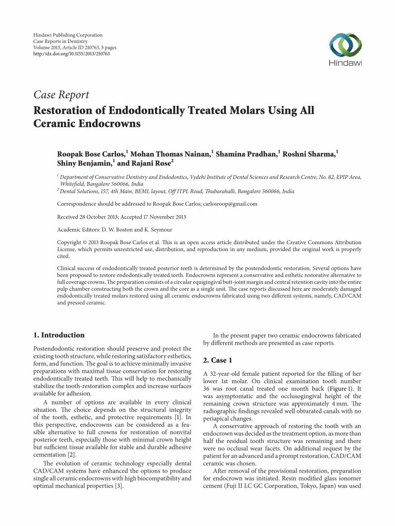
Figure 5: Occlusal view following final cementation.
Lithium-disilicate glass ceramic ingots (Ivoclar/Vivadent,Schaan/Liechtenstein) were used for the press technology.The restoration was fabricated according to the lost waxtechnique of investing and wax pattern burnout followed bypressing of the ceramic ingot in the pressable furnace at apress temperature of 915–920∘C. It was then finished andpolished with Proxyt pink polishing paste (Ivoclar/Vivadent,Schaan/Liechtenstein). The endocrown was cemented usinga dual cure resin luting agent (Variolink, Ivoclar/Vivadent,Schaan/Liechtenstein). Clinical and radiographic evaluationwas done and a 28-month followup showed no secondarycaries, fracture, discoloration or loosening/decementation ofthe crown (Figures 7, 8, 9, 10, 11, and 12).
4. Discussion
A successful endodontic treatment has to be complementedwith an appropriate postendodontic restoration to integratethe pulpless tooth with the masticatory apparatus [4]. Whenup to one half of the coronal tooth structure is missing,complete occlusal coverage is achieved conservatively usingendocrown [5].
The concept of a conservative protective restoration forposterior endodontically treated teeth is not new. Amalcore,inlays, and onlays are based on this principle. The amalcoreharnessed, the large and retentive contours of the root canalorifices, and the pulp chamber to provide a monoblockfoundation. Inlays and onlays promoted the concept of asupragingival finish line and conservative preparations. Theendocrown is an esthetic and conservative addition to thiscontinuum.
All ceramic systems have gained popularity in recenttimes as they offer both esthetics and function [6]. Thedevelopment of CAD/CAM systems and software offersseveral advantages in clinical practice. Custom shaping andprecise milling of ceramic restorations is now a reality;furthermore, the adaptation of the inner surface of therestoration and the replication of the occlusal morphologyare better. Restorations can be produced chairside and seated
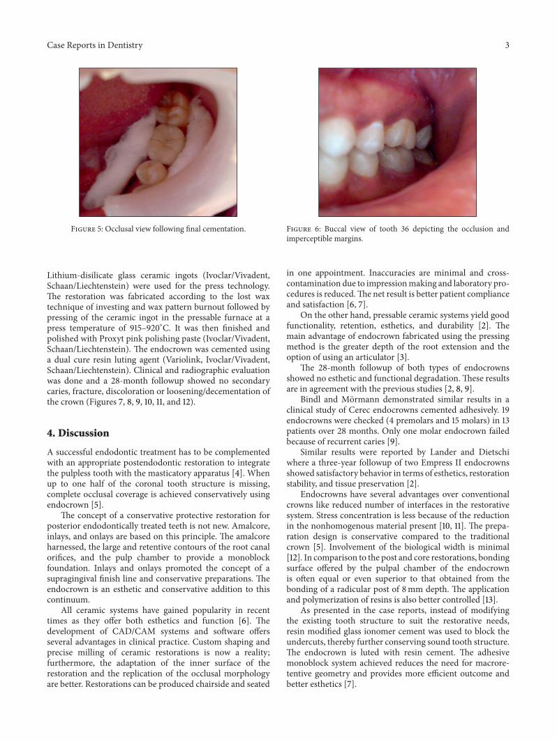
Figure 6: Buccal view of tooth 36 depicting the occlusion andimperceptible margins.
in one appointment. Inaccuracies are minimal and cross-contamination due to impressionmaking and laboratory pro-cedures is reduced.The net result is better patient complianceand satisfaction [6, 7].
On the other hand, pressable ceramic systems yield goodfunctionality, retention, esthetics, and durability [2]. Themain advantage of endocrown fabricated using the pressingmethod is the greater depth of the root extension and theoption of using an articulator [3].
The 28-month followup of both types of endocrownsshowed no esthetic and functional degradation.These resultsare in agreement with the previous studies [2, 8, 9].
Bindl and Mormann demonstrated similar results in aclinical study of Cerec endocrowns cemented adhesively. 19endocrowns were checked (4 premolars and 15 molars) in 13patients over 28 months. Only one molar endocrown failedbecause of recurrent caries [9].
Similar results were reported by Lander and Dietschiwhere a three-year followup of two Empress II endocrownsshowed satisfactory behavior in terms of esthetics, restorationstability, and tissue preservation [2].
Endocrowns have several advantages over conventionalcrowns like reduced number of interfaces in the restorativesystem. Stress concentration is less because of the reductionin the nonhomogenous material present [10, 11]. The prepa-ration design is conservative compared to the traditionalcrown [5]. Involvement of the biological width is minimal[12]. In comparison to the post and core restorations, bondingsurface offered by the pulpal chamber of the endocrownis often equal or even superior to that obtained from thebonding of a radicular post of 8mm depth. The applicationand polymerization of resins is also better controlled [13].
As presented in the case reports, instead of modifyingthe existing tooth structure to suit the restorative needs,resin modified glass ionomer cement was used to block theundercuts, thereby further conserving sound tooth structure.The endocrown is luted with resin cement. The adhesivemonoblock system achieved reduces the need for macrore-tentive geometry and provides more efficient outcome andbetter esthetics [7].

4 Case Reports in Dentistry
Figure 7: Occlusal view showing the amount of residual toothstructure postobturation.
Figure 8: Tooth preparation for pressable ceramic endocrown.
Figure 9: Tissue surface of pressed endocrown.
Endocrowns have their own disadvantages like, debond-ing and risk of root fracture because of the difference in themodulus of elasticity between the harder ceramic and softerdentin [3].Hence case selection is critical for ensuring clinicalsuccess with endocrowns [14]. Endocrowns are indicated in
Figure 10: Occlusal view following final cementation.
Figure 11: Buccal view of tooth 36 highlighting the excellent shadematch and finish.
Figure 12: Radiographic view, postcementation. The supragingivalfinish line is clearly visible.
cases where there are minimal functional and lateral stresses.When there is evidence of increased functional and lateralstresses as evident with steep occlusal anatomy, wear facetsor parafunction, full coverage crown with or without post isthe treatment of choice [12].

Case Reports in Dentistry 5
Based on current evidence, endocrowns fabricated usingCAD/CAM and pressable ceramic technology can be con-sidered as a reliable option for the restoration of moderatelymutilated endodontically treated posterior teeth. However,long-term followup and longitudinal clinical studies areneeded to ensure their overall success.
References
[1] K. Gulabivala, “Restoration of the root treated tooth,” inEndodontics, C. J. R. Stock, K. Gulabivala, and R. Walker, Eds.,pp. 279–305, Elsevier, 3rd edition, 2004.
[2] E. Lander and D. Dietschi, “Endocrowns: a clinical report,”Quintessence International, vol. 39, no. 2, pp. 99–106, 2008.
[3] V. Veselinovic, A. Todorovic, D. Lisjak, and V. Lazic, “Restoringendodontically treated teeth with all ceramic endo-crowns casereport,” Stomatoloski Glasnik Srbije, vol. 55, pp. 54–64, 2008.
[4] B. Suresh Chandra and V. Gopi Krishna,Grossman’s EndodonticPractice, Wolters Kluwer, New Delhi, India, 12th edition, 2010.
[5] D. Dietschi, O. Duc, I. Krejci, and A. Sadan, “Biomechanicalconsiderations for the restoration of endodontically treatedteeth: a systematic reviewof the literature—part 2 (Evaluation offatigue behavior, interfaces, and in vivo studies),” QuintessenceInternational, vol. 39, no. 2, pp. 117–129, 2008.
[6] T. S. Vinothkumar, D. Kandaswamy, and P. Chanana,“CAD/CAM fabricated single unit all ceramic post corecrown restoration,” Journal of Conservative Dentistry, vol. 14,no. 1, pp. 86–89, 2011.
[7] C.-Y. Chang, J.-S. Kuo, Y.-S. Lin, and Y.-H. Chang, “Fractureresistance and failure modes of CEREC endo-crowns andconventional post and core-supported CEREC crowns,” Journalof Dental Sciences, vol. 4, no. 3, pp. 110–117, 2009.
[8] J. Bernhart, A. Brauning, M. J. Altenburger, and K.-T.Wrbas, “Cerec3D endocrowns—two-year clinical examinationof CAD/CAM crowns for restoring endodontically treatedmolars,” International Journal of ComputerizedDentistry, vol. 13,no. 2, pp. 141–154, 2010.
[9] A. Bindl andW.H.Mormann, “Clinical evaluation of adhesivelyplaced cerec endo-crowns after 2 years—preliminary results,”Journal of Adhesive Dentistry, vol. 1, no. 3, pp. 255–265, 1999.
[10] F. Zarone, R. Sorrentino, D. Apicella et al., “Evaluation of thebiomechanical behavior of maxillary central incisors restoredby means of endocrowns compared to a natural tooth: a 3Dstatic linear finite elements analysis,” Dental Materials, vol. 22,no. 11, pp. 1035–1044, 2006.
[11] C.-L. Lin, Y.-H. Chang, and C.-A. Pai, “Evaluation of failurerisks in ceramic restorations for endodontically treated premo-lar with MOD preparation,” Dental Materials, vol. 27, no. 5, pp.431–438, 2011.
[12] D. Dietschi, S. Bouillaguet, and A. Sadan, “Restoration of theendodontically treated tooth,” in Cohen’s Pathways of the Pulp,K. M. Hargreaves and S. Cohen, Eds., pp. 777–807, ElsevierMosby, 10th edition, 2011.
[13] G. T. Rocca and B. Serge, “Alternative treatments for the restora-tion of non vital teeth,” Revue d’Odonto Stomatologie, vol. 37, pp.259–272, 2008.
[14] G. T. Rocca and I. Krejci, “Crown and post- free adhesiverestorations for endodontically treated posterior teeth: fromdirect composite to endocrowns,” European Journal of EstheticDentistry, vol. 8, pp. 156–179, 2013.

Submit your manuscripts athttp://www.hindawi.com
Hindawi Publishing Corporationhttp://www.hindawi.com Volume 2014
Oral OncologyJournal of
DentistryInternational Journal of
Hindawi Publishing Corporationhttp://www.hindawi.com Volume 2014
Hindawi Publishing Corporationhttp://www.hindawi.com Volume 2014
International Journal of
Biomaterials
Hindawi Publishing Corporationhttp://www.hindawi.com Volume 2014
BioMed Research International
Hindawi Publishing Corporationhttp://www.hindawi.com Volume 2014
Case Reports in Dentistry
Hindawi Publishing Corporationhttp://www.hindawi.com Volume 2014
Oral ImplantsJournal of
Hindawi Publishing Corporationhttp://www.hindawi.com Volume 2014
Anesthesiology Research and Practice
Hindawi Publishing Corporationhttp://www.hindawi.com Volume 2014
Radiology Research and Practice
Environmental and Public Health
Journal of
Hindawi Publishing Corporationhttp://www.hindawi.com Volume 2014
The Scientific World JournalHindawi Publishing Corporation http://www.hindawi.com Volume 2014
Hindawi Publishing Corporationhttp://www.hindawi.com Volume 2014
Dental SurgeryJournal of
Drug DeliveryJournal of
Hindawi Publishing Corporationhttp://www.hindawi.com Volume 2014
Hindawi Publishing Corporationhttp://www.hindawi.com Volume 2014
Oral DiseasesJournal of
Hindawi Publishing Corporationhttp://www.hindawi.com Volume 2014
Computational and Mathematical Methods in Medicine
ScientificaHindawi Publishing Corporationhttp://www.hindawi.com Volume 2014
PainResearch and TreatmentHindawi Publishing Corporationhttp://www.hindawi.com Volume 2014
Preventive MedicineAdvances in
Hindawi Publishing Corporationhttp://www.hindawi.com Volume 2014
EndocrinologyInternational Journal of
Hindawi Publishing Corporationhttp://www.hindawi.com Volume 2014
Hindawi Publishing Corporationhttp://www.hindawi.com Volume 2014
OrthopedicsAdvances in


