Int J Clin Exp Pathol 2015;8(9):11741-11746www.ijcep.com /ISSN:1936-2625/IJCEP0011898
Case Report Rare coexistence of mediastinal hepatoid adenocarcinoma, idiopathic azoospermia and horseshoe kidney: a case report and review of the literature
Chun-Hong Hu1, Qiu-Li Li2, Hai-Peng Li2, Song-Qing Fan3, Hai-Xia Zhang1, Xian-Ling Liu1, Yan He1, Ming Huang1, Min Lu1, Si-Si Wang1, Fang Wu1
Departments of 1Oncology, 3Pathology, The Second Xiangya Hospital of Central South University, 139 Middle Ren-min Road, Changsha 410011, China; 2Depatment of Neurology, The First People’s Hospital of Chenzhou, Univer-sity of South China, 102 Luojiajing Road, Chenzhou 423000, China
Received June 25, 2015; Accepted July 29, 2015; Epub September 1, 2015; Published September 15, 2015
Abstract: Hepatoid adenocarcinoma (HAC) is the term proposed for a special type of extrahepatic tumors, which is similar to the hepatocellular carcinoma (HCC) both in the histopathology and immunohistochemistry. HAC has been observed in the stomach, colon, pancreas, gall bladder, lung and female genital tract, but rarely in the mediastinum. Now we describe a case of a 28-year-old Chinese male with primary mediastinal HAC with lung and liver metastasis. In this patient, HAC was associated with horseshoe kidney and idiopathic nonobstructive azoospermia. It seemed derivation abnormalities during organogenesis in the embryo stage played a significant role in the pathogenesis of HAC, horseshoe kidney and idiopathic nonobstructive azoospermia. Even the pathogenesis was still unknown; it may merit consideration of HAC together with horseshoe kidney and idiopathic nonobstructive azoospermia as a syndrome rather than as a spectrum of coincidental diseases. Furthermore, we found the HAC is a neoplasm with unfavorable outcomes despite aggressive and multi-protocol strategies. The serum alpha fetoprotein (AFP) should be regarded as a useful marker for diagnostic purposes and therapeutic response evaluation of HAC.
Keywords: Hepatoid adenocarcinoma, hepatocellular carcinoma, horseshoe kidney, idiopathic nonobstructive azoospermia, alpha fetoprotein, chemotherapy
Introduction
Hepatoid adenocarcinoma (HAC) is the term proposed for a rare type of extrahepatic aggres-sive tumor, which shows histological resem-blance to hepatocellular carcinoma (HCC) [1, 2]. The tumor cells differentiate into liver cells due to certain abnormalities in differentiation during embryonic development [3, 4]. The origin and biology of these tumors is unclear. Only one case of HAC in the thymus has been report-ed, which had no elevated level of serum AFP [4]. In this case, the patient present a rare AFP-producing mediastinal HAC. Meanwhile, we report the coexistence of HAC, horseshoe kid-ney and idiopathic nonobstructive azoosper-mia, and raise the hypothesis that these may be a syndrome rather than a serious of coinci-dental diseases.
This study was approved by the ethics commit-tee of the Second Xiangya Hospital of Central South University (Changsha, China). Patient pro-vided written informed consent.
Case presentation
A 28-year-old Chinese male was admitted to The Second Xiangya Hospital of Central South University (Changsha, China) due to chest and shoulder pain on May 2013. He has no Jaundice, spider angiomas, abdominal pain and disten-sion. The patient’s medical history was remark-able, including idiopathic nonobstructive azo-ospermia and horseshoe kidney. He had no smoking or drinking habits. He had no hepatitis and tuberculosis. Liver and spleen were impal-pable. Markers for hepatitis virus were all nega-tive. Results of laboratory workup revealed

Hepatoid adenocarcinoma with high serum AFP in the mediastinum
11742 Int J Clin Exp Pathol 2015;8(9):11741-11746
aspartate aminotransferase and alanine ami-notransferase level were within the normal range. Endocrinological data showed that Follicle stimulating hormone (FSH) was 25.350 U/L↑, Luteinizing hormone (LH) was 9.420 U/L↑, Testosterone (T) was normal. Serum-human chorionic gonadotropin (HCG) was undetect-able at <1 mIU/mL. Before the surgery, the level of serum AFP was significantly elevated at 155 278.00 ng/ml. Chromosome karyotyping showed 46XY. Computed tomography (CT) showed single tumor (approx. 18 cm in greatest diameter) in the anterior mediastinum and mul-tiple nodules suspected for metastasis in the liver and bilateral lungs (Figure 1). The first diagnosis seemed to be mediastinal carcinoma with lung and liver metastasis. The mediastinal tumor removal and the left upper lung and the middle lobe of right lung wedge resection were performed. The patient also underwent a US-guided liver biopsy. One month after the
operation, the serum AFP level was 7347 ng/ml. Combined with the histological and immu-nohistochemical findings, the final diagnosis was HAC in the mediastinum with lung and liver metastasis. Adjuvant therapy with CAP (cyclo-phosphamide/epirubicin/cislatin) was given every 3 weeks for 4 cycles (June 2013-october 2013). The decline of serum AFP level was noticed (from 7347.47 ng/ml to 402 ng/ml). Meanwhile, the CT revealed multiple tumors in the liver and lung were partly remission. On November 2013, a progression of liver and lung metastasis, along with elevated serum AFP level (105 504 ng/mL) was found. He was switched to second-line chemotherapy with gemcitabine and paclitaxel, after two courses of chemotherapy, serum AFP level decreased to 34 686 ng/ml. However, two cycles later, CT scan demonstrated a progression of liver, bilat-eral pulmonary metastasis and newly devel-oped hilum of lungs metastasis. At the same
Figure 1. Computed tomography scan before surgery in a 28-year old man. A, B: Preoperative CT scan revealing 18 cm in diameter in the anterior mediastinum and horseshoe kidney. C: Chest CT shows multiple tumor in the bialat-eral lung. D: Abdominal CT reveals multiple nodular, high-density contrast mass in the liver lobe.

Hepatoid adenocarcinoma with high serum AFP in the mediastinum
11743 Int J Clin Exp Pathol 2015;8(9):11741-11746
time, the serum AFP level reach up to 121 000 ng/mL. He was then put under the third-line chemotherapy (Gemcitabine and Vinorelbine). However, the level of serum AFP rised to 306 081 ng/ml after one course of chemotherapy (Figure 2). The CT findings in liver, hilum of lung show progressive disease. Hepatomegaly was
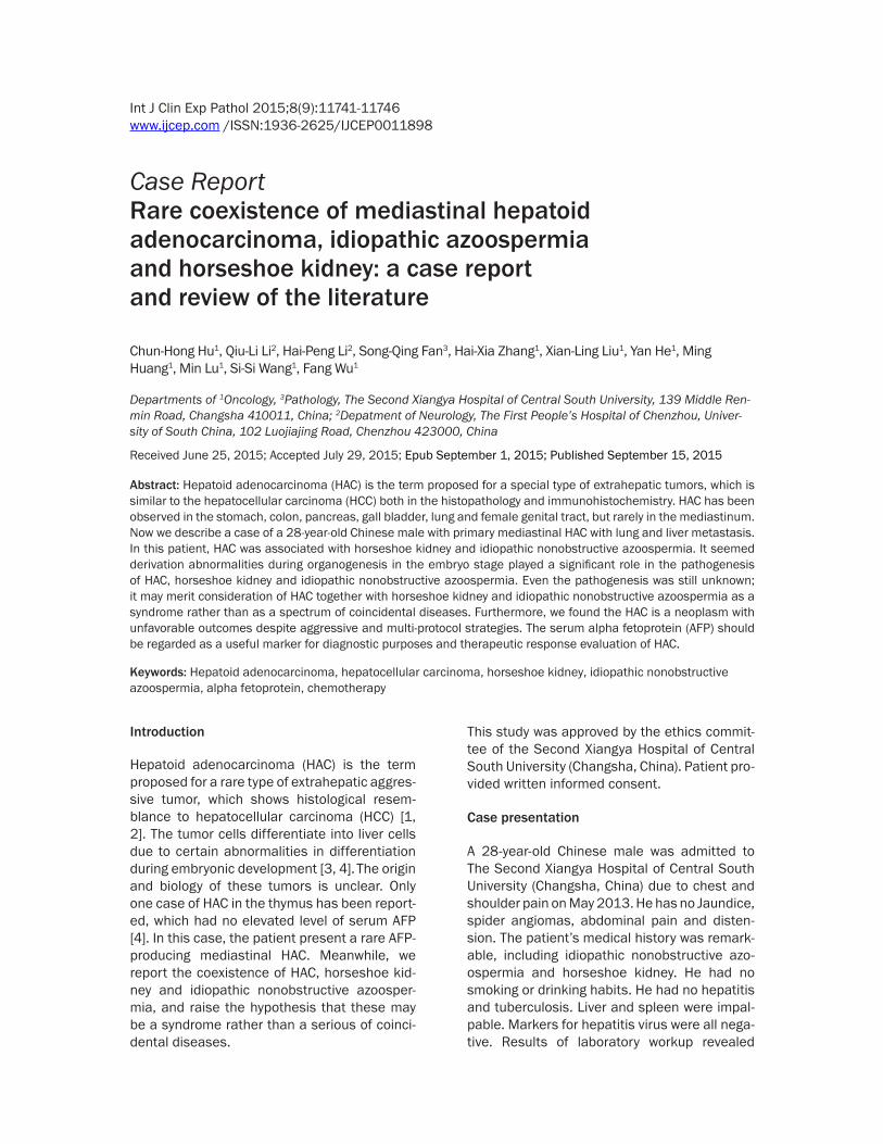
Light microscopy of mediastinal HACs showed the medium-sized, cuboidal and polygonal tumor cells with rather abundant esosinophilic cytoplasm and prominent nucleoli, arranged in sheet-like growth pattern or predominantly pseudoglandular formation, and merging with solid foci of hepatoid differentiation (Figure 3A). Biopsy of hepatic nodules was virtually indistinguishable from HCC. The hepatic tumors were poorly differentiated (Figure 3B) and the non-tumorous liver tissue showed no hepatitis and cirrhosis. Bile drops was not observed. What’s more, there were no neuroendocrine or germ cell component.
Immunohitochemical findings
Immunohistochemistry was performed on mediastinal HACs as well as hepatic metasta-sis, and both stained positive for hepatocellular markers, such as AFP (Figure 4A), Glypican-3 (Figure 4B). Immunostaining for cytokeratins (CK) and CK18 are helpful in defining HAC. Staining for CK7 is negative. A characteristic marker of about 50% of thymic carcinomas, CD5, was not detected. In additional, mediasti-nal HACs displayed epithelial membrane anti-gen (EMA) was focally positive, but negative for β-human chorionic gonadotropin (β-HCG) (Figure 4C), CD30 (Figure 4D), CD15, CD57, LCA, CD20, CD45R0 and CD56. Proliferative activity by Ki67 labeling index was 40%. Both the mediastinal HACs and liver metastasis showed p53 strongly immunoreactivity.
Figure 2. Serial changes in the serum AFP during the entire course of che-motherapy up to his death. A marks the operation. B refers to the first-line chemotherapy. C indicates the second-chemotherapy. D reveals the third-line chemotherapy.
Table 1. Antibodies used in immunohistochemi-cal stainingAntibody Source Dilution Mediastinum LiverAFP Dako 1:2000 + +Glypican3 Dako 1:100 + +CK Dako 1:200 ++ ++CK18 Progen 1:100 + +HCG Dako 1:1000 - -CEA Immunoteck 1:5000 - -EMA Dako 1:100 + +CD30 Dako 1:200 - -CD55 Biocare 1:50 - -LCA Dako 1:500 - -CD20 Dako 1:500 - -CD45R0 Dako 1:400 - -CD56 Novocastra 1:50 - -S100 Dako 1:100 - -TTF-1 Cell Marque 1:200 - -CK7 Progen 1:100 - -Vimentin Dako 1:100 - -CEA Dako 1:2300 - -CD5 Novocastra 1:100 - -
pronounced, with the liver pal-pable 5-7 cm fingerbreadths below the costal margin. He died on April 1, 2014, eleven months after the diagnosis.
Materials and methods
Sections were stained with hematoxylin and eosin, peri-odic acid-Schiff. Immunohisto- chemical staining was per-formed on the mediastinal and liver tumor cells by the avidin-biotin-peroxidase tech-nique. The panel of primary antibodies revealed in Table 1. Clinical data were obtained from the patients’ record.
Histological findings
Hepatoid adenocarcinoma with high serum AFP in the mediastinum
11744 Int J Clin Exp Pathol 2015;8(9):11741-11746
Discussion
We describe an extremely unique HAC in the mediastinum with a markedly elevated serum AFP. Generally, HAC was described in the stom-ach, colon, gallbladder, extrahepatic duct, gall bladder, lung and ovary [3, 5-11]. Only one case of HAC in the thymus has been reported, which had no elevated level of serum AFP [4]. Since
serum AFP has long been used for the screen-ing and diagnosis of HCC, HCC with mediastinal metastasis was highly suspected at this patient. However, primary HCC often appears as a single tumor, and usually accompanied with underlying risk factors, such as liver cirrho-sis, chronic hepatitis B or chronic hepatitis C infection. The preoperative examination revealed the patient was negative for chronic hepatitis B and C. Microscopically, polygonal tumor cells proliferating in both trabecular and intestinal-like structures in HE staining indi-cates HAC [4, 11]. Furthermore, all HAC tumors were positive for both CK18 and glypican 3 (GPC-3) staining (100%) [11]. Therefore, the final diagnosis of HAC in the mediastinum with liver and lung metastasis was established.
The patient had a remarkable history including horseshoe kidney and idiopathic nonobstruc-tive azoospermia. A horseshoe kidney is the most common congenital renal fusion anomaly. It usually occurs with other congenital anoma-lies. Approximately 29% of azoospermic men have underlying genetic abnormalities, com-
monly including chromosomal or gene defects and epigenetic alteration [12]. An epidemiolog-ic research showed there was a high incidence of cancer in azoospermic men [13]. The hypoth-esis revealed that impairment of the process of meiosis and mitosis occurred during fetal life would affect spermatogenesis and increase the possibility of carcinogenesis. Many litera-tures also manifested the occurrence of HAC resulted from certain abnormalities in endoder-mal derivation [3, 4]. Meanwhile, genital appa-ratus and urinary system are both mesoderm-derived [14]. It seemed that derivation abnormalities during organogenesis would be the cause for this unique association. The patient may have a underlying genetic basis that occurred during the fetal life, and the asso-ciation may be a constellation of diseases with the same syndrome rather than a coincidence.
Up to now, HAC is characterized by aggressive clinical course and poor prognosis because of its extensive hematogenous metastasis to the liver and early metastasis to regional lymph nodes [15-17]. One-year survival was 55% and median overall survival 11 months [17]. As lacking of data and convincing-hypothesis of about the origin and biology of these tumors and no standard treatment exists. Based on its location, it was reported that adjuvant thera-pies consisting of Paclitaxel and Carboplatin in gynecological tumors, mostly 5-fluorouracil (5-FU), Cisplatin, and Irinotecan in gastrointes-tinal HAC were performed [17]. Since chemo-
Figure 3. Hematoxylin-eosin and microscopic image of mediastinal mass (A) and liver metastasis (B). (A) Medium-sized, cuboidal and polygonal tumor cells with prominent nucleoli, with pseudoglandular formation. (B) The hepatic tumor were poorly differentiated and focally showed glandular structure arrangement and with hepatoid differentia-tion.

Hepatoid adenocarcinoma with high serum AFP in the mediastinum
11745 Int J Clin Exp Pathol 2015;8(9):11741-11746
therapy for HAC in mediastinum has not been reported, cyclophosphamide, Epirubicin, Cisplatin (CAP) was administered based on its location. The patient had temporary improve-ment but relapsed shortly after multi-protocol chemotherapy (Gemcitabine, paclitaxel and Vinorelbine). He died 11 months after the diag-nosis. Considering of sharing behavior with the primary liver tumor cells, in a recently reported case of HAC originating in the peritoneal cavity, Metzgeroth et al. administered Sorafenib, given at a daily dose of 400 mg. Despite temporary clinical improvement, the patient died 6 months after the diagnosis [17]. Whether to treat it as a cancer of the mediastinal tumor or rather a cancer of the liver from the onset, clinical ben-efit is poor. In conclusion, HAC is a another enti-ty with a poor prognosis despite an aggressive and multi-protocol strategy. Since the poor diagnosis of HAC, it is extremely important to make an earlier diagnosis to evaluate treat-
ment method. Furthermore, the serum AFP should be regarded as a vital marker for diag-nostic purposes and therapeutic response evaluation of HAC.
In summary, HAC is a new group of tumor with unfavorable prognosis, showing the similarity of histological appearance of HCC and often occurring in the stomach. This report describes the first case of mediastinal HAC with markedly elevated level of serum AFP. We also describe an extremely rare case of HAC in the mediasti-num, with horseshoe kidney and idiopathic nonobstructive azoospermia. In terms of the poor prognosis of HAC, further studies about pathogenesis and medical genetics are needed to improve the clinical benefit.
Disclosure of conflict of interest
None.
Figure 4. Immunoreactive characteristics of HAC in the mediastinum. A: Positively-stained AFP (200× original mag-nification). B: Positively-stained focally Glypican-3 (200× original magnification). C: Negatively-stained HCG. D: Neg-atively-stained CD30.
Hepatoid adenocarcinoma with high serum AFP in the mediastinum
11746 Int J Clin Exp Pathol 2015;8(9):11741-11746
Address correspondence to: Dr. Fang Wu, Department of Oncology, The Second Xiangya Hospital of Central South University, 139 Middle Renmin Road, Changsha 410011, China. Tel: 0086-13574858332; Fax: 0086-731-85295929; E-mail: [email protected]
References
[1] Terracciano LM, Glatz K, Mhawech P, Vasei M, Lehmann FS, Vecchione R and Tornillo L. Hep-atoid adenocarcinoma with liver metastasis mimicking hepatocellular carcinoma: an im-munohistochemical and molecular study of eight cases. Am J Surg Pathol 2003; 27: 1302-1312.
[2] Shim J, Go H, Lim YS, Moon KC, Ro JY and Cho YM. Hepatoid differentiation in renal cell carci-noma: a rare histologic pattern with clinical significance. Ann Diagn Pathol 2014; 18: 363-368.
[3] Che YQ, Wang S, Luo Y, Wang JB and Wang LH. Hepatoid adenocarcinoma of the lung: Pre-senting mediastinal metastasis without trans-fer to the liver. Oncol Lett 2014; 8: 105-110.
[4] Franke A, Strobel P, Fackeldey V, Schafer R, Goller T, Becker HP, Schoneich R, Muller-Her-melink HK and Marx A. Hepatoid thymic carci-noma: report of a case. Am J Surg Pathol 2004; 28: 250-256.
[5] Emir S, Karakurt N, Karakus E, Senel E, Kir-saclioglu C, Demir HA and Orhan D. Alpha-feto-protein-producing hepatoid gastric adenocarci-noma in a child presenting with spontaneous gastric perforation. Turk J Pediatr 2014; 56: 88-91.
[6] Chen Y, Schaeffer DF and Yoshida EM. Hepa-toid adenocarcinoma of the colon in a patient with inflammatory bowel disease. World J Gas-troenterol 2014; 20: 12657-12661.
[7] Dettoni JB, Andrauss W, Theise ND, Rocha MS, Mello ES, Rocha CH, Pfiffer TE and Alves VA. Combined hepato-cholangiocarcinoma arising in a gallbladder intracystic papillary neoplasm. A new view on so-called “hepatoid adenocarci-noma of the gallbladder”. Pathologica 2014; 106: 7-13.
[8] Wang Y, Liu YY and Han GP. Hepatoid adeno-carcinoma of the extrahepatic duct. World J Gastroenterol 2013; 19: 3524-3527.
[9] Sekino Y, Mochizuki H and Kuniyasu H. A 49-year-old woman presenting with hepatoid adenocarcinoma of the urinary bladder: a case report. J Med Case Rep 2013; 7: 12.
[10] Liu XL, Wang X and Zhu FF. Hepatoid carcino-ma of the ovary: A case report and review of the literature. Oncol Lett 2012; 4: 947-950.
[11] Su JS, Chen YT, Wang RC, Wu CY, Lee SW and Lee TY. Clinicopathological characteristics in the differential diagnosis of hepatoid adeno-carcinoma: a literature review. World J Gastro-enterol 2013; 19: 321-327.
[12] Lee JY, Dada R, Sabanegh E, Carpi A and Agar-wal A. Role of genetics in azoospermia. Urology 2011; 77: 598-601.
[13] Eisenberg ML, Betts P, Herder D, Lamb DJ and Lipshultz LI. Increased risk of cancer among azoospermic men. Fertil Steril 2013; 100: 681-685.
[14] Nachankar A, Krishnatry R, Munshi A, Prab-hash K, Pradip PB, Pramesh CS and Agarwal JP. Unusual coexistence of a renal anomaly and germ cell tumor: An embryonal happen-stance? Indian J Med Paediatr Oncol 2012; 33: 179-181.
[15] Lucas ZD, Shah M, Trivedi A and Dailey ME. Hepatoid adenocarcinoma of the peritoneal cavity: Prolonged survival after debulking sur-gery and 5-fluorouracil, leucovorin and oxalipl-atin (FOLFOX) therapy. J Gastrointest Oncol 2012; 3: 139-142.
[16] Lin CW, Hsu CC, Chang HC, Sun YC, Sun PL, Hsu CY and Perng DS. Hepatoid adenocarci-noma of the stomach with liver metastasis mimicking hepatocellular carcinoma: a case report. Cases J 2009; 2: 6317.
[17] Metzgeroth G, Strobel P, Baumbusch T, Reiter A and Hastka J. Hepatoid adenocarcinoma-re-view of the literature illustrated by a rare case originating in the peritoneal cavity. Onkologie 2010; 33: 263-269.


