Case ReportIntravascular Hemolysis and Septicemia due toClostridium perfringens Emphysematous Cholecystitis andHepatic Abscesses
Justin Cochrane, Lacie Bland, and Mary Noble
Internal Medicine Residency of Spokane, University of Washington Medical School, Spokane, WA 99204, USA
Correspondence should be addressed to Justin Cochrane; [email protected]
Received 6 May 2015; Revised 22 June 2015; Accepted 23 June 2015
Academic Editor: Bruno Megarbane
Copyright © 2015 Justin Cochrane et al.This is an open access article distributed under theCreative CommonsAttribution License,which permits unrestricted use, distribution, and reproduction in any medium, provided the original work is properly cited.
Context. Clostridium perfringens septicemia is often associated with translocation from the gastrointestinal or gastrourinary tractand occurs in patients who have malignancy or are immunocompromised. Clostridium perfringens septicemia is usually fatalwithout early identification, source control, and antibiotics. Case. We present a case of a 65-year-old female with Clostridiumperfringens septicemia secondary to emphysematous cholecystitis, with progression to hepatic abscesses. Conclusion. Septicemiasecondary to Clostridium perfringens is generally fatal if not detected early. Source control with surgery or percutaneous drainageand early antibiotic therapy is imperative. Hyperbaric oxygen therapy may reduce mortality. Clinicians caring for patients withsepsis and intravascular hemolysis must haveClostridium perfringens septicemia on their differential diagnosis with a low thresholdfor starting antibiotics and pursuing source of infection.
1. Introduction
Clostridium perfringens is an anaerobic gram positive rod ofthe human bowel and genital tract. Clostridium septicemiaoccurs mostly in patients with malignancy or an immuno-compromised state. Clostridium septicemia is usually fatalwithout early identification, source control, and antibiotics.We present a case of a 65-year-old female with Clostridiumperfringens septicemia secondary to emphysematous chole-cystitis with progression to hepatic abscesses and discuss itsrelevance to practicing clinicians.
2. Case
A 65-year-old female with type 2 diabetes mellitus, coronaryartery disease, peripheral arterial disease, and a prior episodeof pancreatitis presented with abdominal pain, nausea, andvomiting for 8 hours prior to admission. The abdominalpain was diffuse, cramping, and unrelenting 10 out of 10severity. She stated that the pain was similar to pain of herprior pancreatitis of unknown etiology 18 months earlier.She had one episode of mild diarrhea with no fever, shakes,
or chills prior to admission. Initial exam demonstratednormal vital signs, diffuse abdominal pain to palpationwithout rebounding our guarding, and negative Murphy’ssign. Laboratory evaluation demonstrated lipase 754U/L(normal level 0–160U/L), AST 128 IU/L (normal level 0–56 IU/L), ALT 72 IU/L (normal level 7–56 IU/L), and normalcomplete blood count (CBC) and basic metabolic panel(BMP).Abdominal ultrasounddemonstrated no cholecystitisor cholelithiasis and a normal diameter of intrahepatic andextrahepatic bile ducts. Computed tomography (CT) of theabdomen with contrast demonstrated amild fatty infiltrate ofthe liver and no gallstone, splenomegaly, or pancreatitis. Pan-creatitis was diagnosed, based upon the lipase and abdominalpain, accordant with practice guidelines by the AmericanCollege of Gastroenterology [1]. Initial treatment consisted ofbowel rest, intravenous fluids, and analgesics.
Over the next 36 hours she showed little improve-ment, developed red tinged urine, and became anemic,and evaluation revealed hemolysis. Hemoglobin declinedfrom 13 to 8.1 g/dL, lactate dehydrogenase was found tobe 5290 IU/L (normal level 140 to 280 IU/L), haptoglobinwas less than 10mg/dL (normal level 45 to 165mg/dL),
Hindawi Publishing CorporationCase Reports in MedicineVolume 2015, Article ID 523402, 3 pageshttp://dx.doi.org/10.1155/2015/523402
2 Case Reports in Medicine
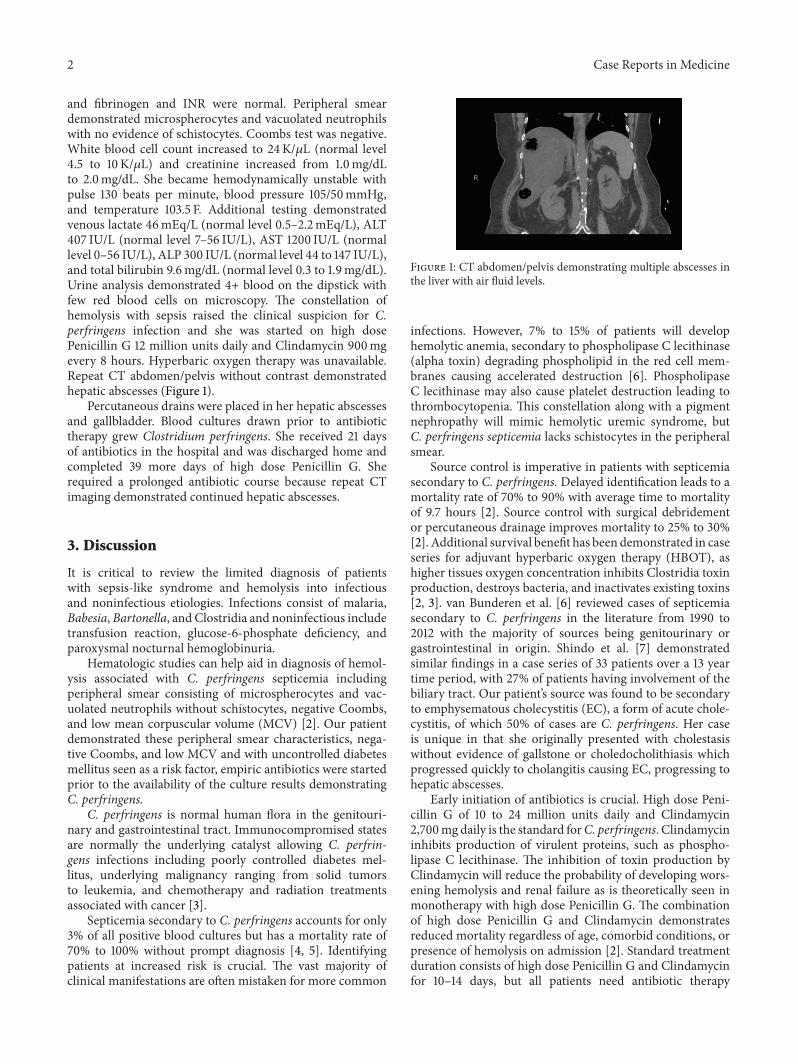
and fibrinogen and INR were normal. Peripheral smeardemonstrated microspherocytes and vacuolated neutrophilswith no evidence of schistocytes. Coombs test was negative.White blood cell count increased to 24K/𝜇L (normal level4.5 to 10 K/𝜇L) and creatinine increased from 1.0mg/dLto 2.0mg/dL. She became hemodynamically unstable withpulse 130 beats per minute, blood pressure 105/50mmHg,and temperature 103.5 F. Additional testing demonstratedvenous lactate 46mEq/L (normal level 0.5–2.2mEq/L), ALT407 IU/L (normal level 7–56 IU/L), AST 1200 IU/L (normallevel 0–56 IU/L), ALP 300 IU/L (normal level 44 to 147 IU/L),and total bilirubin 9.6mg/dL (normal level 0.3 to 1.9mg/dL).Urine analysis demonstrated 4+ blood on the dipstick withfew red blood cells on microscopy. The constellation ofhemolysis with sepsis raised the clinical suspicion for C.perfringens infection and she was started on high dosePenicillin G 12 million units daily and Clindamycin 900mgevery 8 hours. Hyperbaric oxygen therapy was unavailable.Repeat CT abdomen/pelvis without contrast demonstratedhepatic abscesses (Figure 1).
Percutaneous drains were placed in her hepatic abscessesand gallbladder. Blood cultures drawn prior to antibiotictherapy grew Clostridium perfringens. She received 21 daysof antibiotics in the hospital and was discharged home andcompleted 39 more days of high dose Penicillin G. Sherequired a prolonged antibiotic course because repeat CTimaging demonstrated continued hepatic abscesses.
3. Discussion
It is critical to review the limited diagnosis of patientswith sepsis-like syndrome and hemolysis into infectiousand noninfectious etiologies. Infections consist of malaria,Babesia, Bartonella, and Clostridia and noninfectious includetransfusion reaction, glucose-6-phosphate deficiency, andparoxysmal nocturnal hemoglobinuria.
Hematologic studies can help aid in diagnosis of hemol-ysis associated with C. perfringens septicemia includingperipheral smear consisting of microspherocytes and vac-uolated neutrophils without schistocytes, negative Coombs,and low mean corpuscular volume (MCV) [2]. Our patientdemonstrated these peripheral smear characteristics, nega-tive Coombs, and low MCV and with uncontrolled diabetesmellitus seen as a risk factor, empiric antibiotics were startedprior to the availability of the culture results demonstratingC. perfringens.
C. perfringens is normal human flora in the genitouri-nary and gastrointestinal tract. Immunocompromised statesare normally the underlying catalyst allowing C. perfrin-gens infections including poorly controlled diabetes mel-litus, underlying malignancy ranging from solid tumorsto leukemia, and chemotherapy and radiation treatmentsassociated with cancer [3].
Septicemia secondary to C. perfringens accounts for only3% of all positive blood cultures but has a mortality rate of70% to 100% without prompt diagnosis [4, 5]. Identifyingpatients at increased risk is crucial. The vast majority ofclinical manifestations are often mistaken for more common
Figure 1: CT abdomen/pelvis demonstrating multiple abscesses inthe liver with air fluid levels.
infections. However, 7% to 15% of patients will develophemolytic anemia, secondary to phospholipase C lecithinase(alpha toxin) degrading phospholipid in the red cell mem-branes causing accelerated destruction [6]. PhospholipaseC lecithinase may also cause platelet destruction leading tothrombocytopenia. This constellation along with a pigmentnephropathy will mimic hemolytic uremic syndrome, butC. perfringens septicemia lacks schistocytes in the peripheralsmear.
Source control is imperative in patients with septicemiasecondary to C. perfringens. Delayed identification leads to amortality rate of 70% to 90% with average time to mortalityof 9.7 hours [2]. Source control with surgical debridementor percutaneous drainage improves mortality to 25% to 30%[2]. Additional survival benefit has been demonstrated in caseseries for adjuvant hyperbaric oxygen therapy (HBOT), ashigher tissues oxygen concentration inhibits Clostridia toxinproduction, destroys bacteria, and inactivates existing toxins[2, 3]. van Bunderen et al. [6] reviewed cases of septicemiasecondary to C. perfringens in the literature from 1990 to2012 with the majority of sources being genitourinary orgastrointestinal in origin. Shindo et al. [7] demonstratedsimilar findings in a case series of 33 patients over a 13 yeartime period, with 27% of patients having involvement of thebiliary tract. Our patient’s source was found to be secondaryto emphysematous cholecystitis (EC), a form of acute chole-cystitis, of which 50% of cases are C. perfringens. Her caseis unique in that she originally presented with cholestasiswithout evidence of gallstone or choledocholithiasis whichprogressed quickly to cholangitis causing EC, progressing tohepatic abscesses.
Early initiation of antibiotics is crucial. High dose Peni-cillin G of 10 to 24 million units daily and Clindamycin2,700mg daily is the standard forC. perfringens. Clindamycininhibits production of virulent proteins, such as phospho-lipase C lecithinase. The inhibition of toxin production byClindamycin will reduce the probability of developing wors-ening hemolysis and renal failure as is theoretically seen inmonotherapy with high dose Penicillin G. The combinationof high dose Penicillin G and Clindamycin demonstratesreduced mortality regardless of age, comorbid conditions, orpresence of hemolysis on admission [2]. Standard treatmentduration consists of high dose Penicillin G and Clindamycinfor 10–14 days, but all patients need antibiotic therapy
Case Reports in Medicine 3
duration tailored based on improvement of infection throughclinical and radiographic resolution [8, 9].
4. Conclusion
Septicemia secondary to Clostridium perfringens is fatal ifnot detected early. Source control with surgery or percuta-neous drainage and early antibiotic therapy with high dosePenicillin G and Clindamycin to decrease toxin production isimperative. Adjuvant HBOT has also shown survival benefitand should be considered if available. Clinicians caring for apatient with sepsis and intravascular hemolysis must suspectClostridium perfringens septicemia.
Appendix
See Figure 1.
Conflict of Interests
Justin Cochrane, Lacie Bland, and Mary Noble declare thatthere is no conflict of interests regarding the publication ofthis paper.
Acknowledgment
Thanks are due to Dr. Jeremy Graham for editing and paperrevisions.
References
[1] P. A. Banks, M. L. Freeman, and Practice Parameters Com-mittee of the American College of Gastroenterology, “Practiceguidelines in acute pancreatitis,” The American Journal ofGastroenterology, vol. 101, no. 10, pp. 2379–2400, 2006.
[2] T. G. Simon, J. Bradley, A. Jones, and G. Carino, “Massiveintravascular hemolysis from clostridium perfringens sep-ticemia: a review,” Journal of Intensive Care Medicine, vol. 29,no. 6, pp. 327–333, 2014.
[3] G. Rajendran, P. Bothma, and A. Brodbeck, “Intravascularhaemolysis and septicaemia due toClostridium perfringens liverabscess,” Anaesthesia and Intensive Care, vol. 38, no. 5, pp. 942–945, 2010.
[4] W.-C. Huang, W.-S. Lee, T. Chang, T.-Y. Ou, and C. Lam,“Emphysematous cholecystitis complicating liver abscess dueto Clostridium baratii infection,” Journal of Microbiology,Immunology and Infection, vol. 45, no. 5, pp. 390–392, 2012.
[5] S.-T. Law and M. K. Lee, “A middle-aged lady with a pyogenicliver abscess caused by Clostridium perfringens,”World Journalof Hepatology, vol. 4, no. 8, pp. 252–255, 2012.
[6] C. C. van Bunderen, M. K. Bomers, E. Wesdorp, P. Peerbooms,and J. Veenstra, “Clostridium perfringens septicaemia withmassive intravascular haemolysis: a case report and review ofthe literature,” Netherlands Journal of Medicine, vol. 68, no. 9,pp. 343–346, 2010.
[7] Y. Shindo, Y. Dobashi, T. Sakai, C. Monma, H. Miyatani, andY. Yoshida, “Epidemiological and pathobiological profiles ofClostridium perfringens infections: review of consecutive seriesof 33 cases over a 13-year period,” International Journal of
Clinical & Experimental Pathology, vol. 8, no. 1, pp. 569–577,2015.
[8] V. L. Yu, Antimicrobial Therapy and Vaccines, vol. 1, LippincottWilliams &Wilkins, Philadelphia, Pa, USA, 2nd edition, 2002.
[9] D. L. Longo, A. S. Fauci, D. L. Kasper, S. L. Hauser, J. J. Jameson,and J. Loscalzo,Harrison’s Principles of Internal Medicine, vol. 1,McGraw-Hill, 18th edition, 2012.

Submit your manuscripts athttp://www.hindawi.com
Stem CellsInternational
Hindawi Publishing Corporationhttp://www.hindawi.com Volume 2014
Hindawi Publishing Corporationhttp://www.hindawi.com Volume 2014
MEDIATORSINFLAMMATION
of
Hindawi Publishing Corporationhttp://www.hindawi.com Volume 2014
Behavioural Neurology
EndocrinologyInternational Journal of
Hindawi Publishing Corporationhttp://www.hindawi.com Volume 2014
Hindawi Publishing Corporationhttp://www.hindawi.com Volume 2014
Disease Markers
Hindawi Publishing Corporationhttp://www.hindawi.com Volume 2014
BioMed Research International
OncologyJournal of
Hindawi Publishing Corporationhttp://www.hindawi.com Volume 2014
Hindawi Publishing Corporationhttp://www.hindawi.com Volume 2014
Oxidative Medicine and Cellular Longevity
Hindawi Publishing Corporationhttp://www.hindawi.com Volume 2014
PPAR Research
The Scientific World JournalHindawi Publishing Corporation http://www.hindawi.com Volume 2014
Immunology ResearchHindawi Publishing Corporationhttp://www.hindawi.com Volume 2014
Journal of
ObesityJournal of
Hindawi Publishing Corporationhttp://www.hindawi.com Volume 2014
Hindawi Publishing Corporationhttp://www.hindawi.com Volume 2014
Computational and Mathematical Methods in Medicine
OphthalmologyJournal of
Hindawi Publishing Corporationhttp://www.hindawi.com Volume 2014
Diabetes ResearchJournal of
Hindawi Publishing Corporationhttp://www.hindawi.com Volume 2014
Hindawi Publishing Corporationhttp://www.hindawi.com Volume 2014
Research and TreatmentAIDS
Hindawi Publishing Corporationhttp://www.hindawi.com Volume 2014
Gastroenterology Research and Practice
Hindawi Publishing Corporationhttp://www.hindawi.com Volume 2014
Parkinson’s Disease
Evidence-Based Complementary and Alternative Medicine
Volume 2014Hindawi Publishing Corporationhttp://www.hindawi.com


