
Cardiovascular Research, 1977, 11, 427-433
Cardiac hypertrophy and antihypertensive therapy' S. SEN2, R. C. T A R A Z I , A N D F. M. B U M P U S
From the Research Division, Cleveland Clinic Foundation, Cleveland, Ohio
SUMMARY Biochemical (myocardial DNA, RNA, and hydroxyproline) and humoral (plasma [PRA] and kidney [KRA] renin activity) factors were determined in spontaneously hypertensive rats (SHR) and normotensive Wistar controls (NR) before and following treatment with minoxidil or propranolol. Minoxidil ( I 50 mg-litre-1 drinking water) effectively controlled blood pressure (17.3 kPa vs 24.9 kPa [130 mmHg vs 187 mmHg], P <0.001) despite marked and sustained increases in both PRA and K R A ventricular weight which were not reduced and myocardial DNA, RNA, and hydroxyproline which were increased by minoxidil (P < 0.01). In contrast propranolol did not reduce blood pressure in SHR but ventricular weight was reduced somewhat (3.1 *0.4 mg-g-l vs 3.4 0.09 m g p , P < 0.05); in both S H R and NR, KRA, and PRA were lowered by pranolol. Methyldopa which controlled blood pressure and lowered PRA led to a reversal of hypertrophy. Thus, although blood pressure control is obviously important for reversing cardiac hypertrophy, it may not be the sole factor for the development and reversal of cardiac hypertrophy.
Spontaneous hypertension in rats offers a unique model to study the natural development of cardiac hypertrophy and its reversal by medical therapy. Its systematic investigation led to the suggestion that
biochemical composition of myocardium and in plasma renin activity.
Methods other factors beside arterial pressure are involved in the development of cardiac hypertrophy in hyper- tension (Sen et al., 1974). and uncovered the different effects of antihypertensive drugs and their reversal (Sen et al., 1974). We have shown that although treatment with either a-methyldopa or hydralazine controlled blood pressure effectively in SHR, cardiac hypertrophy was reversed only by a-methyldopa and not by hydralazine (Sen et at., 1974). This discrepancy in response to ventricular weight to blood pressure control by different antihypertensive agents could be attributed to differences in their effect on haemodynamic, humoral, and/or neuro- genic factors. A biochemical effect of the drug on cardiac muscle remained another possibility.
In order to evaluate the relative contribution of these various factors, this study was performed using another vasodilator (minoxidil instead of hydrala- zine) and especially investigating the effect of beta- adrenergic blockade alone and in combination with other drugs. Blood pressure response was correlated with the changes observed in heart weight, in the
'This work was supported by NHLl grants HL-6835 and
*Address for correspondence: Dr Subha Sen, Research Division, Cleveland Clinic Foundation, 9500 Euclid Avenue, Cleveland, Ohio 44106, USA.
HL-I 5837.
Details of materials and methods used have been published previously (Sen et al., 1974). (1) Materials The spontaneously hypertensive rats used in the study were of Okamoto-Aoki strain (SHR). They were born in Carworth Labs (F-26) and brought to the Cleveland Clinic at weaning age. They were kept in a separate room under veterinarian supervision. Male SHR used in this study were of 12 to 14 weeks of age and proven to havee stablished hypertension. Normal controls we used were male rats of the same age of Kyoto Wistar strains. Both normal and SHR were kept under the same conditions, properly housed and fed (Purina rat chow), and handled by the same person. Ten rats were used in each group and subgroup for each drug therapy. (2) Antihypertensive therapy The drugs used in these studies were minoxidil, propranolol, and a combination of minoxidil and propranolol, and of minoxidil and a-methyldopa. They were given in drinking water in the following dosages: (a) Minoxidil 80 mg.litre-l (b) Propranolol 1.5 g.litre-' (c) Minoxidil i- 80 mglitre-'
Propranolol 1.5 g.litre-' (d) Minoxidil + 80 mg.litre-'
a-met hyldopa 2.5 glitre-l 427
by guest on Novem
ber 30, 2014D
ownloaded from

428 S. Sen, R. C. Tarazi, and F. M . Bumpirs
The criteria used to establish dosage were effective blood pressure control in SHR; this did not apply to propranolol because it did not lower blood pressure. The criteria used for propranolol were therefore suppression of plasma renin activity. The dosages used were found by pilot experiments in which SHR were given different doses of minoxidil ranging from 20 mg.litre-l to 120 mg.litre-I; administration of 80 mg.1itre-I in drinking water resulted in effective blood pressure control. Similarly the appropriate dose for propranolol was chosen after determination of plasma renin activity at various levels of drug intake; 1.5 gelitre-' was found to suppress PRA significantly.
All rats (both normal and SHR) were treated for 6 weeks, because previous experience (Sen et al., 1974) showed the need for at least this duration of treatment to result in clear cut changes in cardiac weight. In addition, to investigate the effects of minoxidil given alone, 3 other groups of SHR were treated for periods of 2, 4, and 8 weeks. The route of administration was chosen because it had been shown to lower blood pressure effectively and was more convenient than intermittent intramuscular injections (Sen et a/., 1974). ( 3 ) Blood pressure measurement In all rats, arterial pressure was measured using a tail-cuff, similar to the method described by Friedman and Freed (1 949). The blood pressure was measured twice a week in each rat by the same person and approximately the same time of the day. The rats were weighed twice weekly and the last determination of blood pressure and weight was taken immediately before sacrifice.
For plasma renin activity (PRA) determination of a blood sample (0.5 cm3) was obtained under light ether anaesthesia from each rat 1 d before sacrifice. The rats were killed by decapitation and ventricles immediately taken out, cleaned, weighed, and prepared for biochemical determinations as described in detail previously (Sen et a/., 1974). The upper part of the ventricles was used to deter- mine dry weight / wet weight ratio and the lower for biochemical analysis (Sen et a/. , 1976). (4) Analytical methods RNA was determined according to the method of Fleck and Munro (1962) using ultraviolet absorption method. DNA was determined by measuring colour density produce between DNA and indole heated in presence of HC1 as described by Ceriotti (1952). Hydroxy- proline was determined in the ventricular tissue extract by colourimetric method using diethyl amino benzaldehyde (Bergman and Loxley, 1963). Plasma and kidney renin activity were measured according to the micromethod of Boucher et al. ( I 967) using exogenous substrate prepared from 48 h bilaterally nephrectomised male rats.
Results
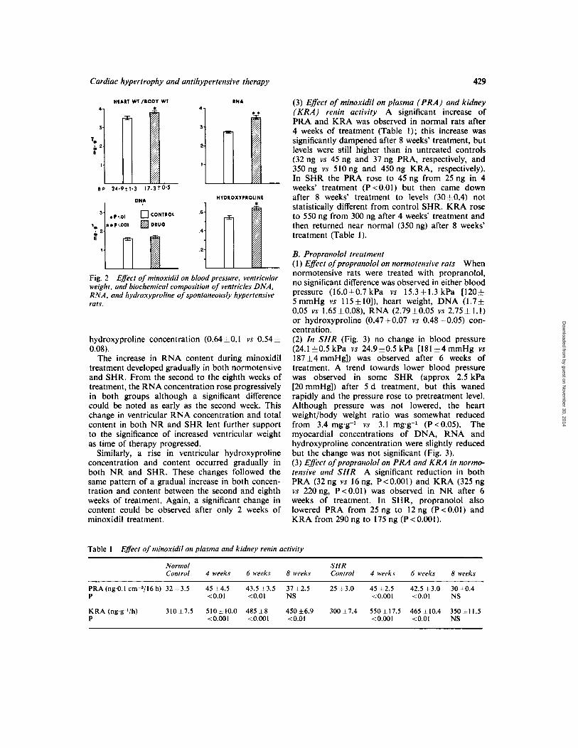
B L O O D PRESSURE CONTROL A N D MYOCARDIAL COMPOSITION A . Minoxidil therapy (1) Effect on normal rats (Fig. 1) Minoxidil treat- ment for 6 weeks had no significant effect on blood pressure level; these averaged 122k3 (CE) and 122 f 8 in untreated and treated rats, respectively, despite a significant increase in ventricular weight/ body weight ratio (2.6 vs 2.9, P ~ 0 . 0 1 ) . Myocardial DNA concentration was increased slightly (P <0.05), and the concentration of hydroxyproline (OHp) rose from 0.51 to 0.58 mg.g-l (P<O.OI). The most significant difference was found in RNA concentra- tion which was increased from 2.6 mg.g-l to 3.2 mg.g-l (P <O.OI). Obviously the total ventricular content of all 3 constituents (DNA, RNA, and OHp) was significantly higher in treated rats both because of increased concentration of each and increased ventricular mass. (2) Effect on S H R (Fig. 2) After 6 weeks of minoxidil therapy, the blood pressure was reduced from 24.91-tl.3 kPa to 17.33~0.5 kPa ( l87+ 10 mmHg to 1 3 0 ~ t 4 mmHg) (P <O.OOl). However, despite the lowering of the blood pressure, ventricu- lar hypertrophy was not reversed, in fact the heart weight/body weight ratio rose from 3.5 mg.g-l to 3.8 mg.g-l (P <0.01). The biochemical pattern associated with the increased ventricular weight was similar to that observed in normotensive rats: minimal increase in DNA concentration (1.7 10.1 vs 1.640. I), but highly significant (P ~ 0 . 0 1 ) increase in both RNA (3.5 <0.08 vs 2.75 <0.05) and
3
'4 t
I
HEAR1 W l /BODY W1
WYDROXYPROLINE
0,751
DNA
0 CONlROL
DRUG
2.0
1.0
Fig. 1 weight and biochemical composition of ventricles, namely DNA, RNA, and hydroxyproline of normal rars.
Effect of minoxidil on blood pressure, ventricular.
by guest on Novem
ber 30, 2014D
ownloaded from
Cardiac hypertrophy and antihypertensive therapy 429
HEAR1 W1 /BODY WT RNA
41
HVDROXYPROLINE
6
.4
2
Fig. 2 weight. and biochemical composition of ventricles DNA, RNA. and hydroxyproline of spontaneously hypertensive rats.
Effect of minoxidil on blood pressure, ventricular
hydroxyproline concentration (0.64 i O . 1 vs 0.54 =t 0.08).
The increase in RNA content during minoxidil treatment developed gradually in both normotensive and SHR. From the second to the eighth weeks of treatment, the RNA concentration rose progressively in both groups although a significant difference could be noted as early as the second week. This change in ventricular RNA concentration and total content in both NR and SHR lent further support to the significance of increased ventricular weight as time of therapy progressed.
Similarly, a rise in ventricular hydroxyproline concentration and content occurred gradually in both NR and SHR. These changes followed the same pattern of a gradual increase in both concen- tration and content between the second and eighth weeks of treatment. Again, a significant change in content could be observed after only 2 weeks of minoxidil treatment.
(3) Efec t of minoxidil on plasma ( P R A ) and kidney ( K R A ) renin activity A significant increase of PRA and KRA was observed in normal rats after 4 weeks of treatment (Table 1); this increase was significantly dampened after 8 weeks’ treatment, but levels were still higher than in untreated controls (32 ng vs 45 ng and 37 ng PRA, respectively, and 350ng vs 510ng and 450ng KRA, respectively). In SHR the PRA rose to 45 ng from 25 ng in 4 weeks’ treatment (P<O.Ol) but then came down after 8 weeks’ treatment to levels (30h0.4) not statistically different from control SHR. KRA rose to 550 ng from 300 ng after 4 weeks’ treatment and then returned near normal (350 ng) after 8 weeks’ treatment (Table 1) .
Table 1 Effect of minoxidil on plasma and kidney renin activity
B. Propranolol treatment ( I ) Effect of propranolol on normotensive rats When normotensive rats were treated with propranolol, no significant difference was observed in either blood pressure (16.0i0.7 kPa vs 15 .3k1 .3 kPa [120;1, 5 mmHg vs 115 *lo]), heart weight, DNA (1.7f 0.05 vs 1.65*0.08), RNA (2.7910.05 vs 2.75-C 1 . 1 ) or hydroxyproline (0.47 50.07 vs 0.48 *0.05) con- centration. (2) In SHR (Fig. 3) no change in blood pressure (24.1 10.5 kPa vs 24.9h0.5 kPa [I81 k 4 mmHg vs 18714mmHgl) was observed after 6 weeks of treatment. A trend towards lower blood pressure was observed in some SHR (approx 2.5 kPa [20 mmHg]) after 5 d treatment, but this waned rapidly and the pressure rose to pretreatment level. Although pressure was not lowered, the heart weight/body weight ratio was somewhat reduced from 3.4 mg.g-’ vs 3.1 mg.g-’ (P<O.O5). The myocardial concentrations of DNA, RNA and hydroxyproline concentration were slightly reduced but the change was not significant (Fig. 3). (3) Effect of propranolol on PRA and K R A in normo- tensive and S H R A significant reduction in both PRA (32 ng vs 16 ng, P<O.OOI) and KRA (325 ng vs 220 ng, P<O.OI) was observed in NR after 6 weeks of treatment. In SHR, propranolol also lowered PRA from 25 ng to 12 ng (P <0.01) and KRA from 290 ng to 175 ng (P<O.OOI).
Normal S H R Control 4 weeks 6 weeks 8 weeks Control 4 weeks 6 weeks 8 weeks
PRA (ng.O.1 ~ r n - ~ / 1 6 h) 32 2-33 45 5 4 . 5 43.5 5 3 . 5 37 i 2 . 5 25 1 3 . 0 4 5 5 2 . 5 42.5 k 3 . 0 30 10.4 P < 0.0 I 10.01 NS ~ 0 . 0 0 1 ~ 0 . 0 1 NS
K R A (ng,g-’/h) 310*7.5 510ztlO.O 485zt8 450f6 .9 3 0 0 1 7 . 4 550*17.5 4 6 5 1 1 0 . 4 3504-11.5 P <O.oOl C O . 0 0 1 <O.OI <0.001 <0.01 NS
by guest on Novem
ber 30, 2014D
ownloaded from

430 S . Sen, R . C . Tarazi, and F. M . Bumpiis
HEART W1 /OODV WT DNA 4.
Fig. 3 Effect of propranolol on blood pressure, ventricular weight, DNA, R N A , and hydroxyproline on SHR.
HEAR1 W l /OODY WT DNA
0 CONTROL
DRUG
HVDROXYPROLtNE RNA 3
6 3 - 4 2 'm
i 2 1 p .4
t 2 1
Fig. 5 on blood pressure, ventricular weight, and biochemical composition of SHR.
Effect of combined minoxidil-propranolol therapy
C . Combined minoxidil-propranolol therapy ( 1 ) Effect in norrnotensive rats (Fig. 4) Addition of propranolol to minoxidil did not prevent the development of cardiac hypertrophy; the heart weight/body weight ratio increased to practically the same level (2.5*0.1 vs 2310.05) as it did with minoxidil alone. Similarly, both hydroxyproline and RNA concentration also increased (Fig. 4); no significant difference in DNA content was observed. (2) Effect on SHR Exactly the same biochemical results were observed in treated SHR as in treated
HEART WT /OODY W l DNA
3 - 2 2 E
1
B P 14.7t0.7 15.3 t0.7
HYDROXVPROUNE RNA
Fig. 4 Effect of combined minoxidil-propranolol therapy on blood pressure, ventricular weight, and biochemical composition of normal rats.
RNA
normotensive rats (Fig. 5) although there were significant changes in blood pressure. The latter was initially reduced from 25.3i1.3 kPa (190 i 10mmHg) to an average of 18.0+1.3 kPa (1355 10 mmHg) (P <O.OI) after the first week of treatment but it subsequently rose in many rats so that average levels showed less satisfactory control (20.111.3 kPa [I51~10mmHg], P<O.O5) at the end of 8 weeks' treatment. Concomittantly, the heart weight/body weight ratio increased (3.6 mg.g-' vs 3.85 mg.g-I, P < 0.05), as did the myocardial concentration of RNA (2.75iO.l mg.g-l vs 3 .01 0.1 mg.g-l) and of hydroxyproline (0.51 10.1 mg.g-' vs 0.58f0.1 mgg-I). DNA concentration was not significantly altered.
D. Combined minoxidil-methyldopa therapy (1) Effect on normotensive rats (Fig. 6) In contrast to propranolol, methyldopa prevented the minoxidil- induced ventricular hypertrophy. Neither heart weight/body weight ratio (2.57 vs 2.55, P=NS) nor average blood pressure (14.9 f0.5 kPa vs 14.77 i- 0.44 kPa [112+4 mmHg vs 110.813.3 mmHg]) were significantly different from untreated controls. Similarly, DNA concentration remained unchanged while RNA and hydroxyproline concentration were only slightly elevated: 2.5 ng.g-l vs 2.7 ngg-I and 0.58 ngag-l vs 0.60 ngsg-' (P < 0.05), respectively. (2) Effect on SHR Treatment of SHR with com- bined therapy of minoxidil and a-methyldopa, produced a significant reduction in blood pressure (25.310.7 kPa vs 16.7il .OkPa [19015 mmHg
by guest on Novem
ber 30, 2014D
ownloaded from

Cardiac hypertrophy and antihypertensive therapy 43 I
M A R 1 Wl/11OOY W l ONA Discussion
3.
m 8 2-
I.
3 i
P - N S 0 CONTROL
DRUG The results clearly suggest that factors other than blood pressure control play a significant role in the reversal of cardiac hypertrophy by antihypertensive therapy. They further demonstrated that the hyperdynamic circulation induced by potent vaso- dilators, such as minoxidil, can lead to significant cardiac hypertrophy, irrespective of the blood pres-
HYOROXYPIOLINE RNA
Fig. 6 Efect of combined minoxidil-methyldopa therapy on blood pressure, ventricular weight, and biochemical composition of ventricles of normal rats.
vs 125 ..7.5 mmHg]), heart weight/body weight ratio (3.550.15 vs 3.2+0.1, PcO.OI), when com- pared with untreated control. A significant reduction in RNA concentration, 3.5 vs 3.06, was also obtained. When compared with minoxidil treated SHR, however, the latter value 3.06 was still slightly, although not significantly, higher in the untreated controls. Although both heart weight/body weight ratio and RNA were reduced, they did not regress completely to levels found in NR, despite main- tained good blood pressure control. An increase in hydroxyproline concentration was observed (0.53 mg.g-l vs 0.59 mg.g-') when compared with untreated controls. The biochemical changes caused by combined therapy with minoxidil and aldomet are summarised in Table 2.
Table 2 Eflect of combination therapy in SHR
Minoxidil ( M x ) Minosidil -1 I Methyldopa ( M d ) Propranolof ( P ) M x M x + M x Ms I alone Md alone P
Heart weight (mg.g-') 3 . 5 i 3 . 2 ~ 3 . 6 i 3.85* Body weight 0.1 0.05' 0.1 +0.15
PRA 4 3 + 3 3 5 4 3 + 38* L (ngO.1 ~ m - ~ / 1 5 h) 0.1 0.2* 0.1 0.05
Blood pressure (kPa) 17.3 i 17.1 f 17.3 i 18.0r 0.5 1.3 0.5 1.3
*Changes were significant, P <0.01. Conversion: I kPa=7.519 mmHg.
sure effects of the drug. In SHR, minoxidil controlled blood pressure effectively, yet the ventricles became even heavier while in normotensive rats cardiac hypertrophy developed, although blood pressure remained at essentially the same levels. These effects are analogous to those obtained with hydrala- zine. both in SHR (Sen et al., 1975) and in rats with renovascular hypertension (Masson et al., 1958).
Many factors could play a significant role in this paradox; these include different effects of the drugs used on haemodynamic functions, on adrenergic drive to the heart, and in the renin-angiotensin system (Sen e t a / . , 1974; Tarazi e t al., 1977). Avail- able indices might favour the first 2 rather than a renin effect but is not sufficient to establish definitely the relative role of all factors involved. Also to be considered are possible direct biochemical effects of some drugs on the myocardium. The constellation of alterations produced by various antihypertensive regimen is outlined in Table 3. Although it was previously suggested that stimulation of the renin- angiotensin system might play some role in the development and reversal of cardial hypertrophy in hypertension (Sen et al., 1974), it would appear from the present results that reflex sympathetic stimula- tion and haemodynamic factors might play a more important role. This tentative conclusion is based on results obtained with minoxidil and with beta- adrenergic blockade. As regards the first, renin
Table 3 drugs in S H R
Summary of the ejects of amtihypertensive
Effect on t f e c t on Eflecr on
pressure in Iiypertrophp SHR SHR in SHR
Drug blood ventricular PRA in
Minoxidil J. T t c Propranolol -
Methyldopa c c e Minoxidil 4
propranolol J t 1 Minoxidil t
c J. J.
-* j. *
methyldopa
'Changes statistically significant (P <0.05) but small in absolute number (14%).
by guest on Novem
ber 30, 2014D
ownloaded from

432 S. Sen, R. C. Tarazi. and F. M . Bi~mpiis
stimulation was not sustained with chronic ad- ministration of minoxidil; yet, although PRA levels slowly returned to control after a few weeks’ treatment, cardiac hypertrophy progressed (Table 1).
Used alone, beta-adrenergic blockade had only minimal effects on ventricular weight in SHR; although the reduction was significant (P < 0.05), the actual numbers involved were very small. In rabbits, beta-adrenergic blockade resulted in severe loss of ventricular weight comparable with that following severe myocardial infarctions (Williams et al., 1975). The discrepancy between these results and ours might be caused by dosage or species differences. Neither in normotensive rats nor in spontaneously hypertensive rats did we observe any important loss in ventricular weight, any difference in the behaviour of the animals, nor any post-mortem evidence of cardiac failure. It might have been expected that sustained bradycardia and prolonged withdrawal of sympathetic support to the heart might have favoured ventricular hypertrophy. This, however, was not observed, possibly because of the relatively short-term experiments (6 to 8 weeks). As has been reported by other investigators (Ebihara and Nakamura, 1979, propranolol did not control blood pressure in SHR. The fact that cardiac hyper- trophy was not accentuated and might even have been slightly reduced, indicates that the degree of beta-adrenergic blockade achieved with our doses did not apparently interfere with cardiac compensa- tion. Evidence for that blockade was derived from the lowering of plasma renin activity obtained by propranolol therapy. The adequacy of that criterion in the absence of haemodynamic measurements is suggested by the many reports that the cardiac effects of propranolol parallel closely its reduction of plasma renin activity (Pettinger et a/., 1973).
More surprising than the effects of propranolol alone were the opposite effects of methyldopa and of beta-blockade when used in combination with a vasodilator agent. Whereas addition of propranolol to minoxidil did not prevent the development of cardiac hypertrophy, the association of methyldopa with minoxidil reserved cardiac hypertrophy in SHR and prevented the minoxidil-induced hypertrophy in normotensive rats. The apparent lack of effect of propranolol could be ascribed to the inadequate beta-adrenergic blockade for the degree of cardiac stimulation induced by the potent vasodilator. Experience in man suggests that the addition of propranolol quietens the hyperkinetic circulation but often does not quite restore it to pre-minoxidil levels (Tarazi et al., 1976). However, both methyldopa and propranolol lowered PRA to approximately the same levels in rats. In contrast to
reports by Pettinger and co-workers (1973), the addition of propranolol did not lead to better blood pressure control by minoxidil nor to smooth and consistent reduction in plasma renin activity. This is similar to our experience in hypertensive patients (Dustan et al., 1975) and to the results obtained by Velasco et a/. (1974). Similarly, variations of equal magnitude in PRA levels were also noted following the addition of methyldopa to minoxidil. Despite these similarities as regards PRA, blood pressure control was somewhat less satisfactory with the minoxidil-propranolol combination than with min- oxidil-methyldopa. The favourable effect of methyl- dopa on this cardiac hypertrophy might, therefore, be ascribed more to its cardiovascular action in the dosage used than to its effects on PRA.
This divergence in the results of the 2 combina- tions on ventricular weight suggests that haemo- dynamic factors and adrenergic suppression might be more important than reduction of plasma renin activity in determining the degree of reversal of cardiac hypertrophy by medical treatment. The addition of either drug to minoxidil led to wide variations in plasma renin activity. Because of these variations any conclusion as to the possible role of the renin-angiotensin system can only be speculative. More definite answers must await experiments with more frequent determination of plasma renin activity during the experimental period instead of 1 measurement only at the end of the experiments. The effects of anaesthesia on plasma renin activity might also be differently affected by the various medications used and repeat studies without the use of ether anaesthesia might help obtain a more complete picture.
Despite these restrictions. the close parallelism between plasma and kidney renin activity in the different groups investigated suggest that the vari- ation in plasma levels did reflect the effect of therapy and were not only transitory fluctuations. The use of single drugs appeared to confirm our previous suggestion regarding the possible role of the renin- angiotensin system and the reversal of cardiac hypertrophy. Changes in ventricular weight and plasma renin levels paralleled each other; both were reduced by propranolol as well as by methyldopa, whereas both minoxidil and hydralazine increased plasma renin, and left cardiac hypertrophy remained unchanged or was even further accentuated despite the reduction in afterload. However, the divergent effects of drug combinations on hypertrophy, despite their similar dampening of plasma renin activity, suggest that further experiments are needed before an exact relationship between the various factors involved can be established.
Another possibility for the difference in effect of
by guest on Novem
ber 30, 2014D
ownloaded from

Cardiac hypertrophy and antihypertensive therapy 433
propranolol and methyldopa is a hypothetical, direct biochemical effect of one drug on myocardial metabolism. Experiments are in progress to evaluate the role of other sympathetic blockers in order to determine whether the reversal of cardiac hyper- trophy by methyldopa is peculiar to that compound or is shared by other sympathetic antagonists. Similar to our experience with methyldopa alone, the prevention of increase in cardiac weight did not mean that the biochemical composition of the myocardium was not altered. Hydroxyproline concentration was slightly but significantly elevated in the small hearts obtained by combined treatment with minoxidil and methyldopa. Possible implica- tions of this relative increase in fibrous tissue await haemodynamic determinations to decide whether reversal of hypertrophy is always and unequivocally beneficial.
We are grateful to Miss Carolee Hollinger and Mrs Rita Block for their expert technical assistance. We are also grateful to Upjohn Co for the generous gift of minoxidil and Ayerst Lab for propranolol.
References
Bergman, I., and Loxley, R. (1963). Two improved and simplified methods for the spectrophotometric determina- tion of hydroxyproline. Analytical Chemistry, 35, 1961- 1965.
Boucher, R., Menard, J., and Genest, J. (1967). Micromethod for measurement of renin in the plasma and kidney of rats. Canadian Journal of Physiology and Pharmacology.
Ceriotti, B. (1952). Micromethod of determination of de- oxyribonucleic acid. Journal of Biological Chenlistry, 198,
Dustan, H. P., Tarazi, R. C., and Bravo, E. L. (1975). Hemodynamic adjustments during long term antihyper-
45, 881-890.
297-303.
tensive drug therapy. In Recent Advances in Hypertension. pp. 247-258. Edited by P. Millez and M. Safar. Boehringer: Ingelheim.
Ebihara, A., and Nakamura, Y. (1975). Effect of oral ad- ministration of propranolol on spontaneously and renal hypertensive rats. Japanese Heart Journal, 16, 342-343.
Fleck, A., and Munro, H. N. (1962). The precision of ultra- violet absorption measurements into Schmidt-Thanhauser procedure of nucleic acid estimation. Biochetnica Bio- physics Acta. 51, 571-583.
Friedman, M., and Freed, S. C. (1949). Microphonic mano- meter for indirect determination of systolic blood pressure in the rat. Proceedings of the Society for Experimental Biology and Medicine. 10, 67C672.
Masson, G. M. C., McCormack, L. J., Dustan, H. P., and Corcoran, A. C. (1958). Hypertensive vascular disease as a consequence of increased arterial pressure. American Journal of Pathology, 34, 817-823.
Pettinger, W. A., Campbell, W. B., and Keeton, K. (1973). Adrenergic component of renin release induced by vasodi- lating antihypertensive drugs in the rat. Circulation Research, 33, 82-86.
Sen, S., Tarazi, R. C., Khairallah, P. A., and Bumpus, F. M. (1974). Cardiac hypertrophy in spontaneously hyperten- sive rats. Circulation Research. 35, 775-782.
Sen, S., Tarazi. R. C., and Bumpus, F. M. (1976). Biochemi- cal changes associated with development and reversal of cardiac hypertrophy in spontaneously hypertensive rats. Cardiovascular Research. 10, 254-26 I .
Tarazi, R. C., Dustan, H. P., Bravo, E. L., and Niarchos, A. P. (1976). Vasodilating drugs contrasting hemodynamic effects. Clinical Science and Molecular Medicine (In press).
Tarazi, R. C., Ferrario, C. M., and Dustan, H. P. (1977). The heart in hypertension. In Hypertension: Physiology and Treatment. Editors J. Genest, E. Kiow and 0. Kuchel. McGraw Hill: New York.
Velasco, M., O’Malley, K., Robie, N. W., Wells, J., Israili, Z. H., and McNay, J. L. (1974). Differential effects of propranolol on heart rate and plasma renin activity in patients with minoxidil. Clinical Pharmacology and Therapeutirs, 16, 103 1-1 038.
Vaughan Williams, E. M., Raine, A. E. G., Cabrera, A. A., and Whyte, J. M. (1975). The effects of prolonged (3- adrenoceptor blockade on heart weight and cardiac intracellular potentials in rabbits. Cardiovascular Re- search. 9, 579-592.
by guest on Novem
ber 30, 2014D
ownloaded from


