Syllabus (2014-2015)
B.Sc Radiology & Imaging Technology
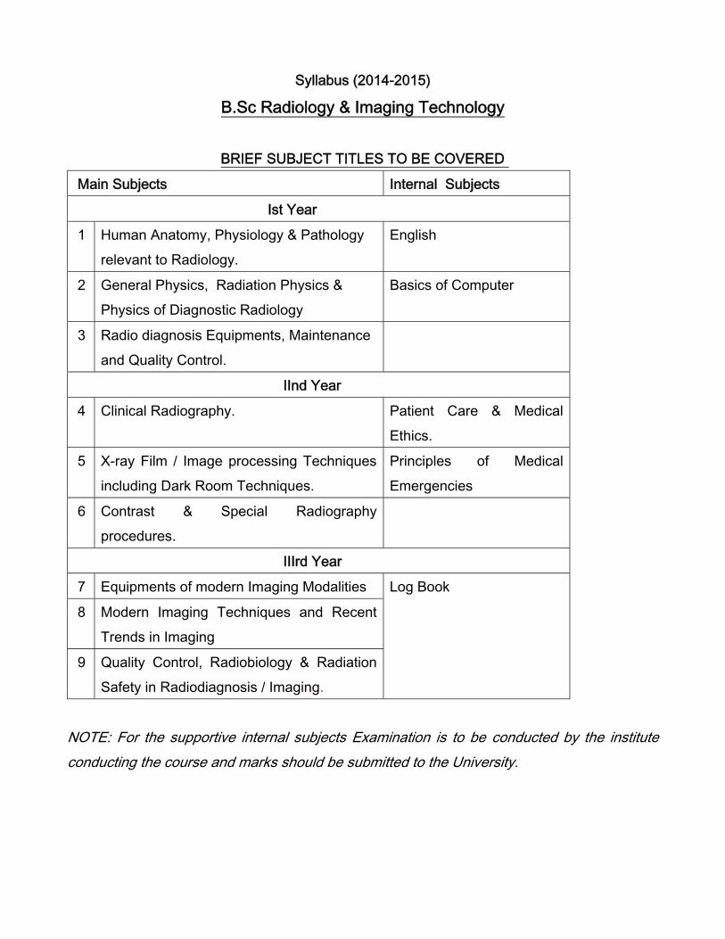
BRIEF SUBJECT TITLES TO BE COVERED
Main Subjects Internal Subjects
Ist Year
1 Human Anatomy, Physiology & Pathology relevant to Radiology.
English
2 General Physics, Radiation Physics & Physics of Diagnostic Radiology
Basics of Computer
3 Radio diagnosis Equipments, Maintenance and Quality Control.
IInd Year
4 Clinical Radiography. Patient Care & Medical Ethics.
5 X-ray Film / Image processing Techniques including Dark Room Techniques.
Principles of Medical Emergencies
6 Contrast & Special Radiography procedures.
IIIrd Year
7 Equipments of modern Imaging Modalities Log Book
8 Modern Imaging Techniques and Recent Trends in Imaging
9 Quality Control, Radiobiology & Radiation Safety in Radiodiagnosis / Imaging.
NOTE: For the supportive internal subjects Examination is to be conducted by the institute conducting the course and marks should be submitted to the University.
Syllabus for B.SC Radiology & Imaging Technology
FIRST YEAR Internal Paper
ENGLISH
SYLLABUS DETAILS
DESCRIPTION
The course is designed to enable students to enhance ability to comprehend spoken and written English (and use English) required for effective communication in their professional work. Students will practice their skills in verbal ad written English during clinical and classroom experience.
OBJECTIVES
At the end of the course, the student will develop
1. Ability to speak and write grammatically correct English.2. Effective skill in reading and understanding the English language.3. Skill in reporting
CONTENT
1. COMMUNICATION
• Role• Definition• Communication• Classification of communication
• Purpose• Major difficulties• Barriers• Characteristics – The seven Cs• Communication at the work place• Human needs and communication “Mind mapping”• Information communication
2. COMPREHENSION PASSAGE
• Reading purposefully• Understanding what is read• Drawing conclusion• Finding and analysis
3. EXPLAINING
• How to explain clearly• Defining and giving reasons• Explaining differences• Explaining procedures• Giving directions
4. WRITING BUSINESS LETTERS
• How to construct correctly• Formal language• Address• Salutation• Body and Conclusion
5. REPORT WRITING• Reporting and accident• Reporting what happened at a session• Reporting what happened at a meeting
PRACTICUM • The clinical experience in the wards and bed side nursing will provide opportunity for
students to fulfill the objectives of learning language.• Assignment on writing and conversation through participation in discussion debates
seminars and symposia. The students will gain further skills in task orientedcommunication.
METHODS OF TEACHING 1. Lecture2. Pair and Group work3. Role plays4. Oral presentations5. Decoding & production grammar exercise6. Comprehension exercise7. Writing assignments8. Word puzzles & Quizzes9. Communicative games & fluency activities
METHODS OF EVALUATION 1. Individual oral presentations2. Group discussion3. Answering questions from the prescribed English text.4. Summary / Essay / Letter writing5. Grammar exercises6. Medical / General vocabulary exercises
Internal Assessment in Year 1: English (Total 50 marks) Theory: English Theory Paper for internal assessment in First Year to be combined with computer science paper as follows-
English-25 + Computer Science-25 marks Viva: 25 marks
Reference Books 1. Selva Rose. 1997, Career English for Nurses. Published by: Orient Blackswan Ltd2. Oxford advanced Leaners Dictionary, 19963. Quirk Randolph and Greenbaum Sidney, 1987. A University Grammar of English,
Hong Kong: Longman group (FE) Ltd/ Pearson.4. Thomson A.J. and Maituiet A.V. 1987, A Practical English Grammar, Delhi: Oxford
University Press.5. Gimson A.C.1989, An Introduction to pronunciation of English. Hodder Arnold; 4th
Revised edition (1 May 1989).6. O’Connor J.D, 1986. Better English pronunciation. Cambridge: University Press7. By water F.V.A. 1982, Proficiency Course in English. London: 1-lodder and Strongliton.8. Roget S.P. 1960, Thesaurus of English Words & Phrases, London: Lowe & Brydone
Ltd. 1960.
2. Basics of Computer
Digital electronics and computers fundamental Number systems: Binary, octal, decimal & Hexa-decimal, conversions from one system to another, Analog to Digital Converter and Digital to Analog Converter. Computer fundamentals: Central Processing Unit, Memory RAM and ROM, Arithmetic and Logic Unit, Display devices, Hard copy devices, Input devices. Computer Applications related to Radiography with examples. Internal Assessment in Year 1: Computer Science [Total 50 marks] Theory: Theory Paper in Computer Science for internal assessment in First Year to be combined with English paper as follows-
English-25 + Computer Science-25 (Total 50 marks)
Practical / Viva for internal Assessment in Computer Science (25marks)
Paper–I. Human Anatomy, Physiology & Pathology relevant to Radiology .
1. General structure of the human body, anatomic terminology, planes of section-Structure and function of human cell with special reference to mitochondria and ribosomes. 2. Elementary tissues of human body- Epithelial tissue, muscular tissue, connectivetissues and nervous tissue.
3. Cardio Vascular System - Anatomy of heart and functions- Structure and functions ofvarious parts of the heart, arterial and venous system, brief account on common cardiovascular disorders. Blood pressure and its recording. Anatomy and function of arteries, capillaries and Arterial system, Venous system. 4. Hematology-Composition of Blood - functions of blood elements –Blood Group andcoagulation of blood, disorders of blood.
5. Lymphatic system - Name and function of lymph glands, Lymphatics and Lymphaticpathway outline.
6. Respiratory System: various parts of respiratory system and their functions, Anatomy ofupper respiratory tract, Structure and functions of lungs, Anatomy of bronchial tree, Physiology of Respiration. 7. Digestive System - names and various parts of digestive system-Buccal Cavity,Pharynx, Oesophagus, Stomach, intestine etc.-physiology of digestion and absorption, Structure functions salivary glands. Enzymes, Structure and functions of pancreas, Anatomy of teeth, Pharynx, Oesophagus, Functions of Stomach and duodenum, Small & Large intestine structure & functions. Anatomy and function of liver, LFT, Physiology of Jaundice. Anatomy of Portal circulation and portal hypertension. Gall bladder, structure and function, Physiology of digestion and food components. 8. Urinary System: various parts of urinary system and its function-structure and functionof kidneys- Anatomy of ureters, bladder and urethra -physiology of urine formation, its constituents- pathophysiology of renal disease and edema.
9. Reproductive System physiology and anatomy of Male & Female reproductive system-Prostate & Uterus & Ovaries etc. The Mammary glands –anatomy & physiology and & its importance in imaging.
10. Musculoskeletal System: Classification of bones & joints, structure of skeleton –structure of skeletal muscle – physiology of muscle contraction, Structure and classification of joints, movements at the joints. Bones & Joints of upper extremity, Bones of thoracic cage, Clavicle and scapula, Joints of shoulder girdle, Bones of pelvis, Bones & Joints of lower extremity, Bones of skull and Fontanelles, Base of skull, Bones of face, Cervical spine and atlanto axial joints, Dorsal spine, Lumbo Sacral spine, Mandible and TM joints, Mastoids and PNS.
11. Eye & ENT: Anatomy of eye, Orbits including orbital fissure and optic foramina.Anatomy of Ear, Nose, Throat- Elementary knowledge on functions of taste, smell, hearing, vision.
12. Nervous System various parts of nervous system- Brain and its parts Divisions of brainand its functions–functions of nervous system - Spinal Cord & Nerves, Cranial nerves, Anatomy of nerves, sensory pathway Spinal cord and spinal nerves. The méninges and ventricles of brain and the CSF.
13. Endocrine System: Endocrine glands, their hormones and functions-Thyroid,Parathyroid, Suprarenal, Pituitary, pituitary and Thymus).
14. Surface Anatomy & Surface Markings of Human Body.
Practical 1. Study of human skeleton.2. Study with the help of charts and models of the following systems and organs.
a) Digestive system e) Reproductive systemb) Respiratory system f) Nervous systemc) Cardio-vascular system g) Eyed) Urinary system h) Ear
3. Microscopic examination of epithelial tissue, cardiac muscle, smooth muscle,skeletal muscle, connective tissue and nervous tissue.
4. Examination of blood films for TLC, DLC and malarial parasite.5. Determination of clotting time of blood, erythrocyte sedimentation rate and
hemoglobin value.6. Recording of body temperature, pulse, heart rate, blood pressure and ECG.
Reference Books 1. Anatomy and Physiology for Radiographers- C.A. Werrick2. Imaging Atlas of Human Anatomy – Jamie Weir et all (Mosby-Elsevier)3. An Atlas of Normal Radiographic Anatomy – Richard and Alwin.4. Anatomy and Physiology for Nurses
5. Comprehensive Radiographic Pathology. Ronald L. Eisenberg, NancyM. Johnson
6. Surface and Radiological Anatomy – Hamilton et al (Heffer)7. An Atlas of normal radiographic Anatomy – Ross and Wilson.
Paper-II General Physics, Radiation Physics & Physics of Diagnostic Radiology.
1. Basic concepts: Basic Units, Heat, Acoustics etc. Basic concepts of power, work, force,energy - Einstein’s formula - Electronics, Electricity & Magnetism, -electromagnetic waves -Units and measurements - temperature and heat-SI units of above parameters-Atomic structure- Nucleus - Atomic Number, Mass Number electron orbit and energy levels-Periodic table -Isotopes-Isobars-Ionisation and excitation. 2. Electromagnetic induction: Electric charges-electric induction - electric potential-capacitance and capacitors. electrical energy and power - unit of current-resistance and Ohm’s law - circuit laws - heating effect of current - sources of electrical energy - e.m.f. Magnetism-Magnetic effect of an electric current - applications of magnetic field. Electro-magnetic induction, laws of mutual induction and self induction. Alternating current-transformers theory and losses - practical aspects-reactance –resonance - impedance and power factors.
3. Radioactivity: Natural and artificial radioactivity-alpha decay-beta decay and spectra –
gamma emission-positron decay electron capture and internal conversion-Exponential decay-Half life-Unit of activity-specific activity. Nuclear Fission-Nuclear reactor. Radiation sources-Natural and artificial-production of radio isotopes-reactor produced isotopes-Fission products-Gamma ray source for Medical uses.
4 Interaction of X-and Gamma rays: Attenuation of X-ray or Gamma rays-absorption and
scattering-half value layer-coherent scattering-Photo electric absorption-compton scattering-pair production and photoelectric disintegration. X-Ray transmission through medium-linear and mass attenuation coefficients. HVT - TVT and interaction of charged particle and neutrons with matter. Interaction of X-and Gamma rays in body-fat-soft-tissue-bone-contrast medium-Total attenuation coefficient. Relative important of different types of interactions.
5. Physics of Diagnostic Radiology : X-ray Tube: Anode & Cathode - Thermionic diode – X-
ray valves and tubes –principle and practical aspects – semiconductors – triode valves – cathode ray oscilloscopes – X-ray circuits – self rectifying circuits – half wave pulsating voltage circuits – full valve pulsating voltage circuits - measurement of high voltage – control of KV circuit – mA circuit. X-ray beam quality.
X-Ray generators and circuits-Filament current and voltage, X-Ray circuits -primary circuit- auto transformer-switch and timers- principle of automatic exposure control and practical operation - filament circuit -high voltage circuits - half wave & full wave rectification -three

phase circuits. Types of generators, 3 phase, 6 and 12 pulse circuits- falling load generators-capacitors discharge and grid control systems. X-ray tables-floating top table & variable height table. X-Ray Grids /Bucky Scattered Radiation -Significance of scatter – Beam limiting devices.-Grid principle and structure – Types of Grids - vertical bucky- versatile bucky -Stationary grid, parallel grid, focused grid – crossed grid, moving grid – Potter Bucky Diaphragm- Control of scattered radiation and grids/Bucky - Methods of minimizing formation of scatter radiation, types of grids and grid ratio- use of cones – diaphragm/ light beam devices - effectiveness of collimation - limitations of the primary beam/the light beam diaphargm -Effects of scatter radiation on radiograph image quality, patient dose and occupational exposure. X-Ray Casettes & Intensifying screens: Fluorescence – constituents of intensifying screens –types of screens-intensification factors-speed of screen-screen unsharpness. Cassette-construction-types of cassettes- use of fluorescent screen in radiology, effect of screen in reduction of patient dose.
Practical Practical involving not less than 20 numbers must be prescribed to the students. The title and nature of practical may be framed by the respective institution conducting the course. Study with charts, models & power point presentations Atomic structure, X-ray tubes, X-ray circuits involving students to present and discuss. Topics:- 1. Congruence of Radiation and Optical field and beam.2. Determination of focal spot size of diagnostic X-ray tube.3. K.V. and Exposure time testing.4. Linearity testing of the Timer.5. Consistency of M.A. loading.6. Consistency of Radiation Output.7. Evaluation of Total filtration of the tube.8. Film screen contact testing.9. Table top Exposure rate measurement in fluoroscopy.10. Radiation protection survey, in and around of diagnostic installations.

Reference Books 1. Physics for Radiography - Hay and Hughs2. Ball and mores essential physics radiographers, IV edition, Blackwell publishing.3. Basic Medical Radiation physics – Stanton.4. Christensen’s Physics of Diagnostic Radiology – Christensen.
Paper-III. Radio Diagnosis Equipments, Maintenance and Quality Control 1. X-ray machines – X-Ray tube: historical aspects - early X-Ray tubes (coolidge tubes) -construction of X-Ray tubes, requirements for X-Ray production (electron source, target and anode material), anode angulation and rotating tubes- tube voltage, current - space charge -tube envelop and housing - cathode assembly, X-Ray production efficiency, advances in X-Ray tubes, Common factors affecting thermionic emission -specialized types- grid controlled and high speed tubes. Inherent filtration, radiation leakage and scattered radiation. Heat dissipation methods- Interlocking and X-Ray tube overload protection -tube rating, heat units - operating conditions, maintenance and Q.A procedures. 2. Portable/Mobile X-ray units- Equipment for mobile radiography-principle- uses- mobileimage intensifiers– Capacitor discharge unit- advantages and limitations -positioning differences-skill in using mobile units - - radiation protection.- mobile units types-differences- Cordless mobiles-selection of equipment. 3. Fluoroscopy: Fluoroscopic equipment-Direct fluoroscopy – The serial changer (spot filmdevice) - Fluoroscopic screen -fluoroscopic image -factors affecting the Fluoroscopic image. Image intensifier tubes – principle construction and function regarding intensified image- cine flurography-mode of operation - Types of day light film handling system-optical coupling and methods of viewing- Automatic brightness control- tilting tables - over and under couch tubes-safety features. The television process – television camera tube– the Cathode ray tube – ielevision image-CCTV. Quality assurance tests for fluoroscopic equipment. 4. Computed Radiography (C.R) –equipment parts –advances- principle of imaging –applications- advantages & disadvantages.
5. Digital Radiography– principle - photostimulable phosphors-image acquisition-digital spotimaging - equipment parts –advances-imaging– advantages & disadvantages. Picture characteristics - archiving possibilities-transfer system and designs- Image recording devices-laser imager and multiformatter-Future developments. 6. Mammography -basic principle, equipment & image acquisition-conventional & digitalmammo studies- Mammotomogram. 7. Dental Radiography – Equipment Basics –types of equipments- Intra oral radiography unit- orthopantomograph unit -imaging techniques- Dental films-film types and processing. 8. Tomography: Theory of tomography – multi section radiography- Tomography equipment-Basic requirements and controls, attachments. Computed tomography – Scanning principle – Reconstruction of image – storing the image – viewing the image – evaluation of the image. Types of movements and applications-Effect on image of variation in focus object distance-Object film distance, exposure angle, and tube movement pattern. 9. Computed Tomography- Basic physics – Tomography principle - detectors technology- digital fundamentals- Basic data acquisition concepts -Scanning principle - basics of plain studies- Image reconstruction- artifacts- contrast studies,-special procedures – image quality- storing the image – viewing the image – evaluation of the image- Equipment for computed tomography – Table, scanning gantry X-Ray generator – CT control console. Scanner types - technologic considerations of sequential /spiral volume zoom -computer hard wire of software– CT computer and image processing system- Options and accessories for CT systems.-Tools for use in CT guided Interventional procedures-Dosimetry- Future developments. 10. Angiography Equipments- Basic physics and principle of image acquisition-conventionalangio- DSA-Cardiac Cath lab. Equipments- advantages-limitations – Dosimetry – Maintenance.
Practicals
Demonstration of basic procedures with all radiographic equipments
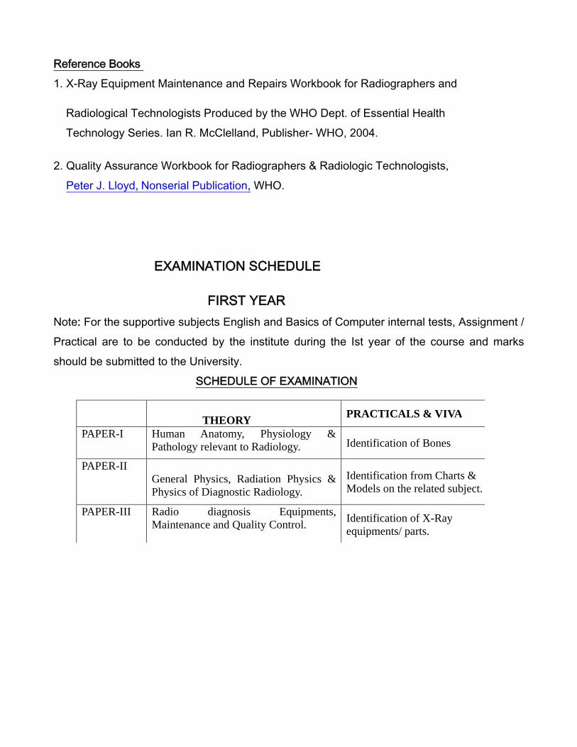
Reference Books 1. X-Ray Equipment Maintenance and Repairs Workbook for Radiographers and
Radiological Technologists Produced by the WHO Dept. of Essential HealthTechnology Series. Ian R. McClelland, Publisher- WHO, 2004.
2. Quality Assurance Workbook for Radiographers & Radiologic Technologists,Peter J. Lloyd, Nonserial Publication, WHO.
EXAMINATION SCHEDULE
FIRST YEAR Note: For the supportive subjects English and Basics of Computer internal tests, Assignment / Practical are to be conducted by the institute during the Ist year of the course and marks should be submitted to the University.
SCHEDULE OF EXAMINATION
THEORY PRACTICALS & VIVA
PAPER-I Human Anatomy, Physiology & Pathology relevant to Radiology. Identification of Bones
PAPER-IIGeneral Physics, Radiation Physics & Physics of Diagnostic Radiology.
Identification from Charts & Models on the related subject.
PAPER-III Radio diagnosis Equipments, Maintenance and Quality Control. Identification of X-Ray
equipments/ parts.
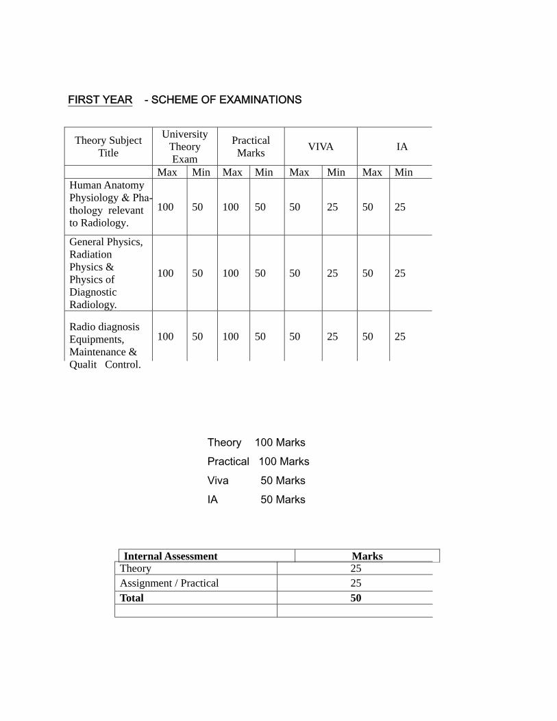
FIRST YEAR - SCHEME OF EXAMINATIONS
Theory 100 Marks Practical 100 Marks Viva 50 Marks IA 50 Marks
Internal Assessment Marks
Theory Subject Title
University Theory Exam
Practical Marks VIVA IA
Max Min Max Min Max Min Max MinHuman Anatomy Physiology & Pha- thology relevant to Radiology.
100 50 100 50 50 25 50 25
General Physics, Radiation Physics & Physics of Diagnostic Radiology.
100 50 100 50 50 25 50 25
Radio diagnosis Equipments, Maintenance & Quality Control.
100 50 100 50 50 25 50 25
Theory 25Assignment / Practical 25 Total 50
B.Sc Radiology & Imaging Technology Second Year
Internal Subjects
(1) Patient care & Medical Ethics: Patient vital signs - temperature, pulse, respiration and blood pressure - normal values and methods of taking and recording them. Development of communication skills with patient- general comfort and reassurance to the patient-patient education and explaining about the study-drugs used in the preparation of the patient. Handling of an unconscious patient-shifting of patients - hazards of lifting and maneuvering patients - rules for correct lifting- transfer from chair/wheel chair or trolley to couch and vice-versa - safety of patient and worker while lifting & shifting of patients- handling of geriatric, pediatric and trauma patients -handling female patients-pregnant women. Communicable diseases - hygiene in the department-cross infection and prevention-handling of infectious patients in the department -application of asepsis. Ethics of medical practice- Radiography professionalism-essential qualities of the radiographer-improving professional and personal qualities- Radiographer as a part of Hospital /Organization-responsibilities. Medico-legal considerations - radiographers clinical and ethical responsibilities- misconduct and malpractice.
(2) Principles of Medical Emergencies Trauma care & Emergency Radiography: procedures in the event of an accident- Special positioning procedures & projections - modification of techniques needed for seriously injured patients. Radiographic factors - patient care & responsibilities-Search of profession confidence-maintenance decorum of the job responsibility - the importance of records maintenance. Fluoroscopy and its application in emergency radiology - Medicolegal aspects of the radiographers work. Common medical emergencies-helping in first aids & zero hour care / know to help in critical hour care -Trauma patients handling – trauma ward bed X-rays – mass casualty managements-selection of study / procedures & radiographic views. Knowing the emergency care places in the hospital & preplanning- checking & readiness of mobile units in functioning status -screening of the high risk patients in various procedure-supportive facilities to encounter emergency-practical training.

Reference Books:
1. Notes on Radiological Emergencies – Ansell and Churchill2. Care of patient in diagnostic Radiography – Chesney & Chesney.3. First Aid – Haugher and Gardner.4. Practical Nursing and First Aid – Ross and Wilson.
Internal Assessment in Year 2 : Patient care & Medical Ethics (Total 50 marks) Principles of Medical Emergencies (Total 50 marks)
Theory Paper for internal assessment in Second Year to be combined as follows- Patient care & Medical Ethics 25 marks + Principles of Medical Emergencies 25marks
Viva Patient care & Medical Ethics 25 marks + Principles of Medical Emergencies 25marks
Paper-I - Clinical Radiography
• Techniques, Preparations, Instructions, Positioning of patient for conventional and digital radiography in the imaging of following-
Conventional Non contrast radiography- Extremities Radiography – Hand- Finger –MCP- Wrist joint- Forearm -Elbow joint – humerus - shoulder joint. Foot – Toes- Tarsal bones -Ankle joint - Knee joint – patella – tibia- femur – Hip joint – pelvis -sacroiliac joint. Spine Radiography -Vertebral column – Atlanta occipital articulation- cervical spine- dorsal spine - lumbar spine – sacrum -vertebral canal- vertebral foramen. Skull Radiography – general, sella – temporal bone – mastoid – optic foramen – Internal auditory canal – Superior and inferior orbital fissure – base of skull – facial bones – petrous apex – Zygomatic bone, nasal bone, sinuses of skull – mandible – Tempro-mandibular joint –Paranasal sinuses Radiography.

Chest Radiography –Basic views (PA & AP) - inspiratory & expiratory films- special chest views & their significance – larynx- trachea- thoracic inlet -Sternum - Ribs – Heart and great vessels – mediastinum -Diaphram – double exposure technique. Abdomen & Pelvic Radiography – all projection – the acute abdomen investigation. Soft tissue radiography: Preparations, Instructions, Various techniques, positioning of patient for conventional and digital mammography, High and low KV Technique – differential filtration – multiple radiography – technique for steep range radiography – Duplication – arrangement of intensifying screen. Stereo Radiography: Principle – tube shifting relation of patient – correct making and viewing of stereo radiographs – application.Macro radiography: Principle sizes of focal spot its limitation in its application. High kv technique: technique & usefulness. Foreign body localization: Preparation – Anatomical localization – various projections – use of skin markers – Tangential projection – uses – opaque – foreign bodies. Dental radiography-types of equipments –techniques- indications-films-dental radiography in trauma patients.
Practical Practical involving patients not less than 10 numbers must be prescribed to students. The title and nature of practical may be framed by the respective institution conducting the course.
Rreference Books: 1. Clark’s Handbook for Radiographers – Charles Sloane, Ken Holmes & Craig
Anderson, Hodder Educations, UK2. Diagnostic Radiography – A concise practical Manual – Glenda J. Bryan (4th edn),
Churchill Livingstone.
Paper-II X-ray Film / Image processing Techniques (including Dark Room Techniques)
1. X-Ray filmX-ray film construction and film characteristics – Composition of single and double coated radiographic films -structure of emulsion- film characteristics; speed, base fog, gamma, latitude -effect of grain size on film response to exposure, interpretation of characteristics curve- exposure to x-rays. 2. Types of Radiographic Films- applications -advantages/limitations of different types Structure, properties of different parts- Film storage - handling -film wrappings- andling of exposed and unexposed films -safe light requirements.
3. Radiographic Image: Meaning of radiographic image contrast, density, resolution,
sharpness, magnification and distortion of image, noise and blur. Primary radiological image formation- Image quality – unsharpness- resolution – fog and noise - use of contrast media- density- contrast – brightness- optical density measurements- Image recording devices.
4. Image processing– Film developing principles- acidity, alkalinity, pH, the processing cycle-
process of film developing - development -developer solution- constituents of developer. Fixing- fixer solution- composition of fixer –washing – drying replenishment -checking and adjusting replenishment rates - other processing solution – effect of temperature and development time - film processing methods - common errors and faults while processing manual and automatic processing-latent image formation– silver recovery and economics.
5. Film archieving systems- Image recording devices-Laser imager/camera functioning.Multiformatter- Optical Disc. System Film archieving systems - MOD/disc/PACS etc.

6. Automatic processing - Automatic film handling systems -Automated Processors -equipment for Film Processing-functions of various components- film roller transport - transport time -film feed system-Importance and relation to temp, fixed and variable time cycles-Care and maintenance -cleaning routine and methods of cleaning. 7. Radiographic illuminators: and viewing conditions, visual acuity and resolution.8. Dark Room- Site – layout - dark room design- construction- processing area– illumination- safe light compatibility - entrance safe lighting – types- storage- shelving of films-cleaning and maintenance.
Practicals Practical involving not less than 10 numbers must be prescribed to the students. The title and nature of practical may be framed by the respective institution conducting the course. Study with charts, models & power point presentations involving students to present and discuss.
Rreference Books 1. Radiographic latent image processing – W. E. J Mckinney2. Diagnostic Radiography – A concise practical Manual – Glenda J. Bryan (4th edn),
Churchill Livingstone.

Paper –III Contrast & Special Radiography procedures.
Non-contrast Special radiography-
1. Paediatric Imaging:special needs of patient and radiographer- use of dedicated equipment and accessories- modified technical considerations - selection of exposure factors-image quality considerations – radiation protection of the patient - special techniques in children for contrast studies.2. Geriatric radiographyEquipment and accessories – exposure factor considerations in special care. Elderly patients profile - difficulties during radiography – technical considerations-projections with unconventional special positioning. 3. Trauma/Emergency RadiographySelection of suitable X-Ray equipment – patient position -radiographic projections and sequence for each patient – modification of routine positioning– radiation protection – patient care. 4. Operation theatre radiographyO.T procedures-Operative cholangiography – orthopaedic procedures –maintenance of asepsis – preparation of radiographer and equipment/accessories – careful safe use of mobile and fluoroscopic equipment – radiation protection – patient care – rapid availability of radiographic image-cooperation with OT staff-type of studies done -clinical applications - clinical applications- per operative radiographs- peroperative fluoroscopy studies -patient care-radiation protection of all staff. Contrast radiography Radiological contrast media – classification -need for radiological contrast media - methods of administration-dosage-reactions to contrast media- role of radiographer in management of patient with contrast reaction. For all contrast investigations-patient preparation, positioning, patient care during the study-post procedural patient care-types of contrast media used and dosage-alternative contrast used-side effects and its identification-treatment of complication during the procedure -pathological conditions- indications and contraindications- injection procedure –techniques for radiographic projections - radiographic appearances– radiation protection.
5. Sialogram6. Barium studies- different types – Barium swallow Barium meal study of upper GIT, Bariummeal follow through, Barium enema, small bowel enema, distal colography, defaecography. 7. Percutaneous Transhepatic Cholangiogram, ERCP, T-Tube cholangiography,
per-operative cholangiography.8. IVP-rapid sequence-infusion pyelography-high dose urography, Cystogram,
Anterior Urethrogram RGU, MCU, RCP9. Angiography, Diagnostic & therapeutic, venography, Lymphangiogram10. Orthography, Discography11. Myelogram,12. Hysterosalphingography.13. Sinography.14. Fistulogram,15. Ductogram.
Practical: Practical involving patients not less than 10 numbers must be prescribed to the students.The title and nature of practical may be framed by the respective institution conducting the course.
Reference Books:
Text book of radiology for residents & technicians – 4th edition, Satish K. Bhargave Radiological patient care – Jensen Chesney. Atlas of dental and maxillofacial radiological imaging – Brownie
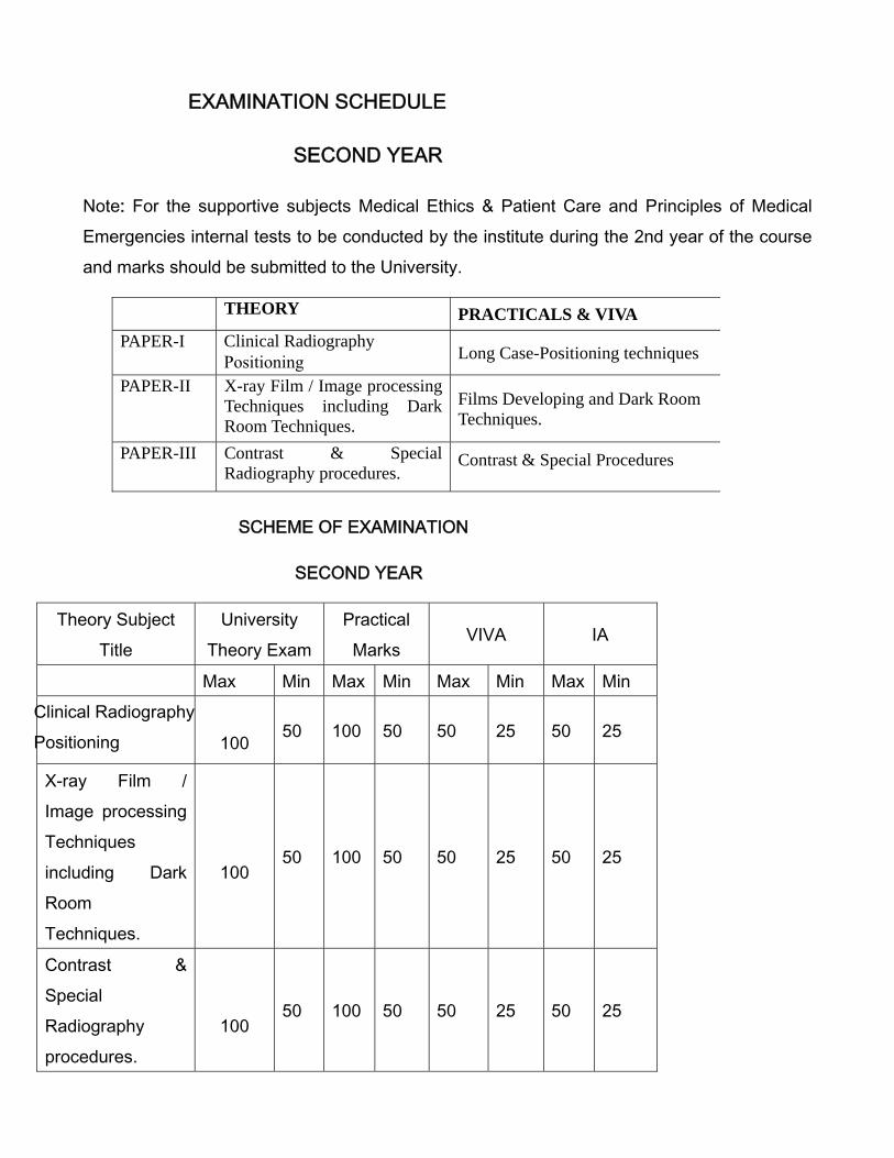
EXAMINATION SCHEDULE
SECOND YEAR
Note: For the supportive subjects Medical Ethics & Patient Care and Principles of Medical Emergencies internal tests to be conducted by the institute during the 2nd year of the course and marks should be submitted to the University.
SCHEME OF EXAMINATION
SECOND YEAR
Theory Subject Title
University Theory Exam
Practical Marks
VIVA IA
Max Min Max Min Max Min Max Min Clinical Radiography Positioning 100 50 100 50 50 25 50 25
X-ray Film / Image processing Techniques including Dark Room Techniques.
100 50 100 50 50 25 50 25
Contrast & Special Radiography procedures.
100 50 100 50 50 25 50 25
THEORY PRACTICALS & VIVA PAPER-I Clinical Radiography
Positioning Long Case-Positioning techniques
PAPER-II X-ray Film / Image processing Techniques including Dark Room Techniques.
Films Developing and Dark Room Techniques.
PAPER-III Contrast & Special Radiography procedures.
Contrast & Special Procedures
Theory 100 Marks Practical 100 Marks Viva 50 Marks IA 50 Marks
Internal Assessment Marks
Theory 25
Practical 25Total 50
B.Sc Radiology & Imaging Technology
THIRD YEAR
PAPER I Equipments of modern Imaging Modalities
1. Mammography system:History - Imaging requirements- Mammography system - construction/types accessories - tube, compression, grids, AEC etc.- nature of X-Ray beam suitable – accessories for immobilization - film processing - image quality - image recording devices -interventional procedures – accessories-biopsy equipment attachments - radiation dose- -mammo tomogram-Sonomammography-future developments.
2. Ultrasonography/ Doppler systems:Basic acoustics principle- Basic physics of sound propagation in different media, production of Ultrasound (piezoelectric effect), ultrasound terminologies – interaction of ultrasound with matter – ultrasound properties propagation in tissue, absorption, scattering, reflection and refraction- acoustic impedence – piezo electric effect – transducer – Pulsar – receiver –beam/sensitivity and gain - generators- A, B and M scanning & echo modes- transducers- techniques of sonography-equipment selection- display methods – ultrasound image formation - data storage and display – image and artifacts – doppler instrumentation – doppler equation – transducer – quality assurance and performance tests – bio effects and safety considerations. Types of machines –portable systems- acoustic coupling agents- ingredients/preparation.
3. CT scan systems:History- generations of scanners-CT technology -helical/spiral & multi slice C.T- ultra fast scanners-system components - performance parameters - image quality and methods of image reconstruction- radiation dose measurements and technical aspects of Q.A -calibration and image acquisition-
4. MRI Scanners: History - basic physical principle - Physical principles -NMR signals–instrumentation- hard ware-MR system components- magnet system- Magnetic shielding- RF shielding- bioeffects of MRI- site selection and safety -reconstruction system - different coils used -NMR signals advantage -imaging methods – pulse imaging sequences - spectroscopy parameters -calibration and image acquisition - reconstructions- 3D images- - image contrast – factors affecting image quality - artifacts - difference between CT and MRI images- hostcomputer -viewing archiving- hard copy - image formation and storage device.
5. Angiography and Cine Studies /DSAAngiography equipments history –Conventional angiography X-Ray equipment - Equipment construction-principle - DSA system basics - digital techniques -subtraction process- procedures for subtraction - care, choice and installation of the equipment – equipment, pitfalls and complications -pressure injectors- contrast media -accessories-catheters, guide wires-uses of serial imaging devices- cine camera - video-recorder -film processing-radiation protection.
6. Nuclear Medicine EquipmentsNuclear Physics - basics in Nuclear Medicine- Nuclear medicine equipments - Gamma Cameras- rectilinear scanners- radioisotope generators-SPECT-CT & PET-CT- introduction-basic physics and principle involved- equipments basic structure—differences- fusion techniques- image formation-storage devices– advantages-limitations.
7. Recent Advances in Imaging SystemsMobile units of Computer Radiography & Digital Radiography system. 3D/4D Sonography systems 128 slice & higher slice C.T equipments. 3 Tesla & higher T MRI scanners Image processing & Display systems-Recent advances, concepts and applications in processing of images in digital form using computer based systems.

8. Picture Archiving and Communication Systems (PACS)-newer advancements – updates -systems designs-transfer restrictions.
Practical Practical involving not less than 10 numbers must be prescribed to the students. The title and nature of practical may be framed by the respective institution conducting the course.
Reference Books
Step by Step CT; Step by Step MRI and MRI made Easy for beginners – Govind B. Chavhan – Jaypee brothers and Medical Publishers (p) Ltd, New Delhi CT & MRI protocol – Satish K. Bhargava, CBS publishers. Text Book of Radiology for Residents & Technicians – 4th Edition – Satish K. Bhargava CBS publishers & Distributor (p) ltd.
PAPER-II Modern Imaging techniques and recent trends in imaging.
1. Mammography:The Mammography as a clinical diagnostic tool- immobilization and identification techniques- positioning techniques for various projections - exposure factors- Conventional & Digital studies- quality and advantage- diagnosis and screening- Characteristics of benign and malignant lesions – patient care – female attendant - interventional procedures - radiation dose- recent advances in mammography techniques -mammo tomogram & Sonomammography procedures- advantages & limitations. 2. Ultrasonography/ Doppler studies:Techniques of sonography-selection- Preparations - instructions and positioning of patient for TAS, TVS, TRUS, neck USG and extremities- patient care and maintenance protocols- clinical applications display methods –quality image reproducible extend -assurance to patients.
3. CT scan studies acquisition/ protocols /techniques:CT of head and neck – thorax – abdomen – pelvis – musculo skeletal system – spine – PNS. Anatomy – clinical indications and contraindications – patient preparation – technique – contrast media-types, dose, injection technique; timing, sequence - image display – patient care – utilization of available techniques & image processing facilities to guide the clinician- CT anatomy and pathology of different organ systems.
4. MRI Scanners:Methods of MRI imaging methods – Head and Neck ,Thorax, Abdomen, Musculoskeletal System imaging - Clinical indications and contraindications- types of common sequences- effects of sequence on imaging - Protocols for various studies- slice section- patient preparation-positioning of the patient -patient care-calibration - paramagnetic agents and dose, additional techniques and recent advances in MRI - image acquisition-modification of procedures in an unconscious or un co-operative patient -plain studies- contrast studies -special procedures- reconstructions- 3D images- MRS blood flow imaging, diffusion/perfusion scans - strength and limitations of MRI- role of radiographer.
5. Angiography and Cine Studies /DSAConventional / DSA studies- Abdominal, visceral, peripheral, cerebral and cardiac angiography - arterial/venous anatomy, physiology-clinical indications and contraindications - patient preparation-positioning of the patient -patient care-contrast media - types of contrast - dosage - accessories catheters, guide wires- pressure injection- control of radiographic and fluoroscopic equipment - exposure factors for serial programmes-programming-injection protocols- outline on each radiological procedure- radiographer’s role- patient management before -during and after the procedure - venography- interventional angiography in hepatobiliary, GIT, urology and vascular system- coils/stents etc- indications and
contraindications - role of radiographer-radiation safety.
6. Nuclear Scintiscan procedures:SPECT-CT & PET-CT studies, protocols, Basics of common clinical Nuclear Medicin procedures/techniques–comparison with different structural imaging studies-advantages and limitations.
7. Recent Advances in ImagingDynamic CT & MRI studies Per operative application of various imaging systems including detector probes application in Nuclear Medicine Imaging guidance in therapeutic procedures-IGRT, TACE & TARE etc.
Practical Practical involving not less than 10 numbers must be prescribed to the students. The title and nature of practical may be framed by the respective institution conducting the course.
Reference Books 1. Concepts in Medical Radiographic Imaging – Marianne Tortoice2. Radiographic Imaging - Derrick3. Processing and Quality Control – William
PAPER-III Quality Control, Radiobiology and Radiation Safety in Radiodiagnosis /Imaging.
1. Radiation Quantities and UnitsRadiation- Radioactivity- Sources of radiation - natural radioactive sources -cosmic rays- terrestrial radiation - - man made radiation sources. Units of radiation - Quality factor - Flux- Fluence-Kerma- Exposure- Absorbed dose- Equivalent Dose- Weighting Factors-Effective Dose - Occupational Exposure Limits - Dose limits to public.
2. Biological Effects of radiationIonization, excitation and free radical formation, hydrolysis of water, action of radiation on cell -Chromosomal aberration and its application for the biological dosimetry- Effects of whole body and acute irradiation, dose fractionation, effects of ionizing radiation on each of major organ system including fetus -Somatic effects and hereditary effects- stochastic and deterministic effects-Acute exposure and chronic exposure-LD50 - factors affecting radio-sensitivity. Biological effects of non-ionizing radiation like ultrasound, lasers, IR, UV and magnetic fields.
3. Radiation detection and Measurements: Ionization of gases- Fluorescence andPhosphorescence -Effects on photographic emulsion. Ionization Chambers – proportional counters- G.M counters- scintillation detectors – liquid semiconductor detectors – Gamma ray spectrometer. Measuring systems – free air ionization chamber – thimble ion chamber – condenser chamber – Victorian electrometer – secondary standard dosimeters – film dosimeter – chemical dosimeter- thermoluminescent Dosimeter. -Pocket dosimeter-Radiation survey meter- wide range survey meter -zone monitor-contamination monitor -their principle- function and uses. Advantages & disadvantages of various detectors & its appropriateness of different detectors for different type of radiation measurement.
4. Radiation protection:Radiation protection of self and patient- Principles of radiation protection, time - distance and shielding, shielding - calculation and radiation survey –ALARA- personnel dosimeters (TLD and film batches)- occupational exposure.
5. Q.A in Diagnostic RadiologyQuality assurance (Q.A), acceptance testing and quality control tests in Radiology- Meaning of the terms used and aspects of a QA programme, equipment and staff requirements, benefits of QA procedures in an imaging department –NABH guidelines. Verification of Optical & Radiation field congruence, Beam alignment, Focal spot size, Linearity of tube current mA and Timer, applied potential, HVT and total tube filter, Contact between film and intensifying screen, contrast resolution, Grid alignment, Special techniques like mammography, CT - CT Dose Modulation-Patient dose management.
6. Radiation Hazard evaluation and controlPhilosophy of Radiation protection, effects of time, Distance & Shielding. Calculation of Work load, weekly calculated dose to radiation worker & General public Good work practice in Diagnostic Radiology. Planning consideration for radiology, including Use factor, occupancy factors, and different shielding material.
7. Regulatory Bodies & regulatory Requirements:International Commission on Radiation Protection (ICRP) / National Regularity body (AERB - Atomic Energy Regulatory Board) - Responsibilities, organization, Safety Standard, Codes and Guides, Responsibilities of licenses, registrants & employers and Enforcement of Regulatory requirements.
8. Role of Radiographer in Planning, QA & Radiation Protection:Role of technologist in radiology department - Personnel and area monitoring., Setting up of a new X-Ray unit, staff requirement, AERB specifications for site planning and mandatory guidelines – Planning of X-ray rooms, dark rooms – Inspection of X-Ray installations - Registration of X-Ray equipment installation- Certification -Evaluation of workload versus radiation factors – Occupational exposure and protection Tools/devices. ICRP, NRPB, NCRP and WHO guidelines for radiation protection, pregnancy and radiation protection.
Practical Practical involving not less than 10 numbers must be prescribed to the students. The title and nature of practical may be framed by the respective institution conducting the course as follows- 1. Time, Dose, Shielding, Measurement of HVT & TVT2. Familiarization of Radiation Survey meters and their functional performance
checks
3. Radiological Protection Survey of Diagnostic X-Ray installation4. Diagnostic Imaging: Quality Assurrance – M. M Rehani5. AERB safety requirements- Atomic Energy Act, Radiation protection rules.
Reference Books:
1. Radiologic science for technologist – 9th edition (2008) Stewart Carlyle Bushong,
Mosby Elsevier, UK.
2. Text Book of Radiological Safety – K. Thaylan (2010) Jaypee Brothers and
medical Publishers, New Delhi.
3. Quality Control in Diagnostic Imaging J.E.Gray
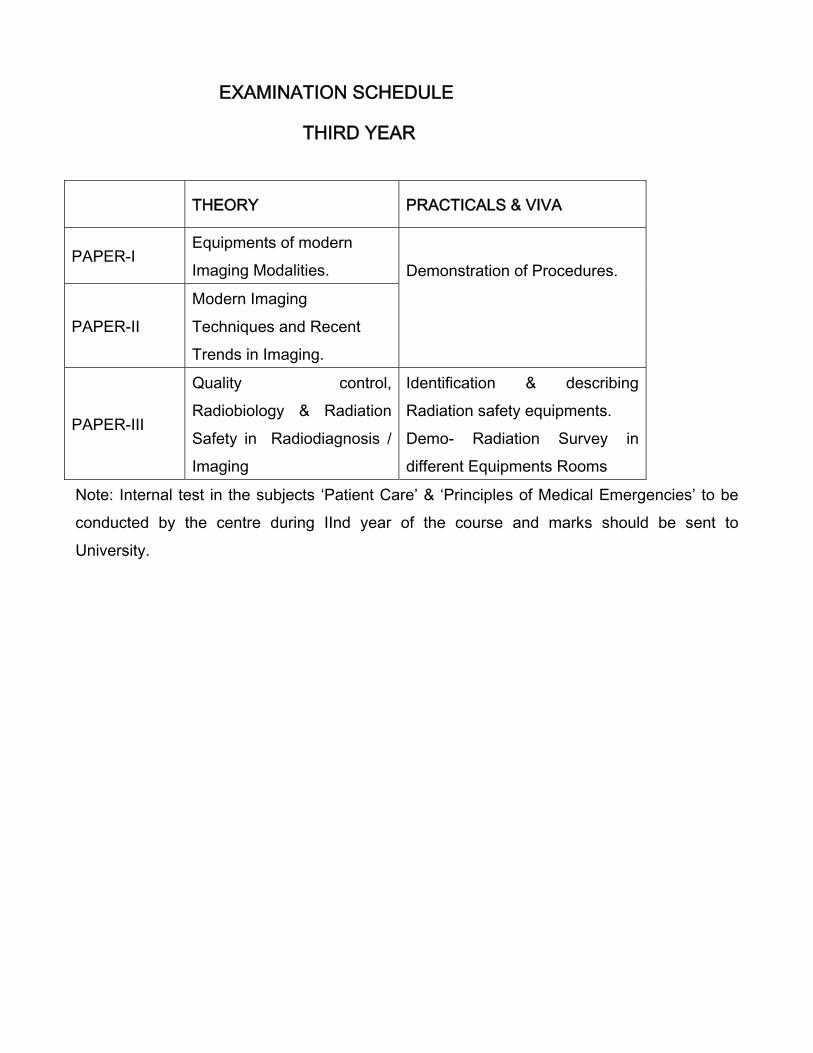
EXAMINATION SCHEDULE
THIRD YEAR
THEORY PRACTICALS & VIVA
PAPER-I Equipments of modern Imaging Modalities. Demonstration of Procedures.
PAPER-II Modern Imaging Techniques and Recent Trends in Imaging.
PAPER-III
Quality control, Radiobiology & Radiation Safety in Radiodiagnosis / Imaging
Identification & describing Radiation safety equipments. Demo- Radiation Survey in different Equipments Rooms
Note: Internal test in the subjects ‘Patient Care’ & ‘Principles of Medical Emergencies’ to be conducted by the centre during IInd year of the course and marks should be sent to University.
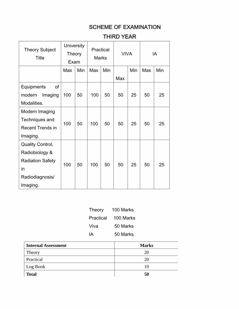
SCHEME OF EXAMINATION THIRD YEAR
Theory Subject Title
University Theory Exam
Practical Marks
VIVA IA
Max Min Max Min Max
Min Max Min
Equipments of modern Imaging Modalities.
100 50 100 50 50 25 50 25
Modern Imaging Techniques and Recent Trends in Imaging.
100 50 100 50 50 25 50 25
Quality Control, Radiobiology & Radiation Safety in Radiodiagnosis/ Imaging.
100 50 100 50 50 25 50 25
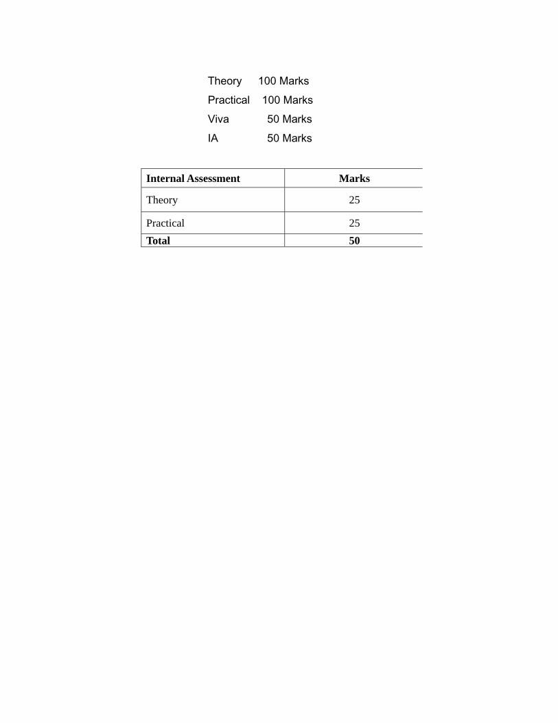
Theory 100 Marks Practical 100 Marks Viva 50 Marks IA 50 Marks
Internal Assessment Marks Theory 20Practical 20Log Book 10 Total 50
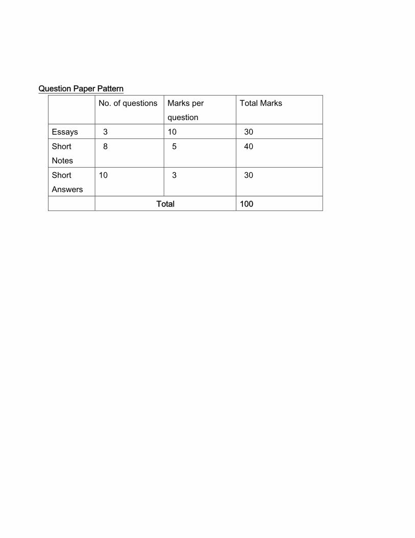
Question Paper Pattern
No. of questions Marks per question
Total Marks
Essays 3 10 30
Short Notes
8 5 40
Short Answers
10 3 30
Total 100


