dopaminergic neurons modulate locomotion in caenorhabditis ... · dopaminergic neurons modulate...
TRANSCRIPT
Dopaminergic neurons modulate locomotion in
Caenorhabditis elegans
Mohamed Abdelhacka,b,c
aOkinawa Institute of Science and Technology, Graduate University, 1919-1 Tancha,Onna-son, Okinawa 904-0495, Japan
bGraduate School of Informatics, Kyoto University, Yoshida Honmachi, Sakyo-ku, Kyoto606-8501, Japan
cATR Computational Neuroscience Laboratories, Kyoto 619-0288, Japan
Abstract
Adaptation in the sensory-mechanical loop during locomotion is a powerful
mechanism that allows organisms to survive in different conditions and envi-
ronments. For example, humans can walk on earth gravity, but they switch
to hopping on, for example, moon gravity. The nematode Caenorhabditis
elegans also shows adaptability by employing thrashing behaviour in low
viscosity media and crawling in high viscosity media. The mechanism that
enables this adaptability is yet unknown. It has been attributed previously
to neuro-modulation by dopamine and serotonin.
The aim of this study is to physiologically investigate the neuronal mech-
anisms of modulation of locomotion by dopamine. It unravels a new role for
the dopaminergic mechanosensory neurons, which is sensing the mechanical
impact of the environment. The significance of such characterization is im-
proving our understanding of dopamine gait switching which gets impaired
in Parkinsons disease.
Keywords: Caenorhabditis elegans , Dopamine, Gait, Locomotion
Preprint submitted to May 31, 2016
.CC-BY-NC-ND 4.0 International licensecertified by peer review) is the author/funder. It is made available under aThe copyright holder for this preprint (which was notthis version posted May 31, 2016. . https://doi.org/10.1101/056192doi: bioRxiv preprint
1. Introduction
In nearly all the living organisms, locomotion is an important strategy for
survival. It is important for feeding, avoiding predators, and finding optimal
environmental conditions for survival. In order to maximize efficiency of
locomotion in different environments and also in different situations (e.g.,
the presence of a predator), organisms need to have an adaptive strategy
which needs a sensory system.
C. elegans modulates its locomotion pattern. This appears to be a mod-
ulation of undulation frequency, undulation wavelength, and velocity of loco-
motion. There has been a debate as to whether this modulation translates to
two distinct locomotion patterns of swimming and crawling [Mesce & Pierce-
Shimomura, 2010, Pierce-Shimomura et al., 2008, Vidal-Gadea et al., 2011];
or whether it is one locomotion pattern that is continuously modulated [Berri
et al., 2009, Boyle et al., 2011, Fang-Yen et al., 2010, Korta et al., 2007].
Dopamine is a biogenic amine that has been shown to be associated with
the process. In C. elegans hermaphrodite, there are 8 dopaminergic neurons
distributed into 3 neuron types, namely CEP, ADE, and PDE. To date, five
dopamine receptors have been identified in C. elegans. Two of them are
homologous to mammalian D1-type receptors (dop-1 and dop-4 ). The other
three are homologues to D2-type receptors (dop-2, dop-3, and dop-6 ). These
receptors are widely expressed in C. elegans nervous system.
Dopamine is responsible for a spectrum of behaviours. It has been shown
that it is responsible for slowing of locomotion on the encounter of food [Sawin
et al., 2000] and in local search behaviour along with serotonin [Hills et al.,
2004]. Dopaminergic neurons are known to be mechanosensory [Bettinger
2
.CC-BY-NC-ND 4.0 International licensecertified by peer review) is the author/funder. It is made available under aThe copyright holder for this preprint (which was notthis version posted May 31, 2016. . https://doi.org/10.1101/056192doi: bioRxiv preprint
& McIntire, 2004, Duerr et al., 1999, Hills et al., 2004, Liu & Sternberg,
1995, Loer & Kenyon, 1993, Sanyal et al., 2004, Sawin et al., 2000] that have
been shown to respond to harsh touch stimulation [Sanders et al., 2013]. A
study has shown that dopamine is also responsible for gait switching from
swimming to crawling [Vidal-Gadea et al., 2011]. They have also shown that
serotonin is important and sufficient to switch from crawling to swimming.
In humans, gait dysfunction is a main symptom of the Parkinson’s disease.
It causes failure to modulate locomotion pattern by gait switching [Jankovic,
2008]. Other symptoms include trembling, stiffness, slowness of movement,
and walking difficulty. It is caused by death of dopaminergic neurons in the
substantia nigra. The cause of death of these cells is so far unclear [Obeso
et al., 2010].
In this study, I measure the calcium activity of dopaminergic neurons
as the worms switch between crawling and swimming due to crossing a vis-
cosity separation to demonstrate that the worms respond to environment
sensation by change in basal activity level and show the bidirectional na-
ture of the dopamine effect. While the previous study [Vidal-Gadea et al.,
2011] has shown that dopamine is responsible for switching from crawling
to swimming as dopaminergic neurons are active at switching. Here, I show
that dopaminergic neurons also respond to the environment as the worm is
switching from crawling to swimming by a decrease in their basal activation
which further supports the idea that dopaminergic neurons sense the pressure
induced by the environment.
3
.CC-BY-NC-ND 4.0 International licensecertified by peer review) is the author/funder. It is made available under aThe copyright holder for this preprint (which was notthis version posted May 31, 2016. . https://doi.org/10.1101/056192doi: bioRxiv preprint
2. Methods
2.1. Behavioural assay
dat-1p::GCaMP3 worms (Gift from Prof. Dr. David Biron, The Univer-
sity of Chicago) are placed between two glass slides with a paper separator
of thickness 0.1-0.2 mm. The paper is coated with grease to secure the slides
together (Figure 1). Worms are moved to a foodless plate before assay while
the glass cassette is prepared. A tiny droplet of 65% dextran is placed on
a glass slide so that the whole droplet is visible under microscope’s field of
view. The worm is picked and added to this droplet by a platinum wire worm
pick. The second glass slide is then placed on top with the separator in the
middle. The top plate is sheared by some distance from the lower plate to
ease addition of the low viscosity medium later. Imaging starts while the
worm is confined in the tiny droplet (Figure 1a). After the imaging starts,
30% dextran is added with a micropipette to fill the area around the 65%
dextran droplet and then tracking starts (Figure 1b,c). Imaging continues
until the whole worm’s body passes through the viscosity separation to the
lower viscosity region(Figure 1d). 30% dextran is used as opposed to pure
buffer in order to ease the imaging as the worms swim in this medium but are
slower compared to pure buffer. Ten worms were assayed for this experiment.
The point when the droplet fills in the gaps until half the droplet is cho-
sen to be the zero time point and after that 70 seconds lengths of calcium
imaging data are analysed. Controls are obtained through worms that stay
confined within the small droplet as the area surrounding is not filled in order
to compensate for photo-bleaching. Ten worms were assayed for the control
experiment. Image sequences are imported to ImageJ software [Schindelin
4
.CC-BY-NC-ND 4.0 International licensecertified by peer review) is the author/funder. It is made available under aThe copyright holder for this preprint (which was notthis version posted May 31, 2016. . https://doi.org/10.1101/056192doi: bioRxiv preprint
et al., 2012] and the fluorescent signal is tracked manually using manual
tracking tool [Cordelieres, 2005]. A 30 pixel region of interest around the
tracking point is used to extract fluorescence signal. This is a bigger area
than the cell size, but it is used to capture blurred signals due to quick move-
ment during swimming. The signal is then smoothed to remove movement
artefacts. A point not containing the worm is chosen as a background signal
and the average of 100 points before zero is used for normalization (Figure
1).
2.2. Imaging
In this chapter. Ca+2 imaging was done under Nikon A1R high-speed
confocal microscope illuminated by a mercury lamp Nikon Intensilight C-
HGFIE with GFP filter. Acquisition is done by Andor Zyla 5.5 high res-
olution camera (Andor Technology Ltd., US) with Micromanager software
[Edelstein et al., 2014]. Exposure time was set to 100 ms so the resulting
frame rate is 10 FPS and 4 x 4 binning was done on each image so that the
final resolution is 640 x 540 pixels. Autotracking was done using Hawkvision
tracking system (Hawkvision Co. Ltd., Japan) with software developed by
Saitama university in the whole body tracking mode to keep the worm in the
field of view.
3. Results and Discussion
3.1. Dopaminergic neurons sense the physical environment
PDE neurons have shown deactivation response when the worms cross
the viscosity separation (Figure 2a). Frequency of head undulation of C.
5
.CC-BY-NC-ND 4.0 International licensecertified by peer review) is the author/funder. It is made available under aThe copyright holder for this preprint (which was notthis version posted May 31, 2016. . https://doi.org/10.1101/056192doi: bioRxiv preprint
elegans increases accordingly, however even if it does not increase to reach
the swimming typical frequency (Figure 2b), still the corresponding PDE re-
sponse goes down. This suggests that PDE neurons respond by deactivation
to the environmental pressure and not to the body bends. However, they
still showed response to body bends (data not shown).
The mean calcium response to crossing the separation is shown in figure
2c,d where zero time point refers to the time when 30% dextran is visible
to have surrounded half of the 65% dextran droplet (Figure 1). I also show
the response up to 70 seconds after the zero time point since the worm’s
body, and hence the PDE neurons, takes 10-60 seconds to fully move from
the 65% dextran and be fully swimming in 30% dextran which is apparent
in the gradual increase in undulation frequency (Figure 2d). The transition
time is variable among worms, so the mean calcium signal shows gradual
change until it stabilizes at about 60 seconds to about 75% of its value
in high viscosity. In some cases we observe a sudden drop while in other
cases the drop is gradual (Figure 2a) depending on the speed of crossing
the separation. The separation described is also designed to be a very steep
gradient of viscosity but as time passes and due to diffusion and stirring effect
caused by the worm movement, the gradient can get smoother and hence the
transient in the physical forces sensed gets smoother. The signals from 60 to
70 seconds are significantly different (p=0.000) compared to signals from 10
seconds before addition of low viscosity medium (Figure 2e,f) .
3.2. Calcium signal of PDE and frequency of undulation correlate negatively
In order to find a relation between frequency of undulation and calcium
signal, the unsmoothed calcium signals were plotted against frequency of
6
.CC-BY-NC-ND 4.0 International licensecertified by peer review) is the author/funder. It is made available under aThe copyright holder for this preprint (which was notthis version posted May 31, 2016. . https://doi.org/10.1101/056192doi: bioRxiv preprint
undulation. First, paired-sample t-test was performed on the data and the
null hypothesis was rejected (p=0.000). Linear fitting was done by fitting a
first degree polynomial f(x) = px + c where p is the correlation coefficient.
The fit showed a correlation coefficient (p = -0.2419) with a goodness of fit of
r2=0.1232 which is low due to fitting of all animals’ data together (Figure 2e).
The same fit was attempted on each time series which shows better results
since signals are more self-consistent (Table 1). Another reason for the non-
uniformity of distribution is the movement artefacts caused by the neuron
sensing body contractions since the dataset used in the fit is unsmoothed.
Some vertical clusters and diagonal lines are observed also in the dataset in
figure 2g. These are caused by the cubic spline interpolation process that is
done to fill in the gaps in fluorescence levels data and frequency caused by
the lack of reliability of measurement mentioned in the methods section.
The study shows that mechanosensation has an effect on normal loco-
motion behaviour agreeing with a previous report [Li et al., 2006]. This is
the first report to attribute such role to PDE neurons through direct mea-
surement of activity. I show that viscosity differences can be measured by
mechansensory neurons and it yields significant change in basal activation
level of the neurons which can imply a change in dopamine release.
3.3. Hypothesized circuit for forward locomotion
Previous reports have shown that activation of dopaminergic neurons
causes switching from swimming to crawling and activation of serotoner-
gic neurons causes switching from crawling to swimming [Vidal-Gadea et al.,
2011]. Here, it is shown that the latter has an associated decrease in dopamine
level. Thus, it can be suggested that the balance between dopamine and sero-
7
.CC-BY-NC-ND 4.0 International licensecertified by peer review) is the author/funder. It is made available under aThe copyright holder for this preprint (which was notthis version posted May 31, 2016. . https://doi.org/10.1101/056192doi: bioRxiv preprint
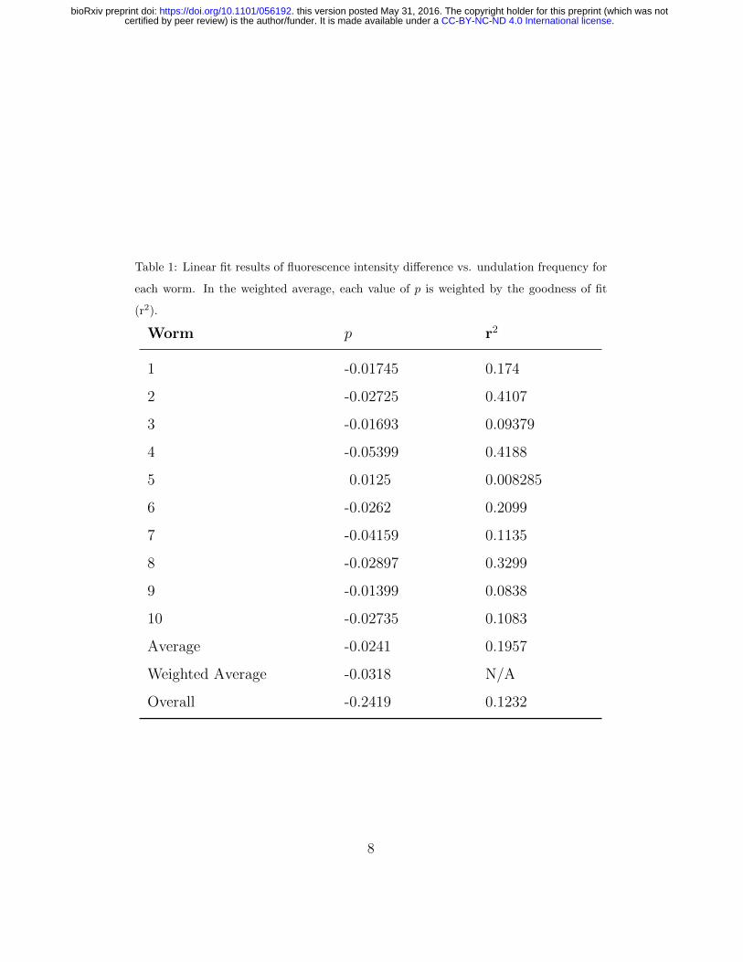
Table 1: Linear fit results of fluorescence intensity difference vs. undulation frequency for
each worm. In the weighted average, each value of p is weighted by the goodness of fit
(r2).
Worm p r2
1 -0.01745 0.174
2 -0.02725 0.4107
3 -0.01693 0.09379
4 -0.05399 0.4188
5 0.0125 0.008285
6 -0.0262 0.2099
7 -0.04159 0.1135
8 -0.02897 0.3299
9 -0.01399 0.0838
10 -0.02735 0.1083
Average -0.0241 0.1957
Weighted Average -0.0318 N/A
Overall -0.2419 0.1232
8
.CC-BY-NC-ND 4.0 International licensecertified by peer review) is the author/funder. It is made available under aThe copyright holder for this preprint (which was notthis version posted May 31, 2016. . https://doi.org/10.1101/056192doi: bioRxiv preprint
tonin levels is responsible for mediating crawling-swimming switching. This
can be further tested by simultaneous imaging of activation level of both
dopaminergic and serotonergic neurons. Moreover, in a previous study, it was
found that RID, RIS, and PQR are the neurons responsible for dopamine-
mediated gait switching from swimming to crawling [Topper, 2013]. RID and
RIS also express serotonin receptors ser-2 and ser-4 respectively so it can be
suggested that they are the ones that compute the dopamine-serotonin bal-
ance and modulate locomotion accordingly. RIS neuron is connected to the
head motor neurons and has been shown before to inhibit head movement
during sleep-like state [Turek et al., 2013], so it might be a good candidate
for regulating head undulation frequency. RID has gap junctions with the
forward locomotion command interneurons AVB that activates body motor
neurons. Calcium imaging of these two neurons would be a logical next step.
Hypothesized circuit for locomotion modulation based on these observations
is shown in figure 3.
4. Acknowledgements
I would like to first thank Prof. Ichiro Maruyama for his advice through-
out the study. I would also like to thank the Okinawa Institute of Science
and Technology, Graduate University for providing the support and funding
throughout the whole study period. I am very grateful to Dr. Bernd Kuhn
for his advice with fluorescence imaging, and Steve Aird, the technical ed-
itor at Okinawa Institute of Science and Technology, for help with editing
sections of this manuscript. I would like to thank Prof. Dr. David Biron for
sending the dat-1p::GCaMP3.
9
.CC-BY-NC-ND 4.0 International licensecertified by peer review) is the author/funder. It is made available under aThe copyright holder for this preprint (which was notthis version posted May 31, 2016. . https://doi.org/10.1101/056192doi: bioRxiv preprint
The author declares that there is no conflict of interest regarding the
publication of this paper.
References
Berri, S., Boyle, J. H., Tassieri, M., Hope, I. A., & Cohen, N. (2009). Forward
locomotion of the nematode C. elegans is achieved through modulation of
a single gait. HFSP Journal , 3 (3), 186–193.
Bettinger, J., & McIntire, S. (2004). State-dependency in c. elegans. Genes,
Brain and Behavior , 3 (5), 266–272.
Boyle, J. H., Berri, S., Tassieri, M., Hope, I. A., & Cohen, N. (2011). Gait
modulation in C. elegans : It’s not a choice, it’s a reflex! Frontiers in
behavioral neuroscience, 5 , 10.
Cordelieres, F. P. (2005). Manual tracking. Institut Curie, Orsay (France).
Duerr, J. S., Frisby, D. L., Gaskin, J., Duke, A., Asermely, K., Huddleston,
D., Eiden, L. E., & Rand, J. B. (1999). The cat-1 gene of caenorhabdi-
tis elegansencodes a vesicular monoamine transporter required for specific
monoamine-dependent behaviors. The Journal of neuroscience, 19 (1), 72–
84.
Edelstein, A. D., Tsuchida, M. A., Amodaj, N., Pinkard, H., Vale, R. D.,
& Stuurman, N. (2014). Advanced methods of microscope control using
µmanager software. Journal of biological methods , 1 (2).
Fang-Yen, C., Wyart, M., Xie, J., Kawai, R., Kodger, T., Chen, S., Wen, Q.,
& Samuel, A. D. (2010). Biomechanical analysis of gait adaptation in the
10
.CC-BY-NC-ND 4.0 International licensecertified by peer review) is the author/funder. It is made available under aThe copyright holder for this preprint (which was notthis version posted May 31, 2016. . https://doi.org/10.1101/056192doi: bioRxiv preprint
nematode Caenorhabditis elegans. Proceedings of the National Academy of
Sciences , 107 (47), 20323–20328.
Hills, T., Brockie, P. J., & Maricq, A. V. (2004). Dopamine and gluta-
mate control area-restricted search behavior in Caenorhabditis elegans. The
Journal of neuroscience, 24 (5), 1217–1225.
Jankovic, J. (2008). Parkinsons disease: clinical features and diagnosis. Jour-
nal of Neurology, Neurosurgery & Psychiatry , 79 (4), 368–376.
Korta, J., Clark, D. A., Gabel, C. V., Mahadevan, L., & Samuel, A. D.
(2007). Mechanosensation and mechanical load modulate the locomotory
gait of swimming C. elegans. Journal of Experimental Biology , 210 (13),
2383–2389.
Li, W., Feng, Z., Sternberg, P. W., & Xu, X. S. (2006). A C. elegans stretch
receptor neuron revealed by a mechanosensitive trp channel homologue.
Nature, 440 (7084), 684–687.
Liu, K. S., & Sternberg, P. W. (1995). Sensory regulation of male mating
behavior in caenorhabditis elegans. Neuron, 14 (1), 79–89.
Loer, C. M., & Kenyon, C. J. (1993). Serotonin-deficient mutants and male
mating behavior in the nematode caenorhabditis elegans. The Journal of
neuroscience, 13 (12), 5407–5417.
Mesce, K. A., & Pierce-Shimomura, J. T. (2010). Shared strategies for be-
havioral switching: understanding how locomotor patterns are turned on
and off. Frontiers in behavioral neuroscience, 4 .
11
.CC-BY-NC-ND 4.0 International licensecertified by peer review) is the author/funder. It is made available under aThe copyright holder for this preprint (which was notthis version posted May 31, 2016. . https://doi.org/10.1101/056192doi: bioRxiv preprint
Obeso, J. A., Rodriguez-Oroz, M. C., Goetz, C. G., Marin, C., Kordower,
J. H., Rodriguez, M., Hirsch, E. C., Farrer, M., Schapira, A. H., & Hall-
iday, G. (2010). Missing pieces in the parkinson’s disease puzzle. Nature
medicine, 16 (6), 653–661.
Pierce-Shimomura, J. T., Chen, B. L., Mun, J. J., Ho, R., Sarkis, R., &
McIntire, S. L. (2008). Genetic analysis of crawling and swimming loco-
motory patterns in C. elegans. Proceedings of the National Academy of
Sciences , 105 (52), 20982–20987.
Sanders, J., Nagy, S., Fetterman, G., Wright, C., Treinin, M., & Biron,
D. (2013). The Caenorhabditis elegans interneuron ala is (also) a high-
threshold mechanosensor. BMC neuroscience, 14 (1), 156.
Sanyal, S., Festa, F., Sakano, S., Zhang, Z., Steineck, G., Norming, U.,
Wijkstrom, H., Larsson, P., Kumar, R., & Hemminki, K. (2004). Poly-
morphisms in dna repair and metabolic genes in bladder cancer. Carcino-
genesis , 25 (5), 729–734.
Sawin, E. R., Ranganathan, R., & Horvitz, H. R. (2000). C. elegans locomo-
tory rate is modulated by the environment through a dopaminergic path-
way and by experience through a serotonergic pathway. Neuron, 26 (3),
619–631.
Schindelin, J., Arganda-Carreras, I., Frise, E., Kaynig, V., Longair, M., Piet-
zsch, T., Preibisch, S., Rueden, C., Saalfeld, S., & Schmid, B. (2012). Fiji:
an open-source platform for biological-image analysis. Nature methods ,
9 (7), 676–682.
12
.CC-BY-NC-ND 4.0 International licensecertified by peer review) is the author/funder. It is made available under aThe copyright holder for this preprint (which was notthis version posted May 31, 2016. . https://doi.org/10.1101/056192doi: bioRxiv preprint
Topper, S. M. (2013). Gait transitions in C. elegans . Ph.D. thesis, University
of Texas at Austin.
Turek, M., Lewandrowski, I., & Bringmann, H. (2013). An ap2 transcription
factor is required for a sleep-active neuron to induce sleep-like quiescence
in C. elegans. Current Biology , 23 (22), 2215–2223.
Vidal-Gadea, A., Topper, S., Young, L., Crisp, A., Kressin, L., Elbel, E.,
Maples, T., Brauner, M., Erbguth, K., & Axelrod, A. (2011). Caenorhab-
ditis elegans selects distinct crawling and swimming gaits via dopamine
and serotonin. Proceedings of the National Academy of Sciences , 108 (42),
17504–17509.
13
.CC-BY-NC-ND 4.0 International licensecertified by peer review) is the author/funder. It is made available under aThe copyright holder for this preprint (which was notthis version posted May 31, 2016. . https://doi.org/10.1101/056192doi: bioRxiv preprint
30% dextran 65% dextran
Stage autmatic trackingis turned on
c) Worm crossing viscosity separation
Add 30% dextran
b) Dextran is added while imaging is running
a) Imaging starts as worm is trapped in the small droplet
30% dextran 65% dextran
Stage autmatic trackingis turned on
d) Worm swimming
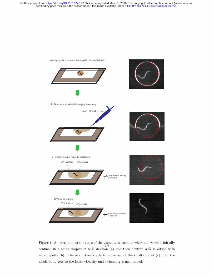
Figure 1: A description of the steps of the viscosity separation where the worm is initially
confined in a small droplet of 65% dextran (a) and then dextran 30% is added with
micropipette (b). The worm then starts to move out of the small droplet (c) until the
whole body gets to the lower viscosity and swimming is maintained.
14
.CC-BY-NC-ND 4.0 International licensecertified by peer review) is the author/funder. It is made available under aThe copyright holder for this preprint (which was notthis version posted May 31, 2016. . https://doi.org/10.1101/056192doi: bioRxiv preprint
Viscosity
ΔF
/F %
120
100
80
60
40
20
0
Error Bars: +/- 2 SE
LowHigh
***
ViscosityLowHigh
Fre
quen
cy
1
0.8
0.6
0.4
0.2
0.0
Error Bars: +/- 2 SE
***
(a)
(b)
(c)
(d)
(e) (f) (g)
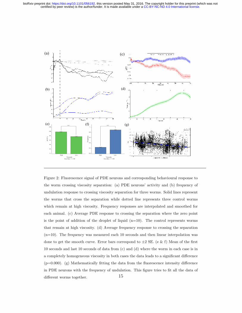
Figure 2: Fluorescence signal of PDE neurons and corresponding behavioural response to
the worm crossing viscosity separation: (a) PDE neurons’ activity and (b) frequency of
undulation response to crossing viscosity separation for three worms. Solid lines represent
the worms that cross the separation while dotted line represents three control worms
which remain at high viscosity. Frequency responses are interpolated and smoothed for
each animal. (c) Average PDE response to crossing the separation where the zero point
is the point of addition of the droplet of liquid (n=10). The control represents worms
that remain at high viscosity. (d) Average frequency response to crossing the separation
(n=10). The frequency was measured each 10 seconds and then linear interpolation was
done to get the smooth curve. Error bars correspond to ±2 SE. (e & f) Mean of the first
10 seconds and last 10 seconds of data from (c) and (d) where the worm in each case is in
a completely homogeneous viscosity in both cases the data leads to a significant difference
(p=0.000). (g) Mathematically fitting the data from the fluorescence intensity difference
in PDE neurons with the frequency of undulation. This figure tries to fit all the data of
different worms together. 15
.CC-BY-NC-ND 4.0 International licensecertified by peer review) is the author/funder. It is made available under aThe copyright holder for this preprint (which was notthis version posted May 31, 2016. . https://doi.org/10.1101/056192doi: bioRxiv preprint

Dopaminergic neurons (ADE,PDE)
Dopamine
DOP-4
RID
DOP-1
RIS
Serotonergic neurons (VC4/5,AIM,NSM)
Serotonin
SER-2 SER-4
AVB
VB/DB
RMD
Body wall muscles
Head and neck muscles
Figure 3: Hypothesized circuit for forward locomotion modulation in C. elegans based on
monoaminergic regulation of locomotory activity.
16
.CC-BY-NC-ND 4.0 International licensecertified by peer review) is the author/funder. It is made available under aThe copyright holder for this preprint (which was notthis version posted May 31, 2016. . https://doi.org/10.1101/056192doi: bioRxiv preprint