dna repair pathways and the effect of radiotherapy in...
TRANSCRIPT

Linköping University Medical Dissertations
No. 1112
DNA repair pathways and the effect ofradiotherapy in breast cancer
Karin Söderlund Leifler
Linköping UniversityDepartment of Clinical and Experimental Medicine
Division of Surgery and Clinical OncologySE-581 85 Linköping, Sweden
Linköping 2009

c© Karin Söderlund Leifler, 2009ISBN 978-91-7393-668-2ISSN 0345-0082
Published articles have been reprinted with permission from therespective copyright holder.Paper I c© Spandidos PublicationsPaper II c© ElsevierPaper III c© ElsevierIllustrations made by the author using OmniGraffle, unless otherwisespecifiedCover illustration: ‘Feather & drop´, courtesy of tanakawhoTypeset using LATEX
Printed by LiU-Tryck, Linköping 2009

Till Ola -luften i mina lungor
Little by little, one travels farJ.R.R. Tolkien


Abstract
A large proportion of breast cancer patients are treated with radiotherapy.Ionising radiation induces different DNA damages, of which double-strandbreaks are the most severe. They are mainly repaired by homologous re-combination or non-homologous end-joining. Different protein complexeshave central roles in these repair processes. In addition to the ability torepair DNA damage, cellular radiosensitivity is also affected by mitogenicsignals that stimulate survival and inhibit apoptosis. The phosphatidylino-sitol 3-kinase (PI3-K)/AKT pathway controls cell proliferation, invasive-ness and cell survival. AKT is regulated by upstream growth factor recep-tors, one of them being HER2 (also called ErbB2). HER2 is overexpressedin 15-30% of all breast cancers and associated with poor prognosis.
In this thesis, we have studied factors that affect tumour cell resistanceto ionising radiation. In Paper I, the role of HER2/PI3-K/AKT signallingin radiation resistance was investigated in two breast cancer cell lines. Theresults support the hypothesis that the HER2/PI3-K/AKT pathway is in-volved in resistance to radiation-induced apoptosis in breast cancer cells inwhich this signalling pathway is overstimulated.
We also investigated if the protein expression of several DNA repair-associated proteins influence the prognosis and treatment response in earlybreast cancer. Moderate/strong expression of the MRE11/RAD50/NBS1(MRN) complex predicted good response to radiotherapy, whereas patientswith negative/weak MRN had no benefit from radiotherapy as comparedto chemotherapy (Paper II). These results suggest that an intact MRN com-plex is important for the tumour-eradicating effect of radiotherapy. In Pa-
v

per III, low expression of the BRCA1/BRCA2/RAD51 complex was asso-ciated with an aggressive phenotype, an increased risk of local recurrenceand good response to radiotherapy.
In Paper IV, we studied if a single nucleotide polymorphism, RAD51135G/C, was related to RAD51 protein expression, prognosis and therapyresistance. We found that genotype was not correlated to neither proteinexpression nor prognosis. Patients who were G/G homozygotes had asignificant benefit from radiotherapy. The results also suggested that theRAD51 135G/C polymorphism predicts the effect of chemotherapy in earlybreast cancer.
In conclusion, DNA repair proteins are potential prognostic and pre-dictive markers. The results indicate that proteins in different repair path-ways may contribute differently to the effect of radiotherapy. Also, theHER2/PI3-K/AKT signalling pathway protects cells from radiation-inducedapoptosis. In the future, it might be possible to target some of these pro-teins with inhibitory drugs to sensitise tumours to radiotherapy.

Contents
Abstract v
Contents vii
List of Figures x
Populärvetenskaplig sammanfattning 1
Abbreviations 3
Original publications 5
Introduction 7Importance . . . . . . . . . . . . . . . . . . . . . . . . . . . . . . . . 7Breast cancer . . . . . . . . . . . . . . . . . . . . . . . . . . . . . . . 8
Risk factors . . . . . . . . . . . . . . . . . . . . . . . . . . . . . 9Prognosis versus prediction of treatment outcome . . . . . . 10
Breast cancer tumour biology . . . . . . . . . . . . . . . . . . . . . 12Tumour suppressors versus oncogenes . . . . . . . . . . . . . 12Tumour heterogeneity . . . . . . . . . . . . . . . . . . . . . . 13
Breast cancer treatment . . . . . . . . . . . . . . . . . . . . . . . . . 14Surgery, chemotherapy and endocrine treatment . . . . . . . 14Radiotherapy . . . . . . . . . . . . . . . . . . . . . . . . . . . 15
Biological effects of ionising radiation . . . . . . . . . . . . . . . . 16Radiation-induced DNA damage . . . . . . . . . . . . . . . . 17
vii

Cellular response to ionising radiation . . . . . . . . . . . . . . . . 18Cell cycle arrest . . . . . . . . . . . . . . . . . . . . . . . . . . 19Cell death . . . . . . . . . . . . . . . . . . . . . . . . . . . . . 20Repair of radiation-induced damages . . . . . . . . . . . . . 21
Tumour cell resistance to ionising radiation . . . . . . . . . . . . . 26HER2 and AKT signalling in breast cancer . . . . . . . . . . . . . . 27
The HER2 receptor . . . . . . . . . . . . . . . . . . . . . . . . 27Role in breast cancer . . . . . . . . . . . . . . . . . . . . . . . 27The PI3-K/AKT signalling pathway . . . . . . . . . . . . . . 28
Connections between AKT signalling and DNA repair . . . . . . . 30DNA repair pathways as targets for cancer therapy . . . . . . . . 32
Aims of the thesis 33General aim . . . . . . . . . . . . . . . . . . . . . . . . . . . . . . . 33Specific aims . . . . . . . . . . . . . . . . . . . . . . . . . . . . . . . 33
Comments on materials and methods 35Cell culture . . . . . . . . . . . . . . . . . . . . . . . . . . . . . . . . 35Patients and tumour material . . . . . . . . . . . . . . . . . . . . . 36Western blot . . . . . . . . . . . . . . . . . . . . . . . . . . . . . . . 37Cell cycle analysis by flow cytometry . . . . . . . . . . . . . . . . . 39Apoptosis detection . . . . . . . . . . . . . . . . . . . . . . . . . . . 39Immunohistochemistry . . . . . . . . . . . . . . . . . . . . . . . . . 40
TMA . . . . . . . . . . . . . . . . . . . . . . . . . . . . . . . . 42Antibodies . . . . . . . . . . . . . . . . . . . . . . . . . . . . . 42Fixation and antigen retrieval . . . . . . . . . . . . . . . . . . 43Advantages and disadvantages . . . . . . . . . . . . . . . . . 43
PCR-RFLP . . . . . . . . . . . . . . . . . . . . . . . . . . . . . . . . 44Statistical analysis . . . . . . . . . . . . . . . . . . . . . . . . . . . . 45
Results & Discussion 47HER2/PI3-K/AKT signalling is important for resistance to radiation-
induced apoptosis (Paper I) . . . . . . . . . . . . . . . . . . . 47Cell cycle regulation after γ-radiation . . . . . . . . . . . . . 48Radiation-induced apoptosis . . . . . . . . . . . . . . . . . . 48
DNA repair-associated proteins in breast cancer (Paper II and III) 50

Reduced or elevated expression of DNA repair proteins inbreast tumours? . . . . . . . . . . . . . . . . . . . . . . 50
Significance of the subcellular localisation . . . . . . . . . . . 51Expression patterns of proteins that form a complex together 53Reduced expression of complexes is associated with clinico-
pathological variables . . . . . . . . . . . . . . . . . . 53Relation to recurrence-free survival . . . . . . . . . . . . . . . 54Can the expression of DNA repair proteins predict the effect
of radiotherapy? . . . . . . . . . . . . . . . . . . . . . 56Does a polymorphism in RAD51 influence protein expression, prog-
nosis and the effect of therapy? (Paper IV) . . . . . . . . . . . 58
Conclusions 61Future perspectives . . . . . . . . . . . . . . . . . . . . . . . . . . . 62
Acknowledgements 63
Bibliography 67
Paper I 87
Paper II 97
Paper III 109
Paper IV 121

List of Figures
1 Breast cancer incidence in the world . . . . . . . . . . . . . . . . 82 Illustration of the ionisation tracks from low LET radiation . . . 173 Illustration of local multiply damaged site in the DNA . . . . . 184 Activation of ATM and downstream networks by radiation-induced
DNA damage . . . . . . . . . . . . . . . . . . . . . . . . . . . . . 205 Non-homologous end-joining . . . . . . . . . . . . . . . . . . . . 236 Homologous recombination . . . . . . . . . . . . . . . . . . . . . 257 Role of HER2 and PI3-K/AKT signalling in tumourigenesis . . 298 Connections between AKT signalling and DNA repair . . . . . . 31
9 Western blot of NBS1 expression . . . . . . . . . . . . . . . . . . 3810 Visualisation of apoptotic cells with M30 staining . . . . . . . . 4111 Immunohistochemical staining of NBS1 in normal breast and
breast tumour . . . . . . . . . . . . . . . . . . . . . . . . . . . . . 4112 Restriction site of the MvaI enzyme . . . . . . . . . . . . . . . . . 44
x

Populärvetenskapligsammanfattning
Bröstcancer är den vanligaste cancerformen som drabbar svenska kvinnor,med ungefär 7000 nya fall per år. En stor del av patienterna med bröst-cancer får strålbehandling som en del av sin behandling. Strålningen dödarceller genom att orsaka olika sorters skador på DNA, varav dubbelsträngs-brotten är de mest allvarliga och svårast för cellen att reparera.
När en cell utsätts för strålning startar en signalkaskad som kan ak-tivera DNA-reparation, hämma celldelning och påverka cellens benägen-het att dö. En sådan signalväg är HER2/fosfatidylinositol 3-kinas (PI3-K)/AKT, som bland annat reglerar celltillväxt, celldelning och en form avcelldöd som kallas apoptos. HER2 är uttryckt i onormalt höga nivåer i 15-30% av alla brösttumörer och är relaterat till sämre prognos.
I den här avhandlingen studeras faktorer som påverkar tumörcellerskänslighet för strålbehandling. PI3-K/AKT-signalering undersöktes i tvåolika bröstcancercellinjer, som skiljer sig åt när det gäller HER2-uttryck(Paper I). När PI3-K hämmades blev celler som hade högt uttryck av HER2mer känsliga för strålningsinducerad apoptos. Celler som hade normalt ut-tryck av HER2 blev mer motståndskraftiga mot strålning när PI3-K/AKT-signalvägen stimulerades. Sammantaget stödjer resultaten hypotesen attdenna signalväg bidrar till strålningsresistens i bröstcancerceller med högtuttryck av HER2.
1

POPULÄRVETENSKAPLIG SAMMANFATTNING
I andra och tredje arbetet studerades uttrycket av flera proteiner somär inblandade i reparation av dubbelsträngsbrott på DNA. Syftet var attundersöka om proteinuttrycket påverkar prognosen och effekten av strål-behandling vid bröstcancer, samt om dessa proteiner har något värde somprediktiva markörer som kan förutsäga vilka patienter som har bäst nyttaav en viss sorts behandling. Ett av DNA-reparationskomplexen kallasMRE11/RAD50/NBS1 (MRN). Resultaten visade att högt uttryck av MRNi brösttumörer predikterade god nytta av strålbehandling, vilket tyder påatt intakt MRN-komplex är viktigt för den tumörcellsdödande effekten avstrålning (Paper II).
I det tredje arbetet undersöktes ett annat proteinkomplex som bestårav BRCA1, BRCA2 och RAD51. Lågt uttryck av komplexet var relaterattill aggressivare tumöregenskaper och ökad risk att få lokalt återfall i ellernära det drabbade bröstet (Paper III). Samtidigt predikterade lågt uttryckav dessa proteiner god effekt av strålbehandling jämfört med cytostatika.
Det fjärde arbetet bygger vidare på resultaten från föregående arbete. Viundersökte om en variation, en så kallad polymorfi, i genen som kodar förRAD51-proteinet hade någon inverkan på proteinuttrycksnivån, patienter-nas prognos eller utfallet av behandling. Resultaten visade att polymorfininte kunde kopplas till uttrycket av RAD51-protein. Förekomst av poly-morfin var inte heller relaterat till prognos. Däremot tycktes patienter sominte hade polymorfin svara bra på strålbehandling. Resultaten antyddeockså att förekomst av polymorfin predikterade god nytta av en viss sortscytostatikabehandling.
Sammanfattningvis pekar resultaten mot att DNA-reparationsproteinerär potentiella prognostiska och prediktiva markörer. Eftersom högt ut-tryck av MRN-komplexet, men lågt uttryck av BRCA1/BRCA2/RAD51-komplexet, predikterade god nytta av strålbehandling verkar det som attproteinerna i olika reparationssystem bidrar på olika sätt till effekten avstrålning. Resultaten visar också att HER2/PI3-K/AKT-signalering skyd-dade bröstcancerceller från strålningsinducerad celldöd. I framtiden kandet bli möjligt att hämma en eller flera av de studerade proteinerna för attgöra tumörer mer känsliga för strålbehandling.
2

Abbreviations
ATLD ataxia-telangiectasia-like disorderATM ataxia-telangiectasia mutatedBARD BRCA1-associated RING domainBASC BRCA1-associated genome surveillance complexBER base excision repairBMI body mass indexBRCA1/2 breast cancer (susceptibility genes) 1/2BSA bovine serum albuminCI confidence intervalCMF cyclophosphamide, methotrexate, 5-fluorouracilCT chemotherapyDAB 3,3’-diamino benzidine tetrahydrochlorideDSB double-strand breakECM extracellular matrixEGFR epidermal growth factor receptorER oestrogen receptorHER2 human epidermal growth factor receptor 2HNSCC head and neck squamous cell carcinomaHR homologous recombinationHRP horseradish peroxidaseHRT hormone-replacement therapyIGF-I insulin-like growth factor IIHC immunohistochemistry
3

ABBREVIATIONS
IR ionising radiationLET linear energy transferLOH loss of heterozygosityMDS multiply damaged sitesNER nucleotide excision repairNHEJ non-homologous end-joiningPARP poly(ADP)-ribose polymerasePCR polymerase chain reactionPI propidium iodidePI3-K phosphatidyl inositol 3-kinasePS phosphatidylserinePTEN phosphatase and tensin homologueRFLP restriction fragment length polymorphismRT radiotherapySCID severe combined immunodeficiencySSA single-strand annealingTGF-β transforming growth factor-βTMA tissue microarrayTNM tumour (size), node (nodal involvement), metastasisUTR untranslated region
4

Original publications
This thesis is based on the following original papers, which will be referredto in the text by their Roman numerals (I-IV).
I Karin Söderlund, Gizeh Pérez-Tenorio and Olle Stål.Activation of the phosphatidylinositol 3-kinase/Akt path-way prevents radiation-induced apoptosis in breast cancercells. International Journal of Oncology 2005; 26:25-32
II Karin Söderlund, Olle Stål, Lambert Skoog, Lars ErikRutqvist, Bo Nordenskjöld and Marie Stenmark Askmalm.Intact Mre11/Rad50/Nbs1 complex predicts good re-sponse to radiotherapy in early breast cancer. Interna-tional Journal of Radiation Oncology Biology & Physics 2007;68(1):50-58
III Karin Söderlund, Lambert Skoog, Tommy Fornander andMarie Stenmark Askmalm.The BRCA1/BRCA2/Rad51 complex is a prognostic andpredictive factor in early breast cancer. Radiotherapy and On-cology 2007; 84:242-251
IV Karin Söderlund Leifler, Anna Asklid, Tommy Fornanderand Marie Askmalm Stenmark.The RAD51 135G/C polymorphism is related to the effectof adjuvant therapy in early breast cancer. Manuscript
5


Introduction
Importance
Over recent decades, the number of available therapeutic options for thetreatment of breast cancer has grown. As a result, it has become increas-ingly important to know whether or not a patient will respond to a spe-cific treatment, so that the optimal therapy combination can be chosen foreach individual patient. One important treatment modality is radiotherapy,which is given to a majority of patients with breast cancer.
The aim of the research presented in this thesis has been to increaseour understanding of the complex mechanisms that govern the cellularresponse to treatment-induced damages. Cellular responses to radiationhave been studied extensively in vitro, but relatively little is known abouthow these mechanisms impact the outcome of cancer treatment. Therefore,it would be valuable to see if knowledge acquired from experimental stud-ies is applicable on patient material.
A second goal of these studies has been to identify markers that canpredict the clinical response of breast tumours to radiotherapy. In the ex-tension, knowledge of predictive markers could be of help in the search forsuitable targets of treatments that aim to increase the sensitivity of tumourcells to radiation. A more immediate use of these markers is to use them asa guide in the choice of treatment, in order to avoid overtreating patientsthat have a good prognosis and, conversely, to prevent the recurrence ofmore aggressive tumours. Validated markers can indicate which patients
7

INTRODUCTION
Figure 1: The age-standardised incidence rate of breast cancer is typically high inthe Western world and lower in Asia and Africa. GLOBOCAN 2002, IARC.
are predicted to benefit from radiotherapy, and which patients will proba-bly need an additional, or different, therapy regimen.
Breast cancer
It is a well known fact that breast cancer is the most common malignancyamong Swedish women today. In 2007, the incidence of breast cancer inSweden (women only) was 7049 cases [1]. Although generally thought ofas a disease of the female breast, approximately 40 Swedish men are diag-nosed with breast cancer each year. Sweden belongs to a group of countriesthat have the highest breast cancer risk in the world (Fig. 1) [2]. More than82,000 people live with the disease, and approximately 1,500 die of breastcancer each year in Sweden [3]. For a Swedish woman the risk of develop-ing breast cancer before the age of 75 is 9.7% [1].
Despite the high incidence, breast cancer mortality has decreased inWestern countries due to mammography mass screenings that find the tu-mours at an early stage and improvements in adjuvant therapy as a com-plement to surgery.
8

Breast cancer
Risk factors
The etiology of breast cancer is surrounded by uncertainty. Still, some riskfactors have been identified. A major risk factor is the cumulative expo-sure of breast tissue to ovarian hormones such as oestrogens. Early ageat menarche, high age at menopause, and hormone-replacement therapy(HRT) in postmenopausal women, all increase the risk of breast cancer byprolonging the duration of exposure to female sex hormones [4, 5].
Risk is also associated with reproductive patterns, since nulliparity (hav-ing no children), as well as a high age at first birth, is related to a higher risk[4]. The influence of pregnancy on breast cancer risk is complex. The highlevels of oestrogens early during a pregnancy might stimulate latent can-cerous cells in the breast, leading to a transient increase in risk for abouta decade after the pregnancy [5]. On the other hand, sex hormones alsoinduce differentiation of breast epithelial cells which make them less sus-ceptible to mutations over the long term.
It has also been suggested that the risk of breast cancer could be affectedby lifestyle factors. Studies on migrants who moved from countries withlow breast cancer incidence (e.g. Japan) to countries with a high incidence(e.g. the United States) have shown that the immigrants adopt the higherrisk of their new country, suggesting that environmental and lifestyle fac-tors play a significant role in breast cancer development.
There are many studies on the possible relation between what peo-ple eat and their risk of developing cancer. A comprehensive review ofprospective studies conducted on diet and breast cancer incidence con-cluded that there is no consistent association, with the exception of alcoholintake, high weight and weight gain [6]. Epidemiological studies consis-tently demonstrate that a high body mass index (BMI) increases the inci-dence of breast cancer in postmenopausal women [7]. A combined ana-lysis of six prospective studies from North America and Europe shows thatalcohol consumption is associated with a linear increase in breast cancerincidence in women [8].
Other factors that have been studied are fat intake, fruit and vegetableintake, dietary intake and serum levels of antioxidants, carbohydrate in-take, consumption of dairy products (which increases endogenous insulin-like growth factor I (IGF-I) levels), soy products and isoflavones (with a
9

INTRODUCTION
chemical structure that resembles oestrogen) and green tea, but the resultsare inconclusive [6].
In addition to environmental factors, the genetic setup also plays a rolein breast cancer etiology. It is well known that having a family history ofbreast cancer increases a woman´s own risk. Approximately 5-10% of allcases are hereditary [9]. Mutations in the breast cancer susceptibility genesBRCA1 and BRCA2 account for 20-40% of the familial cases [10, 11].
Mutations in P53, CHEK2 and PTEN are also found in a small portionof patients with hereditary breast cancer, but not all cases that seem to havea familial component can be explained by mutations in these genes. Thus,it is possible that other genes associated with a more modest influence onbreast cancer risk are also involved, perhaps by modifying the effects ofother risk factors. It might be that mutations or polymorphisms in severallow risk genes act together to increase the risk of breast cancer [12].
Prognosis versus prediction of treatment outcome
When talking about survival rates, it is most common to use 5-year survivalrates. The use of 1-year survival gives a very short term estimation of theprognosis, while 10-year survival rates introduce possible sources of errorssince they refer to patients that were diagnosed long ago and treatmentsmay have improved since then.
One common misconception is that survival rate is the same as curerate. Unfortunately, the survival rates for breast cancer patients continue tofall beyond the first five years after diagnosis. It is thus valuable to have alonger follow up period in studies on patient material.
At present, the most important prognostic indicator is the TNM system,that classifies tumours into five different stages (0, I, II, III and IV) accord-ing to their size, spread to the nearest lymph nodes (nodal involvement)and spread to other organs (metastases). The higher stages have poorerprognosis (Table 2) [13].
The prognosis is also affected by the molecular profile of the tumour.Some biomarkers, such as HER2 overexpression and oestrogen receptor(ER) expression, are associated with clinical outcome. In the treatment sit-uation, it is therefore important to divide patients into different risk groupsto avoid recurrences among patients with high risk by combining several
10

Breast cancer
STAGE 5-YEAR SURVIVALI 96%IIA 85%IIB 78%III 55%IV 16%
Table 2: The approximate 5-year breast cancer-specific survival rate of Swedishbreast cancer patients by combined TNM stages [14].
treatment modalities. A second benefit is avoidance of overtreatment ofpatients that have a good prognosis with low risk of recurrence of the dis-ease.
When evaluating biomarkers it is important to distinguish between thebiological aggressiveness of a tumour versus response to therapy. Bothaffect the clinical outcome, but in different ways. A prognostic factor indi-cates the inherent aggressiveness of a tumour. Nodal status and histologicgrade are examples of prognostic factors. Preferably, a prognostic factoris assessed in patients that are treated by surgery only, without systemictreatment, since it should reflect the natural history of the disease. How-ever, nowadays it is difficult to establish new prognostic markers. Dueto the aggressive nature of breast cancer, and considering the abundanceof treatment options available today, withholding adjuvant treatment frompatients would be unreasonable for ethical reasons.
A predictive factor, on the other hand, is indicative of how well thetumour will respond to a certain treatment. Oestrogen receptor status is anexample of a predictive factor, since it indicates the likelihood of tumourresponse to endocrine treatment with anti-oestrogens. These prognosticand predictive properties of biomarkers are not mutually exclusive, and afactor can have both prognostic and predictive value (e.g. the oestrogenreceptor).
11

INTRODUCTION
Breast cancer tumour biology
The human body can be thought of as a multicellular organism, consistingof more than 1014 cells. Unlike unicellular organisms, e.g. bacteria, the cellsin a body collaborate for the good of the organism. In cancer, however,a clone of mutant cells gains properties that allow them to prosper at theexpense of neighbouring cells. Cancer cells have several characteristics incommon; they divide uncontrollably, are often relatively resistant to apop-tosis, are genetically unstable (with a high mutation rate), and cells in anadvanced tumour have the ability to invade other tissues and proliferatein foreign sites. It is believed that the genesis of a cancer is a multi-stepprocess, that requires the accumulation of several independent mutationsin cancer critical genes.
Cancer cells circumvent many of the constraints that normal cells ad-here to, but they are not independent of the surrounding tissue. Dur-ing tumour formation the tissue microenvironment changes, resulting inincreased number of fibroblasts, formation of new blood vessels and re-modelling of the extracellular matrix (ECM) [15]. A gene expression studyon the different cell types in breast tissue revealed that extensive gene ex-pression changes occur in all cell types during cancer progression and thatmany of the altered genes encode secreted proteins and receptors [16]. Theinteractions and paracrine signalling between the microenvironment andtumour epithelial cells are important in the regulation of the proliferative,angiogenic, invasive and metastatic behaviour of cancer cells [15, 17].
Tumour suppressors versus oncogenes
Genes that are critical for the initiation or development of cancer can be di-vided into two broad groups, depending on whether the cancer risk arisesfrom too much activity of the gene product, or too little. Genes that areassociated with cancer due to gain-of-function mutations are called proto-oncogenes, and their mutant, over active forms are labelled oncogenes.
The other type of cancer critical genes are the tumour suppressors. Lossof function of tumour suppressor genes, due to point mutations, chromo-somal aberrations or epigenetic events, abolish part of the control systemsthat regulate cellular behaviour. Many of the genes that are important intumour biology are involved in the regulation of cell division (e.g. cell cy-
12

Breast cancer tumour biology
cle checkpoints), cell death (e.g. apoptosis), transduction of extracellularsurvival signals, DNA damage detection, repair and telomere stability.
Of the proteins studied in this thesis, HER2 and AKT are well estab-lished as having oncogenic properties. BRCA1 and BRCA2, on the otherhand, are well-known tumour suppressors, whereas there is some contro-versy surrounding the roles of RAD51 and NBS1 in tumour development.
Tumour heterogeneity
An important concept in cancer biology is the understanding of tumourheterogeneity. A single tumour contains several subpopulations of tumourcells, with different genetic and phenotypic characteristics. Two differentmodels to explain the evolution of heterogeneity within a tumour havebeen proposed. The clonal evolution model states that tumourigenesis be-gins in a normal, differentiated cell due to genetic or epigenetic changes.Clonal expansion of this abnormal cell leads to a population of identical tu-mour cells. Some of these cells acquire additional mutations in a multi-stepfashion, independently of each other, and this would explain the hetero-geneity seen in clinically detectable tumours.
The other model, the cancer stem cell theory, hypotheses that thechanges that initiate tumour formation occur in a stem cell. The resultingtumour initiating cell has high self renewal capacity, but it can also matureor differentiate into breast cancer cells that comprise the bulk of the tumour[18]. Such tumour initiating cells have been identified in breast cancer [19].It has been proposed that the tumour initiating cells constitute only a smallsubpopulation of cells in a tumour, but that they nevertheless drive breastcancer initiation, progression and recurrence, and that they are more resis-tant to cancer therapy than the bulk of the tumour [20].
Since subpopulations in a tumour can differ in their expression of genesand proteins, they can have markedly different phenotypes. These differ-ences could influence both the aggressiveness of the tumour cells and theirsensitivity to anti-cancer agents, thereby affecting both prognosis and treat-ment outcome. Tumour heterogeneity adds a layer of complexity to stud-ies of prognostic and predictive markers in clinical material, since it is notknown which subpopulations in a heterogeneous tumour that will have thelargest impact on the outcome.
13

INTRODUCTION
Breast cancer treatment
Surgery, chemotherapy and endocrine treatment
The primary treatment of breast cancer is surgical removal of the tumour.Breast conserving surgery in combination with post-operative radiother-apy is comparable to removal of the whole breast (mastectomy) when itcomes to prevent local recurrence and improve breast cancer specific pa-tient survival, and it is therefore preferred over mastectomy for smallertumours [21].
The basis of all cancer therapies is that they kill tumour cells moreefficiently than normal cells. Chemotherapeutic drugs (also called cyto-static or cytotoxic drugs, depending on their effect on cells) are dividedinto different groups depending on the mechanism by which they kill cells.Chemotherapies commonly used to treat breast cancer include alkylatingagents, antimetabolites, topoisomerase inhibitors, plant alkaloids and ter-penoids (e.g. taxanes and vinca alkaloids). Other anti-tumour agents thatare widely used include monoclonal antibodies (e.g. trastuzumab) and hor-monal/endocrine therapies (e.g. tamoxifen and aromatase inhibitors).
Without going into detail on all therapies mentioned above, the drugsthat have been used in the studies in this thesis (Paper II, III and IV) willbe described briefly. Cyclophosphamide is an alkylating agent that bindscovalently to the DNA, thereby crosslinking the DNA strands. These basemodifications interfere with DNA synthesis, and cyclophosphamide hasalso been shown to induce strand breaks [22].
Methotrexate and 5-fluorouracil (5-FU) both belong to the antimetabo-lite group of chemotherapeutics, and they both target the synthesis of thymi-dine, one of the bases in the DNA. Methotrexate is a folic acid analoguethat inhibits dihydrofolate reductase, which leads to depletion of reducedfolates that are needed for the production of thymidylate [23]. Similarly,5-FU is a pyrimidine analogue of deoxyuridine monophosphate (dUMP),another substrate needed for the generation of thymidylate [24]. Depletionof thymidylate results in inhibition of RNA and DNA synthesis, and slowsthe growth of rapidly dividing cells. Methotrexate enhances the cytotoxiceffects of 5-FU [24].
14

Breast cancer treatment
Radiotherapy
A brief history of radiotherapy
X-rays were discovered in 1895 by the German physicist Wilhelm ConradRöntgen [25]. He called them x-rays, using the mathematical term com-monly used to denominate something unknown. By the turn of the cen-tury, x-rays had been tested in basic therapeutic applications in Europeand America. Notably, the first reports on x-ray treatment of cancer pa-tients came as early as 1896, only seven months after Röntgen´s discovery[26]. Röntgen also demonstrated how the rays could be employed to makea radiograph of the bones of the hand on photographic film, thereby layingthe ground to diagnostic radiology. He was awarded the very first NobelPrize in Physics in 1901.
Parallel to Röntgen´s breakthrough, Becquerel discovered natural ra-dioactivity in 1898 [27]. He also inadvertently found out about the dangerof radiation, after having left a container with 200 mg of radium in his vestpocket [28]. Within two weeks, a rash became evident on the skin of thechest, and an ulceration developed that required several weeks to heal. In1903, he shared the Nobel Prize in Physics with Pierre and Marie Curie.
Radiotherapy was developed in the early decades of the 20th century.During this time, the relation between radiosensitivity and mitotic activ-ity of the tissue was investigated. Also, the advantage of fractionation wasdiscovered. Experiments were performed in which researchers tried to ster-ilise rams by irradiating their testes with x-rays [28]. If the radiation wasdelivered as a single dose, the skin of the scrotum was severely damaged.However, if the same dose was divided and fractionated over a period oftime, it was possible to sterilise the animals and at the same time spare theskin from damage. This formed the basis of fractionation in clinical radio-therapy.
Clinical radiotherapy
Today, radiation therapy is one of the most important treatments of cancer.All patients who have undergone breast conserving surgery, and a majorityof the breast cancer patients who have been mastectomised and are lymphnode positive, are treated with radiotherapy. Even though breast conserv-ing surgery, or the more extensive mastectomy, can remove all disease that
15

INTRODUCTION
has been detected in the breast or regional lymph nodes, there is a riskthat undetected deposits of tumour cells remain. These tumour cells can, ifleft untreated, develop into a local recurrence or set distant metastases andpose a threat to the life of the patient. Radiation therapy is a local treat-ment, as opposed to chemotherapy and endocrine therapies, and it reducesthe risk of locoregional recurrence by killing remaining tumour cells andmicrometastases in the irradiated area [29].
About three-quarters of the risk of local recurrence occurs in the firstfive years after surgery [29]. Distant recurrence and breast cancer mortalitygenerally occur later. The risk of local recurrence is higher if the tumourhad spread to the axillary lymph nodes at the time of surgery (as comparedto lymph node-negative disease). The risk is also affected by age, being ap-proximately twice as high in younger as in older women after breast con-serving surgery. A meta-analysis of the effect of postoperative radiother-apy showed that the absolute risk reduction was 4% if node-negative breastcancer patients were treated with radiotherapy after mastectomy (decreas-ing from a 5-year local recurrence risk of 6% with only surgery, to 2% whensurgery and radiotherapy was combined) [29]. By contrast, the risk of localrecurrence for patients with node-positive disease was 23%, and radiother-apy reduced it to 6%, resulting in an absolute risk reduction of 17%.
Well known side effects of breast cancer radiotherapy are damages tothe skin, radiation fibrosis and changes in the size of the treated breast. Themore serious side effects include damage to the heart and lung tissue, butthe risk of this is reduced as today, radiotherapy dosage is very carefullyplanned.
Biological effects of ionising radiation
In the clinic, linear accelerators are used for the treatment of patients. Theyproduce high energy x-rays at energies of 4-25 MV. Ionising radiation (IR)can also arise through the natural breakdown of radioactive elements, e.g.60Co, that emit γ-rays.
X-rays and γ-rays are uncharged, electromagnetic photon radiations ofvery high energy. When they penetrate tissues in the body, they ionisemolecules in their track. It is this ionisation that results in the biologicaleffects seen after exposure to ionising radiation. During the fraction of a
16

Biological effects of ionising radiation
γ-raysor x-rays
A dose of 1 Gy causes appr.
1000 base damages
1000 single-strand breaks
40 double-strand breaks
Figure 2: Illustration of the ionisation tracks from radiation with low linear en-ergy transfer (LET). One Gy of absorbed radiation dose produces approximately1000 tracks. Ionisation tracks caused by high-LET radiation, from e.g. α-particles,are much more dense.
second that it takes for a photon to pass through a cell, it interacts with elec-trons, ejecting some of them from atoms (ionisation) and elevating others tohigher energy levels within the atom (excitation) (Fig. 2). These secondaryejected electrons may excite or ionise other atoms in their vicinity, causinga chain reaction of ionisations. A dose of 1 Gy produces approximately 105
ionisations/cell [30].
Radiation-induced DNA damage
The most important target of ionising radiation in the cell is the DNA. TheDNA molecule can be damaged by ionising radiation through indirect ordirect ionisation. Indirect ionisation involves the ionisation of some othermolecule in the cell, most often a water molecule. Ionisation of water re-sults in free hydroxyl radicals (OH·), which can then interact with the DNA.The photon itself can also hit the DNA, causing a cascade of ionisations inthe atoms that build up the DNA molecule.
Ionising radiation induces base damages, single-strand breaks anddouble-strand breaks. Similar damages are also produced endogenously
17

INTRODUCTION
Figure 3: Illustration of a local multiply damaged site in the DNA, created by acluster of ionisations. The lesions can be a combination of several different typesof damages.
by reactive oxygen species that are by-products of cellular metabolism, anddouble-strand breaks are created under controlled forms during B- and Tcell receptor recombination. However, due to the localised energy deposi-tion, the damage produced by ionising radiation typically results in clus-tered lesions or multiply damaged sites (MDS) that are different from dam-ages produced by endogenous activities (Fig. 3) [31]. These clustered le-sions can consist of a strand break in combination with a base damage, orseveral strand breaks in close vicinity to each other so that the base-pairingbonds in the area between the breaks are insufficient to hold the DNA partstogether.
The termini of DNA ends at a break caused by ionising radiation arefrequently altered as well, as compared to DNA ends generated by endoge-nous nucleases or bacterial restriction enzymes (often used to induce strandbreaks in experiments designed to study the repair of strand breaks) [31].These altered DNA ends have to be processed before the break can be re-paired. Several enzymes involved in the repair pathways have been foundto possess the ability to process DNA ends; however the excision of dam-aged bases will frequently result in the loss of bases at the break and thepotential introduction of mutations.
Cellular response to ionising radiation
When a cell is faced with DNA damage, it can attempt to repair the damage,delay cell division or die (or a combination of these alternatives). The cel-lular response to radiation-induced damage is influenced by the cell type,
18

Cellular response to ionising radiation
timing (i.e. cell cycle phase in which the damage occurs), the extent ofdamage and the integrity of the pathways that govern these responses. Asmentioned before, tumour cells often have deficient apoptotic, cell cycleregulatory and DNA repair responses due to mutations and other aberra-tions in these pathways. It has been proposed that the DNA damage re-sponse machinery is activated early in tumourigenesis (e.g. by oncogenes),acting as a barrier to tumour development, and that this checkpoint has tobe inactivated by mutations in critical genes before cancer can arise [32].
Cell cycle arrest
A common cellular response to DNA-damaging agents is cell cycle arrestdue to activation of cell cycle checkpoints. These checkpoints act as barriersto prevent propagation of cells with a damaged genome. Especially theG2/M checkpoint is important in the response to ionising radiation, sincecell cycle arrest in the G2 phase allows time for repair of DNA damages inthe replicated genome before cell division.
The ataxia-telangiectasia mutated (ATM) kinase is the most proximalinitiator of signal transduction to the cell cycle machinery after IR-inducedDNA damage. When a cell is exposed to IR, ATM is rapidly activatedand phosphorylates numerous downstream targets, including p53, MDM2,CHK2, NBS1 and BRCA1 [33, 34, 35]. Several of these targets function toblock progression through the cell cycle (Fig. 4).
A transient cell cycle arrest allows time for the cell to try to repair DNAdamage. Cell cycle arrest can also be prolonged and lead to senescence,a state in which the cell ceases to divide. Prolonged cell cycle arrest canultimately result in cell death, but even though the senescent cell does notreplicate it is still metabolically active and produce secreted proteins thatcan have stimulating effects on neighbouring cells [36].
The tumour suppressor p53 has a central role in the cellular responseto DNA-damaging agents, since it coordinates DNA repair with cell cycleprogression and apoptosis [37]. Damage recognition and checkpoint ac-tivation leads to p53 phosphorylation, which alters its conformation andgreatly increases its stability. This is a rapid response and p53 levels in-crease within minutes after DNA damage. p53 has the ability to bind toDNA and functions as a transcription factor. It induces gene transcription
19

INTRODUCTION
G1 S G2 M
ATM p53
BRCA1
CHK1/2
c-Abl
p73
GADD45CDK1
CDC25A/C
NBS1CDK2
Cyclin E
DNA-PK
p21
Mdm2 Mdm2
p21
intra S block
P
P
P
P
T
P
Aneuploid checkpoint
Abortive mitosis
P
T
P
T
Cellcyclephase
T
inhibition stimulation P Tphosphorylation transactivation
DNA damage
P
CDK2
Cyclin B1
14-3-3
T
P
Figure 4: A simplified illustration of the complex networks that are activatedin response to radiation-induced DNA damage. Activation of ATM leads to sig-nalling through several pathways that result in cell cycle arrest. Note that severalproteins that are important for DNA repair also have functions in cell cycle regu-lation (e.g. BRCA1, DNA-PK and NBS1) [40].
of several cell cycle regulators, including p21, GADD45 and members ofthe 14-3-3 family [38]. p53 also induces BAX and other proteins involved inapoptosis, but suppresses DNA repair through down regulation of RAD51[38, 39].
Cell death
Ionising radiation induces two different modes of cell death: apoptosis andmitotic cell death (also called clonogenic cell death or mitotic catastrophe).Mitotic cell death is thought to be the major mechanism by which solidtumours are killed by radiotherapy [41]. It occurs during mitosis, or after
20

Cellular response to ionising radiation
several rounds of cell divisions [42]. This mode of cell death is probablycoupled to failure to completely or accurately repair DNA damage. Also,attempts to enter mitosis before the completion of DNA replication in Sphase can result in mitotic catastrophe [42]. In addition, deficient cell cyclecheckpoints increase the probability of mitotic cell death. Cells that areunable to halt in G2/M after DNA damage might enter repeated roundsof failed cell division that result in aneuploid cells and eventually mitoticcatastrophe. It seems that the apoptotic machinery is activated to executethe mitotic cell death, and that it involves activation of caspase-2, -3 and -9[42].
Radiation also induces apoptosis, a form of strictly controlled cell deathwhich is characterised by nuclear condensation, fragmentation of DNA, theformation of small so called ‘apoptotic bodies´ and subsequent phagocyto-sis by surrounding cells.
The relevance of apoptosis in the response to clinical radiotherapy hasbeen widely debated. Radiation readily induces apoptosis in tumours ofhematopoietic and lymphatic origin, whereas it has been stated that DNAdamage-induced apoptosis seems to be the exception rather than the rulein solid tumours such as breast cancer [43, 44, 45]. On the other hand,low expression of BCL-2, an anti-apoptotic oncogene that is often overex-pressed in breast tumours, was an independent prognostic factor of com-plete response to preoperative chemotherapy [46]. High levels of apoptosisin breast tumours treated with preoperative chemotherapy have also beenshown to correlate with clinical response and survival [47]. The relevanceof radiation-induced apoptosis to clinical effect of radiotherapy remains tobe determined.
Repair of radiation-induced damages
Of the different damages induced by ionising radiation, the double-strandbreaks (DSB) are considered to be the most dangerous. Unrepaired DSBscan lead to loss of chromosomal material, or cell death. If they are repairedincorrectly, DSBs can lead to mutations and chromosomal rearrangements,and thereby contribute to tumourigenesis.
Mammalian cells have evolved several different repair mechanisms,that are specialised in handling different kinds of damages. Base dam-
21

INTRODUCTION
ages are repaired by base excision repair (BER) or nucleotide excision re-pair (NER), depending on the number of bases involved. There are severalrepair mechanisms that take care of double-strand breaks, of which homo-logous recombination (HR) and non-homologous end-joining (NHEJ) arethe best characterised. There is also single-strand annealing (SSA), whichis a form of recombination repair.
Non-homologous end-joining
The repair pathways mentioned above differ in their requirement for ahomologous template, and consequently in the fidelity of the repair. HRuses an intact, homologous DNA sequence as a template, most commonlythe sister chromatide, thereby ensuring accurate restoration of the originalsequence. By contrast, NHEJ does not use a template, and involves the sim-ple re-ligation of the broken DNA ends. NHEJ is mostly precise for simplebreaks with blunt ends, but results in loss of nucleotides when the endsare not compatible. Before ligation, the ends are often trimmed by nucle-ases, and any DNA sequence that is lost at the break site will be fixed as apermanent mutation or deletion after the repair. Although this pathway isdescribed as ‘non-homologous´, a tiny sequence homology of 1-6 bp (a mi-crohomology) near the DNA break facilitates end-joining [48]. HR requireslonger sequences of homology, generally encompassing 100 bp or more.
Repair of double-strand breaks by NHEJ involves detection of thestrand break by the end-binding heterodimer of the Ku70 and Ku80 pro-teins (Fig. 5). The Ku heterodimer forms a ring that encircles two turns ofduplex DNA, thereby providing structural support to broken DNA withoutobscuring the ends [49]. Ku then recruits DNA-PK catalytic subunit (DNA-PKcs) and together they form the DNA-PK complex that holds the brokenends precisely together to allow ligases to fill in the gap [50, 51]. Uponbinding to Ku70/80, DNA-PKcs is activated by autophosphorylation andin turn phosphorylates ligase IV that is responsible for the rejoining step.Damaged ends, or ends with overhang, may have to be processed prior toligation. Several nucleases have been implicated for this step, including theMRE11/RAD50/NBS1 complex, WRN (deficient in the human prematureageing Werner syndrome) and Artemis (deficient in human severe com-bined immunodeficiency) [48].
22

Cellular response to ionising radiation
5´
Ku70/80
DNA-PK
DNA-PKcs
Ligase IV/XRCC4/
XLF
Figure 5: Model of non-homologous end-joining of a double-strand break. First,DNA ends are bound by the Ku70/80 heterodimer, which then attracts DNA-PKcsto form the DNA-PK complex. The ends are ligated by the ligase IV complex,consisting of ligase IV, XRCC4 and XLF.
Interestingly, the Ku heterodimer is also found at the telomeres at theextreme end of the chromosomes, where it probably functions to preventchromosome fusing [52].
Homologous Recombination
There are several subpathways within homologous recombination repair.This rather complex process is illustrated in (Fig. 6). All homology directedrepair pathways are initiated by resection at the double-strand break to cre-
23

INTRODUCTION
ate a 3’-overhang, a process which is facilitated by the MRE11/RAD50/NBS1complex [53].
One of the most important players in HR is the DNA recombinaseRAD51. The single-stranded 3’ end at the break is coated by several RAD51monomers to form a nucleoprotein filament. This oligomerisation pro-cess is regulated by BRCA2 [54]. RAD51 is part of a multiprotein com-plex termed BRCC, which contains seven polypeptides including BRCA1,BRCA2, RAD51 and BRCA1-associated RING domain (BARD1), that pos-sesses ubiquitin E3 ligase activity and enhances cellular survival followingDNA damage [55].
As mentioned previously, this repair mechanism is dependent on se-quence homology between the damaged DNA and the template. RAD51facilitates the search for homology. The next step is strand invasion, inwhich the 3´end invades the homologous sister chromatide and base pairswith the template sequence to form heteroduplex DNA. Strand invasion isfollowed by DNA synthesis that copies the template to restore the missingsequence that was lost at the break point. After synthesis, the invadingstrand is released and anneals to the other side of the break. The repair iscompleted by ligation of the gaps by a DNA ligase.
It is not clear exactly how the DNA repair pathway choice is made. Theimportance of each pathway largely depends on the phase of the cell cycle.Due to the need of a homologous template for HR, this type of repair isconfided to late S and G2 phases of the cell cycle when a sister chromatideis available. NHEJ is dominating in the G1 phase. It has been proposedthat the choice depends on whether it is Ku70/80 or RAD52 that first bindsto the DNA ends [56]. In addition, the involvement of each pathway maybe influenced by the complexity of the break, since complex breaks take alonger time to repair and probably require homologous recombination.
The importance of functional DNA repair is highlighted by the conse-quences of deficiencies of key proteins. Several rare syndromes have beendescribed that are caused by mutations in proteins that are critical in therepair pathways. Some examples are ataxia-telangiectasia (mutated ATM)[57, 58], ataxia-telangiectasia-like disorder (ATLD, mutated MRE11) [59],Nijmegen breakage syndrome (mutated NBS1) [60, 61], severe combinedimmunodeficiency in mice (SCID, mutated DNA-PKcs) [62] and hereditarybreast cancer (mutations in BRCA1 and BRCA2) [63, 64]. Several of these
24

Cellular response to ionising radiation
DSBhomologoustemplate
resection of ends
NRM
RAD51
strandinvasion
Hollidayjunction
DNAsynthesis
sliding ofHollidayjunctions
ligation
strandrelease
7
6
5
4
3
2
1
NRM MRE11/RAD50/NBS1
complex
3´5´
Figure 6: Homologous recombination is initiated by the creation of single-stranded 3´ends. RAD51 binds to the free end, which then invades the templatestrands. New DNA is synthesised. The Holliday junction slides towards the 3´enduntil the end is released and can anneal to the complementary strand. Repair iscompleted by ligation of the gaps. DSB = double-strand break
syndromes are characterised by hypersensitivity to radiation, chromoso-mal instability and predisposition to cancer.
DNA repair-associated proteins investigated in this thesis
Cellular responses to DSBs are initiated by recognition of the damage bysensor proteins. ATM and NBS1 seem to cooperate as primary DSB sensorproteins [65]. DNA damage recognition by these proteins results in post-translational modification of downstream mediators, e.g. BRCA1. The me-diators then amplify the signal, resulting in a signalling cascade that acti-vates effector proteins such as RAD51, p53 and checkpoint proteins whichexecute DNA repair, cell cycle regulation and apoptosis.
25

INTRODUCTION
Many of these proteins work together in larger protein complexes. NBS1forms a complex together with the exonuclease MRE11 and RAD50. TheMRE11/RAD50/NBS1 complex has been implicated in both HR and NHEJrepair of DSBs. BRCA1, BRCA2 and RAD51 function in HR. It has beensuggested that BRCA1 functions as a coordinator of DNA damage responses,since it is also found in a multiprotein complex called BRCA1-associatedgenome surveillance complex (BASC). The BASC contains at least 15 sub-units, including ATM, MRE11, RAD50, NBS1, mismatch repair proteinsand BRCA1 itself [66].
Tumour cell resistance to ionising radiation
IR is an extremely efficient DNA-damaging agent with high spatial speci-ficity. Radiotherapy can be used to treat virtually all types of solid tumours,but there is a relationship between radiocurability and the histological typeof the tumour [30].
Most epithelial tumours, including breast cancer, are more resistant toionising radiation as compared to tumours arising from lymphoid cells orgerm cells. Some tumours, e.g. lymphomas and seminomas, are sensi-tive to radiation and can be cured by low doses, whereas others such asmelanoma and glioblastoma (a brain tumour) are typically radioresistantand continue to grow even after high doses of radiation [34]. As men-tioned before, part of this difference is probably attributable to the ten-dency of lymphomas to undergo apoptosis in response to genotoxic treat-ment, whereas solid tumours seem to be relatively refractory to radiation-induced apoptosis [44]. It is however important to remember that tumoursfrom different patients with the same histological diagnosis can vary intheir responsiveness to radiation.
Other factors, beside tumour origin, that affect radioresistance are tu-mour size, vascular supply (hypoxia) and cell cycle phase (reviewed in[34]). Cells are most sensitive to radiation in the G2/M phase, less sensitivein the G1 phase and the least sensitive during the latter part of S phase. Ithas been suggested, however, that the most important element that deter-mines radioresponsiveness is cellular and genetic factors, and that differen-tial expression of critical genes may result in radiation-resistant phenotypes[34].
26

HER2 and AKT signalling in breast cancer
HER2 and AKT signalling in breast cancer
The HER2 receptor
The human epidermal growth factor receptor-2 (HER2, also called erbB2since it is homologous to the avian erythroblastosis virus v-erbB) belongs toa family of transmembrane growth factor receptors. The other family mem-bers are epidermal growth factor receptor (EGFR or HER1), HER3 (erbB3)and HER4 (erbB4). The receptors in the HER family consists of an extra-cellular ligand-binding domain, a transmembrane domain and an intracel-lular tyrosine kinase domain, and they function by activating intracellularsignalling pathways in response to extracellular signals.
The extracellular domain of HER receptors can adopt two different con-formations: a closed inhibited conformation and an open active one [67].Ligand binding induces a conformational change to the active state, andthis promotes receptor dimerisation and subsequent transphosphorylationof the intracellular kinase domains. These phosphorylated tyrosine residuesdock several intracellular signalling molecules, resulting in the activationof numerous downstream signalling pathways.
Unlike the other family members, HER2 does not alter between the ac-tive and inactive conformation. Instead, it is constitutively active and itlacks the ability to bind a ligand [67]. HER2 also has the strongest catalytickinase activity of the HER receptors, and heterodimers containing this re-ceptor have the strongest signalling functions, especially when being incomplex with HER3 [68].
Role in breast cancer
HER2 is overexpressed in approximately 15-30% of breast tumours [69].Overexpression of the HER2 receptor is due to amplification of a segmenton chromosome 17 at 17q12-q21 that contains many genes, including HER2.HER2 overexpression is related to a worse prognosis of breast cancer [69].
The notion that HER2 amplification is an early event in human breasttumourigenesis is supported by the observation that this alteration is seenin almost half of all in situ ductal carcinomas (a non-invasive growth thatcontains abnormal cells) [70, 71].
27

INTRODUCTION
Besides its function as a growth factor receptor, it has also been pro-posed that HER2 can act as a nuclear transcription factor [72]. Gene expres-sion profile studies indicate that HER2 amplified breast cancer is a specificdisease subtype, with a unique molecular profile that is maintained dur-ing the progression to metastatic disease [73]. In part, this characteristicgene expression profile of these tumours might be a result of the proposedtranscriptional activities of HER2.
Trastuzumab is a recombinant monoclonal antibody against the extra-cellular domains of HER2. Treatment with trastuzumab combined withchemotherapy improves the outcome of women with HER2-positive breastcancer [74, 75], although resistance is not uncommon and it is estimatedthat nearly 15% of the patients receiving trastuzumab based chemotherapywill progress [76].
The molecular mechanism by which trastuzumab works is uncertain,but it contributes to apoptosis, down regulates HER2 expression on the cellsurface, alters downstream signalling and suppresses the production of theangiogenic factor vascular endothelial growth factor (VEGF) [76].
The PI3-K/AKT signalling pathway
The most important function of the HER2/HER3 heterodimer complexseems to be activation of the phosphatidylinositol 3-kinase (PI3-K). Ac-tivation of the PI3-K pathway results in phosphorylation of the ser-ine/threonine kinase AKT mediated by phosphoinositide-dependent ki-nase (PDK) 1 and 2 [77].
AKT functions in the crossroads of multiple signal transduction path-ways that regulate several functions that are critical for normal and can-cerous cells, including cell proliferation, cell survival, glucose metabolism,cell size, epithelial-mesenchymal transition, invasiveness, genome stabilityand angiogenesis (Fig. 7) [78, 79]. Activated AKT stabilises cyclin D1 andenhances degradation of the cyclin-dependent kinase (cdk) inhibitor p27,resulting in uncontrolled proliferation [77, 80]. It also inhibits BAD andcaspase 9, thereby making the cells more resistant to apoptosis [81, 82].
28

HER2 and AKT signalling in breast cancer
EGFR
HER2
HER2
HER2
HER3
HER2
AKT
P
P
PI3-KP
Increased EGFRheterodimer signalling
Increased HER2homodimer signalling
Increased HER3heterodimer signalling
Ras
PI3-K
PLC
invasion G1/Sderegulation
PAR6
aPKC
loss of cell polarity
proliferationsurvival
metabolisminvasion
angiogenesisNucleus
HER2-inducedtranscription
Heregulin+-Trastuzumab
Figure 7: HER2 overexpression increases HER2-containing dimers of all kinds,that contribute to tumourigenesis. HER2/EGFR heterodimers stimulate RAS andPI3-K signalling, thereby driving proliferation and invasion. Activation of PI3-K/AKT signalling through HER2/HER3 heterodimers seems to be the critical tu-mourigenic function of overexpressed HER2. AKT regulates multiple functionsin the cell, including proliferation, evasion of apoptosis, glucose metabolism, cellsize and invasiveness [68].
29
INTRODUCTION
Connections between AKT signalling and DNA repair
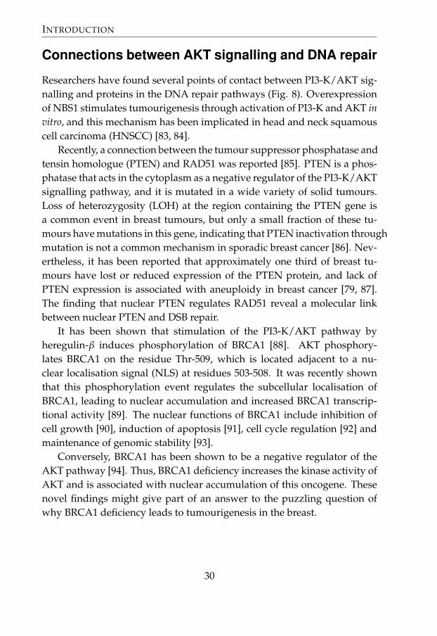
Researchers have found several points of contact between PI3-K/AKT sig-nalling and proteins in the DNA repair pathways (Fig. 8). Overexpressionof NBS1 stimulates tumourigenesis through activation of PI3-K and AKT invitro, and this mechanism has been implicated in head and neck squamouscell carcinoma (HNSCC) [83, 84].
Recently, a connection between the tumour suppressor phosphatase andtensin homologue (PTEN) and RAD51 was reported [85]. PTEN is a phos-phatase that acts in the cytoplasm as a negative regulator of the PI3-K/AKTsignalling pathway, and it is mutated in a wide variety of solid tumours.Loss of heterozygosity (LOH) at the region containing the PTEN gene isa common event in breast tumours, but only a small fraction of these tu-mours have mutations in this gene, indicating that PTEN inactivation throughmutation is not a common mechanism in sporadic breast cancer [86]. Nev-ertheless, it has been reported that approximately one third of breast tu-mours have lost or reduced expression of the PTEN protein, and lack ofPTEN expression is associated with aneuploidy in breast cancer [79, 87].The finding that nuclear PTEN regulates RAD51 reveal a molecular linkbetween nuclear PTEN and DSB repair.
It has been shown that stimulation of the PI3-K/AKT pathway byheregulin-β induces phosphorylation of BRCA1 [88]. AKT phosphory-lates BRCA1 on the residue Thr-509, which is located adjacent to a nu-clear localisation signal (NLS) at residues 503-508. It was recently shownthat this phosphorylation event regulates the subcellular localisation ofBRCA1, leading to nuclear accumulation and increased BRCA1 transcrip-tional activity [89]. The nuclear functions of BRCA1 include inhibition ofcell growth [90], induction of apoptosis [91], cell cycle regulation [92] andmaintenance of genomic stability [93].
Conversely, BRCA1 has been shown to be a negative regulator of theAKT pathway [94]. Thus, BRCA1 deficiency increases the kinase activity ofAKT and is associated with nuclear accumulation of this oncogene. Thesenovel findings might give part of an answer to the puzzling question ofwhy BRCA1 deficiency leads to tumourigenesis in the breast.
30

Connections between AKT signalling and DNA repair
PI3-K
Akt
BRCA1BRCA1
P
P
ubiquitination
P
maintenanceof the
genome
imp.
transcription
Nucleus
Heregulin
c-MYCNBS1
P T
RAD51
PTEN
inhibition stimulation phosphorylation transactivationTP
Figure 8: Connections between PI3-K/AKT signalling and DNA repair. AKT reg-ulates the subcellular localisation and activity of BRCA1. Stimulation of heregulin-β induces AKT-mediated phosphorylation of BRCA1 on Thr-509 next to the nu-clear localisation signal. This phosphorylation event increases the nuclear contentof BRCA1, and at least some of its nuclear functions. Conversely, BRCA1 hasbeen shown to mediate ubiquitination and degradation of AKT. Overexpressionof NBS1 in head and neck tumours stimulates tumourigenesis through activationof PI3-K/AKT signalling. The nuclear fraction of PTEN regulates RAD51 expres-sion, thereby contributing to maintenance of genomic stability.
It also demonstrates that the signalling networks in a cell, regulatinggenomic integrity, cell cycle checkpoints, proliferation, apoptosis and otherfunctions, are interconnected on many levels.
31

INTRODUCTION
DNA repair pathways as targets for cancer therapy
Pharmacological inhibition of DNA repair has the potential to sensitise tu-mour cells to the damaging effects of therapy. Inhibitors have been devel-oped against several of the proteins studied in this thesis, including thePI3-K/AKT pathway [95], DNA-PK and ATM [96, 97, 98].
Recently, a novel strategy using short DNA molecules that mimic DSBsand disorganise DNA damage signalling was shown to sensitise tumoursto radiation in vitro and in mouse models [99]. In addition, inhibitors ofpoly(ADP)-ribose polymerase (PARP) can be used as sensitisers for cancertherapies such as temozolomide and IR. Especially BRCA1- and BRCA2-mutated hereditary breast cancers are sensitive to PARP inhibitors [100,101]. PARP is involved in repair of single-strand breaks by BER. Unre-paired single-strand breaks can be converted to double-strand breaks, andthese lesions can not be repaired by HR-deficient cells. The effect of PARPinhibitors in breast cancer patients with BRCA1/2 mutations is currentlyevaluated in phase II clinical trials. This exemplifies that modulation ofDNA repair has potential to become a future strategy to increase tumoursensitivity to chemo- or radiotherapy.
32

Aims of the thesis
General aim
The general aim of this thesis was to gain deeper insight into the complexmechanisms that affect tumour cell resistance to ionising radiation.
Specific aims
• To investigate the role of the HER2/PI3-K/AKT signalling pathwayin resistance to radiation-induced apoptosis in breast cancer.
• To study the expression of DNA repair-associated proteins in breasttumours.
• To examine if the expression of DNA repair proteins is associatedwith clinicopathological variables and if it is related to local and dis-tant recurrence-free survival.
• To investigate if the expression of DNA repair proteins is related tothe outcome of radiotherapy.
• To examine if the RAD51 135G/C polymorphism is associated withRAD51 protein expression, prognosis and effect of radiotherapy orCMF chemotherapy.
33


Comments on materialsand methods
The following is a discussion on the principles, advantages and disadvan-tages of methods used in the papers included in this thesis. Specific condi-tions and reagents used are described in more detail in the papers.
Cell culture
The use of cancer cell lines have allowed considerable advances in the fieldof cancer biology and have, for a long time, been the principal model sys-tem for breast cancer research [102]. They provide a source of homogenousmaterial that self replicate without limit. Established breast cancer cell linesare easy to culture in standard media and can be manipulated in count-less ways. Under well defined experimental conditions, cell lines generallyyield reproducible and quantifiable results [102].
Cell lines grown in vitro can not fully capture the complexity of clin-ical breast tumours. The advantage of having a homogenous populationof one cell type in the culture dish, at the same time excludes the possi-bility to study the interaction of cancer cells with the microenvironment.Most of the available breast cancer cell lines are derived from metastases,and questions have been raised as to how well they represent primarytumours. However, most investigations have revealed that progressionfrom primary tumour to metastasis is not accompanied by major changesin marker expression or histological grade, and the expression of differ-
35

COMMENTS ON MATERIALS AND METHODS
ent markers is well correlated when cell lines are compared to their corre-sponding archival tumour tissue after being grown in vitro for an extendedperiod of time [103]. The results from experiments performed on cell linescan not be directly translated to in vivo tumours, but they provide impor-tant insights to tumour biology.
The cell lines used in Paper I, MCF-7 and BT-474, are human breastcancer cell lines that have been widely used in experimental research. Wechoose them because they differ in HER2 expression and p53 status.
Patients and tumour material
The breast tumours used in Papers II-IV were collected during two ran-domised clinical trials conducted in 1976-1990 by the Stockholm BreastCancer Study Group [104]. The studies compared postoperative radia-tion therapy (RT) with adjuvant chemotherapy (CT) in pre- and postmeno-pausal patients who were at high risk of recurrence. In Papers II and III,tumours from premenopausal patients were used. In Paper IV, we haveused tumours from both pre- and postmenopausal patients. The postmeno-pausal trial also included a randomisation between tamoxifen versus noadjuvant treatment, which means that the patients were randomised to oneof four groups (RT only, RT + tamoxifen, CT only and CT + tamoxifen). Ta-moxifen was not considered in the analyses of treatment outcome in PaperIV.
All patients included in the two trials had histologically verified inva-sive, unilateral breast cancer. They were treated with modified radical mas-tectomy. Inclusion criteria were node-positive disease, or a tumour dia-meter exceeding 30 mm, as measured on the surgical specimen. Exclusioncriteria were inoperable local disease, distant metastases at the time of pri-mary diagnosis, other concurrent cancers, medical contraindications of thetreatment, or surgery that deviated from the protocol.
Radiotherapy was given at a total dose of 46 Gy with 2 Gy per fraction,5 days per week. Chemotherapy was 12 courses of cyclophosphamide/methotrexate/5-fluorouracil (CMF). More details on the treatments can befound in Papers II-IV, and in the report on the original study [104].
This material has several advantages. It is relatively homogenous interms of patient characteristics and treatment. Patients have been ran-
36

Western blot
domised to receive one of two treatments, and randomised materials simi-lar to this are rare today. The patients have not been treated with neoadju-vant chemotherapy prior to surgery, which means that the protein expres-sion in the tumours is unaffected by treatment.
Some difficulties are that the positive effects of radiotherapy on dis-tant recurrence that has been reported complicate the analyses of predic-tion of treatment effect [105]. Also, these tumours are relatively large andthe frequency of recurrence is high. The patient group is relatively homoge-nous, which makes it difficult to draw generalised conclusions that applyto breast cancer patients with other characteristics.
Western blot
Western blot is a method that allows comparison of protein expression be-tween different samples of cell or tissue extracts (Fig. 9). Similar to im-munohistochemistry (IHC), this technique relies on antibody detection ofthe protein in interest, and the result is dependent on antibody specificity.
In short, samples are lysed and the concentration of proteins in thelysate is measured. The proteins are denatured and separated on a poly-acrylamide gel according to their size. They are then transferred from thegel to a membrane made of polyvinylidine fluoride (PVDF) or nitrocellu-lose, to make the proteins accessible to antibody binding. The membrane isincubated in a blocking solution containing milk or bovine serum albumin(BSA) to prevent nonspecific binding and subsequently incubated with theprimary antibody. The membrane is washed, incubated with a secondaryantibody directed against the primary antibody, and bound antibodies aredetected and analysed.
Correct handling of the lysates is crucial, since proteins are sensitive todegradation. Samples should be kept cold and the lysing medium shouldcontain protease inhibitors. If phosphorylation is to be investigated, phos-phatase inhibitors must be used.
Since the sample consists of concentrated cell lysate, the primary anti-body can be more diluted than in IHC, typically in the range 1:1000-1:10000.Antibody dilution, incubation time (30 min - overnight) and incubationtemperature (warmer temperature resulting in more binding, both specificand nonspecific) have to be optimised for each antibody.
37

COMMENTS ON MATERIALS AND METHODS
Figure 9: Western blot of NBS1 expression in BT-474 and MCF-7 cells using thesame antibody that was used for IHC staining in Paper II. The bands to the left arethe size marker. The molecular weight of NBS1 is 95 kDa.
The size (molecular weight) of a detected protein is approximated bycomparing the distance that the band has migrated in the gel to the bands ina size marker (a ‘ladder´, containing a mixture of proteins of known sizes).Equal loading of the amount of protein in different samples is validated byrepeating the procedure, using an antibody against a structural protein (e.g.β-actin or tubulin) that should not differ between the samples. The relativeamount of protein in different samples can be approximated by measuringthe intensity of the bands, but it is important to remember that Western blotis not a quantitative method.
Western blot is a very common and relatively inexpensive method thatcan be employed to assess the expression of a wide range of proteins towhich antibodies have been developed. Some drawbacks are that the pro-tein expression is analysed in a lysate and not in single cells, which meansthat it gives a mean expression level for all the cells in a sample. Nonspe-cific background staining is sometimes a problem, but can often be avoidedby using the detergent Tween in washing and blocking buffers, optimisingblocking conditions and keeping antibody concentration and incubationtemperature low.
38

Cell cycle analysis by flow cytometry
Cell cycle analysis by flow cytometry
Cell cycle distribution was analysed by DNA flow cytometry. Exponen-tially growing cells were stained with propidium iodide (PI) using a detergent-trypsin method according to Vindelöv [106]. PI is a fluorescent dye thatbinds to DNA and emits light when the cell passes through a laser beamin the flow cytometer. Because PI binds to DNA and cells contain differentamounts of DNA depending on where they are in the cell cycle, the propor-tion of cells in each cell cycle phase can be analysed with this method. Thismethod can also be used to detect populations of cells that have abnormalamounts of DNA, e.g. tetraploid cells.
Flow cytometry can be used in a wide range of applications, includingcell sorting and phenotyping. In addition to DNA staining, different an-tibodies can be used to detect extracellular or intracellular markers, andit is possible to study several markers at the same time by combining an-tibodies that are conjugated with different fluorochromes. Cells are alsocharacterised according to their size and granularity.
Flow cytometry applications that rely on antibodies are afflicted withthe same problems regarding antibody specificity that other immunobasedmethods have. Another disadvantage is that this technique requires rela-tively large amounts of material.
Apoptosis detection
There is a plethora of methods and kits available for apoptosis detection.Some are based on manual evaluation of the morphological changes as-sociated with apoptosis (e.g. DAPI staining), others measure DNA frag-mentation (e.g. TUNEL assay), caspase activation or other alterations inmacromolecules that are unique to apoptotic cells.
In Paper I, apoptosis was detected using an Annexin V assay [107]. Dur-ing the early stages of apoptosis, phosphatidylserines (PS) are translocatedfrom the inner side of the cell membrane to the outer layer. Annexin V is aphosphobinding protein that binds PS with high affinity. Translocation ofPS also occurs during necrosis, so it is necessary to discern apoptotic cellsfrom necrotic. This is achieved by staining the cells with PI. Since the cellmembrane looses its integrity and becomes leaky in necrotic cells, these
39

COMMENTS ON MATERIALS AND METHODS
cells are stained by PI, whereas the membrane remains intact during theinitial stages of apoptosis. Thus, cells that are Annexin V+/PI- are apop-totic, whereas cells that are positive for both Annexin V and PI are necroticor post-apoptotic necrotic. The cells were analysed in a flow cytometer.
We have also used M30, an antibody that recognises caspase-cleavedcytokeratin 18 [108]. In Paper I, we used an unconjugated M30 antibodyfor detection of apoptosis by flow cytometry, and a fluorescein-conjugatedM30 antibody for immunocytochemistry (Fig. 10).
Most methods that detect apoptosis give a notion of the level of apop-tosis at that very moment. However, apoptosis is a process, and it is thusimportant to make measurements at several time points. It is also recom-mended to use at least two different methods. With some methods, it can bedifficult to discern apoptotic cells from necrotic. We chose to use AnnexinV because flow cytometry allows for the rapid analysis of tens of thousandsof cells and the PI staining excludes cells that are necrotic. M30 was cho-sen as a complement to Annexin V, since this method relies on cleavage ofcytokeratin 18, a process that is separate from PS translocation. The pos-sibility to use M30 in both flow cytometry and immunocytochemistry wasconsidered an advantage.
Immunohistochemistry
Immunohistochemistry allows detection of proteins in a tissue by the useof specific antibodies (Fig. 11).
What follows is a brief description of how to perform the method.Paraffin-embedded (or snap frozen) tumours are sectioned and mountedon adhesive microscope slides. The sections are deparaffinised in xyleneand rehydrated in a series of ethanol baths of decreasing concentration. Formost antibodies, antigen retrieval is needed prior to staining (see below).The specimens are incubated with a blocking reagent (see below), washedand the primary antibody is applied. The dilution of the antibody and in-cubation time affect the staining results and must be optimised empiricallyfor each antibody. The slides are washed and incubated with a secondaryantibody, which is enzyme-conjugated (e.g. horseradish peroxidase, HRP)and directed against the primary antibody.
40

Immunohistochemistry
Figure 10: M30 reactivity (green) is confined to the cytoplasm of apoptotic cells.Two cells at different stages of the apoptotic process are shown (left). Cells thathave been successfully transfected with constitutively activated AKT are visu-alised in red (right).
Figure 11: Immunohistochemical staining using an antibody against NBS1. Innormal breast (a), the epithelial cells form ducts in an organised structure with aclear histology. In tumours (b), the tissue is disorganised. The cells have often lostattributes that are characteristic for the different cell types. The size of the cells canalso vary and many tumour cells are enlarged as compared to normal epithelialcells. Original magnification x400.
After washing, the sections are immersed in a substrate reagent solu-tion, e.g. 3,3’-diaminobenzidine tetrahydrochloride (DAB), and the HRPcatalyses a reaction that produces an insoluble, brown end-product in thecells that express the protein of interest. The specimens can be counter-stained; e.g. with heamatoxylin, which stains all nuclei in a blueish colour.The slides are washed, dehydrated in ethanol/water baths of increasingethanol concentration and mounted with cover glasses.
41

COMMENTS ON MATERIALS AND METHODS
TMA
All IHC stainings in Papers II and III were performed on tissue microrarray(TMA) slides. The TMA method was introduced in 1998 by Kononen andcolleagues [109]. The arrays are constructed by taking core needle biop-sies from paraffin-embedded tumours and re-embedding the biopsies ina new paraffin block. In this way, it is possible to collect samples fromhundreds of tumours in a few blocks. Tumour heterogeneity could pose aproblem; however, validation studies have shown that if biopsies are takenin triplicates from each tumour, TMA is comparable to whole-section ana-lysis [110, 111, 112]. This technique has several advantages. In contrastto whole-section staining, which requires the processing and staining ofhundreds of slides, the microarray technique allows for investigation of alarge set of cases by analysing just a few slides. Since it is possible to stainall slides at the same time, the problem of interbatch variation is reduced.The biggest advantage is that this technique reduces the amount of tissueneeded, which is an important aspect when working with archived mate-rial.
Antibodies
Antibodies can be either monoclonal or polyclonal. Monoclonal antibod-ies are typically produced by myeloma cells that are fused with antibody-secreting spleen cells from a mouse that has been immunised with the spe-cific antigen. The resulting cell, a hybridoma, can be cell cultured to pro-duce large quantities of identical monoclonal antibodies. Polyclonal anti-bodies, on the other hand, are secreted from several different clones of cellsand react with multiple epitopes on the antigen. Due to the greater riskof unspecific binding of polyclonal antibodies to molecules other than thetarget, monoclonal antibodies are often preferred. The greatest advantageof polyclonal antibodies is that they are not as sensitive to small conforma-tional changes of the target antigen (caused by fixation, glycosylation orpolymorphisms in the target) since several antibodies with different targetepitopes can bind to the same molecule.
Background staining can be a problem with some antibodies. It stemsfrom hydrophobic or ionic interactions between the antibody and tissuecomponents other than the target antigen. We used Protein Block Serum-
42

Immunohistochemistry
Free (DAKO, Carpinteria, CA) to reduce nonspecific staining. This reagentutilises casein, a hydrophilic protein which has been shown to decreasenonspecific binding of primary and secondary antibodies in immunohisto-chemistry. It is also common to add BSA to the buffer in which the antibodyis diluted and to have a detergent, such as Tween, in the washing buffer toreduce nonspecific staining.
When choosing antibodies, we have reviewed the literature andsearched for antibodies that have been successfully used before and thatare commercially available. We have preferred monoclonal antibodies overpolyclonal, and tried to find antibodies which have been tested for IHC offormalin-fixed, paraffin-embedded sections.
Fixation and antigen retrieval
One of the most important steps in IHC is the fixation of the tissue to pre-serve the structures in a lifelike state and prevent degradation. Formalin isthe most commonly used fixative and it forms stable bonds, or ‘crosslinks´,with macromolecules in the specimen. Fixation can mask the epitopes thatthe antibodies are directed against. It is therefore often necessary to processthe sample to restore the reactivity of many antibodies for tissue epitopes, aprocedure referred to as antigen retrieval. Prior to staining, we have heatedthe slides in citrate buffer (pH 6.0) in a pressure cooker or microwave oven.Other antigen retrieval methods include enzymatic digestion and heatingin an alkaline buffer.
Advantages and disadvantages
Immunohistochemistry is widely used and it is one of a few methods thatcan be used to study protein expression in archived tissue. The method hasseveral advantages, including relatively low cost, no need for expensiveand complicated equipment and the possibility to see the morphologicalstructure of the sample and exactly which cells express a specific protein.
The largest drawback of immunohistochemistry is the risk of subjectiv-ity when grading the staining. To reduce this problem, all stainings werejudged independently by two investigators (K.S. and M.S.A). In cases ofdisagreement, the stainings were re-evaluated until consensus was reached.Grading of the staining was performed blindly.
43

COMMENTS ON MATERIALS AND METHODS
PCR-RFLP
The polymerase chain reaction (PCR) is one of the most widely used labora-tory techniques today. It allows the amplification of a short DNA sequenceinto millions of copies, which can then be visualised on an agarose gel orfurther analysed using different applications.
The usability of DNA from fixed, paraffin-embedded material dependson several factors, including (a) the fixative used, (b) how long the speci-men was kept in the fixative, (c) the age of the specimen and (d) the lengthof the DNA fragment to be amplified. Some fixatives, especially highlyacidic solutions, cause DNA degradation. Fixation with formalin does notseriously compromise amplification efficiency [113]. The probability of suc-cessful amplification of large PCR products decreases with longer fixationperiods. Similarly, DNA from older specimens is more difficult to amplify.When working with older samples, one way to improve the likelihood ofsuccessful amplification is to choose small target fragments (<200 bp).
In Paper IV, a 157 bp fragment of the RAD51 gene was amplified byPCR. The PCR products were subsequently digested by the MvaI restric-tion enzyme, which cleaves DNA at a specific restriction site (Fig. 12). TheRAD51 135G/C polymorphism alters this restriction site. Digested DNA isseparated according to size on an agarose gel containing the DNA-bindingdye ethidium bromide. This restriction fragment length polymorphism(RFLP) method has been used in many previous studies for the investi-gation of this polymorphism [114, 115, 116, 117].
5´- C C A G G -3´
3´- G G T C C -5´
T
A
^^
^ restriction sitepolymorphic site
Figure 12: Restriction site of the MvaI enzyme. Cleavage of the 157 bp ampli-con results in two fragments, 86 and 71 bp long. The restriction site is destroyedby the 135G/C polymorphism, so that the amplicon remains uncleaved in C/Chomozygotes.
44

Statistical analysis
Statistical analysis
In Paper I, we studied the effects of heregulin-β1 and wortmannin on cel-lular sensitivity to radiation-induced apoptosis. The ratio between treatedand untreated cells were calculated. Student´s t-test was used to test thedifference between the mean of the ratios and 95% confidence intervals (CI)were calculated.
In Paper II-IV, the significance of the differences between grouped vari-ables (e.g. the association between the expression of protein x and clinico-pathological variables such as histological grade) were tested with χ2 testfor contingency tables. In Paper III and IV, χ2 test for trend (Spearman) wasused if a variable contained 3 or more categories.
Survival curves were estimated using the product limit method byKaplan-Meier. The role of one (univariate) or a group of variables (mul-tivariate) in survival analysis was estimated using Cox´s proportional haz-ard regression. Cox analysis was also used to test the interaction betweena variable x and treatment. This multivariate Cox analysis included thecovariates x, treatment and the interaction variable x×treatment. p-values≤0.05 were considered statistically significant.
45


Results & Discussion
HER2/PI3-K/AKT signalling is important for resistanceto radiation-induced apoptosis (Paper I)
The gene encoding the HER2 (or erbB2) growth factor receptor is ampli-fied in 15-30% of all breast tumours and overexpression of the receptor isrelated to resistance to several chemotherapeutic agents [69, 118, 119, 120].In the first study, we investigated the hypothesis that the HER2 receptorconfers resistance to radiation-induced apoptosis through the PI3-K/AKTsignalling pathway.
We used two different breast cancer cell lines, which differ in HER2 ex-pression and p53 status. The MCF-7 cell line has normal HER2 expressionand functioning p53, whereas BT-474 has constitutively activated AKT dueto HER2 gene amplification (Table 3). In addition, p53 is mutated in thiscell line.
Cell line HER2 AKT activity p53 statusMCF-7 normal upon stimulation wildtypeBT-474 overexpressed constitutively active mutated
Table 3: Characteristics of breast cancer cell lines used in Paper I.
47

RESULTS & DISCUSSION
Cell cycle regulation after γ-radiation
Cells were irradiated with 10 Gy γ-radiation and analysed for cell cycledistribution. Two days after irradiation, the fraction of MCF-7 cells in Sphase was reduced in irradiated cells as compared to the control (Fig. 1e,Paper I). This decrease of proliferative cells was not seen for the BT-474 cellline, indicating that MCF-7 was more sensitive to radiation. The reductionof MCF-7 cells in S phase was accompanied by an accumulation of cells inG2/M phase, whereas the proportion of cells in G1 phase remained rela-tively constant over time (Fig. 1d-f, Paper I). This suggests that both the G1and the G2/M checkpoint were functional in this cell line. In BT-474 cells,the G1 fraction decreased and the G2/M fraction was increased to the sameextent, indicating that these cells only halted at the G2/M checkpoint (Fig.1a-c, Paper I).
The ability of cells to halt in the G2 phase allows them to repair any sub-lethal DNA damage before entering mitosis, and is thus considered to bean important determinant of radiosensitivity. Whereas a role for p53 in theG1/S checkpoint has been clearly established, its function in G2/M is lessclear [34]. Our results, showing that only the cell line with functional p53entered G1 phase arrest, whereas both cell lines had intact G2/M check-points, are in line with this.
Radiation-induced apoptosis
Both cell lines were treated with different combinations of heregulin-β1 andwortmannin, and half of the samples were exposed to 10 Gy γ-radiation.Radiation-induced apoptosis was higher in MCF-7 cells than in BT-474 cells,again indicating that MCF-7 cells were more sensitive to the effects of IR(Fig. 3, Paper I). Heregulin-β1 protected non-irradiated MCF-7 cells fromspontaneous apoptosis. This effect was not seen in BT-474 cells in whichthe HER2/PI3-K/AKT signalling pathway was already activated by HER2overexpression.
When BT-474 cells were pretreated with the PI3-K inhibitor wortman-nin prior to irradiation, apoptosis increased as compared to treatment withwortmannin alone (Fig. 3, Paper I). We propose that the sensitivity of BT-474 cells to inhibition of PI3-K signalling is due to the dependency of thesecells on HER2-mediated signalling. Transfection of MCF-7 cells with con-
48

Paper I
stitutively active m/p-AKT made the cells more resistant to apoptosis (Fig.5, Paper I).
When assessing radiosensitivity in vitro, it is common to study the abil-ity of cells to form colonies after treatment with radiation (’colony-formingassay’). Despite several attempts, we did not succeed in growing the cellsin soft agar.
PI3-K is structurally related to DNA-PK and ATM, and wortmannin hasbeen shown to inhibit all of these PI3-K superfamily members at differentconcentrations [121, 122, 123, 124]. Since both DNA-PK and ATM have im-portant roles in DNA damage signalling, inhibition of these proteins mightmake cells sensitive to γ-radiation.
For several reasons, we believe that the radiosensitising effect by wort-mannin treatment in BT-474 cells is due to inhibition of the PI3-K/AKTsignalling pathway. The IC50 value of PI3-K inhibition by wortmannin is2-4 nM [121], whereas inhibition of ATM and DNA-PK have higher IC50
values [125]. The dose that we used (100 nM) is considerably lower than instudies on the other kinases, in which the wortmannin dose was 10-50 µM[123, 124, 126].
Wortmannin seemed to only have a sensitising effect in BT-474 cells,which overexpress HER2. Since we have not investigated the expression ofDNA-PK and ATM, we can not rule out the possibility that these kinasesare differently expressed in the cell lines and coincide with the effect ofwortmannin. However, AKT phosphorylation was decreased after treat-ment with wortmannin, indicating that PI3-K was affected (Fig. 2, Paper I).In addition, stimulation with heregulin-β1, as well as transfection of MCF-7cells with activated AKT, made these cells more resistant to apoptosis, thussupporting the hypothesis that the radiosensitising effect of wortmanninwas mediated by PI3-K/AKT signalling.
In conclusion, the results in Paper I support the hypothesis that theHER2/PI3-K/AKT signalling pathway is involved in resistance to radiation-induced apoptosis in breast cancer cells. This pathway has been shown tobe dysregulated on several levels in cancer, including HER2 overexpressionand loss of the tumour suppressor PTEN.
49

RESULTS & DISCUSSION
DNA repair-associated proteins in breast cancer(Paper II and III)
In this thesis, we have studied the protein expression patterns of ATM,MRE11, RAD50 and NBS1 (Paper II), as well as BRCA1, BRCA2 and RAD51(Paper III). We investigated the role of these proteins in prognosis and pre-diction of radiotherapy effect in early breast cancer.
Reduced or elevated expression of DNA repair proteins inbreast tumours?
One of the most elementary questions to address in studies that aim toinvestigate the prognostic or predictive significance of a protein, is in whatway the expression is altered in tumours as compared to normal tissue. Inthe immunohistochemistry studies (Paper II and III), we have used normalbreast epithelium as reference.
NORMAL BREAST TUMOURS
Protein Intensity Percentage a Reducedintensity b
Increased per-centage a,b
ATM moderate 1-25 76% 80%MRE11 strong 51-75 72% 20%RAD50 strong 1-25 79% 61%NBS1 strong 26-50 79% 79%BRCA1 strong 51-75 80% 46%BRCA2 weak 26-50 1% 56%RAD51 strong 26-50% 76% 40%
Table 4: Staining of the different proteins investigated in Paper II and III.a percentage of positively stained nucleib as compared to normal breast epithelium
Five of the seven DNA repair-associated proteins that were investigatedin Paper II and III showed strong staining intensity in normal breast epithe-lium, with the exception of BRCA2 and ATM (Table 4). The staining inten-sity was reduced in more than 70% of the tumours for all proteins exceptBRCA2. The percentage of positive cells in normal breast varied for thedifferent proteins; yet the percentage of positive cells was increased in 40%or more of the tumours for all proteins except MRE11. Taken together, this
50

Paper II and III
indicates that the DNA repair proteins were expressed in more cells, but ata lower level, in breast tumours as compared to normal breast tissue.
There are a couple of reports on the expression of DNA repair proteinsin breast tumours. The results in Paper II, indicating that down regulationor loss of MRE11, RAD50, NBS1 and ATM is a common event in breastcancer, are supported by a previous study by Angele et al [127]. Similarly,BRCA1 expression is often aberrant in sporadic breast cancer due to LOH,methylation of the promotor region and low mRNA and protein expression[128, 129, 130, 131].
As for RAD51, its role in tumour development and progression is notclear. Several studies have reported overexpression of RAD51 in cell linesand tumours of different origin [132, 133, 134, 135]. However, other linesof evidence indicate that RAD51 represses tumour progression [136]. Inbreast tumours, both high and low RAD51 expression has been reported[132, 137], but the mechanism behind deregulated RAD51 expression is asyet unknown.
When the expression of the individual proteins was combined to al-low evaluation of the complexes, 44% of the tumours had negative/weaknuclear staining intensity of the MRN complex and 53% were categorisedas having low expression of the BRCA1/BRCA2/RAD51 complex (PaperII and III). This implies that the protein expression of these DNA repair-associated proteins is reduced in a considerable proportion of breast tu-mours.
Significance of the subcellular localisation
In Paper II, a smaller fraction (10%) of the tumours showed cytoplasmicstaining of NBS1. Similarly, MRE11 was found in the cytoplasm in morethan half of the tumours, but not in normal breast tissue (Paper II). This cy-toplasmic staining correlated with weak nuclear RAD50 staining intensity.This correlation between weak nuclear staining of one protein and cyto-plasmic staining of another might be explained by a previously reportedobservation that alterations in one member of a multiprotein complex candestabilise some or all of its protein components. NBS1 and MRE11 stain-ing is exclusively nuclear in normal fibroblasts, but diffuse in cells frompatients with ATLD (caused by mutations in the MRE11 gene) [59]. Sim-
51

RESULTS & DISCUSSION
ilarly, the nuclear fraction of MRE11 and RAD50 is reduced in NBS cells(lacking NBS1), with a concomitant increase of protein in the cytoplasm[60].
Cytoplasmic staining was also observed for BRCA2, both in normalbreast epithelium and in tumours (Paper III). Reduced cytoplasmic stain-ing of BRCA2 in tumours was correlated to ER negativity (p=0.01), but notto any other clinicopathological factors, nor to the local recurrence-free sur-vival.
RAD51 showed cytoplasmic staining in approximately half of the tu-mours (53%) (Paper III). A small number of tumours were positive for cy-toplasmic staining, but negative for nuclear staining. This expression pat-tern has been associated with germline mutation of BRCA2 [138]. ThoughBRCA2 and RAD51 are primarily nuclear proteins, they have previouslybeen observed in the cytoplasmic compartment [137, 138, 139, 140].
A large fraction of the tumours (64%) showed cytoplasmic stainingof ATM, whereas normal breast epithelial cells had only nuclear expres-sion (Paper II). Cytoplasmic staining of ATM in breast cancer cells hasbeen reported previously [141], but the function of ATM in the cyto-plasm is not known. Cytoplasmic staining of ATM was correlated toER negativity (p=0.03), but not to the rate of local recurrence. Patientswith cytoplasm-positive tumours had no significant benefit of radiother-apy over chemotherapy on local recurrence-free survival (p=0.20), whereascytoplasm-negative patients responded to radiotherapy (p=0.03).
Subcellular relocalisation is a means of regulation of protein function,as can be seen for the oncogenic kinase c-ABL [142], and it is possible thatcytoplasmic accumulation of the MRN proteins (or other enzymes that ex-ert similar activities) impair their function. Since many of the functions ofthe DNA repair enzymes involve DNA damage signalling, cell cycle regu-lation and direct interaction with DNA, we reasoned that nuclear proteinwould have the highest impact on DNA repair efficiency. However, we donot rule out the possibility that cytoplasmic fractions of these proteins mayact as a reserve, which can be shuttled to the nuclear compartment undersome circumstances.
The clinical relevance of cytoplasmic expression of the investigated pro-teins is not clear. In both Paper II and III, it was the nuclear expression thatwas associated with local recurrence-free survival and prediction of radio-
52

Paper II and III
therapy effect. It is however possible that cytoplasmic expression of ATMhas biological relevance, since the results in Paper II indicated that cyto-plasmic ATM staining predicted a poor response to radiotherapy.
Expression patterns of proteins that form a complex together
The nuclear staining intensities for MRE11, RAD50 and NBS1 were highlycorrelated, with strong staining of one of the proteins correlating with strongstaining of the other two (all p<0.001) (Paper II). The observation that lowexpression of MRE11, RAD50 or NBS1 was rarely found alone is in line witha previous finding that the components of the complex stabilise each other.Similarly, low RAD51 expression was correlated to weak BRCA1 staining(p<0.001) (Paper III). We have also found correlations between the expres-sion of DNA-PKcs and Ku70/80, the components of the DNA-PK complexthat is central in NHEJ repair (unpublished results).
It could be that the expression of these proteins is regulated by relatedmechanisms. Another possibility is that the observed correlations are anartefact, caused by fixation problems or differences in tissue quality be-tween tumours, that affect the staining result. The most likely explanation,however, is probably that the similarities in expression patterns of proteinsthat act in complexes with each other are the result of stabilisation when allcomponents of the complex are present, as mentioned above.
Reduced expression of complexes is associated withclinicopathological variables
Negative/weak expression of the MRN complex was correlated with highhistologic grade (p=0.01) and ER negativity (p=0.0001) (Paper II). Simi-larly, low expression of the BRCA1/BRCA2/RAD51 complex was asso-ciated with high histologic grade (p=0.05) (Paper III). This is in line withseveral previous studies, in which a correlation between reduced BRCA1expression and high histologic grade has been a consistent finding [143,144, 145, 146].
When the proteins in Paper III were analysed individually, RAD51 ex-pression in a low fraction of cells (defined as 0-25% positive nuclei) cor-related with high grade and ER negativity (p<0.001 and p=0.006, respec-tively), as did weak RAD51 intensity.
53

RESULTS & DISCUSSION
In contrast to our results, high RAD51 expression (defined as dark stain-ing in >1% of tumour cells) has been shown to correlate with high histo-logic grade and ER negativity in breast cancer specimens [132]. One prob-lem with IHC is that variations in scoring systems make it difficult to com-pare the results from different studies. When evaluating the IHC staining,we chose to categorise the intensity into four groups (-,+,++ and +++), sincethe intensity varied between specimens. Variations in staining intensity ofthese proteins have been observed by others as well [144, 146].
In conclusion, the results from Paper II and III indicate that there is anassociation between reduced expression of these DNA repair complexesand an aggressive tumour phenotype.
Relation to recurrence-free survival
All patients in Paper II-IV have received either local or systemic treatment.In analyses of prognosis, we tried to minimise the effect of treatment byonly including the subgroup of patients that received CMF when analysinglocal recurrence-free survival, and only the radiotherapy treated patientsin analysis of distant recurrence-free survival. However, we can not com-pletely rule out the possible influence of treatment in the prognosis analy-ses.
Local recurrence
The percentage of NBS1-expressing tumour cells was an independent prog-nostic factor, and patients with high expression (>25% positive cells) hadfewer local recurrences (Fig. 2a, Paper II). The MRN complex was not aprognostic factor of local recurrence-free survival.
Similarly, patients with reduced expression of the BRCA1/BRCA2/RAD51 complex had significantly more local recurrences as compared topatients with high expression (Fig. 2a, Paper III). When analysed individu-ally, low RAD51 expression was related to a higher risk of developing localrecurrence (Fig. 2b, Paper III). Both RAD51 and the complex were indepen-dent prognostic factors in multivariate analysis.
54

Paper II and III
Distant recurrence
Moderate/strong expression of the MRN complex was related to longerdistant recurrence-free survival as compared with negative/weak expres-sion (Fig. 2b, Paper II). Similarly, patients whose tumours had moder-ate/strong expression of MRE11 had a better prognosis. MRE11 intensityand expression of the MRN complex were independent prognostic factorsof distant recurrence. In Paper III, the BRCA1/BRCA2/RAD51 complexwas not related to distant recurrence-free survival.
Deregulated expression of DNA repair-associated proteins seems to re-sult in genomic instability, as evidenced by an increased frequency of chro-mosomal rearrangements in cells from NBS patients, chromosome aberra-tions in cells deficient of BRCA1 or BRCA2 and genome instability in cellsafter depletion or overexpression of RAD51 [147, 148, 149, 150, 151, 152].In a recent study, chromosomal instability was shown to predict poor clini-cal outcome in multiple types of cancer, including breast cancer [153]. Ourfindings, that low expression of DNA repair proteins are related to higherrisk of recurrence, are in line with this.
The prognostic significance of some of these proteins have been inves-tigated previously. Studies on the correlation between prognosis and re-duced expression of BRCA1 in spontaneous breast tumours have yieldedcontradicting results [143, 145, 146]. We found no prognostic value forBRCA1 expression alone.
In a previous study on RAD51 expression and prognosis in patientswith non-small cell lung carcinoma (NSCLC) of different stages, the cutoff point was chosen to obtain optimal separation between prognosticallydifferent groups [154]. High RAD51 expression (>10%positive cells) wasfound to correlate to shorter overall survival in that study.
The prognostic significance of NBS1 has been investigated in other can-cer types as well. Overexpression of NBS1 is a marker of poor prognosisin advanced HNSCC and uveal melanoma [84, 155]. This is in contrastwith our results, that suggested that high expression of NBS1 was relatedto good prognosis. As indicated above, moderate/strong expression ofthe MRN complex, and the individual proteins as well, were associatedwith low histologic grade and ER positivity. It should be noted, however,that these studies differ in measured endpoint (local recurrence vs overall
55

RESULTS & DISCUSSION
survival), tumour type and stage of the disease (early vs advanced). It ispossible that both decreased and increased levels of these proteins lead todisturbed cellular functions and affect prognosis.
The results in Paper II suggest that the components of the MRN areprognostic markers in breast cancer. Both RAD51 and the whole BRCA1/BRCA2/RAD51 complex seem to be related to local, but not distant recurrence-free survival. Overall, results from different studies on the possible prog-nostic value of these proteins are not conclusive and further studies areneeded.
Can the expression of DNA repair proteins predict the effect ofradiotherapy?
We hypothesised that reduced expression of DNA repair-associated pro-teins in tumour cells results in impaired repair of radiation-induced dam-ages. This could lead to better eradication of tumour cells by radiotherapyand be reflected as improved local tumour control. For example, inhibi-tion of NBS1 or RAD51 has been shown to increase radiosensitivity in vitro[156, 157, 158, 159, 160].
The MRN complex and ATM
Radiotherapy significantly reduces the risk of local recurrence [29]. Thiseffect was also seen in Paper II and III, in which radiotherapy resulted inimproved local tumour control as compared to chemotherapy (p=0.04). Thegreatest benefit of radiotherapy was seen in patients with moderate/strongMRN expression, whereas patients with negative/weak MRN complex hadno benefit of radiotherapy over CMF on local tumour control (Fig. 3 andTable 4, Paper II).
The finding that high expression of these proteins was important for theclinical effect of radiotherapy was unexpected and differed from our orig-inal hypothesis. The MRN complex acts as a sensor of DSBs and recruitsATM to broken DNA molecules [161]. It is possible that down regulation orloss of the proteins in the MRN complex impairs the ability to detect DNAstrand breaks and results in failure to induce DNA damage signalling, cellcycle arrest and apoptosis, thereby increasing the risk of relapse. In sup-
56

Paper II and III
port of this, a role for NBS1 in apoptosis induction following DNA damagewas recently reported [162].
A benefit of radiotherapy was also seen in patients with moder-ate/strong expression of nuclear ATM (Paper II). We performed analyseson the combined expression of ATM and the MRN complex, and foundthat patients that had moderate/strong expression of MRN responded toradiotherapy, regardless of ATM expression (Fig. 4a-b, Paper II). Amongpatients with negative/weak MRN expression there was no significant dif-ference between radiotherapy and chemotherapy on local recurrence, irre-spective of ATM expression (Fig. 4c-d, Paper II). One interpretation is thatthe MRN complex is more important than ATM in the clinical response toradiotherapy. Experimental studies indicate that binding of ATM to DNAis dependent on MRN, whereas MRN can associate with DNA indepen-dently of ATM [161]. In addition, MRN seems to serve as an amplifier ofATM activity [163].
The BRCA1/BRCA2/RAD51 complex
In Paper III, a good response to radiotherapy was seen in patients whosetumours had low expression of the BRCA1/BRCA2/RAD51 complex (Fig.3a). When analysed individually, there was a significant benefit of radio-therapy in patients with low expression of RAD51 or weak expression ofBRCA2 (Fig. 3 and Table 4, Paper III). Similarly, there was a trend towardsa difference in local recurrence-free survival between the treatments amongpatients with weak BRCA1. In concordance with our results, high expres-sion of RAD51 has been associated with resistance to treatment in differenttumour types [135, 164, 165].
The results from Paper II and III suggest that reduced expression of theMRN complex predicts a poor effect of radiotherapy in patients with earlybreast cancer. Conversely, patients with low expression of the BRCA1/BRCA2/RAD51 complex in their tumours benefit from radiotherapy. Thisindicates that the proteins in different DSB repair pathways contribute dif-ferently to the effect of radiotherapy.
57

RESULTS & DISCUSSION
Does a polymorphism in RAD51 influence proteinexpression, prognosis and the effect of therapy?(Paper IV)
The 135G/C single-nucleotide polymorphism (SNP) in the 5’ UTR of theRAD51 gene has been found to elevate breast cancer risk among BRCA2carriers [114, 116, 166, 167, 168]. We aimed to investigate whether thispolymorphism is related to RAD51 protein expression, prognosis of earlybreast cancer and if it contributes to resistance to therapy. Due to the lowfrequency of C/C homozygotes, C/C and G/C were grouped and labelled’C-allele’ and compared with G/G homozygotes in all analyses.
The frequency of C-allele was 15.4% (including two C/C homozygotes).Presence of C-allele was correlated to ER positivity (p=0.008), but not to anyother clinicopathological variables, nor to the recurrence-free survival.
The functional effect of this SNP in the untranslated region of the gene isstill unclear. The activity of the RAD51 promotor is significantly enhancedwhen G is substituted for a C at this site in vitro [169]. On the other hand,it has been suggested that the SNP affects the alternative splicing withinthe 5’ UTR, and that this would result in a lower abundance of the RAD51protein, but this has not been confirmed on protein level [167]. Since weobserved reduced expression of RAD51 in a majority of breast tumours,we aimed to investigate whether the deregulated expression was coupledto the RAD51 135G/C polymorphism. However, no statistically significantcorrelation between genotype and RAD51 protein expression was found(Paper IV).
In Paper III, patients whose tumours had low expression of RAD51responded well to radiotherapy, in accordance with previous studies onRAD51 and in vitro radioresistance [158, 170]. The results from Paper IVsuggest that patients who had the C-allele had no benefit of radiotherapy,whereas patients with the G/G phenotype had significantly less local re-currences when treated with radiotherapy (Fig. 1, Paper IV). The numberof local recurrences in the C-allele group was, however, small and these re-sults need to be confirmed on a larger group of patients before conclusionscan be drawn.
RAD51 has also been implicated in resistance to different chemothera-peutics, e.g. etoposide, doxorubicin, cisplatin and mitomycin C [135, 165,
58

Paper IV
171]. In Paper IV, CMF significantly reduced the risk of distant recur-rence the first 20 years in patients who had the C-allele. This benefit fromchemotherapy was not observed in patients who had the G/G genotype,indicating that CMF effect was related to genotype (Fig. 2, Paper IV). Therewas a significant interaction between chemotherapy and C-allele (p=0.02).
The results suggest that patients harbouring the C-allele respond toCMF, but not to radiotherapy. Most of the chemotherapeutics that havebeen investigated in respect to RAD51 overexpression and therapy resis-tance, including etoposide, cisplatin and mitomycin C, cause strand breaks,in some ways similar to ionising radiation. Of the drugs in the CMF reg-imen investigated in Paper IV, only cyclophosphamide is a crosslinkingagent, whereas methotrexate and 5-FU inhibits RNA and DNA synthesisand do not induce strand breaks. It is possible that the influence of theRAD51 polymorphism on therapy resistance is different for drugs and ra-diotherapy, and also for chemotherapeutics with different mechanisms ofaction.
Since there was no relation between RAD51 expression and outcome ofCMF treatment in Paper III, it is possible that the connection between the135G/C polymorphism and response to CMF treatment is not mediatedby changes in protein levels. The uncertainty revolving around the conse-quences of this SNP at the protein level makes it difficult to speculate aboutthe molecular mechanism behind the observed connection. It is howevernot implausible that the RAD51 135G/C polymorphism could affect cel-lular phenotype, considering the modifying effect that this SNP seems tohave on BRCA2 mutation penetrance [114, 116, 166, 167, 168].
59


Conclusions
Growing evidence point towards connections between the PI3-K/AKT path-way, DNA repair and genomic stability. A negative regulatory loop involv-ing AKT and BRCA1 indicates that down regulation or loss of DNA repairproteins might affect cell survival through additional mechanisms otherthan impaired DNA repair. Also, NBS1 has been implicated in both apop-tosis stimulation and inhibition. Our studies showed that:
• stimulation of the PI3-K/AKT pathway can prevent induction of apop-tosis in breast cancer cells in vitro. Inhibition of this pathway renderedHER2-overexpressing breast cancer cells more sensitive to radiation-induced apoptosis.
• The expression of the MRE11/RAD50/NBS1 complex and the BRCA1/BRCA2/RAD51 complex on protein level seemed to be reduced in aconsiderable portion of breast tumours.
• The expression of proteins that form a complex together was corre-lated.
• Reduced expression of the complexes was correlated with clinico-pathological variables that are associated with poor prognosis.
• NBS1 alone, but not the MRN complex as a whole, was an indepen-dent prognostic factor of local recurrence-free survival. Similarly, theBRCA1/BRCA2/RAD51 complex and RAD51 had prognostic valuein this patient material.
61
CONCLUSIONS
• The MRN complex, but not the BRCA1/BRCA2/RAD51 complex,was a prognostic marker of distant recurrence-free survival.
• Moderate/strong expression of the MRN complex, but reduced ex-pression of the BRCA1/BRCA2/RAD51 complex, predicted good re-sponse to radiotherapy.
• The RAD51 135G/C polymorphism may be a predictor of therapyresponse. Patients with the G/G genotype tended to respond well toradiotherapy, whereas patients who had the C-allele benefitted fromCMF chemotherapy.
The results could be of clinical importance, since approximately one infive breast cancer patients have tumours that overexpress HER2 and thisreceptor can be targeted by treatment with Trastuzumab. Furthermore, ourresults suggest that the different repair complexes contribute differently totumour cell sensitivity to radiotherapy. This is probably a reflection of thecomplexity of the DNA damage response networks. It would be importantto keep this complexity in mind when developing individualised cancertreatments that target proteins in the DNA damage signalling and repairpathways.
Future perspectives
It would be interesting to study the role of the investigated DNA repair-associated proteins in radioresistance in vitro by using stable transfectionof breast cancer cell lines. Previous studies have primarily used short terminhibition or overexpression of these proteins in cell culture, but it is pos-sible that cells in clinical tumours are adapted to altered expression andrespond differently. Also, the level of expression of key proteins that isneeded for functional repair has as yet not been determined.
Preclinical studies of PARP inhibitors have shown promising results inbreast cancer patients with mutations in BRCA1 or BRCA2. It would beinteresting to study the effect of PARP inhibitors on cells that lack otherkey proteins in double-strand break repair pathways, such as RAD51.
62

Acknowledgements
I would like to express my sincere gratitude to everyone who has helpedand supported me in different ways during these years.
Marie Stenmark Askmalm for all you have taught me and for your greatgenerosity on many levels. Thanks for your never-ending optimism,support and belief in me.
Olle Stål for allowing me to do my Master´s thesis and welcoming me inyour group without any prior knowledge of me. Thank you for yourwise guidance during these years, your fairness and for sharing yourenthusiasm for science. You connect the dots in the map of signallingpathways like no one else.
Bibbi, our very own ‘spider in the web´. Without you the lab would prob-ably look like Ground Zero.
Chatarina Malm You always have time to help with all kinds of things andyou cheer me up with your energy.
Bo Nordenskjöld Thank you for your support in general, and the valuablefeedback on my manuscripts in particular.
My co-authors for critical reading of the article manuscripts.
Jan Rzepecki for fact checking the part about radiotherapy in this thesis,Peter Larsson, Sara Olsson, Lotta Jonson and Erik Angland for your
63

ACKNOWLEDGEMENTS
help with irradiating cells, Najme Wall and Jan Olofsson for classi-fying the tumours according to the NHG system, Torsten Hägerströmfor collecting the archived tumour material, Lilianne Ferraud andBirgitta Holmlund for constructing the TMA and purifying DNA,and Lambert Skoog for sharing your great knowledge about histo-logy and evaluation of IHC stainings.
Anna Asklid for enthusiastically working so hard with the genotyping.
Ben Kersley for the thorough proofreading of my thesis and for curingmy hyphenation disease. Plus, now I know 100% more about ballbearings than I did before.
Dennis Netzell at LiU-Tryck, for excellent help with the printing of thisthesis. I learned many things about cover design and typesettingchoices from working with you.
The Swedish Cancer Foundation, Landstinget i Östergötland and the Li-ons foundation for economical support of this work.
My friends and fellow PhD students:
Åsa my friend, room mate and valuable book critic. Thanks for teachingme (with the help of urbandictionary.com) what a ‘** fo sho´ is (avery dirty expression indeed) and for filling our office with laughter.Also, thanks for providing a warm shoulder to cry on when I neededit the most. I hope our endless discussions on molecular mechanismsin tumour biology, how to hit the notes on ABBA Singstar and, ofcourse, family life and childhood diseases, will continue for a longtime.
Marie A W my friend and room mate, with whom I have shared the upsand downs of PhD studies and research. Thanks for giving me theextra energy needed for thesis writing with your specially designedkit. You ought to patent that and make a living on it, I think it wouldbe a hit. You will always be my ‘seniordoktorand´.
Daniella We shared loss and sorrow not long ago - now it is soon time forus to share joy and pride together when we have reached our goal.
64

Thank you for being such a good and noble friend, both at work andin our free time. I will miss you deeply. Give me a call whenever youneed anything from your ‘sugar daddy´. Good luck ‘over there´!
Gizeh my very own guru, who welcomed me in your very generous waywhen I came to do my Master´s thesis many years ago. Thanks forall the things you have taught me in the lab, the inspiring and oftenphilosophic discussions and all the fun things we have done togetherwith the children. A special thanks for inviting Emma to your heroinparty (or was it ‘Halloween´? Emma was not sure). I know you willbe successful, whatever you choose to do.
Josefine B We have had so much fun together with our four daughters(and men). When I have finished this thesis, we can celebrate withlots of sushi.
Piiha-Lotta for being an inspiring friend and a great travel companion onthe trip to Washington D.C.
Agneta, Tove, Andreas, Annica, Tina, Annelie, Xiao-Feng, Birgitta andLilianne for creating a good atmosphere. Thanks for all our enjoy-able discussions at fredagsfikat and lunches, the many great socialgatherings and for making Onkologen a great place to belong to.
Present and past co-workers at KEF for nice fika discussions and a friendlyatmosphere.
Pia, Håkan and Kerstin for keeping the wheels rolling at KEF.
Present and past members of Syjuntan (Cissi, Daniella, Josefine A, Marie,Charlotte, Hanna, Johanna and Anna) for the chats about kids, career,evolution theories, Eurovision Song Contests, ice hockey and how tomake the embroideries pretty on the back side. I wonder when wasthe last time we finished a cross stitch project?
Bokcirkeln (Agneta, Daniella, Gizeh, Josefine, Piiha-Lotta, Tove and Åsa)for fruitful discussions on relationships, research, how to cast moviesand sometimes even the book we have (tried to) read.
65

ACKNOWLEDGEMENTS
Cissi, Dan, Isabelle, Daniella and Johan for all the board game nights dur-ing these years. Tasty food, good wine, three kids who get tiredat three different time-points during the evening (synchronisation?What does that mean?), challenging boardgames and sweet dessertson top of all that - could there be a better way to spend a Saturdaynight? Nope.
Erik and Maria for being good friends, babysitters and perfect neighboursin the ‘good old days´. I look forward to next New Year´s Eve!
Kicki, Alex, Bettan, Herman, Carro, Ida, Olle, Anne Marie, Gittan andArne for welcoming me into your large and warm family. You havealways room for one more around the table.
Björn and Märit You always offer both delicious dinners and fun discus-sions when we meet. Thank you for encouraging me by showinginterest in both my work and my free time writing projects.
Mum and dad You have always showed that you believe in me. Withoutyour support, this thesis would probably not exist. I love you both sovery much.
Mikael for being the best little brother and for all those fun times that wehave shared. You left me with many happy memories. Thanks forshowing me the importance of not letting obstacles get in the way ofmaking dreams come true. I miss you so much. I think of you everysingle day, and smile.
Emma my witty and very wise daughter, and Tilda, my ever smiling ‘penn-troll´. You have brought so much laughter and joy into my life.
Ola My love and my best friend. ♥
66

Bibliography
[1] Socialstyrelsen - National Board of Health and Welfare. Cancer Inci-dence in Sweden 2007. 2008.
[2] Ferlay, J., Bray, F., Pisani, P., Parkin, D. GLOBOCAN 2002: CancerIncidence, Mortality and Prevalence Worldwide, IARC CancerBaseNo. 5. version 2.0. Lyon. IARCPress, 2004.
[3] Engholm, G., Ferlay, J., Christensen, N. et al. NORDCAN: CancerIncidence, Mortality and Prevalence in the Nordic Countries, Ver-sion 3.2. Association of Nordic Cancer Registries. Danish Cancer Society.(http://www.ancr.nu), 2008.
[4] Kvale, G. Reproductive factors in breast cancer epidemiology. ActaOncol, 31(2): 187–94, 1992.
[5] Hankinson, S. E., Colditz, G. A., Willett, W. C. Towards an integratedmodel for breast cancer etiology: the lifelong interplay of genes, life-style, and hormones. Breast Cancer Res, 6(5): 213–8, 2004.
[6] Michels, K. B., Mohllajee, A. P., Roset-Bahmanyar, E. et al. Diet andbreast cancer: a review of the prospective observational studies. Can-cer, 109(12 Suppl): 2712–49, 2007.
[7] van den Brandt, P. A., Spiegelman, D., Yaun, S. S. et al. Pooled ana-lysis of prospective cohort studies on height, weight, and breast can-cer risk. Am J Epidemiol, 152(6): 514–27, 2000.
67

BIBLIOGRAPHY
[8] Smith-Warner, S. A., Spiegelman, D., Yaun, S. S. et al. Alcohol andbreast cancer in women: a pooled analysis of cohort studies. JAMA,279(7): 535–40, 1998.
[9] Rosen, E. M., Fan, S., Pestell, R. G., Goldberg, I. D. BRCA1 gene inbreast cancer. J Cell Physiol, 196(1): 19–41, 2003.
[10] Serova, O. M., Mazoyer, S., Puget, N. et al. Mutations in BRCA1and BRCA2 in breast cancer families: are there more breast cancer-susceptibility genes? Am J Hum Genet, 60(3): 486–95, 1997.
[11] Hakansson, S., Johannsson, O., Johansson, U. et al. Moderate fre-quency of BRCA1 and BRCA2 germ-line mutations in Scandinavianfamilial breast cancer. Am J Hum Genet, 60(5): 1068–78, 1997.
[12] Antoniou, A. C., Pharoah, P. D., McMullan, G. et al. Evidence forfurther breast cancer susceptibility genes in addition to BRCA1 andBRCA2 in a population-based study. Genet Epidemiol, 21(1): 1–18,2001.
[13] Bland, K. I., Menck, H. R., Scott-Conner, C. E. et al. The NationalCancer Data Base 10-year survey of breast carcinoma treatment athospitals in the United States. Cancer, 83(6): 1262–73, 1998.
[14] Tejler, G., Norberg, B., Dufmats, M., Nordenskjold, B. Survival aftertreatment for breast cancer in a geographically defined population.Br J Surg, 91(10): 1307–12, 2004.
[15] Hu, M., Polyak, K. Molecular characterisation of the tumour mi-croenvironment in breast cancer. Eur J Cancer, 44(18): 2760–5, 2008.
[16] Allinen, M., Beroukhim, R., Cai, L. et al. Molecular characterizationof the tumor microenvironment in breast cancer. Cancer Cell, 6(1):17–32, 2004.
[17] Joyce, J. A., Pollard, J. W. Microenvironmental regulation of metasta-sis. Nat Rev Cancer, 9(4): 239–52, 2009.
[18] Dick, J. E. Breast cancer stem cells revealed. Proc Natl Acad Sci U S A,100(7): 3547–9, 2003.
68

Bibliography
[19] Al-Hajj, M., Wicha, M. S., Benito-Hernandez, A. et al. Prospectiveidentification of tumorigenic breast cancer cells. Proc Natl Acad Sci US A, 100(7): 3983–8, 2003.
[20] Phillips, T. M., McBride, W. H., Pajonk, F. The response of CD24(-/low)/CD44+ breast cancer-initiating cells to radiation. J Natl CancerInst, 98(24): 1777–85, 2006.
[21] ROC. Vårdprogram för cancer mammae - Uppsala/Örebroregionen080310-090310. 2008.
[22] Hengstler, J. G., Fuchs, J., Oesch, F. DNA strand breaks and DNAcross-links in peripheral mononuclear blood cells of ovarian cancerpatients during chemotherapy with cyclophosphamide/carboplatin.Cancer Res, 52(20): 5622–6, 1992.
[23] Tian, H., Cronstein, B. N. Understanding the mechanisms of action ofmethotrexate: implications for the treatment of rheumatoid arthritis.Bull NYU Hosp Jt Dis, 65(3): 168–73, 2007.
[24] Grem, J. L. 5-Fluorouracil: forty-plus and still ticking. A review of itspreclinical and clinical development. Invest New Drugs, 18(4): 299–313, 2000.
[25] Röntgen, W. Eine neue Art von Strahlen. Würzburg, 1895.
[26] Connell, P. P., Hellman, S. Advances in radiotherapy and implica-tions for the next century: a historical perspective. Cancer Res, 69(2):383–92, 2009.
[27] Becquerel, H. Sur les radiations émises par phosphorescence.Comptes Rendus, 122: 420–421, 1896.
[28] Cox, J. D. Moss´ Radiation Oncology: Rationale, Technique, Results.Seventh edition edition. Mosby-Year Book, 1994.
[29] Clarke, M., Collins, R., Darby, S. et al. Effects of radiotherapy and ofdifferences in the extent of surgery for early breast cancer on local re-currence and 15-year survival: an overview of the randomised trials.Lancet, 366(9503): 2087–106, 2005.
69

BIBLIOGRAPHY
[30] Steel, G. G. Basic Clinical Radiobiology. 2nd edition. London. Arnold,1997.
[31] Ward, J. F. Nature of lesions formed by ionizing radiation. TotowaNJ. Human Press, 1998. Vol. 2: DNA Repair in Higher Eukaryotes ofDNA Damage and Repair.
[32] Bartkova, J., Horejsi, Z., Koed, K. et al. DNA damage response as acandidate anti-cancer barrier in early human tumorigenesis. Nature,434(7035): 864–70, 2005.
[33] Canman, C. E., Lim, D. S., Cimprich, K. A. et al. Activation of theATM kinase by ionizing radiation and phosphorylation of p53. Sci-ence, 281(5383): 1677–9, 1998.
[34] Pawlik, T. M., Keyomarsi, K. Role of cell cycle in mediating sensitiv-ity to radiotherapy. Int J Radiat Oncol Biol Phys, 59(4): 928–42, 2004.
[35] Cortez, D., Wang, Y., Qin, J., Elledge, S. J. Requirement of ATM-dependent phosphorylation of brca1 in the DNA damage responseto double-strand breaks. Science, 286(5442): 1162–6, 1999.
[36] Roninson, I. B., Broude, E. V., Chang, B. D. If not apoptosis, thenwhat? Treatment-induced senescence and mitotic catastrophe in tu-mor cells. Drug Resist Updat, 4(5): 303–13, 2001.
[37] Fei, P., El-Deiry, W. S. P53 and radiation responses. Oncogene, 22(37):5774–83, 2003.
[38] Gudkov, A. V., Komarova, E. A. The role of p53 in determining sen-sitivity to radiotherapy. Nat Rev Cancer, 3(2): 117–29, 2003.
[39] Arias-Lopez, C., Lazaro-Trueba, I., Kerr, P. et al. p53 modu-lates homologous recombination by transcriptional regulation of theRAD51 gene. EMBO Rep, 7(2): 219–24, 2006.
[40] Rich, T., Allen, R. L., Wyllie, A. H. Defying death after DNA damage.Nature, 407(6805): 777–83, 2000.
[41] Ross, G. M. Induction of cell death by radiotherapy. Endocr RelatCancer, 6(1): 41–4, 1999.
70

Bibliography
[42] Castedo, M., Perfettini, J. L., Roumier, T. et al. Cell death by mitoticcatastrophe: a molecular definition. Oncogene, 23(16): 2825–37, 2004.
[43] Brown, J. M., Wouters, B. G. Apoptosis, p53, and tumor cell sensitiv-ity to anticancer agents. Cancer Res, 59(7): 1391–9, 1999.
[44] Gewirtz, D. A. Growth arrest and cell death in the breast tumor cellin response to ionizing radiation and chemotherapeutic agents whichinduce DNA damage. Breast Cancer Res Treat, 62(3): 223–35, 2000.
[45] Steel, G. G. The case against apoptosis. Acta Oncol, 40(8): 968–75,2001.
[46] von Minckwitz, G., Sinn, H. P., Raab, G. et al. Clinical response af-ter two cycles compared to HER2, Ki-67, p53, and bcl-2 in indepen-dently predicting a pathological complete response after preopera-tive chemotherapy in patients with operable carcinoma of the breast.Breast Cancer Res, 10(2): R30, 2008.
[47] Shao, Z. M., Li, J., Wu, J. et al. Neo-adjuvant chemotherapy for op-erable breast cancer induces apoptosis. Breast Cancer Res Treat, 53(3):263–9, 1999.
[48] Helleday, T., Lo, J., van Gent, D. C., Engelward, B. P. DNA double-strand break repair: from mechanistic understanding to cancer treat-ment. DNA Repair (Amst), 6(7): 923–35, 2007.
[49] Walker, J. R., Corpina, R. A., Goldberg, J. Structure of the Ku hetero-dimer bound to DNA and its implications for double-strand breakrepair. Nature, 412(6847): 607–14, 2001.
[50] Rivera-Calzada, A., Spagnolo, L., Pearl, L. H., Llorca, O. Structuralmodel of full-length human Ku70-Ku80 heterodimer and its recogni-tion of DNA and DNA-PKcs. EMBO Rep, 8(1): 56–62, 2007.
[51] Spagnolo, L., Rivera-Calzada, A., Pearl, L. H., Llorca, O. Three-dimensional structure of the human DNA-PKcs/Ku70/Ku80 com-plex assembled on DNA and its implications for DNA DSB repair.Mol Cell, 22(4): 511–9, 2006.
71

BIBLIOGRAPHY
[52] Fisher, T. S., Zakian, V. A. Ku: A multifunctional protein involved intelomere maintenance. DNA Repair (Amst), 4(11): 1215–26, 2005.
[53] Paull, T. T., Gellert, M. The 3’ to 5’ exonuclease activity of Mre 11facilitates repair of DNA double-strand breaks. Mol Cell, 1(7): 969–79, 1998.
[54] Pellegrini, L., Yu, D. S., Lo, T. et al. Insights into DNA recombinationfrom the structure of a RAD51-BRCA2 complex. Nature, 420(6913):287–93, 2002.
[55] Dong, Y., Hakimi, M. A., Chen, X. et al. Regulation of BRCC,a holoenzyme complex containing BRCA1 and BRCA2, by asignalosome-like subunit and its role in DNA repair. Mol Cell, 12(5):1087–99, 2003.
[56] Dyck, E. V., Stasiak, A. Z., Stasiak, A., West, S. C. Binding of double-strand breaks in DNA by human Rad52 protein. Nature, 398(6729):728–31, 1999.
[57] Savitsky, K., Bar-Shira, A., Gilad, S. et al. A single ataxia telangiec-tasia gene with a product similar to PI-3 kinase. Science, 268(5218):1749–53, 1995.
[58] Taylor, A. M., Byrd, P. J. Molecular pathology of ataxia telangiectasia.J Clin Pathol, 58(10): 1009–15, 2005.
[59] Stewart, G. S., Maser, R. S., Stankovic, T. et al. The DNA double-strand break repair gene hMRE11 is mutated in individuals with anataxia-telangiectasia-like disorder. Cell, 99(6): 577–87, 1999.
[60] Carney, J. P., Maser, R. S., Olivares, H. et al. The hMre11/hRad50 pro-tein complex and Nijmegen breakage syndrome: linkage of double-strand break repair to the cellular DNA damage response. Cell, 93(3):477–86, 1998.
[61] Varon, R., Vissinga, C., Platzer, M. et al. Nibrin, a novel DNA double-strand break repair protein, is mutated in Nijmegen breakage syn-drome. Cell, 93(3): 467–76, 1998.
72

Bibliography
[62] Blunt, T., Finnie, N. J., Taccioli, G. E. et al. Defective DNA-dependentprotein kinase activity is linked to V(D)J recombination and DNArepair defects associated with the murine scid mutation. Cell, 80(5):813–23, 1995.
[63] Miki, Y., Swensen, J., Shattuck-Eidens, D. et al. A strong candidatefor the breast and ovarian cancer susceptibility gene BRCA1. Science,266(5182): 66–71, 1994.
[64] Futreal, P. A., Liu, Q., Shattuck-Eidens, D. et al. BRCA1 mutationsin primary breast and ovarian carcinomas. Science, 266(5182): 120–2,1994.
[65] Iijima, K., Ohara, M., Seki, R., Tauchi, H. Dancing on damaged chro-matin: functions of ATM and the RAD50/MRE11/NBS1 complex incellular responses to DNA damage. J Radiat Res (Tokyo), 49(5): 451–64,2008.
[66] Wang, Y., Cortez, D., Yazdi, P. et al. BASC, a super complex ofBRCA1-associated proteins involved in the recognition and repair ofaberrant DNA structures. Genes Dev, 14(8): 927–39, 2000.
[67] Sliwkowski, M. X. Ready to partner. Nat Struct Biol, 10(3): 158–9,2003.
[68] Moasser, M. M. The oncogene HER2: its signaling and transform-ing functions and its role in human cancer pathogenesis. Oncogene,26(45): 6469–87, 2007.
[69] Slamon, D. J., Clark, G. M., Wong, S. G. et al. Human breast cancer:correlation of relapse and survival with amplification of the HER-2/neu oncogene. Science, 235(4785): 177–82, 1987.
[70] Park, K., Han, S., Kim, H. J. et al. HER2 status in pure ductal carci-noma in situ and in the intraductal and invasive components of inva-sive ductal carcinoma determined by fluorescence in situ hybridiza-tion and immunohistochemistry. Histopathology, 48(6): 702–7, 2006.
[71] Liu, E., Thor, A., He, M. et al. The HER2 (c-erbB-2) oncogene is fre-quently amplified in in situ carcinomas of the breast. Oncogene, 7(5):1027–32, 1992.
73

BIBLIOGRAPHY
[72] Vadlamudi, R., Adam, L., Tseng, B. et al. Transcriptional up-regulation of paxillin expression by heregulin in human breast cancercells. Cancer Res, 59(12): 2843–6, 1999.
[73] Perou, C. M., Sorlie, T., Eisen, M. B. et al. Molecular portraits ofhuman breast tumours. Nature, 406(6797): 747–52, 2000.
[74] Piccart-Gebhart, M. J., Procter, M., Leyland-Jones, B. et al.Trastuzumab after adjuvant chemotherapy in HER2-positive breastcancer. N Engl J Med, 353(16): 1659–72, 2005.
[75] Romond, E. H., Perez, E. A., Bryant, J. et al. Trastuzumab plus adju-vant chemotherapy for operable HER2-positive breast cancer. N EnglJ Med, 353(16): 1673–84, 2005.
[76] Dean-Colomb, W., Esteva, F. J. Her2-positive breast cancer: herceptinand beyond. Eur J Cancer, 44(18): 2806–12, 2008.
[77] Scheid, M. P., Woodgett, J. R. PKB/AKT: functional insights fromgenetic models. Nat Rev Mol Cell Biol, 2(10): 760–8, 2001.
[78] Kandel, E. S., Hay, N. The regulation and activities of the multifunc-tional serine/threonine kinase Akt/PKB. Exp Cell Res, 253(1): 210–29,1999.
[79] Puc, J., Keniry, M., Li, H. S. et al. Lack of PTEN sequesters CHK1 andinitiates genetic instability. Cancer Cell, 7(2): 193–204, 2005.
[80] Muise-Helmericks, R. C., Grimes, H. L., Bellacosa, A. et al. Cyclin Dexpression is controlled post-transcriptionally via a phosphatidylino-sitol 3-kinase/Akt-dependent pathway. J Biol Chem, 273(45): 29864–72, 1998.
[81] Datta, S. R., Dudek, H., Tao, X. et al. Akt phosphorylation of BADcouples survival signals to the cell-intrinsic death machinery. Cell,91(2): 231–41, 1997.
[82] Cardone, M. H., Roy, N., Stennicke, H. R. et al. Regulation of celldeath protease caspase-9 by phosphorylation. Science, 282(5392):1318–21, 1998.
74

Bibliography
[83] Chen, Y. C., Su, Y. N., Chou, P. C. et al. Overexpression of NBS1 con-tributes to transformation through the activation of phosphatidylino-sitol 3-kinase/Akt. J Biol Chem, 280(37): 32505–11, 2005.
[84] Yang, M. H., Chiang, W. C., Chou, T. Y. et al. Increased NBS1 expres-sion is a marker of aggressive head and neck cancer and overexpres-sion of NBS1 contributes to transformation. Clin Cancer Res, 12(2):507–15, 2006.
[85] Shen, W. H., Balajee, A. S., Wang, J. et al. Essential role for nuclearPTEN in maintaining chromosomal integrity. Cell, 128(1): 157–70,2007.
[86] Bose, S., Wang, S. I., Terry, M. B. et al. Allelic loss of chromosome10q23 is associated with tumor progression in breast carcinomas.Oncogene, 17(1): 123–7, 1998.
[87] Perren, A., Weng, L. P., Boag, A. H. et al. Immunohistochemical evi-dence of loss of PTEN expression in primary ductal adenocarcinomasof the breast. Am J Pathol, 155(4): 1253–60, 1999.
[88] Altiok, S., Batt, D., Altiok, N. et al. Heregulin induces phosphoryla-tion of BRCA1 through phosphatidylinositol 3-Kinase/AKT in breastcancer cells. J Biol Chem, 274(45): 32274–8, 1999.
[89] Hinton, C. V., Fitzgerald, L. D., Thompson, M. E. Phosphatidylino-sitol 3-kinase/Akt signaling enhances nuclear localization and tran-scriptional activity of BRCA1. Exp Cell Res, 313(9): 1735–44, 2007.
[90] Jensen, D. E., Proctor, M., Marquis, S. T. et al. BAP1: a novel ubiqui-tin hydrolase which binds to the BRCA1 RING finger and enhancesBRCA1-mediated cell growth suppression. Oncogene, 16(9): 1097–112, 1998.
[91] Thangaraju, M., Kaufmann, S. H., Couch, F. J. BRCA1 facilitatesstress-induced apoptosis in breast and ovarian cancer cell lines. JBiol Chem, 275(43): 33487–96, 2000.
[92] Somasundaram, K., Zhang, H., Zeng, Y. X. et al. Arrest of the cellcycle by the tumour-suppressor BRCA1 requires the CDK-inhibitorp21WAF1/CiP1. Nature, 389(6647): 187–90, 1997.
75

BIBLIOGRAPHY
[93] Moynahan, M. E., Cui, T. Y., Jasin, M. Homology-directed dna repair,mitomycin-c resistance, and chromosome stability is restored withcorrection of a Brca1 mutation. Cancer Res, 61(12): 4842–50, 2001.
[94] Xiang, T., Ohashi, A., Huang, Y. et al. Negative Regulation of AKTActivation by BRCA1. Cancer Res, 68(24): 10040–4, 2008.
[95] Tenzer, A., Zingg, D., Rocha, S. et al. The phosphatidylinositide 3’-kinase/Akt survival pathway is a target for the anticancer and ra-diosensitizing agent PKC412, an inhibitor of protein kinase C. CancerRes, 61(22): 8203–10, 2001.
[96] Helleday, T., Petermann, E., Lundin, C. et al. DNA repair pathwaysas targets for cancer therapy. Nat Rev Cancer, 8(3): 193–204, 2008.
[97] Zhao, Y., Thomas, H. D., Batey, M. A. et al. Preclinical evaluationof a potent novel DNA-dependent protein kinase inhibitor NU7441.Cancer Res, 66(10): 5354–62, 2006.
[98] Shinohara, E. T., Geng, L., Tan, J. et al. DNA-dependent proteinkinase is a molecular target for the development of noncytotoxicradiation-sensitizing drugs. Cancer Res, 65(12): 4987–92, 2005.
[99] Quanz, M., Berthault, N., Roulin, C. et al. Small-molecule drugsmimicking DNA damage: a new strategy for sensitizing tumors toradiotherapy. Clin Cancer Res, 15(4): 1308–16, 2009.
[100] Farmer, H., McCabe, N., Lord, C. J. et al. Targeting the DNA re-pair defect in BRCA mutant cells as a therapeutic strategy. Nature,434(7035): 917–21, 2005.
[101] Bryant, H. E., Schultz, N., Thomas, H. D. et al. Specific killing ofBRCA2-deficient tumours with inhibitors of poly(ADP-ribose) poly-merase. Nature, 434(7035): 913–7, 2005.
[102] Vargo-Gogola, T., Rosen, J. M. Modelling breast cancer: one size doesnot fit all. Nat Rev Cancer, 7(9): 659–72, 2007.
[103] Lacroix, M., Leclercq, G. Relevance of breast cancer cell lines as mod-els for breast tumours: an update. Breast Cancer Res Treat, 83(3): 249–89, 2004.
76

Bibliography
[104] Rutqvist, L. E., Johansson, H. Long-term follow-up of the Stock-holm randomized trials of postoperative radiation therapy versusadjuvant chemotherapy among ’high risk’ pre- and postmenopausalbreast cancer patients. Acta Oncol, 45(5): 517–27, 2006.
[105] Arriagada, R., Rutqvist, L. E., Mattsson, A. et al. Adequate locore-gional treatment for early breast cancer may prevent secondary dis-semination. J Clin Oncol, 13(12): 2869–78, 1995.
[106] Vindelov, L. L., Christensen, I. J., Nissen, N. I. A detergent-trypsinmethod for the preparation of nuclei for flow cytometric DNA ana-lysis. Cytometry, 3(5): 323–7, 1983.
[107] Vermes, I., Haanen, C., Steffens-Nakken, H., Reutelingsperger, C.A novel assay for apoptosis. Flow cytometric detection of phos-phatidylserine expression on early apoptotic cells using fluoresceinlabelled Annexin V. J Immunol Methods, 184(1): 39–51, 1995.
[108] Leers, M. P., Kolgen, W., Bjorklund, V. et al. Immunocytochemical de-tection and mapping of a cytokeratin 18 neo-epitope exposed duringearly apoptosis. J Pathol, 187(5): 567–72, 1999.
[109] Kononen, J., Bubendorf, L., Kallioniemi, A. et al. Tissue microarraysfor high-throughput molecular profiling of tumor specimens. NatMed, 4(7): 844–7, 1998.
[110] Camp, R. L., Charette, L. A., Rimm, D. L. Validation of tissue mi-croarray technology in breast carcinoma. Lab Invest, 80(12): 1943–9,2000.
[111] Torhorst, J., Bucher, C., Kononen, J. et al. Tissue microarrays forrapid linking of molecular changes to clinical endpoints. Am J Pathol,159(6): 2249–56, 2001.
[112] Fernebro, E., Dictor, M., Bendahl, P. O. et al. Evaluation of the tis-sue microarray technique for immunohistochemical analysis in rectalcancer. Arch Pathol Lab Med, 126(6): 702–5, 2002.
[113] Greer, C. E., Wheeler, C. M., Manos, M. M. Sample preparation andPCR amplification from paraffin-embedded tissues. PCR MethodsAppl, 3(6): S113–22, 1994.
77

BIBLIOGRAPHY
[114] Kadouri, L., Kote-Jarai, Z., Hubert, A. et al. A single-nucleotide poly-morphism in the RAD51 gene modifies breast cancer risk in BRCA2carriers, but not in BRCA1 carriers or noncarriers. Br J Cancer, 90(10):2002–5, 2004.
[115] Costa, S., Pinto, D., Pereira, D. et al. DNA repair polymorphismsmight contribute differentially on familial and sporadic breast cancersusceptibility: a study on a Portuguese population. Breast Cancer ResTreat, 103(2): 209–17, 2007.
[116] Levy-Lahad, E., Lahad, A., Eisenberg, S. et al. A single nucleotidepolymorphism in the RAD51 gene modifies cancer risk in BRCA2but not BRCA1 carriers. Proc Natl Acad Sci U S A, 98(6): 3232–6, 2001.
[117] Poplawski, T., Arabski, M., Kozirowska, D. et al. DNA damage andrepair in gastric cancer–a correlation with the hOGG1 and RAD51genes polymorphisms. Mutat Res, 601(1-2): 83–91, 2006.
[118] Knuefermann, C., Lu, Y., Liu, B. et al. HER2/PI-3K/Akt activa-tion leads to a multidrug resistance in human breast adenocarcinomacells. Oncogene, 22(21): 3205–12, 2003.
[119] Jarvinen, T. A., Holli, K., Kuukasjarvi, T., Isola, J. J. Predictive valueof topoisomerase IIalpha and other prognostic factors for epirubicinchemotherapy in advanced breast cancer. Br J Cancer, 77(12): 2267–73, 1998.
[120] Yu, D., Liu, B., Tan, M. et al. Overexpression of c-erbB-2/neu inbreast cancer cells confers increased resistance to Taxol via mdr-1-independent mechanisms. Oncogene, 13(6): 1359–65, 1996.
[121] Powis, G., Bonjouklian, R., Berggren, M. M. et al. Wortmannin, a po-tent and selective inhibitor of phosphatidylinositol-3-kinase. CancerRes, 54(9): 2419–23, 1994.
[122] Price, B. D., Youmell, M. B. The phosphatidylinositol 3-kinase in-hibitor wortmannin sensitizes murine fibroblasts and human tumorcells to radiation and blocks induction of p53 following DNA dam-age. Cancer Res, 56(2): 246–50, 1996.
78

Bibliography
[123] Chernikova, S. B., Wells, R. L., Elkind, M. M. Wortmannin sensitizesmammalian cells to radiation by inhibiting the DNA-dependent pro-tein kinase-mediated rejoining of double-strand breaks. Radiat Res,151(2): 159–66, 1999.
[124] Hosoi, Y., Miyachi, H., Matsumoto, Y. et al. A phosphatidylinositol3-kinase inhibitor wortmannin induces radioresistant DNA synthesisand sensitizes cells to bleomycin and ionizing radiation. Int J Cancer,78(5): 642–7, 1998.
[125] Vanhaesebroeck, B., Leevers, S. J., Ahmadi, K. et al. Synthesis andfunction of 3-phosphorylated inositol lipids. Annu Rev Biochem, 70:535–602, 2001.
[126] Hashimoto, M., Rao, S., Tokuno, O. et al. DNA-PK: the major targetfor wortmannin-mediated radiosensitization by the inhibition of DSBrepair via NHEJ pathway. J Radiat Res (Tokyo), 44(2): 151–9, 2003.
[127] Angele, S., Treilleux, I., Bremond, A. et al. Altered expression of DNAdouble-strand break detection and repair proteins in breast carcino-mas. Histopathology, 43(4): 347–53, 2003.
[128] Esteller, M., Silva, J. M., Dominguez, G. et al. Promoter hyperme-thylation and BRCA1 inactivation in sporadic breast and ovarian tu-mors. J Natl Cancer Inst, 92(7): 564–9, 2000.
[129] Yoshikawa, K., Honda, K., Inamoto, T. et al. Reduction of BRCA1protein expression in Japanese sporadic breast carcinomas and its fre-quent loss in BRCA1-associated cases. Clin Cancer Res, 5(6): 1249–61,1999.
[130] Sourvinos, G., Spandidos, D. A. Decreased BRCA1 expression lev-els may arrest the cell cycle through activation of p53 checkpoint inhuman sporadic breast tumors. Biochem Biophys Res Commun, 245(1):75–80, 1998.
[131] Chan, K. Y., Ozcelik, H., Cheung, A. N. et al. Epigenetic factorscontrolling the BRCA1 and BRCA2 genes in sporadic ovarian cancer.Cancer Res, 62(14): 4151–6, 2002.
79

BIBLIOGRAPHY
[132] Maacke, H., Opitz, S., Jost, K. et al. Over-expression of wild-typeRad51 correlates with histological grading of invasive ductal breastcancer. Int J Cancer, 88(6): 907–13, 2000.
[133] Raderschall, E., Stout, K., Freier, S. et al. Elevated levels of Rad51recombination protein in tumor cells. Cancer Res, 62(1): 219–25, 2002.
[134] Han, H., Bearss, D. J., Browne, L. W. et al. Identification of differ-entially expressed genes in pancreatic cancer cells using cDNA mi-croarray. Cancer Res, 62(10): 2890–6, 2002.
[135] Hannay, J. A., Liu, J., Zhu, Q. S. et al. Rad51 overexpression con-tributes to chemoresistance in human soft tissue sarcoma cells: a rolefor p53/activator protein 2 transcriptional regulation. Mol CancerTher, 6(5): 1650–60, 2007.
[136] Bertrand, P., Lambert, S., Joubert, C., Lopez, B. S. Overexpres-sion of mammalian Rad51 does not stimulate tumorigenesis while adominant-negative Rad51 affects centrosome fragmentation, ploidyand stimulates tumorigenesis, in p53-defective CHO cells. Oncogene,22(48): 7587–92, 2003.
[137] Yoshikawa, K., Ogawa, T., Baer, R. et al. Abnormal expression ofBRCA1 and BRCA1-interactive DNA-repair proteins in breast carci-nomas. Int J Cancer, 88(1): 28–36, 2000.
[138] Honrado, E., Osorio, A., Palacios, J. et al. Immunohistochemical ex-pression of DNA repair proteins in familial breast cancer differentiateBRCA2-associated tumors. J Clin Oncol, 23(30): 7503–11, 2005.
[139] Bernard-Gallon, D., Latour, M. P. D., Rio, P. et al. BRCA2 proteinexpression in sporadic breast carcinoma with or without allelic lossof BRCA2. Int J Cancer, 86(4): 453–6, 2000.
[140] Davies, A. A., Masson, J. Y., McIlwraith, M. J. et al. Role of BRCA2in control of the RAD51 recombination and DNA repair protein. MolCell, 7(2): 273–82, 2001.
[141] Cuatrecasas, M., Santamaria, G., Velasco, M. et al. ATM gene expres-sion is associated with differentiation and angiogenesis in infiltratingbreast carcinomas. Histol Histopathol, 21(2): 149–56, 2006.
80

Bibliography
[142] Zhu, J., Wang, J. Y. Death by Abl: a matter of location. Curr Top DevBiol, 59: 165–92, 2004.
[143] Taylor, J., Lymboura, M., Pace, P. E. et al. An important role forBRCA1 in breast cancer progression is indicated by its loss in a largeproportion of non-familial breast cancers. Int J Cancer, 79(4): 334–42,1998.
[144] Wilson, C. A., Ramos, L., Villasenor, M. R. et al. Localization of hu-man BRCA1 and its loss in high-grade, non-inherited breast carcino-mas. Nat Genet, 21(2): 236–40, 1999.
[145] Yang, Q., Sakurai, T., Mori, I. et al. Prognostic significance of BRCA1expression in Japanese sporadic breast carcinomas. Cancer, 92(1): 54–60, 2001.
[146] Lambie, H., Miremadi, A., Pinder, S. E. et al. Prognostic significanceof BRCA1 expression in sporadic breast carcinomas. J Pathol, 200(2):207–13, 2003.
[147] The International Nijmegen Breakage Syndrome Study Group. Ni-jmegen breakage syndrome. Arch Dis Child, 82(5): 400–6, 2000.
[148] Tirkkonen, M., Johannsson, O., Agnarsson, B. A. et al. Distinct so-matic genetic changes associated with tumor progression in carriersof BRCA1 and BRCA2 germ-line mutations. Cancer Res, 57(7): 1222–7, 1997.
[149] Yu, V. P., Koehler, M., Steinlein, C. et al. Gross chromosomal re-arrangements and genetic exchange between nonhomologous chro-mosomes following BRCA2 inactivation. Genes Dev, 14(11): 1400–6,2000.
[150] Patel, K. J., Yu, V. P., Lee, H. et al. Involvement of Brca2 in DNArepair. Mol Cell, 1(3): 347–57, 1998.
[151] Richardson, C., Stark, J. M., Ommundsen, M., Jasin, M. Rad51 over-expression promotes alternative double-strand break repair path-ways and genome instability. Oncogene, 23(2): 546–53, 2004.
81

BIBLIOGRAPHY
[152] Sonoda, E., Sasaki, M. S., Buerstedde, J. M. et al. Rad51-deficientvertebrate cells accumulate chromosomal breaks prior to cell death.Embo J, 17(2): 598–608, 1998.
[153] Carter, S. L., Eklund, A. C., Kohane, I. S. et al. A signature of chro-mosomal instability inferred from gene expression profiles predictsclinical outcome in multiple human cancers. Nat Genet, 38(9): 1043–8, 2006.
[154] Qiao, G. B., Wu, Y. L., Yang, X. N. et al. High-level expression ofRad51 is an independent prognostic marker of survival in non-small-cell lung cancer patients. Br J Cancer, 93(1): 137–43, 2005.
[155] Ehlers, J. P., Harbour, J. W. NBS1 expression as a prognostic markerin uveal melanoma. Clin Cancer Res, 11(5): 1849–53, 2005.
[156] Ohnishi, K., Scuric, Z., Schiestl, R. H. et al. siRNA targeting NBS1 orXIAP increases radiation sensitivity of human cancer cells indepen-dent of TP53 status. Radiat Res, 166(3): 454–62, 2006.
[157] Zhang, Y., Lim, C. U., Williams, E. S. et al. NBS1 knockdown by smallinterfering RNA increases ionizing radiation mutagenesis and telom-ere association in human cells. Cancer Res, 65(13): 5544–53, 2005.
[158] Ohnishi, T., Taki, T., Hiraga, S. et al. In vitro and in vivo potentiationof radiosensitivity of malignant gliomas by antisense inhibition of theRAD51 gene. Biochem Biophys Res Commun, 245(2): 319–24, 1998.
[159] Taki, T., Ohnishi, T., Yamamoto, A. et al. Antisense inhibition ofthe RAD51 enhances radiosensitivity. Biochem Biophys Res Commun,223(2): 434–8, 1996.
[160] Collis, S. J., Tighe, A., Scott, S. D. et al. Ribozyme minigene-mediated RAD51 down-regulation increases radiosensitivity of hu-man prostate cancer cells. Nucleic Acids Res, 29(7): 1534–8, 2001.
[161] Lee, J. H., Paull, T. T. ATM activation by DNA double-strand breaksthrough the Mre11-Rad50-Nbs1 complex. Science, 308(5721): 551–4,2005.
82

Bibliography
[162] Iijima, K., Muranaka, C., Kobayashi, J. et al. NBS1 regulates a novelapoptotic pathway through Bax activation. DNA Repair (Amst), 2008.
[163] Horejsi, Z., Falck, J., Bakkenist, C. J. et al. Distinct functional domainsof Nbs1 modulate the timing and magnitude of ATM activation afterlow doses of ionizing radiation. Oncogene, 23(17): 3122–7, 2004.
[164] Connell, P. P., Jayathilaka, K., Haraf, D. J. et al. Pilot study examiningtumor expression of RAD51 and clinical outcomes in human headcancers. Int J Oncol, 28(5): 1113–9, 2006.
[165] Hansen, L. T., Lundin, C., Spang-Thomsen, M. et al. The role ofRAD51 in etoposide (VP16) resistance in small cell lung cancer. Int JCancer, 105(4): 472–9, 2003.
[166] Wang, W. W., Spurdle, A. B., Kolachana, P. et al. A single nucleotidepolymorphism in the 5’ untranslated region of RAD51 and risk ofcancer among BRCA1/2 mutation carriers. Cancer Epidemiol Biomark-ers Prev, 10(9): 955–60, 2001.
[167] Antoniou, A. C., Sinilnikova, O. M., Simard, J. et al. RAD51 135G–>Cmodifies breast cancer risk among BRCA2 mutation carriers: resultsfrom a combined analysis of 19 studies. Am J Hum Genet, 81(6): 1186–200, 2007.
[168] Jara, L., Acevedo, M. L., Blanco, R. et al. RAD51 135G>C polymor-phism and risk of familial breast cancer in a South American popula-tion. Cancer Genet Cytogenet, 178(1): 65–9, 2007.
[169] Hasselbach, L., Haase, S., Fischer, D. et al. Characterisation of thepromoter region of the human DNA-repair gene Rad51. Eur J Gy-naecol Oncol, 26(6): 589–98, 2005.
[170] Vispe, S., Cazaux, C., Lesca, C., Defais, M. Overexpression of Rad51protein stimulates homologous recombination and increases resis-tance of mammalian cells to ionizing radiation. Nucleic Acids Res,26(12): 2859–64, 1998.
83

BIBLIOGRAPHY
[171] Slupianek, A., Hoser, G., Majsterek, I. et al. Fusion tyrosine kinasesinduce drug resistance by stimulation of homology-dependent re-combination repair, prolongation of G(2)/M phase, and protectionfrom apoptosis. Mol Cell Biol, 22(12): 4189–201, 2002.
84