dna origami based assembly of gold nanoparticle … · dna origami based assembly of gold...
TRANSCRIPT
ARTICLE
Received 16 Oct 2013 | Accepted 13 Feb 2014 | Published 13 Mar 2014
DNA origami based assembly of gold nanoparticledimers for surface-enhanced Raman scatteringVivek V. Thacker1, Lars O. Herrmann1, Daniel O. Sigle1, Tao Zhang2, Tim Liedl2, Jeremy J. Baumberg1
& Ulrich F. Keyser1
Plasmonic sensors are extremely promising candidates for label-free single-molecule analysis
but require exquisite control over the physical arrangement of metallic nanostructures. Here
we employ self-assembly based on the DNA origami technique for accurate positioning of
individual gold nanoparticles. Our innovative design leads to strong plasmonic coupling
between two 40 nm gold nanoparticles reproducibly held with gaps of 3.3±1 nm. This is
confirmed through far field scattering measurements on individual dimers which reveal a
significant red shift in the plasmonic resonance peaks, consistent with the high dielectric
environment due to the surrounding DNA. We use surface-enhanced Raman scattering
(SERS) to demonstrate local field enhancements of several orders of magnitude through
detection of a small number of dye molecules as well as short single-stranded DNA oligo-
nucleotides. This demonstrates that DNA origami is a powerful tool for the high-yield creation
of SERS-active nanoparticle assemblies with reliable sub-5 nm gap sizes.
DOI: 10.1038/ncomms4448
1 Cavendish Laboratory, JJ Thompson Avenue, Cambridge CB3 0HE, UK. 2 Center for NanoScience and Department of Physics, Ludwig-Maximilians-Universitat Munchen, Geschwister-Scholl-Platz 1, 80539 Munchen, Germany. Correspondence and requests for materials should be addressed to U.F.K.(email: [email protected]).
NATURE COMMUNICATIONS | 5:3448 | DOI: 10.1038/ncomms4448 | www.nature.com/naturecommunications 1
& 2014 Macmillan Publishers Limited. All rights reserved.
Structural DNA nanotechnology1 and in particular DNAorigami2 has emerged as a versatile ‘bottom-up’ approachfor the assembly of both two- and three-dimensional
nanostructures with tailored geometries3,4. Such structures havebeen typically assembled by folding a long ‘scaffold’ strand of viralsingle-stranded DNA (ssDNA) using multiple short ssDNA‘staple’ strands; however, they can also be assembled without theuse of a scaffold5–7. The strength of this technique has beendemonstrated in a number of applications including control andstudy of molecular transport in cells8–10, drug delivery systems11,as platforms for single-molecule chemical reactions12,13, rulers forsuper-resolution microscopy14,15 as well as nanoporebiosensors16–18. Furthermore, the double helical structure ofDNA offers the possibility of unique binding sites with a regularspacing of B7 nm (21 bp) along the helix and B3 nmperpendicular to the helical axis19 which makes DNA origamiperfectly suited as a platform for the assembly of multi-component nanostructures20,21.
A particularly exciting sensing application utilizing plasmonshas been the creation of metallic nanostructures using double-stranded DNA (dsDNA) linkers22,23 as well as DNA origami24.The oscillations of conduction electrons create localized surfaceplasmons in metal nanoparticles (NPs) which enhance local fieldsin a small volume around the NP25,26. Much DNA assembly workhas therefore focused on creating precise geometries to harnessnovel optical behaviour emerging from the coupling of NPsurface plasmons in chiral assemblies27–29. However, while theseproperties have been demonstrated using bulk measurementssuch as circular dichroism or ultraviolet–visible spectrometry,there have been only a few studies carefully characterizing theplasmonic properties of single nanostructures30 which reveals thecrucially important uniformity in the assembly process. Forfuture applications of DNA-based assemblies, characterization ofthe effect of DNA origami on the plasmonic properties of NPassemblies is thus essential.
DNA origami-assembled NP dimers are also promisingfor single-molecule spectroscopic techniques such as surface-enhanced fluorescence31, as demonstrated by Acuna et al.32
In that work, a DNA origami pillar with docking sites wasused for assembly of NP dimers separated by 23 nm. Thestructures yielded an average enhancement factor of 28 in thefluorescence emission of a dye molecule embedded withinthe DNA pillar. These plasmonic nanoantennas have potentialfor single-molecule Forster resonance energy transfer assays orprotein-binding assays; however, the analytes need to be labelledand embedded within the origami pillar and the structuresproduce a large range of enhancement factors. A label-freealternative to enhanced fluorescence is surface-enhanced Ramanscattering (SERS)33,34. Because Raman scattering cross-sectionsare typically 10 orders of magnitude smaller than fluorescencecross-sections35, a much larger enhancement factor is required.This makes these dimers based on DNA origami pillarsunsuitable for SERS measurements.
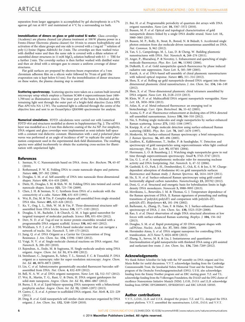
Here we report the successful assembly of 40 nm Au NPdimers with sub-5 nm gaps on a 40� 45 nm2 DNA origamiplatform (Fig. 1a). The innovative design of the origamiplatform ensures a strong plasmonic coupling between the NPswithout occupying the gap between them. Using these structures,we are able to address the specific issues described earlier.Far field scattering measurements of individual structuresunveils a significant red shift in the plasmonic resonancesdue to the underlying DNA origami platform, as well as theirconsistent architecture. The strong optical coupling allowsus to demonstrate SERS measurements of both an externalanalyte as well as ssDNA oligos attached to the NPs usingthese structures.
ResultsAssembly of NP dimers with a sub-5 nm gap. An overview ofthe experimental scheme is depicted in Fig. 1. A multi-layer40� 45 nm2 DNA origami platform is assembled as described inMethods section. By varying the number of layers along theorigami structure, two grooves are created to facilitate the correctpositioning of two 40 nm Au NPs. These grooves are separatedand isolated from each other by a ridge of double helices. At itshighest the ridge is six helices (B15 nm) high. Crucially, in thegap directly between the NPs, the ridge is only two helices(B5 nm) thick, which leaves the gap free for utilization of single-molecule spectroscopic techniques. Gold NPs are attached tocorrectly folded origami structures using a protocol given inKuzyk et al.27 Agarose gel electrophoresis is used to isolate thedimers from aggregates and incorrectly assembled structures aswell as the large excess of free NPs. Based on an intensity analysisof the dimer and aggregate bands in Fig. 1b, the yield is 61±5%.While a small number of structures with only one NP attachedare present within the dimer band, these are easily identifiablefrom the dimers due to their dramatically different spectralresponse. From transmission electron microscope (TEM) imagesof correctly assembled structures (Fig. 1b), we measure an averageNP separation of 3.3±1.0 nm which is one of the shortestcontrollable gaps yet achieved with DNA origami assembly.
2x40 nm NPsa b
c
60
40
20
Coverslip onXYZ nanopositioner
KG/ND LP
BB
Iris
LP
Scattered light
BS
63x NA 1.2 W
Reflectedlight
Incident light
0
Yie
ld (
%)
Aggregates
Dimer pairsDimers
Aggregates
Dim
er pairs
Dim
ers
Free NPs
Figure 1 | Overview of the experimental set-up. (a) A schematic (not to
scale) of the NP dimers assembled on the DNA origami platform. The
NPs are coated with a ssDNA brush to prevent aggregation as well as
facilitate attachment to the origami platform. (b) Correctly formed dimer
structures are separated from free NPs and aggregates by gel
electrophoresis on a 0.7% agarose gel and imaged using a TEM (scale bar,
50 nm). The yield of these structures is around 61±5% (mean±s.d.)
as measured by the intensity of the dimer and aggregate bands.
(c) A schematic of the custom built setup for measuring the scattering
spectra of single dimer nanostructures. KG: heat absorbing filter;
ND: neutral density filter; LP: linear polarizer; BB: beam block; and
BS: beam splitter.
ARTICLE NATURE COMMUNICATIONS | DOI: 10.1038/ncomms4448
2 NATURE COMMUNICATIONS | 5:3448 | DOI: 10.1038/ncomms4448 | www.nature.com/naturecommunications
& 2014 Macmillan Publishers Limited. All rights reserved.

Characterization of plasmonic properties. We examined care-fully the contributions of individual components of the assemblednanostructures to the plasmonic resonance of the NPs. Asupercontinuum laser was employed in a reflective dark-fieldgeometry to collect far-field scattering spectra of individualstructures (Fig. 1c)36,37. We obtained spectra from bothindividual ssDNA-coated NPs as well as single ssDNA-coatedNPs attached to a flat DNA origami sheet and immobilized on apoly-(L)-lysine-coated glass slide. Heat absorbing and neutraldensity filters were used to ensure radiant flux densities are belowthe damage threshold of the DNA origami structures. In all cases,the presence of the DNA changes the dielectric properties of theenvironment around the NPs, allowing an effective refractiveindex for both layers to be calculated38.
Three representative spectra for single ssDNA-coated NPs areshown in Fig. 2a, their similar intensities are further verificationof consistent single NP measurements. The peak intensity of theseNPs (Fig. 2c) already displays an average red shift of 6±1 nm(mean ± s.e.) from bare NPs on glass (scattering peak at530±1 nm, data shown in Supplementary Fig. 1). From TEMimages, the thickness of the ssDNA coating was found to be
2.5±0.5 nm. This value was used in numerical simulations(Supplementary Fig. 2) to estimate the effective refractive indexnssDNA of the ssDNA layer. A value of nssDNA¼ 1.7±0.1 wasfound to best fit the measured spectra (Fig. 2b, SupplementaryFigs 3 and 4, Supplementary Notes 1 and 2).
The controlled attachment of single ssDNA-coated NPs to flatDNA origami sheets allowed for the characterization of any redshift caused by the DNA origami sheet and subsequent extractionof the effective refractive index of DNA origami. Binding of singleNPs was achieved by designing an origami sheet with sufficientDNA docking sites for only a single NP. A lower NP: origamiratio was used and the monomers were carefully isolated using gelelectrophoresis. Once again, the scattering intensities are similarfor each nanostructure (Fig. 2d). The scattering peaks also displaya more pronounced red shift (Fig. 2f) of 20±1 nm and 26±1 nmas compared with the peaks for ssDNA-coated (Fig. 2c) and bareNPs (Supplementary Fig. 1), respectively. This increased red shiftis clearly attributable to the underlying origami structure(Supplementary Note 3). To simplify the simulations, theunderlying origami sheet was modelled as an infinite lateralsheet of 5 nm thickness, with the ssDNA coating described by
1.0
1.0
310
5
0
Cou
nts
8
g h
fed
a b c
4
0
Cou
nts
1424LongitudinalMode
646
554
TransverseMode
554±1 nm
556±1 nm
536±1 nm
634±2 nm
18
12
6
7
0
0
14
7
Cou
nts
Cou
nts
� sca
t (10
–16 m
2 )� s
cat (
10–1
6 m2 )
� sca
t (10
–16 m
2 )
2
9
6
3
0.5
0.5
Inte
nsity
(a.
u.)
Inte
nsity
(a.
u.)
0.0
0.0
1.0
0.5
Inte
nsity
(a.
u.)
0.0
Wavelength (nm)
Red shift Red shift
Experiments Simulations
520 540 560 580 600
536
534
540
556
548
565
Wavelength (nm)
520 540 560 580 600
n=1.5n=1.7n=1.9
n=2.1n=2.3
n=1.9
Wavelength (nm)
520 540 560 580 600
Wavelength (nm)
520 540 560 580 600
Wavelength (nm) Peak (nm)
510 510560 610 610660 710 710
Peak (nm)
510 560 610
Peak (nm)
510 560 610
Figure 2 | Effective refractive index for DNA origami nanostructures. Simulations for the scattering cross-section are presented in (b,e), the other
panels are experimental data. Errors quoted are s.e. of the mean. A single peak is obtained for ssDNA-coated NPs (a) with an average red shift of
6±1 nm from the bare NP peak (c). This corresponds to an effective refractive index of nssDNA¼ 1.7 (b). A single peak (d) with a further red shift of
20±1 nm is obtained for single nssDNA-coated NPs attached to flat origami sheets (f). Using nssDNA¼ 1.7 from (b), the best fit is obtained for norigami¼ 2.1
(e). The two peaks for the NP dimer structures (g) correspond to the transverse and longitudinal modes. The overlaid scattering cross-section
obtained from simulations (red dashed line, no free parameters) has peaks at 554 and 646 nm, in good agreement with the experimental data (g,h).
NATURE COMMUNICATIONS | DOI: 10.1038/ncomms4448 ARTICLE
NATURE COMMUNICATIONS | 5:3448 | DOI: 10.1038/ncomms4448 | www.nature.com/naturecommunications 3
& 2014 Macmillan Publishers Limited. All rights reserved.

nssDNA¼ 1.7. The best fit to the experimental data was obtainedfor norigami¼ 2.1 (Fig. 2e, Supplementary Fig. 4, SupplementaryNote 4). Given the density of the DNA in the sheet comparedwith the ssDNA on the particles, this increased effectivenorigami¼ 2.1±0.05 is not surprising.
Finally, individual dimer structures were characterized. Cor-rectly assembled dimers were easily identifiable due to theircharacteristic polarization-dependent response and enhancedscattering (Fig. 2g and Supplementary Fig. 5). Typically, thelongitudinal mode (excited by light polarized along the dimeraxis) is expected to be strongly red-shifted due to the plasmoniccoupling between the NPs, whereas the transverse mode (lightpolarized perpendicular to the dimer axis) should remain nearlyunchanged from the single NP resonance35. Indeed as clearlyevident, the transverse mode peak for NP dimer structures(Fig. 2g,h) coincides almost exactly with the scattering peak forsingle ssDNA-coated NPs on origami sheets (Fig. 2d,f). The twoparameter values nssDNA¼ 1.7 and norigami¼ 2.1 were used tosimulate the scattering cross-section of the dimer structures. Thedimers were modelled as a pair of NPs covered with a 2.5 nmthick ssDNA layer separated by 3.3 nm on an infinite origamisheet of 5 nm thickness. The simulations are shown by the red-dashed line in Fig. 2g and are in very good agreement with theexperimental data (Fig. 2h). Our values of norigami¼ 2.1 andnssDNA¼ 1.7 are very similar to those reported for 54 bp (n¼ 2.1)and 24 bp (n¼ 1.75) long dsDNA coating around 20 nm goldNPs, also measured using plasmon resonances38.
We also repeated all scattering measurements with thestructures surrounded by buffer instead of air (SupplementaryFig. 6). The resonance peak for ssDNA-coated NPs is red-shiftedby 5±1 nm, as expected given the higher refractive index of watercompared with air. However, for single ssDNA-coated NPs on flatDNA origami sheets, no shift in the resonance peak is observed,whereas for the dimer structures, the longitudinal mode is blue-shifted by 10±2 nm. This blue shift, despite the increasedrefractive index of water as surrounding medium could be causedby an increase in spacing between the NPs as the ssDNA coatinggets hydrated (Supplementary Fig. 7). These measurements arefurther proof of the strong plasmonic coupling in our dimerstructures, and that the architecture can be controlled by flexingof the DNA origami under appropriate stimulation.
SERS measurements of external analytes. After the carefulcharacterization of our dimer structures, we investigated theirpotential for SERS measurements of external analytes. Individualdimer structures are immobilized on a gold-coated silicon waferas described in Methods. The gold coating reduces backgroundRaman emissions from the silicon wafer. The immobilized dimerstructures are incubated briefly in a 100 mM solution of Rhoda-mine 6G to form a monolayer on the dimer structures (Fig. 3a)before the solution is removed39. Rhodamine 6G is a modelRaman analyte with many characteristic peaks40. SERSmeasurements were carried out with a Renishaw inVia Ramanmicroscope using a 100� objective with NA¼ 0.85. Laser-induced damage to the dimers was prevented by illuminatingwith low intensities (see Supplementary Fig. 8). The spectra weretaken on individual dimers which can be identified on the goldsurface. A collection of spectra, each obtained from a differentdimer structure are shown in Fig. 3b. Typical Rhodamine 6Gmodes common to all spectra are highlighted by red-dashed lines.We employed a laser excitation line at 632.8 nm, which is in closeproximity to the longitudinal dimer mode (Fig. 2c). Hence, theobserved enhancement is highly sensitive to the polarization ofthe laser with respect to the dimer axis (Supplementary Fig. 9).Assuming a monolayer coverage41 of Rhodamine 6G (density
of 10� 13 molecules cm� 2) on the NP and the correspondingsensing volume of 10� 25 m� 3, there should be around fivemolecules contributing to the SERS signal (see SupplementaryMethods). Depending on the orientation of the dimer in the laserfield, we calculate surface enhancement factors between five andseven orders of magnitude. This is in good agreement with oursimulations (see Supplementary Fig. 10) and in line with otherSERS systems35. Similar results are obtained for other externalanalytes as shown in Supplementary Fig. 11. Away from the NP(marked as ‘off dimer’ in Fig. 3b), the Raman spectrum of theRhodamine is completely absent for the concentrations used.
Sequence-specific DNA detection using SERS. A powerfulapplication of our dimer structures would be to combine thebinding site possibilities of the origami platform with label-freeSERS detection. To demonstrate this, we performed SERS mea-surements on individual dimer structures without any externallyadded analyte in a similar manner as described above. The gapbetween the NPs is free except for the ssDNA coating. Mea-surements were first performed with NPs coated with 19 bases ofThymine (‘sequence 1’). Typical data for a range of dimers areshown in Fig. 4b. The peak at 1,000 cm� 1 is thought to arisefrom the in-plane rocking mode in Thymine42, and the peak at1,084 cm� 1 corresponds to the phosphate backbone of DNA42,43.An additional peak at 1,576 cm� 1 may arise from the 15 bp poly-Adenine sequence (complementary to the ssDNA coating on theNPs) on the origami platform used for NP attachment43. Toinvestigate if we could detect changes in the oligonucleotidesequence, dimer structures were assembled from a mixture of80% ‘sequence 2’ containing Cytosine and Adenine in addition toThymine and 20% ‘sequence 1’, as shown in the schematic inFig. 4. This combination ensured enough of sequence 1 waspresent in order to attach the NPs to the DNA origami platform.
Rhodamine6G
1,500
a
b
1,000
500
Inte
nsity
(co
unts
/(m
Ws)
)
0600 800 1,000 1,200 1,400
611 1,183 1,310 1,360 1,573
1,6471,507
Wavenumber shift (cm–1)
Off dimer
1,600 1,800
HN O
O
O
NH+Cl–
Figure 3 | SERS measurements of an external analyte. (a) SERS
measurements of a thin layer of Rhodamine 6G adsorbed onto individual
dimer structures plated on a gold-coated silicon wafer. (b) Each spectrum
corresponds to a different dimer structure and typical Rhodamine 6G
peaks are clearly visible (indicated by a dashed line). By contrast, a
spectrum taken from a region away from the dimers (‘off dimers’) is of low
intensity and does not display any peaks.
ARTICLE NATURE COMMUNICATIONS | DOI: 10.1038/ncomms4448
4 NATURE COMMUNICATIONS | 5:3448 | DOI: 10.1038/ncomms4448 | www.nature.com/naturecommunications
& 2014 Macmillan Publishers Limited. All rights reserved.

As can be seen in Fig. 4c, peaks for Adenine (736, 1,260 and1,485 cm� 1)43 and Cytosine (1,260 and 1,485 cm� 1)44 are nowobserved in addition to the peaks already marked in Fig. 4b.
Once again, in comparison with bulk Raman measurements ofthe same oligos, we obtain an average enhancement of 4–5 ordersof magnitude in the scattering cross-section if we assume that asingle DNA molecule with around 20 bases is present in thesensing volume. Such a consistently high enhancement factordemonstrates the potential these structures have for label-freesingle-molecule measurements such as sequence-sensitive DNAdetection in combination with high-throughput techniques. Thepresence of a strong plasmonic gap built over a customisableDNA origami platform makes it an exciting prospect for hybridplasmonic nanopores and many other DNA-based probes.
DiscussionIn summary, DNA origami offers an elegant approach to designmetallic nanostructures with complex optical properties. In this
paper, we demonstrate an innovative origami design that allowsfor the reliable assembly of 40 nm Au NP dimer structures withstrong plasmonic coupling from reliable sub-5 nm gaps. This isreflected in the high uniformity of scattering spectra of individualdimers which obey the characteristic polarization-dependentresponse of strongly coupled NPs. We are also able todemonstrate that the underlying DNA origami platform has asignificant influence on the plasmonic properties of the dimer.The effective refractive index for dense DNA origami estimatedfrom finite-difference time-domain (FDTD) simulations is ingood agreement with similar measurements for dsDNA on goldNPs.
We demonstrate the effectiveness of our dimer structures forSERS measurements with enhancement factors of up to sevenorders of magnitude. We are able to detect not only externalanalytes with only a handful of adsorbed molecules in the sub-5 nm, but also changes in the composition of the ssDNA coatingon the NPs. This effectively demonstrates the combination of aversatile platform for biosensing (DNA origami) with a label-freedetection technique (SERS) and has great potential application fora wide variety of biosensing and single-molecule applications.
MethodsDNA origami structure design and assembly. All DNA origami structures weredesigned using the open source DNA origami software caDNAnano45. All shortssDNA staples were purchased from Integrated DNA Technologies. The precisesequence of staples for as well as the scaffold/staple layout for each design areincluded Supplementary Tables 1 and 2 and Supplementary Figs 12 and 13.
The flat origami design used for attachment of single NPs in Fig. 2b consists of adouble layer with 24 interlinked double-helical DNA domains on each layer on asquare packing lattice. Eight staples on one layer included ssDNA overhangs forattachment to complementary ssDNA on the NPs. These were organized into twodistinct binding sites comprised of four staples each with different ssDNAoverhangs; ttt ttt ttt ttt ttt (complementary to ‘sequence 1’) and atg tag gtg gta gag g(complementary to ‘sequence 2’). Consequently, NPs coated with sequence 1ssDNA could only attach to the staples on one of the binding sites. The 7,249 basesof viral M13mp18 ssDNA was utilized as the scaffold (New England Biolabs). DNAorigami assembly was done by mixing scaffold and staples to a final concentrationof 10 nM and 100 nM, respectively, in a 14 mM MgCl2 solution buffered with1�TE (pH¼ 8.0) and subjecting the mixture to thermal-annealing cycles for 18 h.The heating program utilized was as described in Hernandez-Ainsa et al.46
The NP dimer design consists of multiple layers of double-helical DNAdomains on a square packing lattice such that a ridge of DNA seven helices(17.5 nm thick) separates two large grooves custom designed to fit 40 nm NPs. Ineach groove, six staples are modified to include overhangs complementary tosequence 1. The p8634 mutant of the viral M13 genome was utilized as the scaffoldand the structure requires 251 staple strands of DNA. DNA origami assembly wascarried out in a similar fashion as described above, with a modified thermalannealing programme was follows: 80 �C for 5 min, 79 �C for 4 min, from 79–60 �Cin 19 steps (1 �C per step, 4 min each step), 60 �C for 12 min, from 60 to 15 �C in 90steps (0.5 �C per step, 12 min each step) and finally set at 4 �C.
The assembled structures were purified from the excess staple strands bycentrifugation with 100 kDa MWCO filters (Amicon Ultra, Millipore) in threecycles at a speed of 13,000 g for 5 min at 4 �C in a 0.5 � TBE buffered solution of11 mM MgCl2. The assembled origami structures were then collected at the end ofthe third cycle of filtration.
ssDNA coating of NPs and synthesis of dimer structures. A detailed attach-ment protocol is given in Kuzyk et al.27 Briefly, 40 nm gold NPs (BBI Solutions) areresuspended in 2.5 mM Bis (p-sulfonatophenyl)phenylphosphine dihydratedipotassium (BSPP) solution (Sigma Aldritch) to enable higher NP concentrations.DNA oligos corresponding to ‘sequence 1’ or ‘sequence 2’ are ordered with a50 dithiol modification (Biomers). After incubation in 10 mM Tris(2-carboxyethyl)phosphine (TCEP, Sigma Aldritch) for 30 min, they are added in a4,800-fold excess to the NP solution. Following a protocol given in Zhang et al.47,citrate-HCl buffer (pH¼ 3) is added to a final concentration of 10 mM for 10 min,before HEPES buffer (pH¼ 7) is added at a final concentration of 100 mM. Thisprotocol ensures quick attachment of thiolated ssDNA oligos to the gold NPs.Successful attachment is confirmed by a lack of aggregation when resuspended in100 mM MgCl2.
ssDNA-coated NPs are separated from excess DNA oligos by eight rounds ofcentrifugation with 100 kDa MWCO filters (Amicon Ultra, Millipore) at 10,000 gfor 10 min at 4 �C and finally incubated overnight with the assembled DNAorigami platforms in a 0.5 � TBE buffered solution of 11 mM MgCl2 so that theNPs are in a 10� excess per binding site. Successful formation of dimers and
Sequence 1
Sequence 1
1,000 1,084 1,576
Sequence 2
80% Sequence 2 1,260
736
1,48520% Sequence 1
ttt ttt ttt ttt ttt ttt t
Wavenumber shift (cm–1)
600
b
c
a
400
200
1,200
Inte
nsity
(co
unts
/(m
Ws)
)In
tens
ity (
coun
ts/(
mW
s))
800
400
800 1,000 1,200 1,400 1,600 1,800
Wavenumber shift (cm–1)
600 800 1,000 1,200 1,400 1,600 1,800
ttc ctc tac cac cta cat
Figure 4 | SERS spectra are sensitive to the sequences of the ssDNA
coating. (a,b) The ssDNA coating of 19 bases of Thymine (‘sequence 1’) on
the NPs produces spectra with peaks predominantly corresponding to the
DNA backbone (1,000 cm� 1) and Thymine (1,084 cm� 1), indicated by the
red dashed lines. (b,c) By contrast, the use of an Adenine and Cytosine
containing ‘sequence 2’ in an 80:20 ratio with ‘sequence 1’ shows
significantly more peaks corresponding to Adenine (736, 1,260 and
1,485 cm� 1) and Cytosine (1,260 and 1,485 cm� 1), indicated by the blue
dashed lines.
NATURE COMMUNICATIONS | DOI: 10.1038/ncomms4448 ARTICLE
NATURE COMMUNICATIONS | 5:3448 | DOI: 10.1038/ncomms4448 | www.nature.com/naturecommunications 5
& 2014 Macmillan Publishers Limited. All rights reserved.
separation from larger aggregates is accomplished by gel electrophresis in a 0.7%agarose gel run at 60 V and maintained at 4 �C by a surrounding ice bath.
Immobilization of dimers on glass or gold-coated Si wafer. Glass coverslips(Academy) are plasma cleaned (air plasma treatment at 100 W plasma power in aFemto-Diener Electronic plasma cleaner) for 10 min to ensure hydrophilicity andactivation of the silane groups and one side is covered with a 1 mg ml� l solution ofpoly-(L)-lysine (Sigma Aldritch) for 2 min. The coverslips are then washed twicewith distilled water and then the same side is covered with a dilute solution ofassembled dimer structures in 11 mM MgCl2 solution buffered with 0.5 � TBE fora further 2 min. The coverslip surface is then further washed with distilled waterand then air dried with a nitrogen gun to ensure a uniform coverage of dimerstructures.
Flat gold surfaces are prepared by electron-beam evaporation of a 5 nmchromium adhesion film on a silicon wafer followed by 70 nm of gold (theevaporation rate is kept below 0.5 nm). For the immobilization of dimer structureson these wafers, the plasma cleaning step is omitted.
Scattering spectroscopy. Scattering spectra were taken on a custom built invertedmicroscopy setup which employs a Fianium SC400-4 supercontinuum laser (400–1,700 nm) as illumination source. The inner part of the beam is blocked and theremaining light sent through the outer part of a bright-field objective (Leica HPXPlan APO 63x NA 1.2 W). The scattered light is collected through the centre of theobjective lens and sent to an OceanOptics QE65000 spectrometer for analysis.
Numerical simulations. FDTD calculations were carried out with LumericalFDTD v8.6 and structures modelled as shown in Supplementary Fig. 2. The ssDNAlayer was modelled as a 2.5 nm thin dielectric coating of the Au NP. For simplicity,DNA origami and glass coverslips were implemented as semi-infinite half-spacewith a constant real dielectric constant. Illumination with s and p polarized planewaves was performed at an angle of incidence of 60� to replicate the axial polar-ization component used in the experimental dark-field illumination. The resultingspectra were added incoherently to obtain the scattering cross-section for illumi-nation with unpolarized light.
References1. Seeman, N. C. Nanomaterials based on DNA. Annu. Rev. Biochem. 79, 65–87
(2010).2. Rothemund, P. W. K. Folding DNA to create nanoscale shapes and patterns.
Nature 440, 297–302 (2006).3. Douglas, S. M. et al. Self-assembly of DNA into nanoscale three-dimensional
shapes. Nature 459, 414–418 (2009).4. Dietz, H., Douglas, S. M. & Shih, W. M. Folding DNA into twisted and curved
nanoscale shapes. Science 325, 725–730 (2009).5. Chen, J. H. & Seeman, N. C. Synthesis from DNA of a molecule with the
connectivity of a cube. Nature 350, 631–633 (1991).6. Wei, B., Dai, M. & Yin, P. Complex shapes self-assembled from single-stranded
DNA tiles. Nature 485, 623–626 (2012).7. Ke, Y., Ong, L. L., Shih, W. M. & Yin, P. Three-dimensional structures self-
assembled from DNA bricks. Science 338, 1177–1183 (2012).8. Douglas, S. M., Bachelet, I. & Church, G. M. A logic-gated nanorobot for
targeted transport of molecular payloads. Science 335, 831–834 (2012).9. Derr, N. D. et al. Tug-of-war in motor protein ensembles revealed with a
programmable DNA origami scaffold. Science 338, 662–665 (2012).10. Wickham, S. F. J. et al. A DNA-based molecular motor that can navigate a
network of tracks. Nat. Nanotech. 7, 169–173 (2012).11. Jiang, Q. et al. DNA Origami as a Carrier for Circumvention of Drug
Resistance. J. Am. Chem. Soc. 134, 13396–13403 (2012).12. Voigt, N. V. et al. Single-molecule chemical reactions on DNA origami. Nat.
Nanotech. 5, 200–203 (2010).13. Rajendran, A., Endo, M. & Sugiyama, H. Single-molecule analysis using DNA
origami. Angew. Chem. Int. Ed. 51, 874–890 (2012).14. Steinhauer, C., Jungmann, R., Sobey, T. L., Simmel, F. C. & Tinnefeld, P. DNA
origami as a nanoscopic ruler for super-resolution microscopy. Angew. Chem.Int. Ed. 48, 8870–8873 (2009).
15. Lin, C. et al. Submicrometre geometrically encoded fluorescent barcodes self-assembled from DNA. Nat. Chem. 4, 832–839 (2012).
16. Bell, N. A. W. et al. DNA origami nanopores. Nano Lett. 12, 512–517 (2012).17. Wei, R., Martin, T. G., Rant, U. & Dietz, H. DNA origami gatekeepers for
solid-state nanopores. Angew. Chem. Int. Ed. 51, 4864–4867 (2012).18. Burns, J. R. et al. Lipid-bilayer-spanning DNA nanopores with a bifunctional
porphyrin anchor. Angew. Chem. Int. Ed. 52, 12069–12072 (2013).19. Castro, C. E. et al. A primer to scaffolded DNA origami. Nat. Meth. 8, 221–229
(2011).20. Ding, B. et al. Gold nanoparticle self-similar chain structure organized by DNA
origami. J. Am. Chem. Soc. 132, 3248–3249 (2010).
21. Bui, H. et al. Programmable periodicity of quantum dot arrays with DNAorigami nanotubes. Nano Lett. 10, 3367–3372 (2010).
22. Busson, M. P. et al. Optical and topological characterization of goldnanoparticle dimers linked by a single DNA double strand. Nano Lett. 11,5060–5065 (2011).
23. Busson, M. P., Rolly, B., Stout, B., Bonod, N. & Bidault, S. Accelerated singlephoton emission from dye molecule-driven nanoantennas assembled on DNA.Nat. Commun. 3, 962 (2012).
24. Tan, S. J., Campolongo, M. J., Luo, D. & Cheng, W. Building plasmonicnanostructures with DNA. Nat. Nanotech. 6, 268–276 (2011).
25. Anger, P., Bharadwaj, P. & Novotny, L. Enhancement and quenching of single-molecule fluorescence. Phys. Rev. Lett. 96, 113002 (2006).
26. Dulkeith, E. et al. Gold nanoparticles quench fluorescence by phase inducedradiative rate suppression. Nano Lett. 5, 585–589 (2005).
27. Kuzyk, A. et al. DNA-based self-assembly of chiral plasmonic nanostructureswith tailored optical response. Nature 483, 311–314 (2012).
28. Shen, X. et al. Rolling up gold nanoparticle-dressed DNA origami into three-dimensional plasmonic chiral nanostructures. J. Am. Chem. Soc. 134, 146–149(2012).
29. Shen, X. et al. Three-dimensional plasmonic chiral tetramers assembled byDNA origami. Nano Lett. 13, 2128–2133 (2013).
30. Klein, W. P. et al. Multiscaffold DNA origami nanoparticle waveguides. NanoLett. 13, 3850–3856 (2013).
31. Aslan, K. et al. Metal-enhanced fluorescence: an emerging tool inbiotechnology. Curr. Opin. Biotechnol. 16, 55–62 (2005).
32. Acuna, G. P. et al. Fluorescence enhancement at docking sites of DNA-directedself-assembled nanoantennas. Science 338, 506–510 (2012).
33. Nie, S. Probing single molecules and single nanoparticles by surface-enhancedRaman scattering. Science 275, 1102–1106 (1997).
34. Kneipp, K. et al. Single molecule detection using surface-enhanced Ramanscattering (SERS). Phys. Rev. Lett. 78, 1667–1670 (1997).
35. Moskovits, M. Surface-enhanced Raman spectroscopy: a brief retrospective.J. Raman Spectrosc. 36, 485–496 (2005).
36. Lindfors, K., Kalkbrenner, T., Stoller, P. & Sandoghdar, V. Detection andspectroscopy of gold nanoparticles using supercontinuum white light confocalmicroscopy. Phys. Rev. Lett. 93, 037401 (2004).
37. Herrmann, L. O. & Baumberg, J. J. Watching single nanoparticles grow in realtime through supercontinuum spectroscopy. Small 9, 3743–3747 (2013).
38. Liu, G. L. et al. A nanoplasmonic molecular ruler for measuring nucleaseactivity and DNA footprinting. Nat. Nanotech. 1, 47–52 (2006).
39. Ganbold, E.-O., Park, J.-H., Dembereldorj, U., Ock, K.-S. & Joo, S.-W. Charge-dependent adsorption of rhodamine 6G on gold nanoparticle surfaces:fluorescence and Raman study. J. Raman Spectrosc. 42, 1614–1619 (2011).
40. He, X. N. et al. Surface-enhanced Raman spectroscopy using gold-coatedhorizontally aligned carbon nanotubes. Nanotechnology 23, 205702 (2012).
41. Doni, G. et al. Structural and energetic basis for hybridization limits in high-density DNA monolayers. Nanoscale 5, 9988–9993 (2013).
42. Movileanu, L., Benevides, J. M. & Thomas, G. J. Temperature dependence ofthe Raman spectrum of DNA. II. Raman signatures of premelting and meltingtransitions of poly(dA).poly(dT) and comparison with poly(dA-dT).poly(dA-dT). Biopolymers 63, 181–194 (2002).
43. Barhoumi, A., Zhang, D., Tam, F. & Halas, N. J. Surface-enhanced Ramanspectroscopy of DNA. J. Am. Chem. Soc. 130, 5523–5529 (2008).
44. Rao, S. et al. Direct observation of single DNA structural alterations at lowforces with surface-enhanced Raman scattering. Biophys. J. 104, 156–162(2013).
45. Douglas, S. M. et al. Rapid prototyping of 3D DNA-origami shapes withcaDNAno. Nucleic. Acids. Res. 37, 5001–5006 (2009).
46. Hernandez-Ainsa, S. et al. DNA origami nanopores for controlling DNAtranslocation. ACS Nano 7, 6024–6030 (2013).
47. Zhang, X., Servos, M. R. & Liu, J. Instantaneous and quantitativefunctionalization of gold nanoparticles with thiolated DNA using a pH-assistedand surfactant-free route. J. Am. Chem. Soc. 134, 7266–7269 (2012).
AcknowledgementsWe thank Robert Schreiber for help with the NP assembly on DNA origami and EvaMaria-Roller for useful discussions. V.V.T. acknowledges funding from the CambridgeCommonwealth Trust, the Jawaharlal Nehru Memorial Trust and the Emmy Noetherprogram of the Deutsche Forschungsgemeinschaft (DFG). U.F.K. also acknowledgesfunding from the Emmy Noether program and an ERC starting grant. T.Z. and T.L.acknowledge funding from the Volkswagen Foundation, the DAAD and the DFG cluster ofexcellence Nanosystems Initiative Munich (NIM). L.O.H., D.O.S. and J.L.B. acknowledgefunding from EPSRC EP/G060649/1, EP/K028510/1 and ERC LINASS 320503.
Author contributionsV.V.T., L.O.H., J.L.B. and U.F.K. designed the project. T.Z. and T.L. designed the DNAorigami platform. V.V.T. assembled the nanostructures. L.O.H., D.O.S. and V.V.T.
ARTICLE NATURE COMMUNICATIONS | DOI: 10.1038/ncomms4448
6 NATURE COMMUNICATIONS | 5:3448 | DOI: 10.1038/ncomms4448 | www.nature.com/naturecommunications
& 2014 Macmillan Publishers Limited. All rights reserved.

carried out the measurements and analysed the data. L.O.H. and J.L.B. carried out thesimulations. V.V.T., L.O.H., D.O.S., J.L.B. and U.F.K. wrote the paper. V.V.T. and L.O.H.contributed equally to this work.
Additional informationSupplementary Information accompanies this paper at http://www.nature.com/naturecommunications
Competing financial interests: The authors declare no competing financial interests.
Reprints and permission information is available online at http://npg.nature.com/reprintsandpermissions/
How to cite this article: Thacker, V. V. et al. DNA origami based assembly of goldnanoparticle dimers for surface-enhanced Raman scattering. Nat. Commun. 5:3448doi: 10.1038/ncomms4448 (2014).
NATURE COMMUNICATIONS | DOI: 10.1038/ncomms4448 ARTICLE
NATURE COMMUNICATIONS | 5:3448 | DOI: 10.1038/ncomms4448 | www.nature.com/naturecommunications 7
& 2014 Macmillan Publishers Limited. All rights reserved.