dmd 37:1806–1818, 2009 printed in u.s.a....
TRANSCRIPT

Bioactivation of Minocycline to Reactive Intermediates byMyeloperoxidase, Horseradish Peroxidase, and Hepatic
Microsomes: Implications for Minocycline-Induced Lupusand Hepatitis
Baskar Mannargudi, David McNally, William Reynolds, and Jack Uetrecht
Faculties of Pharmacy (B.M., J.U.) and Medicine (J.U.), and Department of Chemistry (D.M., W.R.), University of Toronto,Toronto, Ontario, Canada
Received February 25, 2009; accepted June 3, 2009
ABSTRACT:
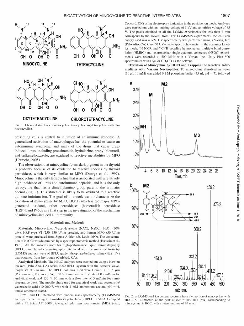
Of the tetracyclines, minocycline is unique in causing a significantincidence of a lupus-like syndrome and autoimmune hepatitis. It isalso unique among the tetracyclines in having a para-N,N-dimeth-ylaminophenol ring. Many drugs that cause autoimmune reactionsare oxidized to reactive metabolites by the myeloperoxidase (MPO)system of macrophages. In this study, we showed that minocyclineis oxidized to reactive intermediates by MPO/H2O2/Cl�, HOCl,horseradish peroxidase/H2O2, or hepatic microsomes. When trappedwith N-acetylcysteine (NAC), two adducts with protonated molecularions at m/z 619 were isolated and analyzed by NMR. One representsattack of the aromatic D ring by NAC meta to the N,N-dimethylaminogroup, which implies that the reactive intermediate was a quinoneiminium ion. The NMR of the other adduct, which was not observed
when minocycline was oxidized by hepatic microsomes, indicatesthat the NAC is attached at the junction of the B and C rings. In theoxidation by HOCl, we found an intermediate with a protonated mo-lecular ion of m/z 510 that represents the addition of HOCl to mino-cycline. The HOCl presumably adds across the double bond of the Bring, and reaction of this intermediate with NAC led to the secondNAC adduct. We were surprised to find that the same NAC adductwas not observed after oxidation of tetracycline with HOCl, eventhough this part of the tetracycline structure is the same as forminocycline. We propose that one or more of these reactive metab-olites are responsible for the idiosyncratic drug reactions that arespecific to this tetracycline.
Minocycline (Fig. 1), a tetracycline antibiotic (Kamel et al., 2002),is used for the treatment of acne (Cullen and Cohan, 1976), infectiousdiseases, and rheumatoid arthritis. However, minocycline is associ-ated with serious adverse reactions, which include hypersensitivityreactions (Antunes et al., 1999), a serum sickness-like reaction (Puy-ana et al., 1990), hepatitis (Hardman et al., 1996), and a drug-inducedlupus-like reaction (Farver, 1997). The incidence of minocycline-induced lupus is approximately 15 cases/100,000 prescriptions and ismore common in women (Borchers et al., 2007). Patients treated withminocycline often develop perinuclear antineutrophilic cytoplasmicantibodies (P-ANCA, usually antimyeloperoxidase) (Dunphy et al.,2000), and in patients with minocycline-induced lupus, P-ANCA ispresent in 80% of cases (Sturkenboom et al., 1999).
Minocycline-induced hepatitis can lead to hepatic failure requiringliver transplantation (Boudreaux et al., 1993). The incidence of mi-
nocycline-induced hepatitis in new users is approximately 1/10,000patients (Seaman et al., 2001). Reports to the World Health Organi-zation of the hepatic adverse drug reactions associated with theminocycline accounted for 6% (493) of all the minocycline-inducedadverse drug reactions (8025) (Lawrenson et al., 2000). The reportedcases of minocycline-induced hepatitis are characterized by autoim-mune characteristics, including antinuclear antibodies and a very longduration of therapy (more than 1 year) (Lawrenson et al., 2000) beforethe onset of clinical signs of hepatitis (Knowles et al., 1996; Teitel-baum et al., 1998). There is a large amount of circumstantial evidenceto support the hypothesis that most idiosyncratic drug reactions arecaused by reactive metabolites (Lavergne et al., 2008). CytochromesP450 (P450s) are likely responsible for the production of more reac-tive metabolites than any other enzyme; however, myeloperoxidase(MPO) is also capable of forming reactive metabolites, especiallywhen the drug has an easily oxidized nitrogen (Uetrecht, 1992). Thecells that contain MPO include neutrophils, macrophages, and otherantigen-presenting cells. The formation of reactive metabolites byneutrophils has obvious implications for drug-induced agranulocyto-sis. In addition, the formation of reactive metabolites by antigen-presenting cells may lead to their activation, and activation of antigen-
This work was supported by the Canadian Institutes of Health Research [GrantMOP-9336].
Article, publication date, and citation information can be found athttp://dmd.aspetjournals.org.
doi:10.1124/dmd.109.027292.
ABBREVIATIONS: P-ANCA, perinuclear antineutrophilic cytoplasmic antibodies; P450, cytochrome P450; MPO, myeloperoxidase; HRP, horse-radish peroxidase; NAC, N-acetyl-L-cysteine; HPLC, high-performance liquid chromatography; LC/MS, liquid chromatography interfaced withmass spectrometry; PBS, phosphate-buffered saline; LC/MS/MS, liquid chromatography interfaced with fragmentation mass spectrometry;HMBC, heteronuclear multiple bond correlation; HSQC, heteronuclear single quantum coherence; amu, atomic mass unit.
0090-9556/09/3709-1806–1818$20.00DRUG METABOLISM AND DISPOSITION Vol. 37, No. 9Copyright © 2009 by The American Society for Pharmacology and Experimental Therapeutics 27292/3501513DMD 37:1806–1818, 2009 Printed in U.S.A.
1806
at ASPE
T Journals on June 1, 2018
dmd.aspetjournals.org
Dow
nloaded from
presenting cells is central to initiation of an immune response. Ageneralized activation of macrophages has the potential to cause anautoimmune syndrome, and many of the drugs that cause drug-induced lupus, including procainamide, hydralazine, propylthiouracil,and sulfamethoxazole, are oxidized to reactive metabolites by MPO(Uetrecht, 2005).
The observation that minocycline forms dark pigment in the thyroidis probably because of its oxidation to reactive species by thyroidperoxidase, which is very similar to MPO (Doerge et al., 1997).Minocycline is the only tetracycline that is associated with a relativelyhigh incidence of lupus and autoimmune hepatitis, and it is the onlytetracycline that has a dimethylamino group para to the aromaticphenol (Fig. 1). This structure is likely to be oxidized to a reactivequinone iminium ion. The goal of this work was to characterize theoxidation of minocycline by MPO, HOCl (which is the major MPO-generated oxidant), other peroxidases [horseradish peroxidase(HRP)], and P450s as a first step in the investigation of the mechanismof minocycline-induced autoimmunity.
Materials and Methods
Materials. Minocycline, N-acetylcysteine (NAC), NaOCl, H2O2 (30%w/v), HRP type VI (250–330 U/mg protein), and human MPO (50 U/mgprotein) were purchased from Sigma-Aldrich (St. Louis, MO). The concentra-tion of NaOCl was determined by a spectrophotometric method (Hussain et al.,1970). All the solvents used for high-performance liquid chromatography(HPLC), and liquid chromatography interfaced with the mass spectrometry(LC/MS) analysis were of HPLC grade. Phosphate-buffered saline (PBS; 1�)was obtained from Invitrogen (Carlsbad, CA).
Analytical Methods. The HPLC analyses were carried out using a HewlettPackard (Palo Alto, CA) series 1050 HPLC system with the detector wave-length set at 254 nm. The HPLC columns used were Gemini C18, 5 �m(Phenomenex, Torrance, CA), 150 � 2 mm with a flow rate of 0.2 ml/min foranalytical work and 150 � 10 mm with a flow rate of 5 ml/min for semi-preparative work. The mobile phase used for analytical work was acetonitrile/water/acetic acid (10:90:0.7, v/v) with 2 mM ammonium acetate, pH � 4,unless otherwise stated.
LC/MS and LC interfaced with tandem mass spectrometry (LC/MS/MS)were performed using a Shimadzu (Kyoto, Japan) HPLC LC-10AD coupledwith a PE Sciex API 3000 triple quadruple mass spectrometer (MDS Sciex,
Concord, ON) using electrospray ionization in the positive ion mode. Analyseswere carried out with an ionizing voltage of 5 kV and an orifice voltage of 65V. The peaks obtained in all the LC/MS experiments for less than 2 mincorrespond to the solvent front. For LC/MS/MS experiments, the collisionenergy used was 40 eV. UV spectrometry was performed using a Varian, Inc.(Palo Alto, CA) Cary 50 UV-visible spectrophotometer in the scanning kinet-ics mode. 1H NMR and 13C-1H coupling heteronuclear multiple bond corre-lation (HMBC) and heteronuclear single quantum coherence (HSQC) experi-ments were recorded at 500 MHz with a Varian, Inc. Unity Plus 500spectrometer with D2O or CD3OD as the solvent.
Oxidation of Minocycline by HOCl and Trapping the Reactive Inter-mediates with Various Nucleophiles. To minocycline dissolved in water(10 �l, 10 mM) was added 0.1 M phosphate buffer (75 �l, pH � 7), followed
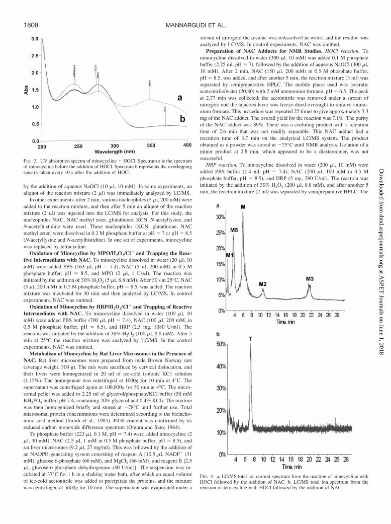
FIG. 2. a, LC/MS total ion current spectrum from the reaction of minocycline withHOCl. b, LC/MS/MS of the peak at m/z � 510 amu (M2) corresponding tominocycline � HOCl with a retention time of 10 min.
FIG. 1. Chemical structures of minocycline, tetracycline, oxytetracycline, and chlo-rotetracycline.
1807BIOACTIVATION OF MINOCYCLINE TO REACTIVE INTERMEDIATES
at ASPE
T Journals on June 1, 2018
dmd.aspetjournals.org
Dow
nloaded from
by the addition of aqueous NaOCl (10 �l, 10 mM). In some experiments, analiquot of the reaction mixture (2 �l) was immediately analyzed by LC/MS.
In other experiments, after 2 min, various nucleophiles (5 �l, 200 mM) wereadded to the reaction mixture, and then after 5 min an aliquot of the reactionmixture (2 �l) was injected into the LC/MS for analysis. For this study, thenucleophiles NAC, NAC methyl ester, glutathione, KCN, N-acetyllysine, andN-acetylhistidine were used. These nucleophiles (KCN, glutathione, NACmethyl ester) were dissolved in 0.2 M phosphate buffer at pH � 7 or pH � 8.5(N-acetyllysine and N-acetylhistidine). In one set of experiments, minocyclinewas replaced by tetracycline.
Oxidation of Minocycline by MPO/H2O2/Cl� and Trapping the Reac-tive Intermediates with NAC. To minocycline dissolved in water (20 �l, 10mM) were added PBS (163 �l, pH � 7.4), NAC (5 �l, 200 mM) in 0.5 Mphosphate buffer, pH � 8.5, and MPO (2 �l, 1 U/�l). The reaction wasinitiated by the addition of 30% H2O2 (5 �l, 8.8 mM). After 30 s at 25°C, NAC(5 �l, 200 mM) in 0.5 M phosphate buffer, pH � 8.5, was added. The reactionmixture was incubated for 30 min and then analyzed by LC/MS. In controlexperiments, NAC was omitted.
Oxidation of Minocycline by HRP/H2O2/Cl� and Trapping of ReactiveIntermediates with NAC. To minocycline dissolved in water (100 �l, 10mM) were added PBS buffer (700 �l, pH � 7.4), NAC (100 �l, 200 mM, in0.5 M phosphate buffer, pH � 8.5), and HRP (2.5 mg, 1080 U/ml). Thereaction was initiated by the addition of 30% H2O2 (100 �l, 8.8 mM). After 5min at 25°C the reaction mixture was analyzed by LC/MS. In the controlexperiments, NAC was omitted.
Metabolism of Minocycline by Rat Liver Microsomes in the Presence ofNAC. Rat liver microsomes were prepared from male Brown Norway rats(average weight, 300 g). The rats were sacrificed by cervical dislocation, andtheir livers were homogenized in 20 ml of ice-cold isotonic KC1 solution(1.15%). The homogenate was centrifuged at 1000g for 10 min at 4°C. Thesupernatant was centrifuged again at 100,000g for 50 min at 4°C. The micro-somal pellet was added to 2.25 ml of glycerol/phosphate/KCl buffer (50 mMKH2PO4 buffer, pH 7.4, containing 20% glycerol and 0.4% KCl). The mixturewas then homogenized briefly and stored at �78°C until further use. Totalmicrosomal protein concentrations were determined according to the bicincho-ninic acid method (Smith et al., 1985). P450 content was confirmed by itsreduced carbon monoxide difference spectrum (Omura and Sato, 1964).
To phosphate buffer (223 �l, 0.1 M, pH � 7.4) were added minocycline (2�l, 30 mM), NAC (2.5 �l, 1 mM in 0.5 M phosphate buffer, pH � 8.5), andrat liver microsomes (9.2 �l, 27 mg/ml). This was followed by the addition ofan NADPH-generating system consisting of reagent A [10.5 �l, NADP� (31mM), glucose 6-phosphate (66 mM), and MgCl2 (66 mM)] and reagent B [2.5�l, glucose-6-phosphate dehydrogenase (40 U/ml)]. The suspension was in-cubated at 37°C for 1 h in a shaking water bath, after which an equal volumeof ice-cold acetonitrile was added to precipitate the proteins, and the mixturewas centrifuged at 5600g for 10 min. The supernatant was evaporated under a
stream of nitrogen; the residue was redissolved in water; and the residue wasanalyzed by LC/MS. In control experiments, NAC was omitted.
Preparation of NAC Adducts for NMR Studies. HOCl reaction. Tominocycline dissolved in water (300 �l, 10 mM) was added 0.1 M phosphatebuffer (2.25 ml, pH � 7), followed by the addition of aqueous NaOCl (300 �l,10 mM). After 2 min, NAC (150 �l, 200 mM) in 0.5 M phosphate buffer,pH � 8.5, was added, and after another 5 min, the reaction mixture (3 ml) wasseparated by semipreparative HPLC. The mobile phase used was isocraticacetonitrile/water (20:80) with 2 mM ammonium formate, pH � 6.5. The peakat 2.77 min was collected; the acetonitrile was removed under a stream ofnitrogen; and the aqueous layer was freeze-dried overnight to remove ammo-nium formate. This procedure was repeated 25 times to give approximately 3.3mg of the NAC adduct. The overall yield for the reaction was 7.1%. The purityof the NAC adduct was 89%. There was a coeluting product with a retentiontime of 2.6 min that was not readily separable. This NAC adduct had aretention time of 3.7 min on the analytical LC/MS system. The productobtained as a powder was stored at �75°C until NMR analysis. Isolation of aminor product at 2.8 min, which appeared to be a diastereomer, was notsuccessful.
HRP reaction. To minocycline dissolved in water (200 �l, 10 mM) wereadded PBS buffer (1.4 ml, pH � 7.4), NAC (200 �l, 100 mM in 0.5 Mphosphate buffer, pH � 8.5), and HRP (5 mg, 290 U/ml). The reaction wasinitiated by the addition of 30% H2O2 (200 �l, 8.8 mM), and after another 5min, the reaction mixture (2 ml) was separated by semipreparative HPLC. The
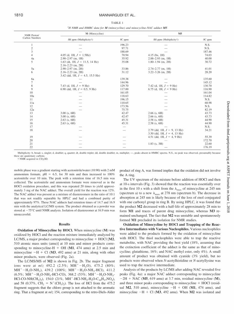
FIG. 3. UV absorption spectra of minocycline � HOCl. Spectrum a is the spectrumof minocycline before the addition of HOCl. Spectrum b represents the overlappingspectra taken every 10 s after the addition of HOCl.
FIG. 4. a, LC/MS total ion current spectrum from the reaction of minocycline withHOCl followed by the addition of NAC. b, LC/MS total ion spectrum from thereaction of tetracycline with HOCl followed by the addition of NAC.
1808 MANNARGUDI ET AL.
at ASPE
T Journals on June 1, 2018
dmd.aspetjournals.org
Dow
nloaded from

FIG. 5. a, 1H NMR spectrum of the minocycline M. b, 1H NMR spectrum of the minocycline-NAC adduct M5, which has a retention time of 3.7 min on LC/MS.
1809BIOACTIVATION OF MINOCYCLINE TO REACTIVE INTERMEDIATES
at ASPE
T Journals on June 1, 2018
dmd.aspetjournals.org
Dow
nloaded from
mobile phase was a gradient starting with acetonitrile/water (10:90) with 2 mMammonium formate, pH � 6.5, for 30 min and then increased to 100%acetonitrile over 10 min. The peak with a retention time of 16.5 min wascollected. The acetonitrile and ammonium formate were removed as in theHOCl oxidation procedure, and this was repeated 20 times to yield approxi-mately 3 mg of the NAC adduct. The overall yield for the reaction was 12%.The NAC adduct was present as a mixture of diastereomers in the ratio of 10:1that was not readily separable by HPLC and had a combined purity ofapproximately 97%. These NAC adducts had retention times of 14.7 and 16.9min with the analytical LC/MS system. The product obtained as a powder wasstored at �75°C until NMR analysis. Isolation of diastereomer at 16.9 min wasunsuccessful.
Results
Oxidation of Minocycline by HOCl. When minocycline (M) wasoxidized by HOCl and the reaction mixture immediately analyzed byLC/MS, a major product corresponding to minocycline � HOCl [M2,510 atomic mass units (amu)] at 10 min and minor products corre-sponding to minocycline-H � OH (M1, 474 amu) at 2.5 min andminocycline �H � Cl (M3, 492 amu) at 21 min, along with otherminor products, were observed (Fig. 2a).
The LC/MS/MS of M2 is shown in Fig. 2b. The major fragmentions were at m/z 492.2 (2.5%; MH��H2O), 475.2 (80%;MH��H2O-NH3), 439.2 (100%; MH��H2O-NH3-HCl), 411.2(6.5%; MH��H2O-NH3-HCl-CO), 368.2 (53%; MH��H2O-NH3-HCl-CO-NH(CH3)2), 154.0 (21%, MH�-HCl-NH3-H2O-C16H17NO4),and 58 (0.37%, CH2 � N�(CH3)2). The loss of HCl from the 475.2fragment suggests that the chloro group is not attached to the aromaticring. That a fragment at m/z 154, corresponding to the retro-Diels-Alder
product of ring A, was formed implies that the oxidation did not involvethe A ring.
The UV spectrum of the mixture before addition of HOCl and thenat 10-s intervals (Fig. 3) showed that the reaction was essentially overin the first 10 s with a shift from the �max of minocycline at 245 nm(spectrum a) to a new �max at 278 nm (spectrum b). The decrease inabsorption at 245 nm is likely because of the loss of enol conjugatedwith one carbonyl group in ring B. By using HPLC, it was found thatthe product M2 decreased with a half-life of approximately 28 min toform M1 and traces of parent drug minocycline, whereas M3 re-mained unchanged. The fact that M2 was unstable and spontaneouslyformed M1 precluded its isolation for NMR studies.
Oxidation of Minocycline by HOCl and Trapping of the Reac-tive Intermediates with Various Nucleophiles. Various nucleophileswere added to the products formed by the oxidation of minocyclinewith HOCl. The thiol nucleophiles were able to trap the reactivemetabolite, with NAC providing the best yield (18%, assuming thatthe extinction coefficient of the adduct is the same as that of mino-cycline; glutathione, 16%; and NAC methyl ester, only 4%). A smallamount of product was obtained with cyanide (3% yield), but noproducts were observed when N-acetylhistidine or N-acetyllysine wasused to trap the reactive intermediate.
Analysis of the products by LC/MS after adding NAC revealed fivepeaks (Fig. 4a): a major NAC adduct corresponding to minocycline�2H � NAC (M5, 619 amu) at 3.7 min, residual minocycline (M),and three minor peaks corresponding to minocycline � HOCl (resid-ual M2, 510 amu), minocycline �H � OH (M1, 474 amu), andminocycline �H � Cl (M3, 492 amu). When M2 was isolated and
TABLE 11H NMR and HMBC data for M (minocycline) and minocycline-NAC adduct M5
NMR Proton/Carbon Numbers
M (Minocycline) M5
�H ppm (Multiplicity†) �C ppm �H ppm (Multiplicity†) �C ppm
1 — 196.23 — N.S.2 — 97.71 — N.S.3 — 188.69 — 187.464 4.05 (d, 1H, J � 1.5Hz) 70.94 4.15 (bs, 1H) 74.034a 2.90–2.97 (m, 1H) 35.92 2.88–2.93 (m, 1H) 40.005 1.63 (dt, 1H, J � 11.5, 14 Hz) 35.08 1.80–1.94 (m, 2H) 30.72
2.16–2.23 (m, 2H)5a 2.90–2.97 (m, 2H) 33.86 2.74–2.77 (m, 1H) 41.016 2.16–2.23 (m, 2H) 31.12 3.22–3.26 (m, 2H) 28.20
3.42 (dd, 1H, J � 4.5, 15.5 Hz)6a — 139.38 — 135.607 — 144.96 — 145.128 7.43 (d, 1H, J � 9 Hz) 129.70 7.42 (d, 1H, J � 9 Hz) 130.709 6.90 (dd, 1H, J � 0.5, 9 Hz) 117.00 6.75 (d, 1H, J � 9 Hz) 116.90
10 — 161.05 — 161.0410a — 118.63 — 114.8211 — 196.15 — N.S.11a — 110.65 — 68.9812 — 173.56 — N.S.12a — 75.75 — 82.1913 3.00 (s, 6H) 42.47 2.66 (s, 6H) 43.7314 3.00 (s, 6H) 42.47 2.66 (s, 6H) 43.7315 2.63 (s, 6H) 45.31 2.58 (s, 6H) 44.9016 2.63 (s, 6H) 45.31 2.58 (s, 6H) 44.9017 — — — N.S.18 — — 2.79 (dd, 1H, J � 9, 13 Hz) 34.21
3.50 (dd, 1H, J � 4, 13 Hz)19 — — 4.51 (dd, 1H, J � 4, 9 Hz) 55.3920 — — — 172.7021 — — 1.83 (s, 3H) 22.6022 — — — 176.19
Multiplicity: b, broad; s, singlet; d, doublet; q, quartet; dt, double triplet; dd, double doublet; m, multiplet; —, peaks absent in HMBC spectra. N.S., no peak was observed, presumably becausethese are quaternary carbons.
† NMR acquired in CD3OD.
1810 MANNARGUDI ET AL.
at ASPE
T Journals on June 1, 2018
dmd.aspetjournals.org
Dow
nloaded from

reacted with NAC, M5 was the major product. When tetracycline (T)was oxidized with HOCl followed by addition of NAC, no NACadducts were observed (Fig. 4b).
NMR of the Minocycline-NAC Adduct M5. The proton NMR
spectra of M5 (Fig. 5; Table 1) shows an almost complete disappear-ance of the H-4 proton at 4.1 ppm, presumably because the H-4 protonis acidic and exchangeable with deuterium of the NMR solvents. Thisresult was confirmed by the exchange of the H-4 proton of minocy-
FIG. 6. HMBC spectrum of the minocycline-NAC adduct M5, which has a retention time of 3.7 min on LC/MS.
1811BIOACTIVATION OF MINOCYCLINE TO REACTIVE INTERMEDIATES
at ASPE
T Journals on June 1, 2018
dmd.aspetjournals.org
Dow
nloaded from

cline with D2O at pH � 7 (data not shown). Except for the additionof the NAC protons, there were only minor changes in the protonNMR of M5 when compared with that of minocycline. This effectimplies that the proton that was lost in forming M5 was an exchange-
able proton. An attempt was made to obtain the spectrum in dimethylsulfoxide, but M5 was found to be unstable in this solvent.
HMBC data for M5 (Fig. 6) revealed that the carbon peak for C-11aat 110.65 ppm in minocycline is missing and was replaced by a peak
FIG. 7. a, ion extraction LC/MS of m/z � 619 amu (M5 and M6) corresponding tothe NAC adducts of minocycline at 3.7 and 14.5 min generated by HOCl. b,LC/MS/MS of the peak at m/z � 619 amu (M5) corresponding to NAC adduct ofminocycline with a retention time of 3.7 min.
FIG. 8. a, ion extraction LC/MS of m/z � 619 amu (M4, M5, and M6) correspond-ing to the NAC adduct of minocycline at 2.6, 3.3, and 14.7 min generated by MPOin presence of NAC. b, LC/MS/MS of the peak at m/z � 619 amu (M6) corre-sponding to NAC adduct of minocycline at 14.7 min.
1812 MANNARGUDI ET AL.
at ASPE
T Journals on June 1, 2018
dmd.aspetjournals.org
Dow
nloaded from
at 68.98 ppm. Furthermore, this carbon interacts with the protons ofH-18 (NAC-CH2 hydrogens), along with H5, H5a, and H6. There is acorrelation of H4a with C-3 at 187.5 ppm, implying NAC is notattached at C-3. The chemical shift of C-4 (74 ppm with correlationsto H-13 and H-14) did not change significantly, implying that substi-tution did not occur at this position. Taken together, the carbon shiftat 68.98 ppm was assigned to C-11a, and hence we conclude thatNAC is attached at this position. From the NMR, it appears that theattack of NAC was stereospecific, probably leading to the more stabletrans ring junction, but we do not have definitive evidence to supportthis assignment.
The extracted ion chromatogram of the NAC adduct, M5 (Fig. 7a),and the LC/MS/MS of M5 generated by HOCl oxidation are shownin Fig. 7b. The major fragment ions were at m/z 602.4 (60%;MH�-NH3), 473.3 (5.9%; MH�-NH3-C5H8NO3), 441.2 (100%;MH�-NH3-C5H8NO3S), 423.2 (1.1%; MH�-NH3-C5H8NO3S-H2O), 395.1 (1.19%; MH�-NH3-C5H8NO3S-H2O-CO), 162.1(55%, C5H8NO3S�), and 154 (3.9%, MH�-NH3-C5H8NO3S-C16H17NO4). The peak at 154 implies that NAC is not attached tothe A ring, and the fragment at m/z 162 implies that NAC is notattached to the aromatic D ring. The fragmentation pattern isconsistent with the structure proposed in Figs. 5 and 6. A traceamount of a minor NAC adduct (M6, 619 amu) at 14.5 min is alsoseen in the extracted ion chromatogram (Fig. 7a).
Oxidation of Minocycline by MPO/H2O2/Cl� and Trapping theReactive Intermediates with NAC. Minocycline was readily oxi-dized by MPO, and in the presence of NAC, three NAC adducts
FIG. 9. LC/MS ion currents of the minocycline-NAC adducts, M5 and M6, whenminocycline was oxidized by MPO in presence of NAC at different pHs.
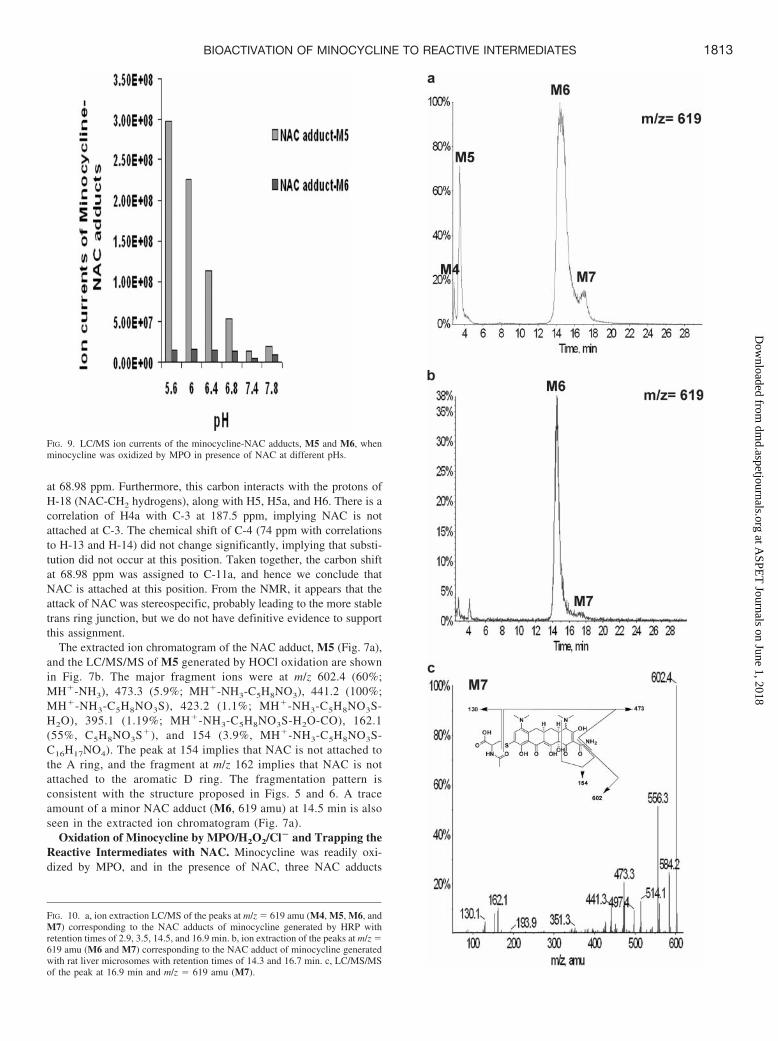
FIG. 10. a, ion extraction LC/MS of the peaks at m/z � 619 amu (M4, M5, M6, andM7) corresponding to the NAC adducts of minocycline generated by HRP withretention times of 2.9, 3.5, 14.5, and 16.9 min. b, ion extraction of the peaks at m/z �619 amu (M6 and M7) corresponding to the NAC adduct of minocycline generatedwith rat liver microsomes with retention times of 14.3 and 16.7 min. c, LC/MS/MSof the peak at 16.9 min and m/z � 619 amu (M7).
1813BIOACTIVATION OF MINOCYCLINE TO REACTIVE INTERMEDIATES
at ASPE
T Journals on June 1, 2018
dmd.aspetjournals.org
Dow
nloaded from

FIG. 11. a, 1H NMR spectrum of the minocycline M. b, 1H NMR spectrum of minocycline-NAC adducts M6 and M7, which have retention times of 14.5 and 16.9 min,respectively, on LC/MS. The primed signal assignment of proton chemical shifts correspond to diastereomer M7.
1814 MANNARGUDI ET AL.
at ASPE
T Journals on June 1, 2018
dmd.aspetjournals.org
Dow
nloaded from

corresponding to minocycline �2H � NAC (m/z 619) were observed(M4, M5, and M6). The major M5 adduct had a retention time of 3.3min, and the other minor adducts, M4 and M6, had retention times of2.6 and 14.7 min, respectively (Fig. 8a). The MS/MS spectrum of theNAC adduct M4 is essentially the same as M5. Because it also has asimilar retention time as M5, it could be concluded that M4 is adiastereomer of M5, formed by slow epimerization of M5, most likelyat C4. At pH 4, we saw a change in the ratio of M5/M4 from 42:1 to0.8:1 over 44 h as determined by HPLC and confirmed by LC/MS. AtpH 8, the ratio of M5/M4 changed from 27:1 to 0.3:1 over 44 h.
The LC/MS/MS of M6 generated by MPO is shown in Fig. 8b. Themajor fragment ions were at m/z 602.3 (100%; MH�-NH3), 584.2(4.4%; MH�-NH3-H2O), 556.4 (4.8%; MH�-NH3-H2O-CO), 512.9(4.8%; MH�-NH3-H2O-CO-NH(CH3)2), 473.3 (56%; MH�-NH3-C5H8NO3), 153.9 (5.2%; MH�-NH3-C5H8NO3S-C16H17NO4), and129.9 (14%; C5H8NO3S�). The prominent fragments of M6 are theresult of loss of NH3 at 602.4 and 473.3 caused by the loss of NACportion of the molecule, with the sulfur still attached to the parent ion,and this is confirmed by the fragment peak at 129.9 amu. The absenceof a significant peak at 441.2 or 162.1 amu, corresponding to breakingthe bond between the NAC sulfur and minocycline, also suggests thatthe NAC is bound to an aromatic carbon.
The ratio of the NAC adducts M5 and M6 formed by the MPOreaction are pH-dependent (Fig. 9). At the lower pH of 5.6, the ratioof the NAC adducts at 3.3 min to 14.7 min was 19:1, and at a higherpH of 7.8, the ratio changes to 2:1.
Oxidation of Minocycline by HRP in the Presence of NAC.Oxidation of minocycline by HRP was performed to determinewhether other peroxidases, which have a lower oxidation potential
than MPO, would also form the same reactive metabolites. In addi-tion, it made it easier to generate sufficient NAC adduct to obtain anNMR spectrum of M6. Oxidation of minocycline by HRP/H2O2 in thepresence of NAC produced four NAC adducts corresponding tominocycline �2H � NAC (M4, M5, M6, and M7). The major M6adduct had a retention time of 14.5 min, and the other minor adducts(M4, M5, and M7) had retention times of 2.9, 3.5, and 16.9 min,respectively (Fig. 10a). The LC/MS/MS spectra of M4 and M5 weresimilar to that of M5 produced by the MPO oxidation, and presumablyM5 is the same as the NAC adduct formed by MPO, and M4 is adiastereomer of M5.
The LC/MS/MS of M7 generated by HRP is shown in Fig. 10c. Themajor fragment ions were at m/z 602.4 (100%; MH�-NH3), 584.2(25%; MH�-NH3-H2O), 556.3 (51%; MH�-NH3-H2O-CO), 513.2(2.5%; MH�-NH3-H2O-CO-NH(CH3)2), 473.3 (21%; MH�-NH3-C5H8NO3), 441.2 (11%; MH�-NH3-C5H8NO3S), 162.1 (10%;C5H8NO3S�), 153.8 (8%; MH�-NH3-C5H8NO3S-C16H17NO4), and130.1 (4.7%; C5H8NO3
�). This MS/MS spectrum is very similar tothat of the NAC adduct M6 obtained by oxidation of minocycline withHRP. Because it has an almost identical retention time and MS/MS asM6, we concluded that M7 is a diastereomer of M6, formed by slowepimerization of M6, most likely at C4. We saw a change in the ratioof M6/M7 from 73:1 to 3:1 over 24 h at pH 4 as seen by HPLC andconfirmed by LC/MS. At pH 8, the ratio of M6/M7 changed from86:1 to 8:1 over 24 h.
NMR of the Minocycline-NAC Adduct M6. From a comparisonof the proton NMR spectra of M6 (Fig. 11; Table 2) and minocycline(Table 1), it is clear that the aromatic H-9 proton at 6.8 ppm is gone.Instead, there are several singlets close to 7.52 ppm. This implies that
TABLE 21H NMR and HSQC data for minocycline-NAC adducts M6 and M7
NMR Proton/Carbon NumbersM6 M7
�H ppm (Multiplicitya) �C ppm �H ppm (Multiplicitya) �C ppm
1 — — — —2 — — — —3 — — — —4 3.80 (s, 1H) 71.05 3.69 (s, 1H) 71.834a 2.62–2.73 (m, 1H) 34.45 2.62–2.73 (m, 1H) 33.275 1.64–1.71 (m, 1H) 34.27 1.64–1.71 (m, 1H) 34.27
2.18–2.33 (m, 1H) 2.18–2.33 (m, 1H)5a 2.75–2.81 (m, 1H) 32.23 2.75–2.81 (m, 1H) 32.236 2.18–2.33 (m, 1H) 30.48 2.18–2.33 (m, 1H) 30.48
2.98–3.12 (m, 1H) 2.98–3.12 (m, 1H)6a — — — —7 — — — —8 7.52 (s, 1H) 132.19 7.43 (s, 1H) 128.159 — — — —
10 — — — —10a — — — —11 — — — —11a — — — —12 — — — —12a — — — —13 2.90 (s, 6H) 40.81 2.93 (s, 6H) 42.3514 2.90 (s, 6H) 40.81 2.93 (s, 6H) 42.3515 2.59 (s, 6H) 44.63 2.68 (s, 6H) 44.7716 2.59 (s, 6H) 44.63 2.68 (s, 6H) 44.7717 — — — —18 2.98–3.12 (m, 1H) 34.59 2.98–3.12 (m, 1H) 33.94
3.45–3.52 (m, 1H) 3.37–3.42 (m, 1H)19 4.16–4.22 (m, 1H) 55.14 4.26–4.29 (m, 1H) 56.1120 — — — —21 1.76 (s, 3H) 21.77 1.70 (s, 3H) 21.6422 — — — —
Multiplicity: b, broad; s, singlet; d, doublet; q, quartet; dt, double triplet; dd, double doublet; m, multiplet; —, peaks absent in HSQC spectra.a NMR acquired in D2O.
1815BIOACTIVATION OF MINOCYCLINE TO REACTIVE INTERMEDIATES
at ASPE
T Journals on June 1, 2018
dmd.aspetjournals.org
Dow
nloaded from
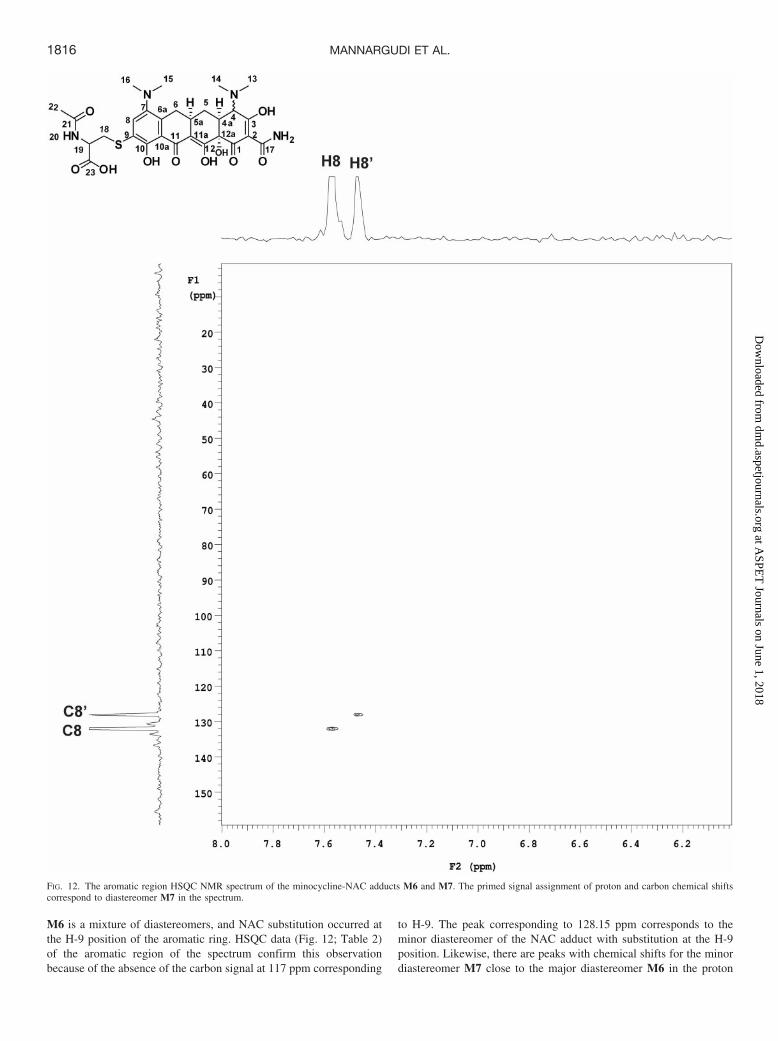
M6 is a mixture of diastereomers, and NAC substitution occurred atthe H-9 position of the aromatic ring. HSQC data (Fig. 12; Table 2)of the aromatic region of the spectrum confirm this observationbecause of the absence of the carbon signal at 117 ppm corresponding
to H-9. The peak corresponding to 128.15 ppm corresponds to theminor diastereomer of the NAC adduct with substitution at the H-9position. Likewise, there are peaks with chemical shifts for the minordiastereomer M7 close to the major diastereomer M6 in the proton
FIG. 12. The aromatic region HSQC NMR spectrum of the minocycline-NAC adducts M6 and M7. The primed signal assignment of proton and carbon chemical shiftscorrespond to diastereomer M7 in the spectrum.
1816 MANNARGUDI ET AL.
at ASPE
T Journals on June 1, 2018
dmd.aspetjournals.org
Dow
nloaded from
and HSQC data. In the nuclear Overhauser effect spectroscopy spec-trum of M6 there is a correlation between H-15/16 protons and theH-8 proton, confirming that the NAC adduct is substituted at the H-9position rather than the H-8 position (data not shown). Other protonand carbon chemical shifts of M6 remained similar to that of mino-cycline (Table 1). Taken together, we conclude that M6 represents theNAC adduct in which the NAC is attached at the H-9 position of thearomatic ring. Similar assignments could be made for M7 (Fig. 11;Table 2).
Metabolism of Minocycline by Rat Liver Microsomes in thePresence of NAC. The major NAC adduct formed by the oxidation ofminocycline by microsomes in presence of NAC corresponds to M6that was formed by MPO and HRP oxidation, along with its minordiastereomer M7. In particular, the HPLC retention time of M6formed from oxidation by hepatic microsomes (Fig. 10b) and itsLC/MS/MS are virtually the same. No NAC adduct with a retentiontime of 3.3 min corresponding to M5 was observed.
Discussion
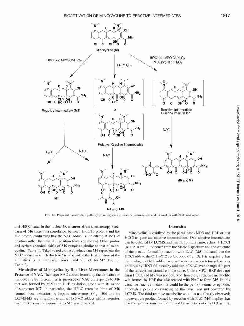
Minocycline is oxidized by the peroxidases MPO and HRP or justHOCl to generate reactive intermediates. One reactive intermediatecan be detected by LC/MS and has the formula minocycline � HOCl(M2, 510 amu). Evidence from the MS/MS spectrum and the structureof the product formed by reaction with NAC (M5) indicated that theHOCl adds to the C11a-C12 double bond (Fig. 13). It is surprising thatthe analogous NAC adduct was not observed when tetracycline wasoxidized by HOCl followed by addition of NAC even though this partof the tetracycline structure is the same. Unlike MPO, HRP does notform HOCl, and M2 was not observed; however, a reactive metabolitewas formed by HRP that also reacted with NAC to form M5. In thiscase, the reactive metabolite could be the peroxy ketone or epoxide,although a peak corresponding to this mass was not observed byLC/MS. The third reactive metabolite was also not directly observed;however, the product formed by reaction with NAC (M6) implies thatit is the quinone iminium ion formed by oxidation of ring D (Fig. 13).
FIG. 13. Proposed bioactivation pathway of minocycline to reactive intermediates and its reaction with NAC and water.
1817BIOACTIVATION OF MINOCYCLINE TO REACTIVE INTERMEDIATES
at ASPE
T Journals on June 1, 2018
dmd.aspetjournals.org
Dow
nloaded from

It is somewhat surprising that we were only able to detect traceamounts of the quinone that would be formed by the hydrolysis of theiminium ion (data not shown). M6 was the major adduct formed afteroxidation by HRP and the only product formed by hepatic micro-somes. This adduct was also formed in small quantities by MPO andHOCl. A small amount of isomeric NAC adducts (M4 and M7) wereobserved with similar retention times and MS/MS spectra as the majorNAC adducts, M5 and M6, respectively. These isomers presumablyrepresent diastereomers formed by slow epimerization of M5 and M6,respectively, most likely at C4.
These results provide a likely explanation for why minocycline isthe only tetracycline that causes a lupus-like syndrome and autoim-mune hepatitis. It also is consistent with the pattern that many drugsthat cause autoimmune syndromes are oxidized to reactive metabo-lites by the MPO of macrophages. It could also explain why P-ANCAs, antibodies against MPO, are observed in patients with mi-nocycline-induced lupus because MPO would likely be a major targetfor covalent binding. However, it cannot be determined from thesestudies which reactive metabolite is more important or whether for-mation by P450 or MPO is more likely to be responsible for theidiosyncratic reactions associated with minocycline. Even though it isusually assumed that reactive metabolites formed by P450 are respon-sible for liver toxicity, reactive metabolites formed by the MPO ofKupffer cells may play an important role, especially in the case ofdrug-induced autoimmune hepatitis.
Acknowledgments. We thank Drs. Henrianna Pang, Ling Xu, andYing Yang for training and support for LC/MS studies.
References
Antunes A, Davril A, Trechot P, Grandidier M, Truchetet F, and Cuny JF (1999) [Minocyclinehypersensitivity syndrome]. Ann Dermatol Venereol 126:518–521.
Borchers AT, Keen CL, and Gershwin ME (2007) Drug-induced lupus. Ann N Y Acad Sci1108:166–182.
Boudreaux JP, Hayes DH, Mizrahi S, Hussey J, Regenstein F, and Balart L (1993) Fulminant
hepatic failure, hepatorenal syndrome, and necrotizing pancreatitis after minocycline hepato-toxicity. Transplant Proc 25:1873.
Cullen SI and Cohan RH (1976) Minocycline therapy in acne vulgaris. Cutis 17:1208–1210,1214.
Doerge DR, Divi RL, Deck J, and Taurog A (1997) Mechanism for the anti-thyroid action ofminocycline. Chem Res Toxicol 10:49–58.
Dunphy J, Oliver M, Rands AL, Lovell CR, and McHugh NJ (2000) Antineutrophil cytoplasmicantibodies and HLA class II alleles in minocycline-induced lupus-like syndrome. Br JDermatol 142:461–467.
Farver DK (1997) Minocycline-induced lupus. Ann Pharmacother 31:1160–1163.Hardman CM, Leonard JN, Thomas HC, and Goldin R (1996) Minocycline and hepatitis. Clin
Exp Dermatol 21:244–245.Hussain A, Trudell P, and Repta AJ (1970) Quantitative spectrophotometric methods for
determination of sodium hypochlorite in aqueous solutions. J Pharm Sci 59:1168–1170.Kamel AM, Fouda HG, Brown PR, and Munson B (2002) Mass spectral characterization of
tetracyclines by electrospray ionization, H/D exchange, and multiple stage mass spectrometry.J Am Soc Mass Spectrom 13:543–557.
Knowles SR, Shapiro L, and Shear NH (1996) Serious adverse reactions induced by minocycline.Report of 13 patients and review of the literature. Arch Dermatol 132:934–939.
Lavergne SN, Park BK, and Naisbitt DJ (2008) The roles of drug metabolism in the pathogenesisof T-cell-mediated drug hypersensitivity. Curr Opin Allergy Clin Immunol 8:299–307.
Lawrenson RA, Seaman HE, Sundstrom A, Williams TJ, and Farmer RD (2000) Liver damageassociated with minocycline use in acne: a systematic review of the published literature andpharmacovigilance data. Drug Saf 23:333–349.
Omura T and Sato R (1964) The carbon monoxide-binding pigment of liver microsomes. I.Evidence for its hemoprotein nature. J Biol Chem 239:2370–2378.
Puyana J, Urena V, Quirce S, Fernandez-Rivas M, Cuevas M, and Fraj J (1990) Serumsickness-like syndrome associated with minocycline therapy. Allergy 45:313–315.
Seaman HE, Lawrenson RA, Williams TJ, MacRae KD, and Farmer RD (2001) The risk of liverdamage associated with minocycline: a comparative study. J Clin Pharmacol 41:852–860.
Smith PK, Krohn RI, Hermanson GT, Mallia AK, Gartner FH, Provenzano MD, Fujimoto EK,Goeke NM, Olson BJ, and Klenk DC (1985) Measurement of protein using bicinchoninic acid.Anal Biochem 150:76–85.
Sturkenboom MC, Meier CR, Jick H, and Stricker BH (1999) Minocycline and lupuslikesyndrome in acne patients. Arch Intern Med 159:493–497.
Teitelbaum JE, Perez-Atayde AR, Cohen M, Bousvaros A, and Jonas MM (1998) Minocycline-related autoimmune hepatitis: case series and literature review. Arch Pediatr Adolesc Med152:1132–1136.
Uetrecht J (2005) Current trends in drug-induced autoimmunity. Autoimmun Rev 4:309–314.Uetrecht JP (1992) The role of leukocyte-generated reactive metabolites in the pathogenesis of
idiosyncratic drug reactions. Drug Metab Rev 24:299–366.
Address correspondence to: Jack Uetrecht, Leslie Dan Faculty of Pharmacy,144 College Street, University of Toronto, Toronto, ON, Canada M5S3M2. E-mail:[email protected]
1818 MANNARGUDI ET AL.
at ASPE
T Journals on June 1, 2018
dmd.aspetjournals.org
Dow
nloaded from