distribution of neuropeptide y-like lmmunoreactivity and its
TRANSCRIPT
0270~6474/85/0507-1729$02 00/O CopyrIght 0 Smety for Neuroscience Prmted I” U.S.A.
The Journal of Neuroscience Vol. 5, No. 7, pp, 1729-1739
July 1985
Distribution of Neuropeptide Y-like lmmunoreactivity and Its Relationship to FMRF-amide-like lmmunoreactivity in the Sixth Lumbar and First Sacral Spinal Cord Segments of the Rat’
CATHRINE A. SASEK AND ROBERT P. ELDE*
Department of Anatomy, University of Minnesota, Minneapolis, Minnesota 55455
Abstract
The present study was aimed at describing the distribution of neuropeptide Y (NPY)-like immunoreactivity in the sixth lumbar (L6) and first sacral segments (Sl) of the rat spinal cord, comparing this distribution to that of FMRF-amide-like immunoreactivity and determining whether NPY- and FMRF- amide-like immunoreactivities are present in the same neu- rons in the dorsal gray commissure (DGC) in L6 and Sl of the rat spinal cord.
For distribution studies tissue from colchicine-treated ani- mals was processed according to the peroxidase-antiper- oxidase technique using anti-NPY as the primary antiserum. For co-localization studies serial 5Km sections were proc- essed for immunofluorescence. Adjacent sections were in- cubated with either anti-NPY or anti-FMRF-amide as the primary antiserum. The number of immunoreactive cells per section was counted and each section was photographed. The sections were then restained with the other antiserum (i.e., tissue first stained with anti-NPY was stained with anti- FMRF-amide and vice versa), the number of cells per section was recounted, and the sections were rephotographed.
NPY-like immunoreactive cells and fibers were identified in the DGC, sacral parasympathetic nucleus, substantia ge- latinosa, marginal zone, nucleus proprius, and ventral horn. Every cell in the DGC that contained NPY-like immunoreac- tivity was found also to contain FMRF-amide-like immuno- reactivity, and the distribution of NPY-like immunoreactive fibers was found to be similar, although denser than FMRF- amide-like immunoreactive fibers.
The distribution of NPY-like immunoreactivity in L6 and Sl of the rat spinal cord suggests that an NPY-like peptide may be involved in regulation of pelvic viscera, processing of primary afferent information, and motor regulation of pelvic muscles. The presence of NPY- and FMRF-amide-like im- munoreactivities in the same neurons in the DGC together with the lack of bona fide FMRF-amide in the rat central nervous system, the presence of NPY in the rat central nervous system, and the cross-reactivity of anti-FMRF-amide with NPY support the hypothesis that the FMRF-amide anti- serum recognizes an NPY-like peptide in the rat spinal cord.
Recerved June 18, 1984; Revised December 10, 1984; Accepted December 11, 1984
’ This work was supported by United States Publrc Health Service Grant DA 02148. The expert technical assrstance of Jean Floyd and the helpful assrstance of Drs. Virginia Seybold and Martin Wessendorf durrng the preparation of thus manuscript is gratefully acknowledged.
* To whom correspondence should be addressed.
Recently, a novel 36-amino acid peptide with potent biological activity (Lundberg et al., 1982; Agnati et al., 1983; Fuxe et al., 1983; Stjernquist et al., 1983), neuropeptide Y (NPY; Tatemoto et al., 1982; Tatemoto, 1982) was isolated from porcine brain. In subsequent studies, NPY-like immunoreactivity was shown to be present in regions throughout the rat nervous system (Allen et al., 1983; Guy et al., 1983; Hokfelt et al., 1983; O’Donohue et al., 1983; Emson and De Quidt, 1984; Everitt et al., 1984; Lundberg et al., 1984); including several that are involved in autonomic regulation. Further- more, an NPY-like immunoreactive substance from rat brain extracts has been shown to elute in the same position as pure porcine NPY after high pressure liquid chromatographic fractionation (Allen et al., 1983; Lundberg et al., 1984). Recently, Emson and De Quidt (1984) noted that NPY-like immunoreactive cells were present in the sub- stantia gelatinosa and sacral regions of the rat spinal cord, and Gibson et al. (1984) described the distribution of NPY-like immuno- reactive fibers in the sprnal cord of several species. There have been no studies, however, in which the distribution of NPY-like immuno- reactive cells in the sixth lumbar (L6) and first sacral (Sl) spinal cord segments of the rat has been described in detail. These segments are of particular interest because they contain the neurons of origin of parasympathetic preganglionic fibers that innervate the pelvic viscera. Thus, a portion of the present study describes the distribu- tion and assesses the number of NPY-like immunoreactive cells in L6 and Sl of the rat spinal cord. In addition, it describes in greater detail the distribution and density of NPY-like immunoreactive fibers in these segments.
The present study was also aimed at comparing the distribution of NPY-like immunoreactivity to the distribution of FMRF-amide-like immunoreactivity in L6 and Sl of rat spinal cord. In a previous study (Sasek et al., 1984) we described the distribution of FMRF-amide- like immunoreactivity in these spinal cord segments and hypothe- sized that the immunoreactivity seen with this antiserum was due to recognition of NPY or its precursor by the FMRF-amide antiserum. We based this hypothesis on several pieces of evidence, most notably the absence of bona fide FMRF-amide in the central nervous system of the rat (Bishop et al., 1983), the cross-reactivity of FMRF- amide antiserum with synthetic NPY (Sasek et al., 1984) and the presence of NPY in the rat central nervous system (Allen et al., 1983; Lundberg et al., 1984). Thus, we have attempted to provide further support for thus hypothesis by determining whether the distributions of NPY- and FMRF-amide-like immunoreactivities in L6 and Si were similar and whether NPY- and FMRF-amide-like immu- noreactivities were localized in the same neurons of the dorsal gray commissure (DGC) in L6 and Sl of the rat spinal cord.
Materials and Methods Surgery. Twenty-two male Sprague-Dawley rats (280 to 340 gm), anesthe-
tized with chloral hydrate (350 mg/kg), were implanted with chronic intrathecal cannulae to the level of approximately L6/Sl (8.5 cm) according to the method of Yaksh and Rudy (1976). The animals were allowed to recover
1729
1730 Sasek and Elde Vol. 5, No. 7, July 1985
from the anesthesia, and any animals showing signs of hindlimb paralysis resulting from the cannulation were discarded. To visualize immunohisto- chemically peptide-containing neurons, colchicine (25 to 50 pg/lO to 20 ~1; Sigma Chemical Co.) in 0.9% saline was administered via the cannulae to the lumbosacral spinal level at 0 and 24 hr. All animals used in the present study received colchicine.
Tissue preparaation. At 48 hr anesthetized animals were perfused transcardially with calcium-free Tyrode’s solution equilibrated with 95% O$ 5% COP followed either by 4% paraformaldehyde in 0.1 M phosphate buffer (pH 6.5; 200 ml) and then 4% paraformaldehyde in 0.1 M borate buffer (pH 9.2; 500 ml; Berod et al., 1981) or by 2% paraformaldehyde and 15% picnc acid in 0.1 M phosphate buffer (Stefanini et al., 1967). L6 and Si were dissected from the animals and allowed to postfix for approximately 2 hr. At this point tissues were inspected with a dissecting microscope, and they showed no signs of damage due to cannula insertion or drug treatment. The tissue was then transferred to 5% sucrose in 0.1 M phosphate buffer (pH 7.2) and stored until sectioning.
lmmunohistochemistry. Tissue was processed according to either the peroxidase-antiperoxidase technique of Sternberger (1979) for distribution studies, or the immunofluorescence technique of Coons (1958) as modified by Hokfelt and Goldstein (1975) for co-localization studies (Hokfelt et al., 1977). Those cord segments to be processed according to the peroxidase- antiperoxidase technique were cut at 50 pm either in a transverse or a horizontal plane on a vibrating microtome and placed in a well containing phosphate-buffered saline (PBS). The sections were then incubated for 10 min in phenylhydrazine hydrochloride (100 pg/ml of PBS; Eastman Organic Chemicals) to inactivate endogenous peroxidase activity (Straus, 1972) rinsed several times in PBS, and incubated at 4°C overnight in antiserum to NPY (l/1000 dilution). The generation and characterization of the NPY antiserum have been described elsewhere (Lundberg et al., 1984). All antisera used in this study were diluted with 0.3% Triton X-l 00 in PBS.
The next day, after rinsing in PBS, the sections were sequentially incubated in sheep anti-rabbit IgG (l/300 dilution for 1 hr; Antibodies Inc.), two PBS rinses (15 min each), and rabbit peroxidase-antiperoxrdase (l/500 dilution for 1 hr; Cooper Biomedical) at room temperature. After further rinsing, the sections in each well were reacted with approximately 1 ml of 3,3’-diaminob- enzidine tetrahydrochloride (50 fig/ml of 0.05 M Tris-buffered 0.9% saline, pH 7.6; Sigma and 0.3% H202 (330 nl/ml of diaminobenzrdine tetrahydro- chloride) for 5 to 10 min. An additional 5 PI of 0.3% H202 was added to each incubation well and sections were incubated for another 5 to 10 min. The reaction was halted by rinsing with Tris-buffered saline. The sections were then rinsed several times in Tris buffer, mounted on gelatin-coated slrdes, dehydrated, cleared, and coverslipped. Sections were viewed and photo- graphed on an Olympus microscope, model BH2.
NPY-like rmmunoreactive cells were found in most regions of the spinal cord. The sacral parasympathetic nucleus (SPN) and DGC were identified as previously described (Sasek et al., 1984) and the boundries of the substantia gelatinosa were identified by using darkfield microscopy. Neurons which were immunoreactive for NPY were counted in randomly selected sections from L6 and Sl from seven animals. Neurons were identified by the presence of either a nucleus or nerve processes. The mean number of cells per nucleus per section and standard error were calculated, and a Mann- Whitney U test was used to determine whether there was a significant difference between the number of immunoreactive cells in L6 and Sl for each region. The numbers of NPY-like immunoreactive cells in the DGC and SPN were compared wrth data previously obtained with an antiserum to FMRF-amide (Sasek et al., 1984) using a Mann-Whitney U test. The densities of immunoreactive fibers were rated according to the following scheme: very dense (++++), dense (+++), moderate (++), and sparse (+). Levels of density are illustrated in Figure 4, a and b (very dense, dorsolateral nucleus; sparse, ventral horn) and Frgure 1 a (dense, substantia gelatinsoa; moderate, nucleus proprius). The density and drstribution of NPY- and FMRF-amide-lrke immunoreactive fibers were compared using tissues from a previous study of the distribution of FMRF-amide (Sasek et al., 1984).
To determine whether NPY- and FMRF-amide-like immunoreactrvitres were localized in the same neurons, experiments were carried out in which tissue was processed for immunofluorescence. In these experiments, identified cord segments were cut serially at 5 pm in a transverse plane on a cryostat and mounted on gelatin-coated slides. After rehydrating, alternate sections were incubated overnight at 4°C with rabbit antisera to either FMRF-amide (l/100 dilution; rabbit 246a) or NPY (l/100 dilution). The antiserum to FMRF- amide was generated in our laboratory, and its generation and characteriza- tion have been described previously (Sasek et al., 1984). The next day the sections were rinsed with PBS and incubated for 1 hr at room temperature with fluorescein-labeled goat anti-rabbit IgG (l/8 dilution; Antibodies lnc). The
sections were rinsed and then counterstained with ethidium bromide (1 pg/ ml of PBS, Sigma; Schmued et al., 1981) a fluorescent NISSI stain. After rinsrng, the slides were coverslipped with PBS/glycerin (3/l). Sections were viewed and photographed on a Zeiss fluorescence microscope equipped with epi-illumination.
The number of cells present in each section was recorded, and each section was photographed. Then the coverslips were removed with PBS and the sections were restained with the other primary antiserum without eluting the first antiserum (i.e., tissue first stained with anti-FMRF-amide was then stained for NPY, and tissue first stained with anti-NPY was then stained for FMRF-amide). The number of cells per section was then recounted and the sections were rephotographed (Hokfelt et al., 1977). The data were analyzed and compared using a Mann-Whitney U test and the photographs. By means of this method a numerical assessment of coexistence can be obtained. If coexistence is complete, no new cells will be detected after restaining. If there is no coexistence, the number of cells present after restaining will equal the sum of the number of cells seen with each antiserum. To determine whether one can sequentially stain two antigens, sections were immuno- stained with anti-vasoactive intestinal polypeptide, anti-substance P, or anti- somatostatin, and the number of immunostained cells was counted. The sections were then restained with anti-FMRF-amide or anti-methionine en- kephalin. An increase in the number of immunoreactive cells was identified with both antisera; thus, it was concluded that processing tissue for immu- nofluorescence does not prevent visualization of additional immunoreactive substances.
Specificity studies. As a control, NPY antiserum at its working dilution was incubated with synthetic porcine NPY (10 pg/ml; Pentnsula Laboratories), and the ability of the antiserum to stain tissue was tested. Incubation of the NPY antiserum with synthetic NPY abolished all immunostaining.
As an additional control, the technique of Larsson (1981) was used to test the ability of the NPY antiserum to recognize heterologous peptides. Briefly, this technique consists of applying small volumes of peptide solutions to Whatman no. 1 filter paper, fixing them to the paper with paraformaldehyde vapors in vacua, and processing the papers according to the peroxidase- antiperoxidase technique of Sternberger (1979). NPY antiserum was tested for its ability to recognize NPY, peptide YY (Bachem), FMRF-amide (Bachem), avian pancreatic polypeptide (Bachem), somatostatin (Sigma), substance P (Sigma), neurotensin (Peninsula), vasoactive intestinal polypeptide (Sigma), cholecystokinrn octapeptide (Vega Biochemicals), methionine and leucine enkaphalin (Sigma), dynorphin l-8 (Peninsula), neurophysins I and II (gener- ously donated by Dr. Virginia Seybold), bombesin (Sigma), and oxytocin (Bachem). Using this technique it was found that the NPY antiserum reacted with the homologous antigen and with peptide YY; however, since peptide YY does not appear to be present in the spinal cord (Hbkfelt et al., 1983; Lundberg et al., 1984; our unpublished observations), the NPY-like immuno- reactivity seen in the present study is most probably not due to cross- reacttvity with peptide YY. The antiserum did not cross-react with any other of the peptides.
Results
Distribution of NPY
The distribution of NPY-like immunoreactive cells and fibers was similar in L6 and St; thus, the descriptions that follow will be for both segments, unless otherwise specified. Table I summarizes the number of NPY-like immunoreactive cells in each segment and region and the distribution and density of immunoreactive fibers. It should be noted that, although colchicine has been found in some instances to decrease the density of immunoreactive fibers, prelim- inary studies in our laboratory indicate that there is little or no difference in the density of NPY- or FMRF-amide-like immunoreactive fibers in colchicine-treated animals as compared to nontreated animals. However, it is possible that some undetected alterations in fiber density or pattern occurred as a result of colchicine treatment; therefore, in comparing the distribution of FMRF-amide- and NPY- like immunoreactivities, colchicine-treated tissue was used for both antisera.
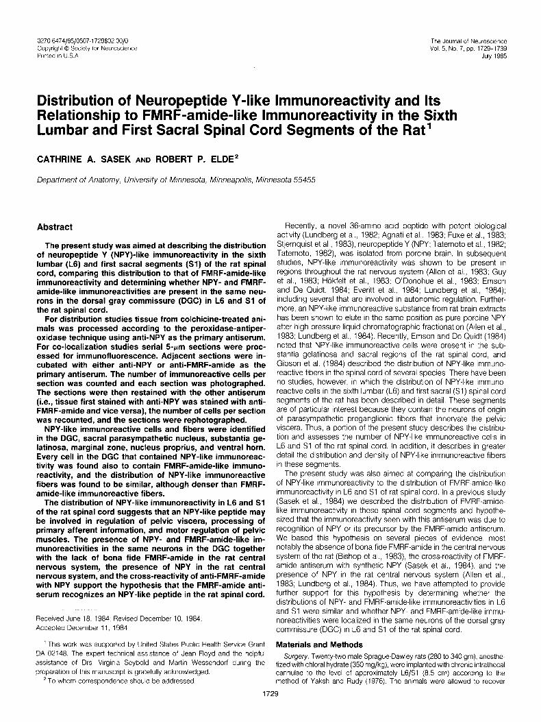
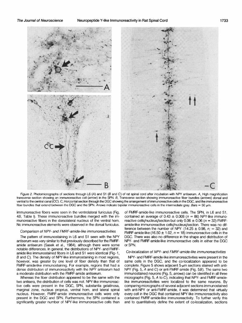
SPN. The SPN contained a dense plexus of NPY-like immuno- reactive fibers (Fig. 1 A, Table I) and an occasional round or triangular NPY-like immunoreactive cell (Fig. ZA, Table I). The SPN in Sl was found to contain slightly fewer immunoreactive cells per section than did the SPN in L6. A prominent band of long, beaded immunoreac- tive fibers extended between the plexus of immunoreactive fibers in
The Journal of Neuroscience Neuropeptide Y-like lmmunoreactivity in Rat Spinal Cord
TABLE I Summary of the di.stribuBon of WY-like immunoreactivity in the sixth lumbar and first sacral spinal cord segments
CELLS/REGION/SECTIONa RegKJn Fibers’
L6 Sl
Sacral parasympathetic nucleusC 0.5 + 0.0 (46) 0.4 f 0.0 (40) +++ Dorsal gray commissure 14.3 f 0.3 (14) 14.2 f 0.4 (18) +++ Substantia gelatinosa and marginal zoneC 3.4 + 0.1 (18) 1.9 -t 0.1 (14) +++ Nucleus proprius 0.8 + 0.1 (18) 0.3 f 0.0 (14) ++ Ventral horn 0.3 + 0.0 (44) 0.2 f 0.0 (24) + Dorsomedial nucleus 0.0 (14) ++d Dorsolateral nucleus 0.0 (14) ++++d Lateral spinal nucleus 0.9 f 0.2 (20) 0.7 + 0.2 (20) ++ Lateral funiculus 0.0 (15) 0.0 (17) ++ Ventrolateral funiculus 0.0 (15) 0.0 (17) +++d
a Values represent mean f SEM; numbers in parentheses are the number of cells studied. b Fiber density was rated as follows: ++++, very dense; +++, dense; ++, moderate; +, sparse. ’ Regions in which a significant difference (p < 0.05) was seen between the number of immunoreactrve cells in L6 and Sl
1731
d Represents fiber densrty in L6.
the SPN and a plexus of immunoreactive fibers present in the DGC perpendicular to these regions (Fig. 3A). In addition, clusters of (Figs. 1A and 2C). In some sections bipolar immunoreactive cells immunoreactive fibers were often present in the lateral part of with medially and laterally directed processes were located along nucleus proprius. The perpendicularly oriented fibers often appeared this band of fibers (Fig. 2C). to terminate in these clusters of fibers.
DGC. Numerous round, triangular, or spindle-shaped NPY-like immunostained cells were present in the DGC (Figs. 1A and 2C, Table I). Long beaded axons from some of the more laterally situated immunoreactive cells could be followed through the intermediate gray and into the SPN. In horizontal sections (Fig. 2C) the immuno- reactive cells in the DGC were seen to be organized in clusters the centers of which were separated by a variable distance of 200 to 300 pm. The clusters of cells were interconnected by bundles of immunoreactive fibers.
Long, varicose NPY-like immunoreactive fibers were also identified medially in the dorsal horn along the lateral edge of the dorsal funiculus (Fig. IA). These fibers extended between the medial edge of the substantia gelatinosa and the DGC, and they merged with the immunoreactive fiber plexus located in the DGC.
The immunoreactive cells in the DGC were surrounded by a dense plexus of NPY-like immunoreactive fibers (Fig. 1 A, Table I). This plexus merged with the band of immunoreactive fibers that extended between the DGC and SPN (Fig. la). Additionally, NPY-like immu- noreactivity was seen in longitudinally oriented fiber bundles located dorsal and ventral to the central canal (Figs. 1A and 2B). The dorsal fiber bundle was present in most sections through Sl but was absent in many through L6. The ventral bundle, which was less dense than the dorsal bundle, was not present in many sections through both L6 and Sl. In those sections in which a distinct dorsal or ventral bundle was not present, it was replaced by a diffuse plexus of immunoreactive fibers.
In some sections a sparse bundle of NPY-like immunoreactive fibers extended between the lateral edge of the substantia gelatinosa and the DGC. An occasional bipolar immunoreactive neuron was located along these fibers. The processes of the bipolar neurons followed the course of the fibers. An additional, larger NPY-like immunoreactive fiber bundle that extended through the lateral funic- ulus from the lateral part of the substantia gelatinosa to the SPN was also present in some sections.
Ventral horn. Only an occasional round or triangular NPY-like immunoreactive cell was present in the ventral horn (Table I). These cells, which were smaller than the large ventral horn motoneurons, were not localized to a specific part of the ventral horn but were scattered throughout its extent.
Numerous immunoreactive fibers were also present along the sides of the central canal (Fig. 2B). These fibers, which ran in a dorsoventral direction, merged dorsally with the dorsal bundle and ventrally with the ventral bundle. In some sections an additional dorsoventrally oriented immunoreactive fiber bundle was located between the DGC and the dorsal longitudinal bundle.
Dorsal horn. Scattered, round NPY-like immunoreactive cells were located throughout the dorsal horn (Figs. IA and 3A). The greatest numbers of immunoreactive cells, however, were present in the substantia gelatinosa and the marginal zone (Fig. 38, Table I), whereas fewer immunoreactive cells were present in the nucleus proprius (Fig. 3A, Table I). A significantly greater number of immu- noreactive cells was present in the substantia gelatinosa and mar- ginal zone of L6 than in the same regions in Sl
Although only a sparse distribution of long varicose NPY-like immunoreactive fibers was present in most of the ventral horn (Fig. 4, A and B; Table I), a very dense accumulation of immunoreactive fibers was seen in the dorsolateral nucleus of the ventral horn (Fig. 4, A and B; Table I), and a moderate distribution of fibers was present in the dorsomedial nucleus of the ventral horn (Fig. 4A, Table I). Both of these nuclei contain neurons that innervate pelvic muscles, and the dorsolateral nucleus is considered to be the rat homologue of Onuf’s nucleus. Neither nucleus is present in Sl (Schroder, 1980). lmmunoreactive fiber bundles that extended be- tween the dorsolateral and dorsomedial nuclei and between the dorsolateral nucleus and the SPN (Fig. 4B) were seen in some sections. The immunoreactive fiber plexuses in both the dorsolateral and dorsomedial nuclei were only present in sections taken through L6.
Dense plexuses of punctate NPY-like immunoreactivity were pres- ent in the marginal zone and outer substantia gelatinosa of the dorsal horn (Figs, IA and 3, A and i3; Table I). In some sections horizontally oriented, varicose immunoreactive fibers formed a cap over the punctate immunoreactivity in the marginal zone and sub- stantia gelatinosa. A moderate number of immunoreactive fibers was present in nucleus proprius (Figs. IA and 3A). Many of these were long, beaded fibers that merged with the immunoreactive fibers in the marginal zone and substantia gelatinosa and had orientations
White matter. A moderate distribution of immunoreactive fibers and an occasional round or triangular immunoreactive cell was localized in the lateral spinal nucleus (Giesler and Elde, 1985) of the dorsolateral funiculus (Fig. 48, Table I). A moderate plexus of fibers was also present just lateral to the SPN in the lateral funiculus, and in many sections immunoreactive fibers could be followed from the SPN into this plexus.
In sections taken through L6, long, beaded immunoreactive fibers were scattered throughout the ventral funiculus (Table I). Similar fibers were present in sections through Sl ; however, their distribution was extremely sparse. In sections through L6, dense bundles of
F/gure 7. A, Photomrcrograph of a transverse sectron through L6 of rat spinal cord after incubation with NPY antiserum. lmmunostaining rn the SPN, DGC, dorsal horn (DH). and intermediate gray can be seen. Arrowheads indicate rmmunostained fibers that extend between the substantia gelatinosa and the DGC. Double so//d arrows Indicate rmmunostained fibers that extend between the DGC and SPN. The open arrow rndrcates the longrtudinally oriented immunoreactrve fiber bundle dorsal to the central canal (CC). 6 and C, Serial transverse sectrons through the dorsal gray commissure n Sl showrng the similar drstributron of NPY- (B) and FMRF-amide-like (C) immunoreactrvrtres. Arrows indicate an example of a neuron, found in both sections, that contains NPY- and FMRF-amide-like rmmunoreactrvities. Bars = 50 pm.
1732
The Journal of Neuroscience Neuropeptide Y-like lmmunoreactivity in Rat Spinal Cord
Figure 2. Photomlcrographs of sections through L6 (A) and Sl (13 and C) of rat spinal cord after incubation with NPY antiserum. A, High magnification transverse section showing an lmmunoreactive cell (arrow) in the SPN. B, Transverse section showing immunoreactive fiber bundles (arrows) dorsal and ventral to the central canal (CC). C, Horizontal section through the DGC showing the arrangement of immunoreactive cells in the DGC, and the immunoreactive fiber bundles that extend between the DGC and the SPN. Arrows indicate bipolar immunoreactive cells in the intermediate gray. Bars = 50 pm.
immunoreactive fibers were seen in the ventrolateral funiculus (Fig. 4B, Table I). These immunoreactive bundles merged with the im- munoreactive fibers in the dorsolateral nucleus of the ventral horn. No immunoreactive elements were observed in the dorsal funiculus.
Comparison of NPY- and FMRF-amide-like immunoreactivities The pattern of immunostaining in L6 and Si seen with the NPY
antiserum was very similar to that previously described for the FMRF- amide antiserum (Sasek et al., 1984), although there were some notable differences. In general, the distributions of NPY- and FMRF- amide-like immunostained fibers in L6 and Sl were identical (Fig. 1, B and C). The density of NPY-like immunostaining in most regions, however, was greater by one level of fiber density than that of FMRF-amide-like immunostaining. For example, regions that had a dense distribution of immunoreactivity with the NPY antiserum had a moderate distribution with the FMRF-amide antiserum.
Whereas the fiber distribution appeared to be the same with the two antisera, the distribution of ceils was not. NPY-like immunoreac- tive cells were present in the DGC, SPN, substantia gelatinosa, marginal zone, nucleus proprius, ventral horn, and lateral spinal nucleus. However, FMRF-amide immunoreactive cells were only present in the DGC and SPN. Furthermore, the SPN contained a significantly greater number of NPY-like immunoreactive cells than
of FMRF-amide-like immunoreactive cells. The SPN, in L6 and Sl, contained an average of 0.43 + 0.008 (n = 86) NPY-like immuno- reactive cells/nucleus/section but only 0.06 + 0.06 (n = 32) FMRF- amide-like immunoreactive cells/nucleus/section. There was no dif- ference between the number of NPY- (14.25 + 0.98, n; = 32) and FMRF-amide-like (16.50 + 1.02, n = 18) immunoreactive cells in the DGC. There was also no difference in the shape and distribution of NPY- and FMRF-amide-like immunoreactive cells in either the DGC or SPN.
Co-localization of NPY- and FMRF-amide-like immunoreactivities NPY- and FMRF-amide-like immunoreactivities were present in the
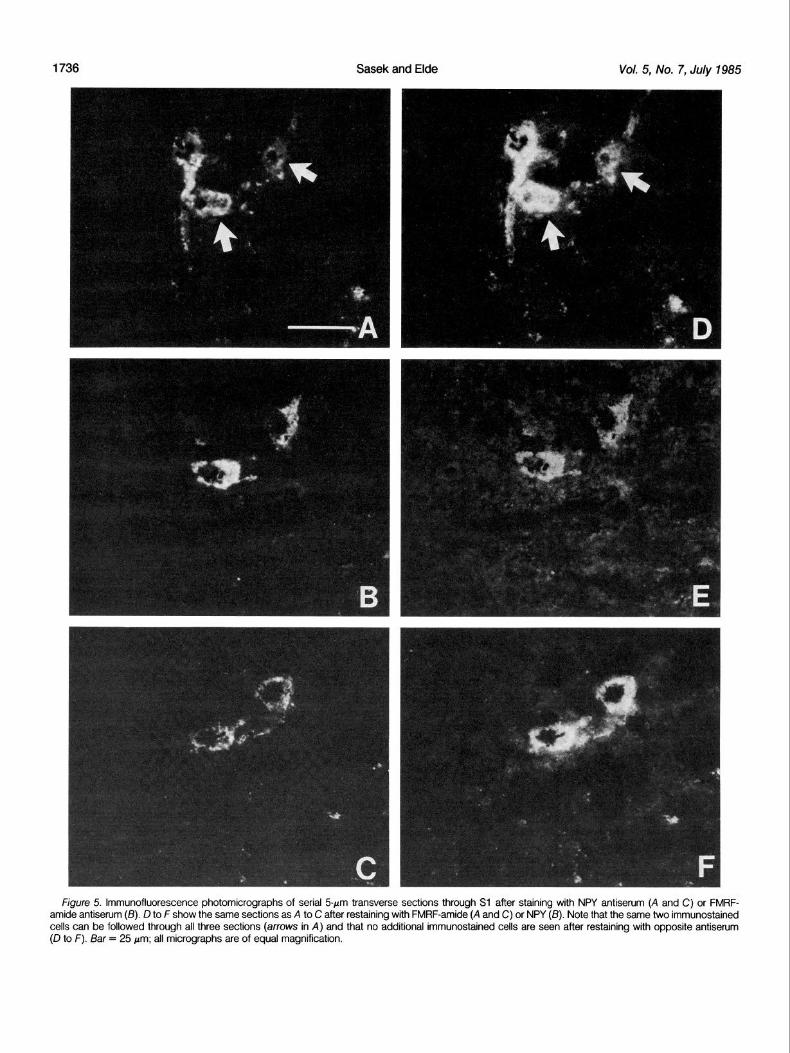
same cells in the DGC, and the co-localization appeared to be complete. Figure 5 shows adjacent 5-&m sections stained with anti- NPY (Fig. 5, A and C) or anti-FMRF-amide (Fig. 5B). The same two immunostained neurons (Fig. 5, arrows) can be identified in all three micrographs (Fig. 5, A to C), indicating that NPY- and FMRF-amide- like immunoreactivities were localized to the same neurons. By comparing micrographs of several adjacent sections immunostained with anti-NPY or anti-FMRF-amide, it was determined that virtually every cell in the DGC that contained NPY-like immunoreactivity also contained FMRF-amide-like immunoreactivity. To further verify this and to quantitatively define the extent of co-localization, sections

Sasek and Elde Vol. 5, No. 7, July 1985
Figure 3. High power photomicrographs of sections stained with NPY antiserum. A, Transverse section through the dorsal horn in L6; B, horizontal section through the substantia gelatinosa in Sl. The arrows in A indicate perpendicularly oriented fibers extending between the substantia gelatinosa and the nucleus proprius. The arrow in B points to an example of an immunoreactive cell. The punctate nature of the immunoreactivity is especially evident in 6. Bars = 50 m.
stained for FMRF-amide were restained for NPY and vice versa. The section after restaining with antiserum to NPY. Similarly, in sections number of immunostained cells found after restaining was then immunostained with NPY antiserum, 2.29 f 0.05 cells/5pm section compared to the number found before restaining. It was found that were found prior to restaining and 2.29 + 0.05 cells/9~m section sections immunostained for FMRF-amide contained 2.21 + 0.04 were found after restaining with FMRF-amide antiserum. There was cells/d~m section prior to restaining and 2.21 & 0.04 cells/dclm no difference in the number of immunoreactive cells per section
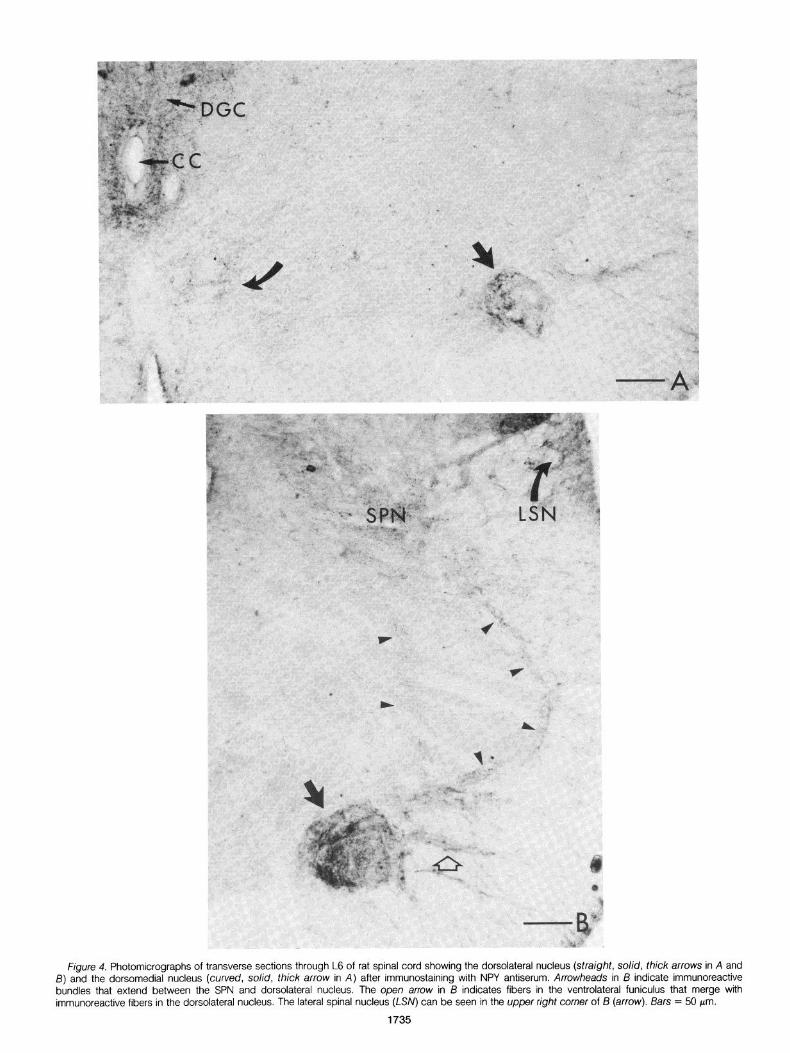
Figure 4. Photomicrographs of transverse sections through L6 of rat spinal cord showing the dorsolateral nucleus (straight, solid, thick arrows in A and 5) and the dorsomedial nucleus (curved, solid, thick arrow in A) after immunostaining with NPY antiserum. Arrowheads in I3 indicate immunoreactive bundles that extend between the SPN and dorsolateral nucleus. The open arrow in 13 indicates fibers in the ventrolateral funiculus that merge with immunoreactive fibers in the dorsolateral nucleus. The lateral spinal nucleus (LSN) can be seen in the upper right corner of B (arrow). Bars = 50 pm.
1735
1736 Sasek and Elde Vol. 5, No. 7, July 1985
figure 5. lmmunofluorescence photomicrographs of serial 5pm transverse sections through Sl after staining with NPY antiserum (A and C) or FMRF- amide antiserum (IS). D to F show the same sections as A to C after restaining with FMRF-amide (A and C) or NPY (6). Note that the same two immunostained cells can be followed through all three sections (arrows in A) and that no additional immunostained cells are seen after restaining with opposite antiserum (D to F). Bar = 25 pm; all micrographs are of equal magnification.
The Journal of Neuroscience Neuropeptide Y-like lmmunoreactivity in Rat Spinal Cord 1737
before and after restaining. Furthermore, in neither case were any additional cells found on photomicrographs after restaining (Fig. 5, cf. A to C with D to F). These results clearly demonstrate that NPY- and FMRF-amide-like immunoreactivities are localized in the same neurons in the DGC of L6 and Sl
Discussion The results of the present studies demonstrate the presence of
an NPY-like immunoreactive substance in L6 and Si of the rat spinal cord. They extend the work of Emson and De Quidt (1984) and Gibson et al. (1984) by providing a detailed description of NPY-like immunoreactivity in L6 and Sl and by quantifying the number of immunoreactive cells in these segments. L6 and Sl are of particular interest because they contain the cell bodies of the parasympathetic preganglionic neurons.
It is not clear what functions are mediated by the NPY-like immunoreactive elements in each of these regions. The distribution of NPY-like immunoreactivity in L6 and Sl , however, suggests that an NPY-like peptide may be involved in several functions, including regulation of pelvic viscera, transmission and processing of primary afferent sensory information, and motor regulation of pelvic muscu- lature.
NPY-like immunoreactive cells and fibers in both the SPN and DGC may be involved in the regulation of pelvic viscera. The NPY- like immunoreactive cells in the SPN are similar in shape and location to those of parasympathetic preganglionic neurons in the rat as described by Hancock and Peveto (1979) and Nadelhaft and Booth (1982). Preganglionic cells are round, triangular, or spindle shaped. The majority of preganglionic cells are located in the SPN, but occasional cells are found more medially in the intermediate gray. Thus, it is likely that at least some of the NPY immunostained neurons in the SPN are preganglionic neurons.
There is evidence to suggest that neurons in the DGC are also involved in autonomic regulation. Primary afferent fibers from pelvic viscera have been demonstrated to form terminal fields in the region in which these cells are localized (Morgan et al., 1981; Nadelhaft and Booth, 1982). In addition, in the present study, processes from NPY immunostained cells in the DGC were found to extend through the intermediate gray into the SPN. Thus, neurons in the DGC are in a prime location to act as interneurons in a reflex pathway between primary afferent and parasympathetic efferent neurons. They may therefore have an important function in integrating incoming visceral afferent information prior to relaying it to autonomic efferent neurons in the SPN. These neurons may also be important for relaying visceral afferent information to higher centers in the central nervous system and, therefore, for modulating descending input for the control of pelvic viscera. Tract tracing studies have shown that cells in the DGC of L6 do project to the brainstem (Nahin et al., 1983); however, it is not known which if any peptides are contained in these projecting neurons.
The presence of NPY-like immunoreactivity in the dorsal horn suggests that any NPY-like peptide may be involved in the perception of sensory information. The marginal zone, substantia gelatinosa, and nucleus proprius are the sites of termination of primary afferent fibers (Light and Perl, 1979a, b; Ralston and Ralston, 1979). NPY- like immunoreactive terminals were identified in each of these re- gions, and the distribution was similar to that of other peptides that are known to be contained in primary afferent fibers (Seybold and Elde, 1980; Hunt et al., 1981) suggesting that some of the NPY-like immunoreactivity present in the dorsal horn may be primary afferent in origin. However, Gibson et al. (1984) detected no change in NPY- like immunoreactive fibers after dorsal rhizotomy at the cervical level, indicating that at cervical spinal cord levels NPY-like immunoreactive fibers are probably not primary afferent in origin. The distribution of NPY-like immunoreactivity is also similar to that of enkephalin, which is present in cells and fibers that are intrinsic to the spinal cord (Elde et al., 1976). Therefore, it is also possible that some or all of the NPY-like immunoreactivity in the dorsal horn, rather than having a
primary afferent origin, represents the terminal fields of neurons that are intrinsic to the brain or spinal cord. Since NPY-like immunoreac- tive cells were also present in the dorsal horn, it is probable that many of the immunoreactive processes in the dorsal horn are from these neurons. Additional studies are necessary to determine the precise origin of these immunoreactive fibers. The NPY-like immu- noreactive neurons in the dorsal horn may be interneurons that receive and modulate primary afferent input or, in the case of those immunoreactive cells in the nucleus proprius, cells of origin of ascending tracts such as the spinocervical or spinothalamic tracts.
An NPY-like peptide may also be involved in the regulation of somatic motor activity. NPY-like immunoreactive fibers were present in the dorsolateral and dorsomedial nuclei of the ventral horn. These nuclei contain motoneurons that innervate pelvic musculature (Schroder, 1980). No immunoreactive neurons were identified in either nucleus. The source of the immunoreactive fibers in the dorsolateral and dorsomedial nuclei is unknown, but their presence indicates that an NPY-like peptide may function to modulate the activity of neurons that innervate pelvic muscles.
lmmunoreactive fiber bundles were identified between the dorso- lateral nucleus and the SPN. Although voluntary, the activity of the pelvic musculature is intimately connected with that of the pelvic viscera. Thus, it might be speculated that the immunoreactive bun- dles consist of axon collaterals of parasympathetic, efferent, NPY- containing neurons the function of which is to synchronize the activity of motoneurons that innervate pelvic muscles with the activity of pelvic viscera.
It should be noted that the present study was conducted using only male rats. Differences between male and female rats in the ventral horn nuclei that innervate the pelvic muscles have been described (Schroder, 1980). Therefore, it is possible that the distri- bution of NPY-like immunoreactivity in the ventral horn of female rats may be different from that of the male.
NPY-like immunoreactive cells and fibers were also identified in the lateral spinal nucleus. Although numerous other peptides have recently been identified in this region (Giesler and Elde, 1985) there is little known about its function. Retrograde tract-tracing studies utilizing horseradish peroxidase have demonstrated that some cells in the lateral spinal nucleus project to the mesencephalon (Menetrey et al., 1980, 1982; Giesler et al., 1981). It is not known, however, what substances are contained in these projecting neurons.
Comparison of NPY- and FMRF-amide-like immunoreactivities. FMRF-amide was first isolated in the clam ganglion by Price and Greenberg (1977). Subsequently, FMRF-amide antisera were shown to recognize a novel neuronal system throughout the mammalian CNS (Boer et al., 1980; Dockray et al., 1981; Weber et al., 1981; Sasek et al., 1983; Williams and Dockray, 1983). There has been much speculation as to the nature of the peptide or peptides recognized by these antisera. In a recent study we described the pattern of staining seen with the FMRF-amide antiserum in the rat spinal cord (Sasek et al., 1984) and hypothesized that the immuno- reactivity was due to recognition of NPY by the FMRF-amide antisera. We based this hypothesis on the following pieces of evidence. The distribution of NPY-like immunoreactivity in the rat CNS appears to be nearly identical to that of FMRF-amide-like immunoreactivity (Hokfelt et al., 1983). The peptide FMRF-amide is not present in the rat CNS (Bishop et al., 1983) whereas NPY is present (Allen et al., 1983). The peptide recognized by anti-FMRF-amide in the mamma- lian CNS appears to be significantly larger than the tetrapeptide isolated from clam ganglion (Dockray et al., 1981). In addition, our FMRF-amide antisera recognize synthetic porcine NPY as well as or better than FMRF-amide in a model immunohistochemical system (Sasek et al., 1984).
In the present study we have attempted to provide additional support for this hypothesis by determining whether FMRF-amide- and NPY-like immunoreactivities are localized in the same neurons in the DGC and whether they are similarly distributed in L6 and Sl of the rat spinal cord. It was found that NPY- and FMRF-amide-like
1738 Sasek and Elde Vol. 5, No. 7, July 1985
immunoreactive fibers were distributed in a nearly identical pattern, and there appeared to be complete co-localization of FMRF-amide- and NPY-like immunoreactivities in neurons in the DGC. These results support the hypothesis that the peptide recognized by the FMRF- amide antiserum is indeed NPY or a related peptide.
There were, however, differences between the two antisera.The NPY antiserum generally stained fibers with a slightly greater intensity than did the FMRF-amide antiserum. In addition, NPY-like immuno- reactive cells were identified in several regions in which FMRF-amide immunoreactive cells were not present. Since anti-FMRF-amide strongly recognizes NPY, these results suggest that some neurons process NPY-like peptides to a form unrecognized by anti-FMRF- amide. Cross-reactivity tests have indicated that the FMRF-amide antiserum only recognizes the amidated carboxy-terminus of NPY. Any processing that changed or masked the carboxy-terminus or its amide group would prohibit the binding of the FMRF-amide antise- rum. Furthermore, the FMRF-amide antiserum would be unable to bind the precursor to the NPY-like peptide since most peptides that terminate in an amide group are synthesized as long molecules that are later cleaved and enzymatically amidated (Bradbury et al., 1982). Presumably the amidation of NPY occurs via this same mechanism.
In contrast, the NPY antiserum probably contains several popula- tions of antibodies that bind antigenic sites along the entire length of the NPY molecule. The NPY antiserum may, therefore, recognize the intact NPY molecule as well as forms that result from processing. Furthermore, the antiserum may recognize biosynthetic precursor molecules since recognition most probably does not depend on amidation.
Thus, the neurons identified immunohistochemically by both the FMRF-amide antiserum and the NPY antiserum most probably con- tain an NPY-like peptide with an amidated carboxy-terminus. Those neurons labeled by only the NPY antiserum may contain a form of NPY that has been processed in such a way as to make it unrecog- nizable by the FMRF-amide antiserum. These results suggest that several different NPY-like peptides exist in the rat CNS and that the processing and distribution of these peptides are not identical. Confirmation of these suggestions awaits further biochemical stud- ies
References Agnati, L. F., K. Fuxe, F. Benfenati, N. Battistini, A. Harfstrand, K. Tatemoto,
T. Hokfelt, and V. Mutt (1983) Neuropeptide Yin vitro selectively increases the number of cup-adrenergic binding sites in membranes of the medulla oblongata of the rat. Acta Phystol. Stand. 118: 293-295.
Allen, Y. S., T. E. Adrian, J. M. Allen, K. Tatemoto, T. J. Crow, S. R. Bloom, and J. M. Polak (1983) Neuropeptide Y distribution in the rat brain. Science (NY) 227: 877-879.
Berod, A., B. K. Hartman, and J. F. Pujor (1981) Importance of fixation in immunohistochemistry: Use of formaldehyde solutions at variable pH for the localrzation of tyrostne hydroxylase. J. Histochem. Cytochem. 29: 844- 850.
Bishop, J. F., W. W. Watson, J. Groome, and T. L. O’Donohue (1983) Distribution and characterization of FMRFamide-like peptrdes tn rat brain and digestive system. Sot. Neurosci. Abstr. 9: 139.
Boer, H. H., L. P. C. Schot, J. A. Veenstra, and D. Reichelt (1980) Immuno- cytochemlcal identification of neural elements in the central nervous system of a snail, some insects, and a mammal, with an antiserum to the molluscan cardioexcitatory tetrapeptide FMRF-amide. Cell Tissue Res. 213: 21-27.
Bradbury, A. F., M. D. A. Finnie, and D. G. Smyth (1982) Mechanism of C- terminal amide formation by pituitary enzymes. Nature 298: 686-688.
Coons, A. H. (1958) Fluorescent antibody methods. In Genera/ Cytochemical Mefhods, J. R. Danielli, ed., pp. 399-422, Academic Press, Inc., New York.
Dockray, G. J., C. Vaillant, and R. G. Williams (1981) New vertebrate brain- gut peptide related to a molluscan neuropeptide and an opioid peptide. Nature 293: 656-657.
Elde, R., T. Hokfelt, 0. Johansson, and L. Terenrus (1976) Immunohistochem- ical studres using antibodies to leucine-enkephalin: Initial observations on the nervous system of the rat. Neuroscience 7: 349-351.
Emson, P. C., and M. E. De Qutdt (1984) NPY-A new member of the pancreatic polypeptide family. Trends Neurosci. 7: 31-35.
Eventt, B. J., T. Hokfelt, L. Terenius, K. Tatemoto, V. Mutt, and M. Goldstein (1984) Differential co-existence of neuropeptide Y (NPY)-like immunoreac- tivity with catecholamines in the central nervous system of the rat. Neuroscience 11: 443-462.
Fuxe, K., L. F. Agnati, A. Harfstrand. I. Zini, K. Tatemoto, E. M. Pith, T. Hokfelt, V. Mutt, and L. Terenius (1983) Central administration of neuro- peptide Y induces hypotension bradypnea and EEG synchronization in the rat. Acta Physiol. Stand. 118: 189-192.
Gibson, S. J.. J. M. Polak. J. M. Allen. T. E. Adrian, J. S. Kellv. and S. R. Bloom (1984) The distribution and origin of a novel brain peptide, neuro- peptide Y, in the spinal cord of several mammals. J. Comp. Neurol. 227: 78-91.
Giesler, G. J., Jr., and R. P. Elde (1985) lmmunocytochemical studies of the peptidergic content of fibers and terminals within the lateral spinal and lateral cervical nuclei. J. Neurosci.. in press.
Gtesler, G. J., Jr., H. R. Spiel, and W. D. Willis (1981) Organization of spinothalamtc tract axons within the rat spinal cord. J. Comp. Neurol. 19.5: 243-252.
Guy, J., Y. S. Allen, J. M. Polak, and G. Pelletier (1983) lmmunocytochemical localization of neuropeptide Y (NPY) in the rat brain, Sot. Neuroscl. Abstr. 9: 291.
Hancock, M. B., and C. A. Peveto (1979) Preganglionic neurons in the sacral spinal cord of the rat: An HRP study. Neurosci. Lett. 11: l-5.
Hokfelt, T., K. Fuxe, and M. Goldstein (1975) Applications of immunohisto- chemistry to studies on monoamine cell systems with special references to nervous tissues. Ann. N. Y. Acad. Sci. 254: 407-432.
Hokfelt, T., L. G. Elfvin, R. Elde, M. Schultzberg, M. Goldstein, and R. Luft (1977) Occurrence of somatostatin-like immunoreactivity in some periph- eral sympathetic noradrenergic neurons. Proc. Natl. Acad. Sci. U. S. A. 74: 3587-3591.
Hokfelt, T., J. M. Lundberg, K. Tatemoto, V. Mutt, L. Terenius, J. Polak, S. Bloom, C. Sasek, R. Elde, and M. Goldstein (1983) Neuropeptide Y (NPY)- and FMRF-amide neuropeptide-like immunoreactivities in catecholamine neurons of the rat medulla oblongata. Acta Physiol. Stand. 117: 315-318.
Hunt, S. P., J. S. Kelly, P. C. Emson, J. R. Kimmel, R. J. Miller, and J.-Y. Wu (1981) An immunohistochemical study of neuronal populations containing neuropeptides or y-aminobutyrate within the superficial layers of the rat dorsal horn. Neuroscience 6: 1883-1898.
Larsson. L. -I. (1981) A novel immunocytochemical model system for speci- ficity and sensitivity screening of antisera against multiple antigens, J. Histochem. Cytochem. 29: 408-410.
Light, A. R., and E. R. Perl(1979a) Reexamination of the dorsal root projection to the spinal dorsal horn including observations on the differential termi- nation of coarse and fine fibers. J. Comp. Neurol. 786: 117-132.
Light, A. R., and E. R. Perl (197913) Spinal termination of functionally identified primary afferent neurons with slowly conducting myelinated fibers. J. Comp. Neurol. 786: 133-150.
Lundberg, J. M., L. Terenius, T. Hokfelt, C. R. Matiling, K. Tatemoto. V. Mutt, J. Polak, S. Bloom, and M. Goldstein (1982) Neuropeptide Y (NPY)-like immunoreactivity in peripheral noradrenergic neurons and effects of NPY on sympathetic function. Acta Physiol. Stand. 116: 477-480.
Lundberg, J. M., L. Terenius, T. Hokfelt, and K. Tatemoto (1984) Comparative immunohistochemical and biochemical analysis of pancreatic polypeptide- like peptides with special reference to presence of neuropeptide Y in central and peripheral neurons. J. Neurosci. 4: 2376-2386.
Menetrey, D., A. Chaouch, and J. M. Besson (1980) Location and properties of dorsal horn neurons at origin of spinoreticular tract in lumbar enlarge- ment of the rat. J. Neurophysiol. 44: 862-877.
Menetrey, D., A. Chaouch, D. Binder, and J. M. Besson (1982) The origin of the spinomesencephakc tract in the rat: An anatomical study using the retrograde transport of horseradish peroxidase. J. Comp. Neurol. 206: 193-207.
Morgan, C., I. Nadelhaft, and W. C. deGroat (1981) The distribution of visceral primary afferents from the pelvic nerve to Lissauer’s tract and the spinal gray matter and its relationship to the sacral parasympathetic nucleus. J. Comp. Neurol. 201: 415-440.
Nadelhaft, I., and A. M. Booth (1982) Preganglionic neurons and visceral afferent fibers in the rat pelvic nerve. Sot. Neurosci. Abstr. 8: 77.
Nahtn, R. L., A. M. Madsen, and G. J. Giesler, Jr. (1983) Anatomical and physiological studies of the gray matter surrounding the spinal cord central canal. J. Comp. Neurol. 220: 321-335.
O’Donohue, T. L., B. M. Chronwall, and D. A. DiMaggio (1983) Distribution
The Journal of Neuroscience Neuropeptide Y-like lmmunoreactivity in Rat Spinal Cord 1739
of neuropeptrde Y-like immunoreactivity in rat brain. Sot. Neurosci. Abstr. 9: 290.
Price, D. A., and M. J. Greenberg (1977) Structure of a molluscan cardioex- crtatory neuropeptide. Science (N. Y.) 797: 670-671.
Ralston, H. J., Ill, and D. D. Ralston (1979) The distribution of dorsal root axons tn lamrnae I, II and Ill of the macaque spinal cord: A quantitative electron microscope study. J. Comp. Neural. 784: 643-684.
Sasek, C. A., R. P. Elde, and V. S. Seybold (1983) Localization of FMRF- NHP-like immunoreactivity in autonomic regions of the brainstem and spinal cord of the rat. Sot. Neurosci. Abstr. 8: 809.
Sasek, C. A., V. S. Seybold, and R. P. Elde (1984) The immunohtstochemical localrzation of nine peptrdes in the sacral parasympathetic nucleus and dorsal gray commissure in rat spinal cord. Neuroscrence 12: 855-887.
Schmued, L. C., L. W. Swanson, and P. E. Sawchenko (1981) Some fluorescent counterstarns for neuroanatomical studies. Sot. Neurosci. Abstr. 7: 417.
Schroder, H. D. (1980) Organization of the motoneurons innervating the pelvic muscles of the male rat. J. Comp. Neurol. 192: 567-587.
Schroder, H. D. (1984) Somatostatin in the caudal spinal cord: An immuno- histochemical study of the spinal centers involved in the innervatron of pelvic organs. J. Comp. Neurol. 223: 400-414.
Seybold, V., and R. Elde (1980) lmmunohistochemical studies of peptidergic neurons in the dorsal horn of the spinal cord. J. Histochem. Cytochem. 28: 367-370.
Stefanini, M., C. DeMartino, and L. Zamboni (1967) Fixation of ejaculated spermatozoa for electron microscopy. Nature 276: 173-l 74.
Sternberger, L. A. (1979) lmmunocyfochemistry, Prentice-Hall, Englewood Cliffs, NJ.
Stjernquist, M., P. Emson, C. Owman, N. -0. Sjoberg, F. Sundler, and K. Tatemoto (1983) Neuropeptide Y in the female reproductive tract of the rat. Distribution of nerve fibres and motor effects. Neuroscr. Lett. 39: 279- 284.
Straus, W. (1972) Phenylhydrazine as inhibitor of horseradish peroxidase for use in immunoperoxrdase procedures. J. Histochem. Cytochem. 20: 949- 951.
Tatemoto, K. (1982) Neuropeptide Y: Complete amino acid sequence of the brain peptide. Proc. Natl. Acad. Sci. U. S. A. 79: 5485-5489.
Tatemoto, K.. N. Carlquist, and V. Mutt (1982) Neuropeptide Y-A novel brain peptide with structural similarities to peptide YY and pancreatic polypeptide. Nature 296: 659-660.
Weber, E., C. J. Evans, S. J. Samuelsson, and J. D. Barchas (1981) Novel peptide neuronal system rn rat brain and pituitary. Science (N. Y.) 274: 1248-1250.
Williams, R. G., and G. J. Dockray (1983) lmmunohistochemical studies of FMRF-amide-like immunoreactivity in rat brain. Brain Res. 276: 213-229.
Yaksh, T. L., and T. A. Rudy (1976) Chronic catheterization of the spinal subarachnord space. Physiol. Behav. 77: 1031.