distal clavicle fractures and acute acromioclavicular ... · pdf fileit is important to...
TRANSCRIPT
VOL.11 NO.5 MAY 2006 Medical Bulletin
20
VOL.15 NO.1 JANUARY 2010
IntroductionClavicle fractures constitute 44% of all shoulder girdle injuries. Most of them are mid-clavicle fractures that unite satisfactorily with non-operative treatment. In contrary, fractures of the distal one third of the clavicle is an exception that carry a high non-union rate. Therefore it is important to recognise this distinct clavicle fracture as different from those commonly encountered mid-clavicle fractures.
Distal clavicle fractures account for 15% of all clavicle fractures. Neer classified this fracture into two types based on the status of the coracoclavicular (CC) ligament.
How to Evaluate this Injury Clinically and Radiologically?Most of the distal clavicle fractures are due to a direct blow or fall on the shoulder. Ecchymosis, abrasion wounds and associated swellings are commonly seen. If
there is significant fracture displacement, the skin may be subjected to too much tension by the bone spike of the proximal fragment that needs urgent attention. In case of high energy trauma such as road traffic accidents, a detailed examination is needed to exclude other associated injuries. Head and neck injuries are found in up to 10% of cases while ipsilateral rib fractures as well as associated chest injuries are not uncommon.
A shoulder trauma series including AP view, scapular Y view and axillary view of the injured shoulder should be the standard radiographs taken. Classification of the fracture could be made based on the fracture displacement. An axillary view is useful in evaluating the AP translation of the fracture. Computer tomography is sometimes needed in cases of complex and comminuted fracture patterns. Ultrasound and MRI are seldom necessary except associated soft tissue injuries such as a rotator cuff tear is suspected.
What will Happen if we Leave the Distal Clavicle Fractures Untreated?Natural history tells us that a Type I fracture of the distal clavicle without disruption of the CC ligament is inherently stable and usually will heal with a favourable outcome after conservative treatment. Only a very small percentage of patients will have residual shoulder symptoms.
Due to the unstable nature of Type II fractures, the risk of fracture non-union is high with an average of 30%. But are these fracture non-unions all symptomatic that necessitate surgical intervention? Nordqvist in his largest series of distal clavicle fractures found that there were 10 nonunions out of an overall 23 type II fractures, only two cases were symptomatic. When we review Neer's report, only one non-union case ultimately required surgery. Other literatures also pointed out that patients could still have satisfactory shoulder functions even when the fractures are not united radiologically.
Neer suggested that Type III fractures are subjected to a higher chance of ACJ arthrosis with late painful symptoms. However Nordqvist did not note any long term problems in his 15 (11-21) years' follow-up results.
What is the Current Management Strategy?
The type I fracture is a stable injury with an intact CC ligament that prevents the fragments from significant displacement. In Type II fractures, the distal clavicle fragment is subjected to the distal pull by the weight of the arm and medial pull by the strong pectorii muscles as well as the Latissmus dorsi muscle while the proximal fragment is dragged posteriorly by the trapezius. These disturbing forces contribute to the fracture displacement and the unstable nature of Type II fractures.
The type II fracture is further sub-categorised into Type IIA in which the fracture occurs medial to the CC ligament and Type IIB in which the fracture occurs more laterally with the CC ligament disrupted from the proximal fragment. With identification of more subtypes, the Neer classification of distal clavicle fractures was later modified to include up to five types.
Type I - Minimally displaced fractures that occur lateral to the CC ligament
Type II - Displaced fractures that the proximal fragment is detached from the CC ligament
Distal Clavicle Fractures and Acute Acromioclavicular Joint InjuriesDr. Wilkie Wai-kee NGAI
Dr. Wilkie Wai-kee NGAI
MBBS(HK), MScSEM(Bath), MRCS(Edin), FRCSEd(Orth), FHKAM(Orthopaedic Surgery), FHKCOS
Type III - Fracture extension into acromioclavicular joint (ACJ)
Type IV - Fracture with periosteal disruption occurring in children
Type V - Avulsion fracture of the distal clavicle with the inferior cortical fragment remains attached to the CC ligament
Resident Consultant in Orthopedics and Traumatology, Hong Kong Baptist Hospital
Medical Bulletin
21
VOL.15 NO.1 JANUARY 2010
There is no doubt that the initial treatment for Type I and Type III fractures will be non-operative. An arm sling is offered to support the weight of the arm. A figure of eight splint is not necessary. Pendulum exercises could be started as pain is tolerated. The arm sling could be taken off when the pain has subsided with active assisted and passive mobilisation exercises to start shortly thereafter. Strengthening exercises can be commenced when a pain-free full shoulder range-of-motion achieved.
Despite the excellent prognosis of Type I and III fractures, patients should be informed about the remote chance of late residual shoulder symptoms; especially in type III fractures, patients will have a higher chance in the development of ACJ arthrosis. For those patients who have significant residual symptoms, operative treatment with distal clavicle resection may be needed to alleviate the symptoms.
Management of Type II fractures is always the focus of debate. With a high risk of non-union, some clinicians will suggest operative treatment for all type II fractures while proponents for non-operative treatment will argue that a lot of these non-unions are asymptomatic. For patients who have marked fracture displacements, potential overlying skin compromise and open injuries, they are clearly indicated for surgery. Others may have the right to opt for non-operative treatment.
When non-operative treatment is selected, patients must be carefully counselled and informed of the followings: Rehabilitation after Operation
The arm is placed in a sling for 3 weeks after operation in order to avoid early failure. Rigid fixation using plate and screws allows earlier active mobilisation without protection.
Pendulum shoulder mobilisation exercises should be started as the pain is tolerated. We will not wait for radiological bone union as it may take up to 12 weeks and passive and active assisted shoulder mobilisation exercises should be commenced once the sling has been taken off. Contact sports should be avoided until evidence of solid bony union. Removal of implants such as plate and screws is needed once the fracture has united.
Judging from the above, each and every patient who has Type II fractures should be considered individually. For young and active patients who do not accept the risk of late reconstruction surgery will tend to have operative treatment early. In contrary, patients who have multiple medical co-morbidities with associated high peri-operative risks would best choose an initial conservative approach.
Operative Treatment for Distal Clavicle FracturesThere are numerous operative techniques reported in the past. The methods of surgical treatment could be summarised as follows:
Different surgical techniques have their own advantages and disadvantages as tabulated (Table 1). With more than 20 techniques described so far, no single fixation is ideal and perfect. There is still no consensus regarding the best surgical method to fix these fractures. Whichever method is chosen, careful operative planning and familiarity with the features of that particular operative technique are essential for the best clinical outcomes.
Method of Fixation Advantages Disadvantages
The average risk of non-union is 30% Most distal clavicle fracture non-unions will end up with mild symptoms and functional loss is usually lowFor those patients who do develop symptomatic non-unions, late reconstructive surgery such as the Weaver Dunn procedure may be necessary
Transacromial or intramedullary fixation in terms of K-wires and different pins such as the Steinmann pin, Hagie pin and Knowles pin. This fixation could also be augmented with concomitant tension band wiresCoracoclavicular indirect fixation with the use of screws, suture anchors, Dacron graft or Mersilene tapeOpen reduction and clavicular plate fixation with the use of different plate systems such as the Balser plate, Wolter hook plate, AO hook plate, etc.
Transacromial or Intramedullary
Coracoclavicular Indirect Fixation
Open reduction and clavicular plate fixation
SimpleCould be augmented with other fixation devices such as tension band wires / suture anchorsMinimally invasive
More rigid fixation Does not disturb the fracture site Does not cross the ACJNo need to remove the implant (except screw) Allows minimally invasive techniquesMost rigid fixationAllows early active mobilisation of the shoulder High union rate
Fixation not rigid enough - loss of reduction risk, slow rehabilitationMigration of the implant with lethal outcomes had been reported in the past Premature ACJ arthrosisRemoval of the implant needed Pull out of the implant reported Osteolysis of the clavicle or coracoid
Extensive dissection neededShoulder impingement Rotator cuff tear Acromion fractureHook cut out Removal of the implant necessary
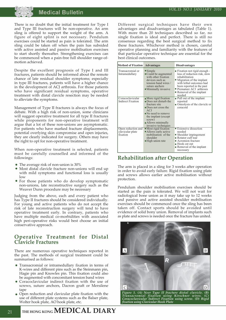
Figure 1. (A) Neer Type II fracture distal clavicle. (B) Transacromial fixation using Kirschner wires. (C) Coracoclavicular Indirect Fixation using screw. (D) Rigid fixation using Clavicular Hook Plate
VOL.11 NO.5 MAY 2006 Medical Bulletin
22
VOL.15 NO.1 JANUARY 2010
IntroductionThe acromioclavicular joint (ACJ) is the articulation between the clavicle and the acromion of the scapula. It is covered by a thin capsule with a meniscus-like disc inside. The joint is reinforced by the surrounding acromioclavicular (AC) ligament. This is further strengthened by the overlying delto-trapezius fascia and the coracoclavicular (CC) ligament. As it is the key linkage between the clavicle and the scapula which couples the glenohumeral and scapulothoracic motion, integrity of the ACJ is important for a smooth coordinated shoulder motion.
ACJ injuries could be considered as the "soft tissue" injury counterparts in which the clavicle remains intact with variable degrees of traumatic disruption around the ACJ. They account for about 12% of all shoulder girdle injuries. The true prevalence is expected to be even more as many patients with minor ACJ sprains may not seek medical advice. This incomplete ACJ disruption is actually even commoner than complete dislocations.
Pathology and ClassificationInjuries to the ACJ should be regarded as a spectrum of injuries instead of a single entity, from minor sprains to complete ACJ dislocations and even complex injuries that involve the disruption of the delto-trapezius fascia. It is important to be familiar with the different forms of injuries as their management are different.
Three types of ACJ injuries were initially described by Tossy et al. and was later modified into six types by Rockwood. This Rockwood classification is mainly based on the extent of ACJ disruption as well as the state of the CC ligament and the resulting clavicular displacement. All six types of ACJ injuries could be differentiated using standard radiography as shown in Table 2 below:
Clinical and Radiological DiagnosisThe clinical presentations of ACJ injuries are quite similar to fractures of the distal clavicle as both of them share similar injury mechanisms. Most of the patients fell on their shoulder with direct contusion. Local abrasions, ecchymosis and swellings are commonly noted in acute ACJ injuries. Depending on the types of injury, the position of the distal clavicle will be different as described above. For ACJ subluxations/dislocations (II-V), the distal clavicle will look prominent and will be mobile with the so-called "piano-key" sign. Despite that there is a prominent lateral clavicular end, acute threatening to the overlying skin is not common. Concomitant injuries to surrounding neurovascular structures are also not a usual presentation as well.
Standard radiographs of a shoulder trauma series are essential for the initial diagnosis of ACJ injuries. For better delineation of the vertical displacement, a true AP view of the ACJ (Zanca view) with the X-ray beam tilted 10 degrees cephalad centring on the ACJ is sometimes necessary. Concerning the use of weight-bearing view to classify ACJ injuries, some clinicians may not use it especially for those minor ACJ sprains and incomplete subluxations. An axillary view is important for the assessment of AP displacement of the clavicle which could be easily missed. CT scan and MRI are seldom needed in acute ACJ injuries.
Could ACJ Injuries be Treated Conservatively?For mild degrees of ACJ injuries (i.e. Type I and II), these are invariably treated with non-operative treatment. Rest, ice therapy and the use of analgesics is helpful in relieving the acute symptoms. Protection with an arm sling is useful in supporting the weight of the arm and hence reduces the stress on the ACJ. In contrary, a collar and cuff tends to apply additional traction force to the ACJ and therefore should not be used. Sophisticated shoulder immobilisers are deemed not necessary as there is no evidence showing that they could help in reducing the subluxation.
Concerning the rehabilitation, the arm sling could be taken off at 1-2 weeks after injury when acute symptoms have subsided. Pendulum exercises of the shoulder should be started as early as the pain can be
Acute Acromioclavicular Injuries
Rockwood Types
Features X-ray appearance
I
II
III
IV
V
VI
ACJ sprain with incomplete disruption the AC ligamentACJ subluxation with complete disruption of the AC ligament and incomplete disruption of the CC ligamentComplete ACJ dislocation with complete disruption of the AC ligament as well as the CC ligament Complete ACJ dislocation with clavicle displaced posteriorlyComplete ACJ dislocation with disruption of overlying the delto-trapezial fascia Complete ACJ dislocation with the distal clavicle displaced anteriorly to lie underneath the coracoid process
Normal
ACJ subluxation with increase in CC distance <25%
ACJ dislocation with increase in CC distance at 25-100%
ACJ dislocation with evidence of significant displacement of clavicle posteriorlyACJ dislocation with increase in CC distance by more than 100% and up to 300% ACJ dislocation with the clavicle displaced anteriorly to lie below the coracoid process
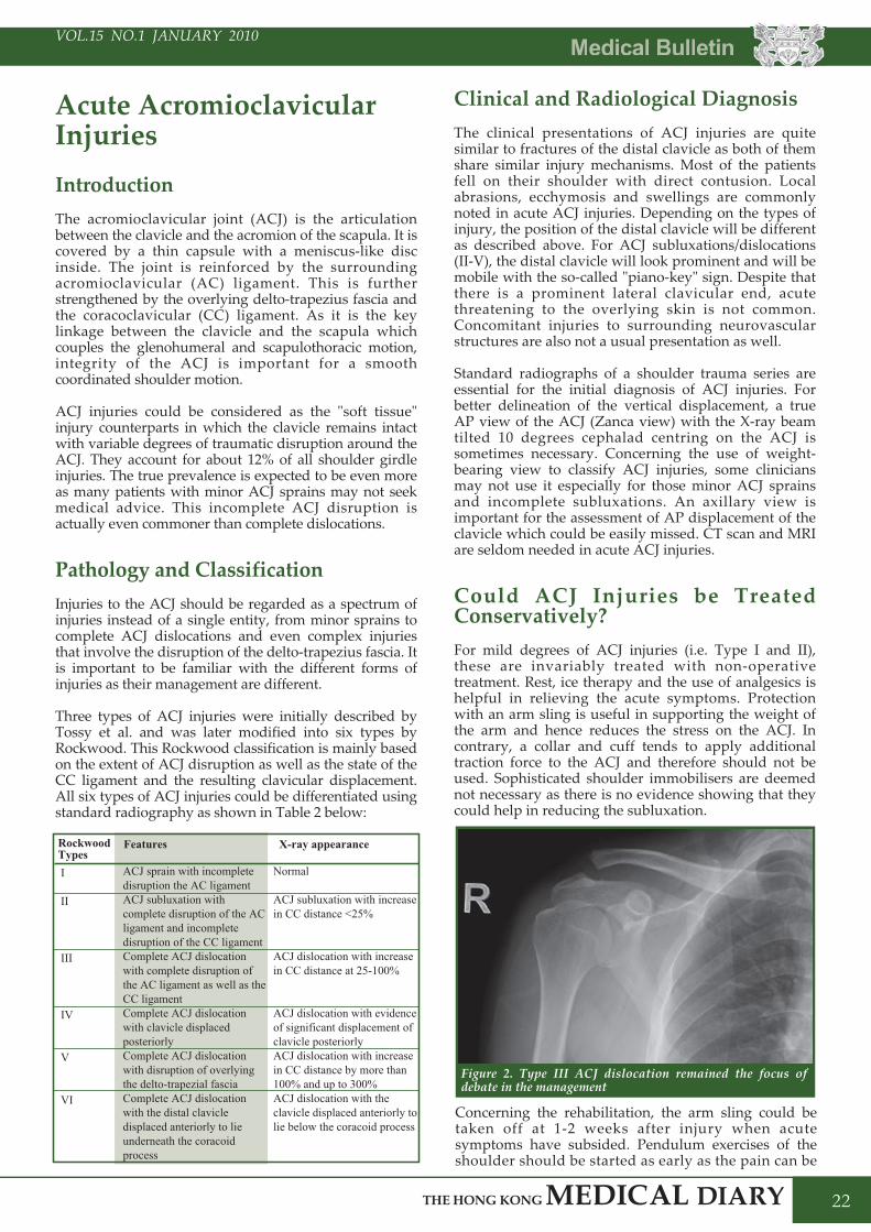
Figure 2. Type III ACJ dislocation remained the focus of debate in the management
Medical Bulletin
23
VOL.15 NO.1 JANUARY 2010
tolerated. Active assisted mobilisation and passive mobilisation of the shoulder is to be commenced afterwards. Patients should avoid contact sports for at least 3 months after injury.
The prognosis of this mild form of ACJ injuries is excellent. Stiffness and weakness of the shoulder is uncommon. Patients sometimes may complain of residual discomfort and pain on activity around the ACJ. This could be due to the resulting ACJ instability and arthritis. Only to those with significant persistent symptoms will further operative treatment be needed.
For Type IV, V and VI ACJ injuries, operative treatment is indicated as the soft tissue damage around the shoulder girdle is more severe. Management of Type III ACJ dislocations still remains controversial as it stays somewhere in between. Again we will need to ask ourselves what will happen if we treat all Type III i n j u r i e s c o n s e r v a t i v e l y . D e f i n i t e l y t h e subluxation/dislocation will persist with prominence over the distal end of the clavicle. Whether it will pose any cosmetic problems is rather subjective, but the functional outcome is usually satisfactory. Literature shows that injured shoulder strength and endurance are comparable to the contralateral side after rehabilitation, and most patients could return to their previous level of activities. This reminds us the wisdom shared by Allman in 1967:
Proponents for conservative treatment opine that operative anatomical reduction is not necessary for achieving adequate shoulder function, yet surgery will pose possible operative complication risks to our patients. However others argue that we should treat those patients with high shoulder demands differently as a delayed reconstruction is associated with less favourable outcomes. Despite the lack of hard evidence, it is often suggested that this group of patients should receive surgery early.
Therefore each patient should be considered individually in Type III ACJ dislocations. When conservative treatment is chosen, patients should be well informed about possible persistent ACJ deformity but that the functional result is usually satisfactory. Still there is the risk of persistent shoulder symptoms. Under such circumstances, a delayed reconstruction with excision of the distal part of the clavicle may be needed.
Is There Any Gold Standard for Operative Treatment?There are hundreds of operative techniques described to treat high grade ACJ dislocations. In other words there is no technique that could be singled out with significant superior operative results. Conventional open reductions in the form of a shoulder strap incision is the most common surgical approach. As the ACJ is inherently unstable, internal fixation to maintain reduction and to stabilise the joint is necessary. With different kinds of internal fixation reported, the fixation techniques could be categorised as below:
All these techniques have their own pros and cons as listed in the previous Table 1. Orthopedic surgeons should be familiar with the techniques used in order to achieve good clinical results and prevent complications.
Do we Need to Repair the Disrupted Ligaments During Acute ACJ Reconstruction?
Open ACJ reconstructions that are done within the first two weeks theoretically offer the opportunity to repair the torn AC and CC ligaments. In practical scenarios, complicated attempts at repairs in a sophisticated manner is often difficult because the ligaments are usually contused, shredded and even sometimes not recognisable. Some have reported the technique of transferring the coracoacromial (CA) ligament for substitution. On the other hand, any damage to the delto-trapezial fascia and muscle should be repaired as this contributes to significant stability.
Rehabilitation after SurgeryAs soft tissue repair takes a longer time to heal, the operated shoulder should be supported with an arm sling for 4-6 weeks. Passive and active assisted mobilisation could be started afterwards. Any rigid fixations such as Bosworth screws and plate should be removed once the biological healing of ACJ has consolidated as implant failures may result. However too early removal of the implant will result in re-displacement of the ACJ. Therefore the average timing of removal is around 12 weeks after surgery.
Is There Any Operative Innovation Recently? With technological innovation plus better implants, new minimally invasive and arthroscopic-assisted operative techniques have been reported. Development of these techniques reduces the surgical trauma that was introduced by conventional open techniques. With theoretical advantages of a smaller scar, better cosmesis and less postoperative pain, it offers an attractive option
"Patients with poor anatomical results may have no symptoms, whereas anatomical restoration of the joint does not always relieve symptoms".
Transacromial fixation across the ACJ using pins, K-wiresCoracoclavicular indirect fixation using Bosworth screws, suture anchors or endobutton system. Other coracoclavicular cerclage techniques with the use of autografts such as hamstring tendons and toe extensors and synthetic materials such as Mersilene tape and Dacron graft have also been reported.Rigid fixation by plating across the ACJ using a hook plate
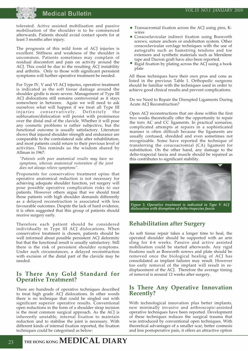
Figure 3. Operative treatment is indicated in Type V ACJ dislocation with disruption of delto-trapezius fascia
VOL.11 NO.5 MAY 2006 Medical Bulletin
24
VOL.15 NO.1 JANUARY 2010
for both surgeons and patients. Although their early reports are quite encouraging, more clinical outcome studies are needed to prove their long term efficacy.
ConclusionDistal clavicle fractures and acromioclavicular joint injuries form a distinct group of shoulder girdle injuries. Natural history shows that conservative treatment may still give satisfactory functional outcomes even in some unstable Neer Type II fractures and Type III ACJ dislocations. This reminds us the importance of "Treat the patient, not the X-rays". The management plan of each patient should be individualised. For operative treatments, there are numerous surgical techniques described while no single one stands up to be the gold standard.
Coracoclavicular screw fixation for unstable fractures of the distal clavicle. A report of five cases.F T Ballmer. J Bone J Surg 1991; 73-B(2) : 291-4The natural course of lateral clavicle fracture. 15 (11-21) year follow-up of 110 cases.A Nordqvist. Acta Orthop Scand 1993; 64(1): 87-91Surgical Treatment of Distal Clavicle Fractures using the Clavicular Hook Plate.M Kashii. CORR 2006; 447: 158-164Type II distal clavicle fractures: a retrospective review of surgical treatment. J Kona. J Orthop Trauma 1990; 4(2):115-120Migration of K-wire from the shoulder region into the lung.R Mazel. J Bone J Surg (Am) 1943; 25(2): 477-83Intraspinal migration of a Kirschner wire 3 months after Clavicular fracture fixation.J P Regel. Neurosurg Rev 2002; 25(1-2):110-2.Treatment of distal clavicle fracture using Kirschner wires and tension-band wires.F C Kao. J Trauma 2001; 51(3): 522-5.Results of Bosworth method for unstable fractures of distal clavicle. H Yamaguchi. Int Orthop 1998; 22(6):366-8.Surgical Treatment of unstable fractures of the distal clavicle: A comparative study of Kirscher wire and Clavicular hook plate fixation.T Flinkkila. Acta Orthop Scand 2002; 73(1): 50-3Fractures of distal third of the clavicle.Neer CS II. CORR 1968; 58: 43-50Simple minimal invasive surgical technique for treatment of type II fractures of distal clavicle.O Levy. J Shoulder Elbow Surg 2003; 12(1): 24-8Intramedullary fixation of Neer Type II fractures of the distal clavicle with an AO/ASIF screw.J E Scadden. Injury 2005; 36: 1172-75Surgical treatment of fractures of the distal clavicle with polydioxanone suture tension band wiring: an alternative osteosynthesis.J W Mall. J Orthop Sci 2002; 7(5): 535-7Surgical treatment of distal clavicle fracture associated with coracoclavicular ligament rupture using a cannulated screw fixation technique.Z J Cheng. J Trauma 2006; 60: 1358-1361Transacromial Knowles Pin in the treatment of Neer Type II Distal Clavicle Fractures: A prospective evaluation of 32 cases.Y F Cheng. J Trauma 2004; 56: 1102-1106Treatment of Distal clavicle Fractures with coracoclavicular ligament disruption. A report of 10 cases.M Bezer. J Orthop Trauma 2005; 19: 524-528Nonoperative treatment of Neer Type II distal clavicle fractures: a prospective study.M K Deafenbaugh. Contemp Orthop 1990; 20(4): 405-13A comparison of nonoperative and operative treatment of type II distal clavicle fractures.A S Rokito. Bulletin Hosp Joint Dis 2002; 61(1-2): 32-9Distal Clavicle Physeal Injury.J A Ogden. Clin Orthop 1984; 188: 68-73Dynamic Fixation of the avulsed Clavicle. A Katznelson. J Trauma 1976; 16(10): 841-4
1.
2.
3.
4.
5.
6.
7.
8.
9.
10.
11.
12.
13.
14.
15.
16.
17.
18.
19.
20.
Reference for Distal Clavicle Fractures
Acromioclavicular and sternoclavicular jointsR Emery. 1997. In Shoulder surgery, Copeland S (Ed.). London: WB Saunders. Disorders of the acromioclavicular jointC A Rockwood. 1990. In the Shoulder, Rockwood CA Jr (Ed.). Philadelphia: WB SaundersAcromioclavicular separations: useful and practical classification for treatmentJ D Tossy. Clin Orthop. 1963. Vol. 28; 111-19Rockwood CA. 1991. Rockwood and Green's fractures in adults (Third ed.), Philadelphia: Lippincott. Shoulder pain: involvement of the acromioclavicular joint Analysis of 1,000 casesP Zanca. Am J Roentgenol Radium Ther Nucl Med. 1971. Vol. 112; 493-506Lack of efficacy of 'weighted' radiographs in diagnosing acute acromioclavicular separationP J Bossart. Ann Emerg Med. 1988. Vol. 17; 20-4Evaluation of the acromioclavicular joints following first- and second-degree sprainsJ A Bargfeld. Am J Sports Med. 1978. Vol. 6; 153-9Acromio-clavicular separations treated conservatively: a 5-vear follow-up studyH Bjemeld. Acta Orthop Scand. 1983. Vol. 54; 743-5A prospective evaluation of untreated acute grade III acromioclavicular separationsT F Schlegal. Am J Sports Med. 2001. Vol. 29; 699-703Treatment of grade III acromioclavicular joint injuries: a systematic reviewE E Spencer. Clin Orthop. 2007. Vol. 455; 38-44Long-term results of conservative treatment for acromioclavicular dislocationM L Rawes. J Bone Joint Surg Br. 1996. Vol. 78; 410-412The management of acute acromioclavicular dislocation: a randomized prospective controlled trialG C Bannister. J Bone Joint Surg [Br]. 1989. Vol. 71; 848-50The conservative treatment of acromioclavicular dislocation: review after five yearsJ Dias. J Bone Joint Surg [Br]. 1987. Vol. 69; 719-22Arthroscopic reconstruction of the acromioclavicular joint disruption: surgical technique and preliminary results.B Chernchujit. Arch Orthop Trauma Surg. 126(9):575-81, 2006 Nov.Minimally invasive coracoclavicular ligament augmentation with a flip button/polydioxanone repair for treatment of total acromioclavicular joint dislocationM Wellmann. Arthroscopy. 23(10):1132.e1-5, 2007 Oct.Arthroscopic treatment of acute and chronic acromioclavicular joint dislocation.L Lafosse. Arthroscopy. 21(8):1017, 2005 Aug.The treatment of acromioclavicular joint dislocation Tossy grade III with a clavicle hook plate.T De Baets. Acta Orthopaedica Belgica. 70(6):515-9, 2004 Dec.Arthroscopic treatment of acute acromioclavicular joint dislocation.P R Rolla. Arthroscopy. 20(6):662-8, 2004 Jul. Fractures and ligamentous injuries of the clavicle and its articulation. F L Allman, Jr., J. Bone Joint Surg. (Am.) 49A (1967), pp. 774-784.Acromioclavicular dislocations: treatment by coracoacromial ligamentoplasty.C Dumontier. Journal of Shoulder & Elbow Surgery. 4(2):130-4, 1995 Mar-Apr.The surgical treatment of complete acromioclavicular joint dislocation.W P Ho. Orthopaedic Review. 17(11):1116-20, 1988 Nov.Acute acromioclavicular joint dislocation: results of operative treatment with the Bosworth screw.G P Lowe. Australian & New Zealand Journal of Surgery. 47(5):664-7, 1977 Oct. Acromioclavicular separation: new method of repairB M Bosworth. Surg Gynecol Obstet. 1941. Vol. 73; 866-71Percutaneous cannulated screw coracoclavicular fixation for acute acromioclavicular dislocationsP M Tsou. Clin Orthop. 1989. Vol. 243; 112-21Stabilization of acute acromioclavicular dislocation by a modified Bosworth technique: a long-term follow-up studyN Sundaram. Injury. 1992. Vol. 23; 189-93Complete acromioclavicular dislocations: treatment with a Dacron ligamentL Stem. Injury. 1991. Vol. 22; 173-6Treatment of acromioclavicular joint separation: suture or suture anchors?M J Brailow. J Shoulder Elbow Surg. 2002. Vol. 11; 225-9Arthroscopic reconstruction for acromioclavicular joint dislocationE M Wolf. Arthroscopy. 2001. Vol. 17; 558-63Clinical outcomes of coracoclavicular ligament reconstructions using tendon graftsS J Nicholas. Am J Sports Med. 2007. Vol. 35; 1912-17Acromioclavicular separation: reconstruction using synthetic loop augmentationD S Morrison. Am J Sports Med. 1995. Vol. 23; 105-10A crook plate for treatment of acromioclavicular joint separation: indication, technique, and results after one yearH Habernek. J Trauma. 1993. Vol. 35; 893-901Repair of complete acromioclavicular separations using the acromioclavicular-hook plateE Sim. Clin Orthop. 1995. Vol. 314; 134-42Clavicular Hook Plate: Complications of retaining the implant.R Nadarajah. Injury 2005; 36: 681-3
1.
2.
3.
4.
5.
6.
7.
8.
9.
10.
11.
12.
13.
14.
15.
16.
17.
18.
19.
20.
21.
22.
23.
24.
25.
26.
27.
28.
29.
30.
31.
32.
33.
Reference for Acromioclavicular Injuries