disparate effects of nitric oxide on lung ischemia-reperfusion injury
TRANSCRIPT

T H O R A C I C SURGERY DIRECTORS A S S O C I A T I O N A W A R D
The Thoracic Surgery Directors Association (TSDA) Resident Research Award, sponsored by Medtronic, Inc, was established in 1990 to encourage resident research in cardiothoracic surgery. Abstracts submitted to The Society of Thoracic Surgeons (STS) Program Committee representing research performed by residents were forwarded to the TSDA to be considered for this award. The abstracts were reviewed by the TSDA Executive Committee consisting of: Gordon F. Murray, President; John R. Benfield, President-Elect; Mark B. Orringer, Secretary/Treasurer; Gordon N. Olinger, Executive Committeeman; and Sidney Levitsky, Executive Committeeman.
The f~fth TSDA Resident Research Award was given to Dr Michael J. Eppinger, a general surgery resident in the Department of Surgery, Wright State University, Dayton, Ohio, who was a University of Michigan thoracic surgery research fellow. He received a monetary award of $2,500 and an engraved desktop award, and had his expenses paid to the STS meeting.
The TSDA, with support by Medtronic, Inc, makes this award annually, using the above selection procedure. The resident author of the selected study is recognized at the STS meeting.
Disparate Effects of Nitric Oxide on Lung Ischemia-Reperfusion Injury Michael J. Eppinger, MD, Peter A. Ward, MD, Michael L. Jones, Steven F. Bolling, MD, and G. Michael Deeb, MD Section of Thoracic Surgery and Department of Pathology, University of Michigan Medical Center, Ann Arbor, Michigan
Background. Inhaled nitric oxide (,NO) has been found to be a potent pulmonary vasodilator. We assessed whether ,NO, through this function or others, could alleviate lung reperfusion injury.
Methods. Rats underwent thoracotomy, with clamps used to create left lung ischemia. After 90 minutes of ischemia, clamps were released, permitting reperfusion for either 30 minutes or 4 hours. Additional animals received inhaled ,NO via the ventilator to determine its effects on reperfusion injury.
Results. Lung injury, measured by increased vascular permeability using iodine-125-1abeled bovine serum al- bumin leakage, was significantly increased in ischemic- reperfused animals compared with time-matched shams not undergoing ischemia. Inhaled ,NO delivered at the
start of reperfusion worsened injury at 30 minutes but was protective at 4 hours. The increased injury could be avoided either by delaying ,NO for 10 minutes or by treating the animals with superoxide dismutase before reperfusion. ,NO reversed postischemic pulmonary hy- poperfusion at 4 hours, as measured by labeled micro- spheres. Lung neutrophil content was significantly re- duced at 4 hours in ,NO-treated animals.
Conclusions. ,NO is toxic early in reperfusion, due to its interaction with superoxide, but is protective at 4 hours of reperfusion, due to reversal of postischemic lung hypoperfusion and reduction of lung neutrophil sequestration.
(Ann Thorac Surg 1995;60:1169-76)
T he use of lung transplantation as therapy for end- stage lung disease has made a strong resurgence
since the late 1980s. However, difficulties in maintaining optimal graft function continue to compromise patient outcome. Lung dysfunction during the intraoperative or early postoperative period caused by technical difficulties or lung reperfusion injury threatens graft viability as well as patient survival [1]. Furthermore, there is evidence that significant reperfusion injury predicts a worse out- come for long-term graft survival [2]. An important factor
Presented at the Thirty-first Annual Meeting of The Society of Thoracic Surgeons, Palm Springs, CA, Jan 30-Feb 1, 1995.
Address reprint requests to Dr Deeb, University of Michigan Hospitals, 1500 E. Medical Center Dr, 2124F Taubman Center TC/0344, Ann Arbor, MI 48109.
in reperfusion injury may be dysfunction of the pulmo- nary vascular endothelium, as manifested by pulmonary hypertension and increased vascular permeability, re- sulting in pulmonary edema and impaired gas exchange. The recent identification of nitric oxide (.NO) as endo- thelium-derived relaxing factor [3, 4] has resulted in much research regarding the contribution of endogenous • NO to processes involved in the maintenance of vascu- lar endothelial integrity.
Driven by the finding that inhaled .NO is a potent pulmonary vasodilator, its application in clinical settings has been rapidly employed. A major attraction is its pulmonary selectivity due to inactivation by hemoglobin in the circulation [5, 6]. Inhaled -NO has been used in treating pulmonary hypertension after surgical correc-
© 1995 by The Society of Thoracic Surgeons 0003-4975/95/$9.50 SSDI 0003-4975(95)00697-4

1170 TSDA AWARD EPPINGER ET AL Ann Thorac Surg NITRIC OXIDE IN LUNG REPERFUSION INJURY 1995;60:1169-76
Table 1. Groups
Vascular Permeability Determination With 125I-BSA
Reperfusion Positive .NO Early Time Control Sham .NO Early .NO Delayed + SOD
Relative Pulmonary Flow
Myeloperoxidase Assay
Positive Positive Control -NO Early Control *NO Early
None . . . . . . . . . . . . . . . J(n = 4) . . . 30 Minutes A (n = 6) C (n = 4) E (n = 4) G (n - 4) I (n = 4) K (n = 4) M (n = 4) 4 Hours B (n = 6) D (n - 4) F(n - 2) H (n = 5) . . . L(n - 4) N(n - 4) O(n - 4) P(n = 4)
BSA bovine se rum albumin; , N O - nitric oxide; SOD - superoxide dismutase.
tion of congenital cardiac defects [7], for p r imary pu lmo- nary hyper tens ion in the newborn [8, 9], and for adults undergoing cardiac operat ions [10]. It has also been of value in the reversal of pu lmonary hyper tens ion second- ary to hypercapnia [5, 11, 12]. There has been at least one repor t deal ing with the use of inhaled *NO in the t rea tment of postopera t ive graft dysfunction after lung t ransplanta t ion [13].
In the mids t of clinical successes using inhaled *NO, there have been isolated reports of deleter ious effects associated with its use [14, 15]. These mixed results point out the need for careful considerat ion of the spec t rum of activity of *NO. The vascular relaxing proper t ies of *NO are due pr imar i ly to the activities of constitutive *NO synthase, an endothel ia l -der ived, ca lc ium-dependen t en- zyme, which is a low level p roducer of *NO [3]. Nitric oxide is also p roduced as an inf lammatory media tor by the inducible form of *NO synthase, a macrophage- derived, calcium independen t enzyme, which is a high output p roducer of , N O [16]. In contrast to the beneficial effects of inhaled *NO in settings of abnormal ly high pu lmonary vascular resistance, some models of lung injury have po in ted out the toxic effects of endogenous ly p roduced . N O in inf lammation [17].
We have previously descr ibed an in vivo rat lung model of i schemia-reperfus ion demonst ra t ing a b imoda l pat tern of injury with peaks at 30 minutes and 4 hours of reperfusion [18]. Using this model , we sought to deter- mine the effects of inhaled . N O on lung ischemia- reperfus ion injury. We a t t empted to identify causes for both beneficial and toxic effects of inhaled . N O to pro- vide some insight into strategies for the rat ional use of inhaled -NO in reperfus ion lung injury.
Material and M e t h o d s
Ischemia-Reperfusion Injury Model Pathogen-free adul t male Long-Evans rats (300 to 350 g; Charles River Laboratories or Har lan Industries) were used for all experiments . Exper imental protocols were approved by The Universi ty of Michigan Commit tee on the Use and Care of Animals. Animals were initially anes thet ized with 32 mg int raper i toneal pentobarbi ta l . The animals were then shaved, in tuba ted orotracheal ly with a 14-gauge intravenous catheter, and placed on a Harvard rodent venti lator with supp lementa l oxygen (60%) at a rate of 75 cycles per minute, max imum peak
pressure of 10 cm H20, and max imum posit ive end- expiratory pressure of 2 cm H20. These factors ( inspired oxygen fraction, vent i la tory rate, and vent i la tory pres- sure) were he ld constant through all exper iments to el iminate them as variables in lung injury. All animals received 0.4 mg in t ramuscular a t ropine after being anes- thetized. Animals received halothane via the venti lator to mainta in anesthesia as it became necessary.
The animals were p laced on their r ight sides, and a left anterolateral thoracotomy via the fifth interspace was carr ied out. The left pu lmonary hi lum was s t r ipped of all neura l , vascular , lymphat ic , and connect ive t issue, thereby skeletonizing the left bronchus, pu lmonary ar- tery, and pu lmonary vein. The inferior pu lmonary liga- ment was d iv ided as it en tered the hilum. All dissection was carried out under an opera t ing microscope. Each animal received 50 units of hepar in in saline solution in t ravenously (total volume 500/xL) via the penile vein.
After we wai ted 5 minutes for circulation of the hepa- rin, the left pu lmonary artery, bronchus, and pu lmonary vein were sequent ia l ly occluded with noncrushing mi- crovascular clamps. The lungs were kept moist with intermit tent appl icat ion of warm normal saline solution, and the wounds were kept covered with plastic film to prevent excessive fluid loss. Periods of ischemia were held constant at 90 minutes (see Table I for a descr ipt ion of all groups used in these experiments) .
At the end of the ischemic period, the c lamps were removed from the vein, bronchus, and artery, respec- tively, and the lungs were al lowed to venti late and reperfuse for ei ther 30 minutes (group A) or 4 hours (group B). These t ime points were chosen based on injury peaks seen in our previous studies [18]. Animals received 0.5 mL of normal saline solution via subcutaneous injec- tion for each hour of reperfus ion time. Rats were sacri- ficed at the end of the reperfus ion t ime by c lamping of the r ight and left super ior venae cavae and the inferior vena cava. The left atrial appendage was amputa t ed and the lungs were f lushed with 30 mL of saline solution by gravity infusion at 30 cm H20 via the right ventricle. Blood samples were obta ined from the inferior vena cava just before sacrifice. The left lung was then removed for analysis as out l ined below. T ime-matched control ani- mals (shams) unde rwen t the identical p rocedure except that the microvascular c lamps were not appl ied to the hilar structures (group C, 30 minutes; and group D, 4 hours). Due to the nature of the model, sequent ia l data

Ann Thorac Surg TSDA AWARD EPPINGER ET AL 1171 1995;60:1169-76 NITRIC OXIDE IN LUNG REPERFUSION INJURY
were not available on individual animals, and thus each animal was operated on as a separate experiment.
Lung Permeability Assessment To quantitate injury to the lung as a result of ischemia and reperfusion, lung permeability was measured in the following manner. Iodine-125-1abeled bovine serum al- bumin, prepared by a standard chloramine-T method, was diluted to approximately 3 to 5 ~Ci/mL in 1% bovine serum albumin/phosphate-buffered saline solution. Eight hundred thousand counts per minute of 125I-bovine serum albumin was then bought to a final volume of 500 ~L in phosphate-buffered saline solution for injec- tion. At the time of unclamping of the hilar structures, or at the comparable time in the sham animals, the mix was then injected into the animal via the penile vein. Imme- diately before sacrifice, 1 mL of blood was drawn from the inferior vena cava for counting. After the animal was sacrificed and the pulmonary vasculature was flushed, the left lung was excised for counting. Each lung speci- men was weighed, and the lungs and blood were counted separately in a gamma counter. All results are expressed as counts per minute ratio of 1 g of lung tissue per milliliter of blood (permeability index). This ratio pro- vided a reliable measure of microvascular permeability as a marker for lung injury (see Table I for a summary of groups used for permeability comparisons).
Nitric Oxide Intervention Nitric oxide, 240 ppm in N 2 (Airco Gas and Gear, Ann Arbor, MI), was provided via the ventilator with supple- mental oxygen (60%) at a concentration of 80 ppm either immediately with the start of reperfusion (Groups E and F) or beginning 10 minutes after the start of reperfusion (Groups G and H). The nitric oxide was then continued for the duration of the reperfusion time course, either 30 minutes (Groups E and G) or 4 hours (Groups F and H). Injury was assessed as for standard model, described above.
Superoxide Dismutase Treatment A subset of animals undergoing 90 minutes of ischernia and 30 minutes of reperfusion with early delivery of eNO (group I, n = 4) also received 10,000 units of superoxide dismutase (from bovine erythrocytes) in 50 ~L of saline solution via penile vein injection immediately before the start of reperfusion. These animals were evaluated for lung injury using 125I-bovine serum albumin as de- scribed above.
Microsphere Analysis of Relative Pulmonary Flow Chromium-51Mabeled microspheres (NEN-DuPont, Bos- ton, MA) were used to determine the effect of left lung ischemia-reperfusion on relative pulmonary flow. Ani- mals not undergoing the ischemia-reperfusion protocol were evaluated for relative pulmonary flow comparing the left lung with the middle and lower lobes of the right lung (chosen because of an approximately equal lung mass). Normal rats (group J, n = 4) were anesthetized, intubated, and placed on the ventilator in a supine
position. They were then injected via the inferior vena cava with approximately 1 ~Ci of ~lCr-labeled micro- spheres (mean diameter, 15 ~m) in 500 rnL of normal saline solution with 0.02% Tween-20 (Bio-Rad, Hercules, CA) as a detergent. After 5 minutes, the rats were sacrificed as above (lungs flushed via right ventricle with 10 mL of saline solution). The left lungs and middle and lower lobes of the right lung were harvested separately and counted in a gamma counter. The ratio of micro- sphere sequestration was then recorded as representing the ratio of pulmonary blood flow to these lung segments (flow ratio = 5lCr counts/rain of left lung/counts/min of middle and lower lobes right lung). To assess whether the effect of inhaled .NO on lung reperfusion injury could in part be explained by a reversal of postischemic hypoperfusion, animals undergoing the ischernia-reper- fusion protocol were also subjected to pulmonary flow measurements using microspheres. Four groups of ani- mals were used: 30 minutes (group K) or 4 hours (group L) of reperfusion with supplemental oxygen alone, and 30 minutes (group M) or 4 hours (group N) of reperfusion with inhaled -NO (early) added via the ventilator (n = 4 for each group). Five minutes before the end of the reperfusion period and with the animals completely supine, radiolabeled microspheres were injected via the inferior vena cava. At sacrifice the lungs were flushed as described above and the left lung and middle and lower lobes of the right lung were counted separately in a gamma counter.
Myeloperoxidase Assay Myeloperoxidase assay was used to compare the relative neutrophil sequestration in lung tissue of experimental animals. Myeloperoxidase, an enzyme found primarily within neutrophils, has been shown to be a sensitive marker for quantifying neutrophil content in tissue. The procedure was adapted from a published method for quantitating neutrophils using myeloperoxidase as a marker [19]. We have previously found neutrophils to b e important for the development of reperfusion injury at the 4-hour time point, with no significant neutrophil sequestration or contribution to injury at the 30-rninute time point [18[. Therefore, animals undergoing the isch- emia-reperfusion protocol with 90 minutes of ischemia and 4 hours of reperfusion either with supplemental oxygen alone (group O) or with delayed .NO (group P, n = 4 each) were sacrificed as described above at the end of the reperfusion period. Left (ischemic-reperfused) lungs were then removed, frozen in liquid nitrogen, and stored at -80°C until the assay was performed. Lung samples were homogenized in 3 mL of 0.5% hexadecyl- trimethylammoniurn bromide, 5 mmol/L ethylenedi- aminetetraacetic acid in 50 mmol/L potassium phosphate buffer, pH 6.0. Samples were sonicated to disrupt the granules and solubilize the myeloperoxidase in the hexa- decyltrirnethylammonium bromide. Samples were then centrifuged at 2,300 g for 30 minutes at 4°C. Assay buffer comprised 0.0005% H202, 0.167 mg/mL o-dianisidine dihydrochloride in 100 mrnol/L potassium phosphate buffer, pH 6.0. Fifty rnicroliters of each sample was mixed
1172 TSDA AWARD EPPINGER ET AL Ann Thorac Surg NITRIC OXIDE IN LUNG REPERFUSION INJURY 1995;60:1169-76
into 1.45 mL of assay buffer at room temperature, and the change in absorbance at 460 nm over 1 minute was recorded. Results are expressed as relative change in absorbance per minute at 460 nm.
Statistical Ana ly s i s
All data are presented as mean _+ standard error of the mean. All comparisons except for myeloperoxidase data were made using a one-way analysis of variance, with Tukey's procedure used to determine significant differ- ences between groups at individual time points. Com- parisons between groups on myeloperoxidase data were made using a two-tailed, unpaired Student's t test. Sta- tistical significance for all tests was set at p less than 0.05.
Results
Lung I schemia-Reper fus ion Injury: Controls
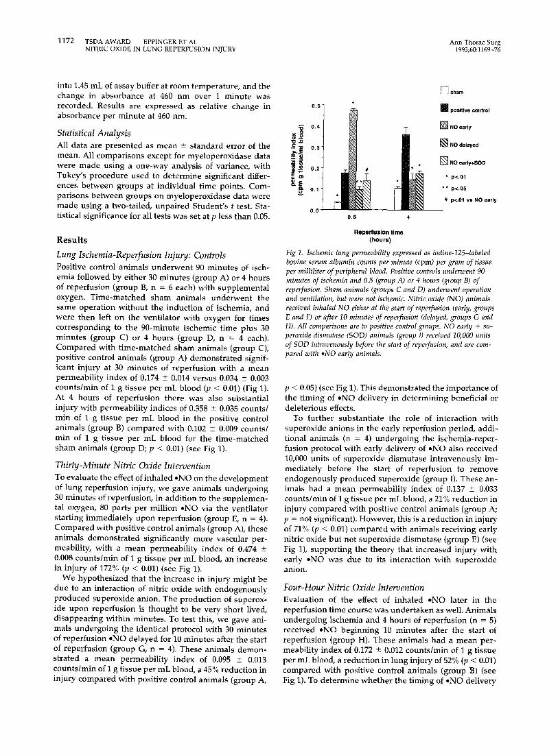
Positive control animals underwent 90 minutes of isch- emia followed by either 30 minutes (group A) or 4 hours of reperfusion (group B, n = 6 each) with supplemental oxygen. Time-matched sham animals underwent the same operation without the induction of ischemia, and were then left on the ventilator with oxygen for times corresponding to the 90-minute ischemic time plus 30 minutes (group C) or 4 hours (group D, n = 4 each). Compared with time-matched sham animals (group C), positive control animals (group A) demonstrated signif- icant injury at 30 minutes of reperfusion with a mean permeability index of 0.174 _+ 0.014 versus 0.034 _+ 0.003 counts/min of I g tissue per mL blood (p < 0.01) (Fig 1). At 4 hours of reperfusion there was also substantial injury with permeability indices of 0.358 _+ 0.035 counts/ min of 1 g tissue per mL blood in the positive control animals (group B) compared with 0.102 +_ 0.009 counts/ min of 1 g tissue per mL blood for the time-matched sham animals (group D; p < 0.01) (see Fig 1).
T h i r t y - M i n u t e Nitr ic Oxide Intervent ion
To evaluate the effect of inhaled eNO on the development of lung reperfusion injury, we gave animals undergoing 30 minutes of reperfusion, in addition to the supplemen- tal oxygen, 80 parts per million oNe via the ventilator starting immediately upon reperfusion (group E, n = 4). Compared with positive control animals (group A), these animals demonstrated significantly more vascular per- meability, with a mean permeability index of 0.474 -+ 0.008 counts/min of I g tissue per mL blood, an increase in injury of 172% (p < 0.01) (see Fig 1).
We hypothesized that the increase in injury might be due to an interaction of nitric oxide with endogenously produced superoxide anion. The production of superox- ide upon reperfusion is thought to be very short lived, disappearing within minutes. To test this, we gave ani- mals undergoing the identical protocol with 30 minutes of reperfusion eNO delayed for 10 minutes after the start of reperfusion (group G, n = 4). These animals demon- strated a mean permeability index of 0.095 _+ 0.013 counts/min of I g tissue per mL blood, a 45% reduction in injury compared with positive control animals (group A,
0
' ; , E
J~ w O . -
D,.
0 .57
0.5 4
~--] sham
m positive control
[ ] N O early
[ ] N O delayed
~ ] N O early+SOD
* p<.01
* * p<.05
# p<,01 vs NO early
Reperfusion time (hours)
Fig 1. Ischemic lung permeability expressed as iodine-125-labeled bovine serum albumin counts per minute (cprn) per gram of tissue per milliliter of peripheral blood. Positive controls underwent 90 minutes of ischemia and 0.5 (group A) or 4 hours (group B) of reperfusion. Sham animals (groups C and D) underwent operation and ventilation, but were not ischemic. Nitric oxide (NO) animals received inhaled NO either at the start of reperfusion (early, groups E and F) or after 10 minutes of reperfusion (delayed, groups G and H). All comparisons are to positive control groups. NO early + su- peroxide dismutase (SOD) animals (group I) received I0,000 units of SOD intravenously before the start of reperfusion, and are com- pared with aNO early animals.
p < 0.05) (see Fig 1). This demonstrated the importance of the timing of eNO delivery in determining beneficial or deleterious effects.
To further substantiate the role of interaction with superoxide anions in the early reperfusion period, addi- tional animals (n = 4) undergoing the ischemia-reper- fusion protocol with early delivery of -NO also received 10,000 units of superoxide dismutase intravenously im- mediately before the start of reperfusion to remove endogenously produced superoxide (group I). These an- imals had a mean permeability index of 0.137 + 0.033 counts/min of I g tissue per mL blood, a 21% reduction in injury compared with positive control animals (group A; p = not significant). However, this is a reduction in injury of 71% (p < 0.01) compared with animals receiving early nitric oxide but not superoxide dismutase (group E) (see Fig 1), supporting the theory that increased injury with early eNO was due to its interaction with superoxide anion.
Four-Hour Nitr ic Oxide Intervent ion
Evaluation of the effect of inhaled oNe later in the reperfusion time course was undertaken as well. Animals undergoing ischemia and 4 hours of reperfusion (n = 5) received oNe beginning 10 minutes after the start of reperfusion (group H). These animals had a mean per- meability index of 0.172 - 0.012 counts/min of I g tissue per mL blood, a reduction in lung injury of 52% (p < 0.01) compared with positive control animals (group B) (see Fig 1). To determine whether the timing of .NO delivery

Ann Thorac Surg TSDA AWARD EPPINGER ET AL 1173 1995;60:1169-76 NITRIC OXIDE IN LUNG REPERFUSION INJURY
would critically affect the outcome at 4 hours of reperfu- sion (as had been seen at 30 minutes), we performed the identical protocol in a limited number of animals (n = 2), but with early delivery of ,NO (group F). These animals had a mean permeability index of 0.143 +- 0.028 counts/ min of I g tissue per mL blood, not significantly different from that of animals receiving delayed .NO (group H) (see Fig 1). Because there was no observable difference using this permutation, no additional animals were used. In contrast to reperfusion injury at 30 minutes, the timing of .NO delivery had no bearing on the outcome at 4 hours, suggesting that other factors are involved in lung injury over longer reperfusion times.
Microsphere Studies
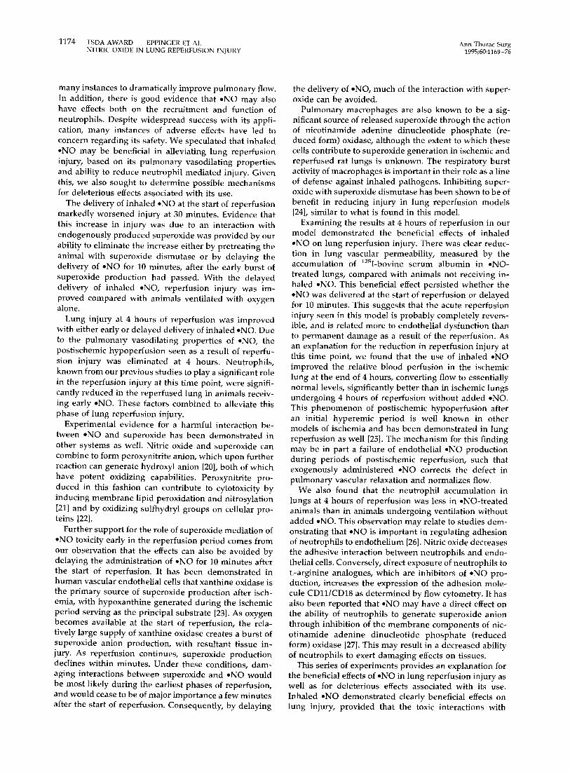
The ratio of pulmonary blood flow (measured by 51Cr counts per minute) of the left lung versus middle and lower lobes of the right lung in normal animals (group J) was 0.84 + 0.05 (Fig 2). In animals undergoing 90 minutes of ischemia and 30 minutes of reperfusion with supple- mental oxygen but not .NO (group K, n = 4), the mean ratio of left (ischemic lung) to right pulmonary flow was 0.61 +_ 0.02, a reduction in relative flow to the ischemic lung to 73% of normal lung values (p < 0.01) (see Fig 2). After 4 hours of reperfusion (group L, n = 4), the mean flow ratio had increased to 0.70 +_ 0.03, 83% of normal values (p = not significant) (see Fig 2). Animals under- going ischernia-reperfusion with early delivery of *NO were evaluated for relative pulmonary flow. At 30 min- utes of reperfusion (group M, n = 4) relative flow to the ischemic lung was reduced to 0.53 _+ 0.05, significantly lower than that in normal animals (group K; p < 0.01) but not significantly different from that in animals undergo- ing reperfusion in the absence of inhaled *NO (group L) (see Fig 2). After 4 hours of reperfusion, animals receiving inhaled .NO (group N, n = 4) had a mean relative flow to
1.0 ¸
0 . 8 ' o
~- 0.6-
~ 0,4"
E
o. 0.2
0.0
T
w
ij 0.5 4
] normal
[ ] 0 2 positive control
~'~ Oz+NO
* p<.01 vs normal
** p<.05 vs 02 control
Reperfusion time (hours)
Fig 2. Measurement of relative pulmonary flow between left lung and middle and lower lobes of the right lung (flow ratio = chro- mium 5I counts per minute for left lung~counts per minute for mid- dle and lower lobes of right lung). Normal animals (group J) were not operated on before measurements. Oxygen (0 2 ) (positive control, groups K and L) and 02 + nitric oxide (NO) (groups M and IV) are animals receiving oxygen without and with the addition of 80 parts per million NO (early) during the reperfusion period.
0.6
0.5
0.4
0.3
0.2
0.1
0.0
I -7-
0 2 only
p<.02
-42%
[
0 2 +NO
n=4 each
Fig 3. Myeloperoxidase (MPO) activi~ in ischemic-repeuCused lung homogenates after 4 hours of reperfusion in animals receiving sup- plemental oxygen (02) without (group O) and with the addition of 80 parts per million nitric oxide (NO) (early, group P) during the reperfusion period.
the ischemic lung of 0.88 -+ 0.04, not significantly different from that of normal animals (group J) but significantly better (p < 0.05) than that of positive control animals (group M) (see Fig 2). This demonstrated the vasodilating effects of inhaled *NO on postischemic hypoperfusion at 4 hours but not at 30 minutes of reperfusion.
Lung Neu t roph i l Conten t
Animals undergoing 4 hours of reperfusion either with or without the delivery of inhaled .NO (early) were evalu- ated for neutrophil content at the end of the reperfusion period, reflected by lung content of myeloperoxidase. Animals undergoing reperfusion with the addition of inhaled .NO (group P) demonstrated 42% less neutrophil sequestration (p < 0.02) in ischemic-reperfused lung tissue at 4 hours of reperfusion than lungs of positive control animals undergoing reperfusion with supple- mental oxygen alone (group O) (Fig 3).
C o m m e n t
We investigated the effects of inhaled eNO in our in vivo rat lung model of ischemia-reperfusion injury. In this model we have previously found that injury during the first 4 hours of reperfusion is bimodal in pattern, with significant injury peaks occurring at 30 minutes and 4 hours. Of note is the finding that there is partial recovery after the 30-minute reperfusion time point, suggesting that the two peaks of injury are separate phenomena, probably mediated by different factors. In addition, we have demonstrated that the 4-hour injury is dependent on neutrophil participation, whereas the 30-minute in- jury is not [18].
Inhaled ,NO, a potent and selective pulmonary vaso- dilator, has recently been introduced as an intervention in conditions associated with acute increases in pulmo- nary vascular resistance. As such, it has been shown in
1174 TSDA AWARD EPPINGER ET AL Ann Thorac Surg NITRIC OXIDE IN LUNG REPERFUSION INJURY 1995;60:1169-76
many instances to dramatically improve pulmonary flow. In addition, there is good evidence that .NO may also have effects both on the recruitment and function of neutrophils. Despite widespread success with its appli- cation, many instances of adverse effects have led to concern regarding its safety. We speculated that inhaled • NO may be beneficial in alleviating lung reperfusion injury, based on its pulmonary vasodilating properties and ability to reduce neutrophil mediated injury. Given this, we also sought to determine possible mechanisms for deleterious effects associated with its use.
The delivery of inhaled ,NO at the start of reperfusion markedly worsened injury at 30 minutes. Evidence that this increase in injury was due to an interaction with endogenously produced superoxide was provided by our ability to eliminate the increase either by pretreating the animal with superoxide dismutase or by delaying the delivery of .NO for 10 minutes, after the early burst of superoxide production had passed. With the delayed delivery of inhaled ,NO, reperfusion injury was im- proved compared with animals ventilated with oxygen alone.
Lung injury at 4 hours of reperfusion was improved with either early or delayed delivery of inhaled .NO. Due to the pulmonary vasodilating properties of .NO, the postischemic hypoperfusion seen as a result of reperfu- sion injury was eliminated at 4 hours. Neutrophils, known from our previous studies to play a significant role in the reperfusion injury at this time point, were signifi- cantly reduced in the reperfused lung in animals receiv- ing early .NO. These factors combined to alleviate this phase of lung reperfusion injury.
Experimental evidence for a harmful interaction be- tween • NO and superoxide has been demonstrated in other systems as well. Nitric oxide and superoxide can combine to form peroxynitrite anion, which upon further reaction can generate hydroxyl anion [20], both of which have potent oxidizing capabilities. Peroxynitrite pro- duced in this fashion can contribute to cytotoxicity by inducing membrane lipid peroxidation and nitrosylation [21] and by oxidizing sulfhydryl groups on cellular pro- teins [22].
Further support for the role of superoxide mediation of • NO toxicity early in the reperfusion period comes from our observation that the effects can also be avoided by delaying the administration of -NO for 10 minutes after the start of reperfusion. It has been demonstrated in human vascular endothelial cells that xanthine oxidase is the primary source of superoxide production after isch- emia, with hypoxanthine generated during the ischemic period serving as the principal substrate [23]. As oxygen becomes available at the start of reperfusion, the rela- tively large supply of xanthine oxidase creates a burst of superoxide anion production, with resultant tissue in- jury. As reperfusion continues, superoxide production declines within minutes. Under these conditions, dam- aging interactions between superoxide and .NO would be most likely during the earliest phases of reperfusion, and would cease to be of major importance a few minutes after the start of reperfusion. Consequently, by delaying
the delivery of .NO, much of the interaction with super- oxide can be avoided.
Pulmonary macrophages are also known to be a sig- nificant source of released superoxide through the action of nicotinamide adenine dinucleotide phosphate (re- duced form) oxidase, although the extent to which these cells contribute to superoxide generation in ischemic and reperfused rat lungs is unknown. The respiratory burst activity of macrophages is important in their role as a line of defense against inhaled pathogens. Inhibiting super- oxide with superoxide dismutase has been shown to be of benefit in reducing injury in lung reperfusion models [24], similar to what is found in this model.
Examining the results at 4 hours of reperfusion in our model demonstrated the beneficial effects of inhaled • NO on lung reperfusion injury. There was clear reduc- tion in lung vascular permeability, measured by the accumulation of 125I-bovine serum albumin in .NO- treated lungs, compared with animals not receiving in- haled *NO. This beneficial effect persisted whether the • NO was delivered at the start of reperfusion or delayed for 10 minutes. This suggests that the acute reperfusion iniury seen in this model is probably completely revers- ible, and is related more to endothelial dysfunction than to permanent damage as a result of the reperfusion. As an explanation for the reduction in reperfusion injury at this time point, we found that the use of inhaled .NO improved the relative blood perfusion in the ischemic lung at the end of 4 hours, converting flow to essentially normal levels, significantly better than in ischemic lungs undergoing 4 hours of reperfusion without added .NO. This phenomenon of postischemic hypoperfusion after an initial hyperemic period is well known in other models of ischemia and has been demonstrated in lung reperfusion as well [25]. The mechanism for this finding may be in part a failure of endothelial .NO production during periods of postischemic reperfusion, such that exogenously administered ,NO corrects the defect in pulmonary vascular relaxation and normalizes flow.
We also found that the neutrophil accumulation in lungs at 4 hours of reperfusion was less in .NO-treated animals than in animals undergoing ventilation without added .NO. This observation may relate to studies dem- onstrating that .NO is important in regulating adhesion of neutrophils to endothelium [26]. Nitric oxide decreases the adhesive interaction between neutrophils and endo- thelial cells. Conversely, direct exposure of neutrophils to L-arginine analogues, which are inhibitors of .NO pro- duction, increases the expression of the adhesion mole- cule CD11[CD18 as determined by flow cytometry. It has also been reported that .NO may have a direct effect on the ability of neutrophils to generate superoxide anion through inhibition of the membrane components of nic- otinamide adenine dinucleotide phosphate (reduced form) oxidase [27]. This may result in a decreased ability of neutrophils to exert damaging effects on tissues.
This series of experiments provides an explanation for the beneficial effects of .NO in lung reperfusion injury as well as for deleterious effects associated with its use. Inhaled -NO demonstrated clearly beneficial effects on lung injury, provided that the toxic interactions with

Ann Thorac Surg TSDA AWARD EPPINGER ET AL 1175 1995;60:1169-76 NITRIC OXIDE 1N LUNG REPERFUSION INJURY
superoxide were avoided. Inhaled -NO can act as a selective pu lmonary vasodilator, as an inhibitor to neu- trophil-mediated lung injury, and as a bronchodilator [28]. Nitric oxide can also act as a mediator of inflam- mation, both directly and through its interaction with superoxide anions. This may be important, as we have demonstrated, in settings of high level superoxide pro- duction, such as in early reperfusion. Because superoxide and .NO have both been incriminated in cytotoxicity mechanisms by leukocytes in settings of inflammation, inhaled .NO may result in an exacerbation of already present mediators of lung injury.
Despite the potential toxicities associated with the administrat ion of inhaled .NO, the therapeutic gain that may be realized through the mechanisms described above suggests that it may prove useful in the clinical setting. Reperfusion injury in the setting of lung trans- plantation, although not generally fatal, contributes to early graft dysfunction and may predispose to the early appearance of rejection or to long-term bronchiolitis obliterans. Given the relatively poor long-term prognosis for lung t ransplant patients compared with the success with other solid organs, insights into the early develop- ment of rejection may improve overall outcome. The use of inhaled .NO during the reperfusion period may min- imize the effects of reperfusion injury, keeping in mind the toxic effects associated with its delivery in the early phase. A clear unders tand ing of the chemistry and phys- iology related to .NO therapy will help ensure that t reatment follows a rational plan, and that problems generated by its use will be minimized.
This work was supported in part by National Institutes of Health grant GM 29507. Doctor Eppinger is supported by the United States Air Force.
Opinions and conclusions in this article are those of the authors and are not intended to represent the official position of the Department of Defense, United States Air Force, or any other government agency.
References
1. Haydock DA, Trulock EP, Kaiser LR, et al. Lung transplan- tation. Analysis of thirty-six consecutive procedures per- formed over a twelve-month period. J Thorac Cardiovasc Surg 1992;103:329-40.
2. Pham S, Yoshida Y, Aeba IL et al. Interleukin-6, a marker of preservation injury in clinical lung transplantation. J Heart Lung Transplant 1992;11:1017-24.
3. Palmer RMJ, Ferrige AG, Moncada S. Nitric oxide release accounts for the biological activity of endothelium-derived relaxing factor. Nature 1987;327:524-6.
4. Ignarro LJ, Buga BM, Wood KS, Byrns RE, Chaudhuri G. Endothelium-derived relaxing factor produced and released from artery and vein is nitric oxide. Proc Nail Acad Sci USA 1987;84:9265-9.
5. Frostell CG, Blomqvist H, Hedenstierna G, Lundberg J, Zapol WM. Inhaled nitric oxide selectively reverses human hypoxic pulmonary vasoconstriction without causing sys- temic vasodilation. Anesthesiology 1993;78:427-35.
6. Rimar S, Gillis CN. Selective pulmonary vasodilation by inhaled nitric oxide is due to hemoglobin inactivation. Cir- culation 1993;88:2884-7.
7. Journois D, Ponard P, Mauriat P, Malh6re T, Vouh6 P, Safran
D. Inhaled nitric oxide as a therapy for pulmonary hyper- tension after operations for congenital heart defects. J Tho- rac Cardiovasc Surg 1994;107:1129-35.
8. Davidson D. NO bandwagon, yet. Inhaled nitric oxide (NO) for neonatal pulmonary hypertension. Am Rev Respir Dis 1993;147:1078 -9.
9. Rossaint R, Pison U, Gerlach H, Falke KJ. Inhaled nitric oxide: its effects on pulmonary circulation and airway smooth muscle cells. Eur Heart J 1993;14:133-40.
10. Rich GF, Murphy GD Jr, Roos CM, Johns RA. Inhaled nitric oxide: selective pulmonary vasodilation in cardiac surgical patients. Anesthesiology 1993;78:1028-35.
11. Puybasset L, Stewart T, Rouby J-J, et al. Inhaled nitric oxide reverses the increase in pulmonary vascular resistance in- duced by permissive hypercapnia in patients with acute respiratory distress syndrome. Anesthesiology 1994;80: 1254-67.
12. Frostell C, Fratacci M-D, Wain JC, Jones R, Zapol WM. Inhaled nitric oxide: a selective pulmonary vasodilator re- versing hypoxic pulmonary vasoconstriction. Circulation 1991;83:2038-47.
13. Adatia I, Lillehei C, Arnold JH, et al. Inhaled nitric oxide in the treatment of postoperative graft dysfunction after lung transplantation. Ann Thorac Surg 1994;57:1311-8.
14. Oriot D, Boussemart T, Berthier M, Bonneau D, Coisne D. Paradoxical effect of inhaled nitric oxide in a newborn with pulmonary hypertension. Lancet 1993;342:364-5.
15. Bocchi EA, Bacal F, Auler JOC Jr, Carmone MJC, Bellotti G, Peleggi F. Inhaled nitric oxide leading to pulmonary edema in stable severe heart failure. Am J Cardiol 1994;74:70-2.
16. Marletta MA, Yoon PS, Iyengar R, Leaf CD, Wishnok JS. Macrophage oxidation of L-arginine to nitrite and nitrate: nitric oxide is an intermediate. Biochemistry 1988;27: 8706 -11.
17. Mulligan MS, Hevel JM, Marletta MA, Ward PA. Tissue injury caused by deposition of immune complexes is L- arginine dependent. Proc Natl Acad Sci USA 1991;88: 6338-42.
18. Eppinger MJ, Jones ML, Deeb GM, Bolling SF, Ward PA. Pattern of injury and the role of neutrophils reperfusion injury of rat lung. J Surg Res (in press).
19. Bradley PP, Priebat DA, Christensen RD, Rothstein G. Mea- surement of cutaneous inflammation: estimation of neutro- phil content with an enzyme marker. J Invest Dermatol 1982;78:206 -9.
20. Freeman B. Free radical chemistry of nitric oxide: looking at the dark side. Chest 1994;105:79S-84S.
21. Radi R, Beckman JS, Bush KM, Freeman BA. Peroxynitrite- induced membrane lipid peroxidation: the cytotoxic poten- tial of superoxide and nitric oxide. Arch Biochem Biophys 1991;288:481-7.
22. Radi R, Beckman JS, Bush KM, Freeman BA. Peroxynitrite oxidation of sulfhydryls. J Biol Chem 1991;266:4244-50.
23. Zweier JL, Broderick R, Kuppusamy P, Thompson-Gorman S, Lutty GA. Determination of the mechanism of free radical generation in human aortic endothelial cells exposed to anoxia and reoxygenation. J Biol Chem 1994;269:24156-62.
24. Yamashita C, Oobo H, Tsuji F, et al. Effect of prostaglandin 12 and superoxide dismutase of reperfusion injury of warm ischemic lung. Ann Thorac Surg 1992;54:921-4.
25. Gilroy RJ Jr, Bhatte MJ, Wickersham NE, Pou NA, Loyd JE, Overholser KA. Postischemic hypoperfusion during unilat- eral lung reperfusion in vivo. Am Rev Respir Dis 1993;147: 276-82.
26. Kubes P, Suzuki M, Granger DN. Nitric oxide: an endoge- nous modulator of leukocyte adhesion. Proc Natl Acad Sci USA 1991;88:4651-5.
27. Clancy RM, Leszczynska-Piziak J, Abramson SB. Nitric ox- ide, an endothelial cell relaxation factor, inhibits neutrophil superoxide anion production via a direct action on the NAPDH oxidase. J Clin Invest 1992;90:1116-21.
28. Dupuy PM, Shore SA, Drazen JM, Frostell C, Hill WA, Zapol WM. Bronchodilator action of inhaled nitric oxide in guinea pigs. J Clin Invest 1992;90:421-8.

1176 TSDA AWARD EPPINGER ET AL Ann Thorac Surg NITRIC OXIDE IN LUNG REPERFUSION INJURY 1995;60:1169-76
D I S C U S S I O N
DR THOMAS M. EGAN (Chapel Hill NC): Did you look at the other lung? In other words, was there any evidence of micro- vascular injury to the other lung that was a result of your ischemic insult?
DR EPPINGER: We have looked at the other lung, and we are presently trying to characterize exactly what is going on in that lung. There is injury that happens to the nonischemic lung, but it is different from what goes on in the ischemic reperfused lung.
DR EGAN: Can you elaborate on that at all?
DR EPPINGER: There is no significant increase in the micro- vascular permeability using the radiolabeled albumin. There are other indices of injury in the lung, such as increased hemor- rhage index in that lung and evidence of neutrophil infiltrate and increased expression of adhesion molecules.
DR MALCOLM M. DECAMP, JR (Boston, MA): I have two questions. One, to support your work in terms of decreased blood flow to the ischemic organ, were these animals hepa- rinized before occlusion of the hilum? The second question is, did you do any dose-response work with the nitric oxide? We have certainly seen clinical benefit at much lower doses, in the range of 20 ppm or less, in terms of abrogating some of the effects of ischemia and reperfusion.
DR EPPINGER: Yes, the animals are heparinized before the clamps are placed. They get 50 units per animal, which is about 150 units/kg. All of the animals, both the shams and the ischemic-reperfused animals, received the same.
As far as dose-response studies, we have not done them at this point. We are mainly interested in finding if we could determine some of the adverse effects that have been reported with nitric oxide as well as the beneficial effects, and we have not really picked out the dose-response relationship yet.
DR THOMAS R. J. TODD (Toronto, Ont, Canada): You did not show us any of your pulmonary artery data. Certainly one of the
salutary effects that you are getting may be strictly through pulmonary artery vasodilatation. Even though your comments about myeloperoxidase and the effects on superoxide are well- founded, they are theoretical and hypothetical. We know that in rats the ability of nitric oxide to significantly lower pulmonary artery pressure is certainly less than it is in humans, and I presume that is why you started with 80 ppm, because some- times in rats you need 120 ppm to produce even a 50% reduction in pulmonary vascular resistance. So my question for you is, did you measure pulmonary artery pressure and would you share those data with us?
DR EPP1NGER: We did not measure pulmonary artery pres- sure. All we looked at was relative pulmonary flow using the microspheres. What we found is that when the animals are ischemic and then reperfused with oxygen ventilation, there is a shift in pulmonary flow to the nonischemic lung, indicating to us that there is a relative imbalance of the pulmonary vascular resistance. By giving the animals the nitric oxide at 4 hours of reperfusion, we equalized the flow back to normal levels. So although the relative flow is normal we do not specifically know what the specific pressures are and whether overall there is a drop in the vascular resistance or not.
DR STEVEN J. MENTZER (Boston, MA): Doctor Eppinger, given that your permeability measures are flow dependent, wouldn' t the hydrostatic differences in pulmonary blood flow account for some of the changes that you observed; in particular, for the radiolabeled bovine serum albumin results?
DR EPPINGER: That certainly is a possibility. I do not think that that would be necessarily a bad thing, to have a drop in the pulmonary vascular resistance such that you get less precapil- lary pressure, and that certainly could account for a large part of the benefit.