diseases of the gall bladder and the biliary...
TRANSCRIPT

Diseasesof thegall bladder and thebiliary tructthebiliary truct
Dr. Fuszek Péter Phd.
Semmelweis Egyetem Kútvölgyi Klinikai Tömb
2016-09-28

Diseases of gallbladder and biliary truct
�Cholelithiasis (stones in GB),
�Cholecystitis (inflamation of GB),
�Cholangitis, �Cholangitis,
�Choledocholithiasis,
�Carcinoma
�Functional gallbladder disorder

Cholelithiasis, (gallstone)
• Gallstone disease may be thought of ashaving the following4 stages:
• Lithogenicstate, in which conditionsfavor gallstone formation
• Asymptomaticgallstones
• Symptomaticgallstones, characterizedby episodesof biliary colicby episodesof biliary colic
• Complicatedcholelithiasis(cholecystitis, cholangitis, choledocholithiasis)
• Symptoms and Complications depend
on where stuck the gallstones.-

Gallstones Causes• The bile contains too much cholesterol.Normally, the bile
contains enough chemicals to dissolve the cholesterol excreted by the liver. But if the liver excretes more cholesterol than the bile can dissolve, the excess cholesterol may form into crystals and eventually into stones.
• The bile contains too much bilirubin. Certain conditions • The bile contains too much bilirubin. Certain conditions cause the liver to make too much bilirubin, including liver cirrhosis, biliary tract infections and certain blood disorders. The excess bilirubin contributes to gallstone formation.
• The gallbladder doesn't empty correctly. If the gallbladder doesn't empty completely or often enough, bile may become very concentrated and this contributes to the formation of gallstones.

Gallstones
• Gallstones are common with prevalences as high asand 10% to 15% in adults of developed countries.
• Types of gallstones, it contains
• Cholesterolgallstones• Cholesterolgallstones
• The most common type of gallstone, called acholesterol gallstone 75%
• Pigment gallstones
• These dark brown or black stones formwhen thebile contains too much bilirubin
• Mixed gallstones

Gallstones

Gallstones Diagnosis
– Diagnosis (Imaging modalities that may be useful include the following)useful include the following)
– Abdominal UH, CT, MRI
– Lab
– ERCP

CASE REPORT 1.
• 50 yr. old slightly overweightmale pts. (referred by afamily doctor). Complaints:Right upper quadrant pain,which developed after ameal. Shoulder blademeal. Shoulder bladeradiating pain, nausea,abdominal discomfort.
• Lab: Sebi: 37, gGT: 56
• ALP 200, WBC: 9, CRP:6
US: cholelith (2 cm), normal gallbladder wall thickness, bile ducts were not wider. Dg: (biliary colic)? Cholelit,? gastritis?, pancreatitis?The pts. was hospitalized for observation inf. Iv splasmolytic, selected time LC?

Gallstones SymtomsGallstones may causeno signs or symptoms20- 50 %
�Biliary colic�Typically after meals�Suddenandrapidly intensifyingpain in the upperright portion�Suddenandrapidly intensifyingpain in the upperright portionof the pts. abdomen�Back pain between pts. shoulder blades, Pain in right shoulder,nausea-vomiting, sweating.�Gallbladder spasms causing the pain (stone stuck in cystic duct)
�Lab: Patients with uncomplicated cholelithiasis or simple biliarycolic typically have normal laboratory test results; laboratory studies aregenerally not necessary unless complications are suspected.�(sebi, ALP, gGT, urin bilirubin, WBC, CRP)


Gallstones differential diagnosis
• Ulcer• GERD• Nephrolithiasis• Colon disease: IBS, Diverticulitis, CRC• Angina,• Angina,• Ao. aneurysmdissection,• Neuralgia,• Pleurisy,• Pericarditis,• Acute intermittent porpyria

Cholecystectomy for asymptomatic gallstones may be indicated in the following patients:
• Those with large (>2 cm) gallstones
• Those who have a nonfunctional or calcified (porcelain) gallbladder on imaging studies and are at high risk of gallbladder carcinoma gallbladder carcinoma
• Those with spinal cord injuries or sensory neuropathies affecting the abdomen
• Those with sickle cell anemia in whom the distinction between
painful crisis and cholecystitis may be difficult
MEDSCAPE: Gallstones (Cholelithiasis) Author: Douglas M Heuman, MD, FACP, FACG, AGAF; Chief Editor: BS Anand, MD 2016

Case riport 2.• 50 yr. old slightly
overweight male pts..(referred by a family doctor)Complaints: Right upperquadrant pain, whichdeveloped after a meal.developed after a meal.Shoulder blade radiatingpain, chills, nausea,abdominal discomfort.
• + fever 38 C
• LAB:
• Sebi: 37, gGT: 56
• ALP 200, Fvs: 11, CRP: 33
UH: cholelith, gallbladder wall isthicker than normal, there is not larger bile ductsThe pts. was hospitalized, infusion, AB, diet selected time LC?Dx: Cholecystitis acuta?,

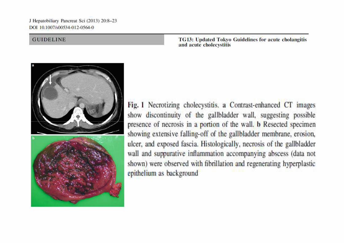
Cholecystitis acuta• Cholecystitis is inflammation of the gallbladder that occurs most commonly
because of an obstruction of the cystic duct by gallstones arising from thegallbladder. (the gallbladder cannot be emptying+ ascending bacterial overgrowth)Uncomplicated cholecystitis has an excellent prognosis; the development ofcomplications such as perforation or gangrene the prognosis less favorable.
• Signs and symptoms• The most common presenting symptom of acute cholecystitis is upper abdominal
pain and tenderness in the RUQ or epigastric region• Fever, tachycardia,• Thephysicalexamination• Thephysicalexamination• Palpable gallbladder or fullness of the RUQ (30-40% of patients)• Jaundice (~15% of patients)• Murphy's sign: If the patient stops breathing in for during an abdominal
examination (gently placing the hand below the right costalmargin)• Lab:
– GOT, GPT, sebi, gGT, ALP, CRP, levels may be elevated in cholecystitis or with common bile duct (CBD) obstruction
• UH– Gallstone, gallbladder wall thicker than normal, not larger bile ducts– fluid around the gallbladder

CHOLECYSTITIS, CHOLANGITIS
• Gram-negativeorganisms
• Escherichia coli 31–44
• Klebsiella spp. 9–20
• Gram-positiveorganisms
• Enterococcus spp. 3–34
• Streptococcus spp. 2–10• Klebsiella spp. 9–20
• Pseudomonas spp. 0.5–19
• Enterobacter spp. 5–9
• Acinetobacter spp. –
• Citrobacter spp. –
• Streptococcus spp. 2–10
• Staphylococcus spp. 0a
• Anaerobes 4–20
• Others –

Treatment of cholecystitis
� Supportive care: In acute cholecystitis, the initial treatmentincludes bowel rest, IVhydration, correction of electrolyteabnormalities, analgesia, and IVantibiotics, emesis can betreated with antiemetics and nasogastric suction.
� AB: Sanford guide – Piperacillin-tazobactam, ampicillin-sulbactam, or meropenem; in severe life-threatening cases,imipenem-cilastatin
� Laparoscopic cholecystectomy (standard of carefor surgical treatment of cholecystitis)

When is the surgery?
�Early: Within 72 hours of the start of symptoms
� (the gallbladder wall is fragile after 72 hours)� (the gallbladder wall is fragile after 72 hours)
�UNFORTUNATELY (surgery postponed)
�Elective: 6-12 weeks later

Chronic cholecystitis
• Chronic cholecystitis is a long-standing gallbladder inflammation almost alwaysdue to gallstones.
• Chronic cholecystitis almost always results from gallstones and prior episodes ofacute cholecystitis. Damage ranges from a modest infiltrate of chronicinflammatory cells to a fibrotic, shrunken gallbladder. Extensive calcification dueto fibrosis is called porcelain gallbladder.
• Symptoms and Signs
• Gallstonesintermittently obstruct the cystic duct and so causerecurrentbiliary• Gallstonesintermittently obstruct the cystic duct and so causerecurrentbiliarycolic. Such episodes of pain are not necessarily accompanied by overt gallbladderinflammation; the extent of inflammation does not correlate with the intensity orfrequency of biliary colic. Upper abdominal tenderness maybe present, but usuallyfever is not.
• Diagnosis: Ultrasonography
• Treatment: Laparoscopic cholecystectomy is indicated to prevent symptomrecurrence and further biliary complications. This procedure is particularlyappropriate for the porcelain gallbladder associated withgallbladder carcinoma.

Case Riport 3.
• 50 yr. old slightly overweightmale pts.. (referred by a familydoctor) Complaints: Right upperquadrant pain, which developedafter a meal. Shoulder bladeradiatingpain,chills, fever,icterus,radiatingpain,chills, fever,icterus,nausea, vomiting, abdominaldiscomfort.
• + icterus
• Lab: Sebi: 55, gGT: 120
• ALP 450, WBC: 15, CRP: 70,
• Amilaz: 76
UH: cholelith, choledocholith, gallbladder wall thickness is normal, larger bile ductsThe pts. was hospitalized, inf. AB, diet Control UH : not larger bile ductsDx: choledocholithiasis, stones in CBD

Choledocholithiasis– Choledocholithiasis is the presence of gallstone in the common bile duct. – (gallstones pass from the gallbladder)Symptoms:– Pain in the right upper or middle upper abdomen for at least 30 minutes.
The pain may be constant or cramping. It can feel sharp or dull.– Fever– Yellowing of skin and whites of the eyes (jaundice)– Loss of appetite– Loss of appetite– Nausea and vomiting– Clay-colored stools– Lab:– Elevated (ALP, gGT), bilirubin
• UH: larger bile duct, stone• Treatment
– Endoscopos Retrograd Cholangio et Pancreatographia ERCP– Endoscopic Sphincterotomy – EST– Surgery

Cholangitis� Etiology: � Biliary stone spontaneously migrate from gallbladder into
the common bile duct where the stone trapped and cause blockage and bacterial infections
� Symptoms� Right upper quadrant pain, fever, jaundice mechanical � Right upper quadrant pain, fever, jaundice mechanical
(Charcot's triad)� Lab� Inflammatory markers, elevated cholestatic liver enzymes� UH: wide bile ducts� Treatment• Antibiotic (well secreted in bile)� Remove stones if stones are in the background: primarily
ERCP


Case Riport
• A 36-year-old woman comes to theoffice because of a 3 day hystory ofyellow skin, fever and abdominalpain. RUQ and her right shoulder.She has had several similar episode inpast,but they were not accompainedpast,but they were not accompainedby fever, and yellowskin.
• Lab: Sebi: 55, gGT: 120
• ALP 450, Fvs: 15, CRP: 70,
• Amilaz: 76
A: acut cholangitisB: acut cholecystitisC: acut hepatitisD: acut pancreatitisE: biliary colic

ERCP • Biliary tract disease including:
– Choledocholithiasis – Malignant and benign biliary strictures – Bile-duct injuries or leaks – Sphincter of Oddi dysfunction
• Pancreatic disease including: • Pancreatic disease including: – Recurrent acute pancreatitis – Chronic pancreatitis – Pancreatic duct leaks – Pancreatic fluid collections such as acute pseudocysts,
chronic pseudocysts, and pancreatic necrosis – Pancreatic cancer and other pancreatic malignancies
• Ampullary adenomas
ASGE guideline: the role of ERCP in diseases of the biliary tract and the pancreas. 2007

Contraindication• Absolute contraindication:
• The uncooperative patient.
• Contraindication
• Recent attack of acute pancreatitis, within the past several weeks.
• Recent myocardial infarction. • Recent myocardial infarction.
• Inadequate surgical back-up
• History of contrast dye anaphylaxis
• Relative contraindications:
• Poor health condition for surgery.
• Severe cardiopulmonary disease.
• Ascites.

Major complications of ERCP and endoscopic sphincterotomy
• Pancreatitis Amylase at least three times normal at more than 24 hours after the procedure, requiring admission or prolongation of planned admission to two to three days Hospitalization of 4 to 10 days Hospitalization of more than ten days, hemorrhagic pancreatitis, phlegmon or pseudocyst, or intervention (percutaneous drainage or surgery)
• Bleeding Clinical, not just endoscopic evidence of bleeding, hemoglobin drop <3 g, and no need for transfusion Transfusion (four units or less), no angiographic intervention or surgery(four units or less), no angiographic intervention or surgery
Transfusion (five units or more), or intervention (angiographic or surgical)
• Cholangitis >38ºC for 24 to 48 hours Febrile or septic illness requiring more than three days of hospital treatment or endoscopic or percutaneous intervention Septic shock or surgery
• Perforation Possible, or only very slight eak of fluid or contrast, treatable by fluids and suction for three days or less Any definite perforation treated medically for 4 to 10 days Medical treatment for more than 10 days, or intervention (percutaneous or surgical)
Adapted with permission from Cotton, PB, et al. Endoscopic sphincterotomy complications and their management: An attempt at cAdapted with permission from Cotton, PB, et al. Endoscopic sphincterotomy complications and their management: An attempt at consonsensus. ensus.
Gastrointest Endosc 1991; 37:383.Gastrointest Endosc 1991; 37:383.

Stones in cystic duct

Choledocholithiasis

Cholelith

Sludge

Pancreas npl.

STONE REMOVAL

LC utáni állapot
Vater papilla stenosis

MIRIZZI SYNDROME
Estimated to occur in 0.7-1.4% of all cholecystectomies

Volume 71, No. 1 : 2010 GASTROINTESTINAL ENDOSCOPY

Gallbladder cancer• Gallbladder cancer is a relatively
uncommon cancer. (The gallbladdercancer is fifth in gastrointestinalcancer)
• Gender—approximately twice morecommon in women than men,usually in seventh and eighthusually in seventh and eighthdecades.
• In our country the incidence is (6-7 /100,000).
• Incidentally discovered gallbladdercancer (adenocarcinoma) following acholecystectomy. (2-3%)
• Compared CRC (70/100,000)

Case riport
• 68 years old, minimallyoverweight males. Patient
• Complaints: Steady pain inthe upper right abdomen
• IndigestionDyspepsia(gas)• IndigestionDyspepsia(gas)
• Weakness, Loss of appetite
• Weight loss
• LAB: not typical
• Sebi: 47, gGT: 56
• ALP 280, Fvs: 11, CRP: 11
UH

Gallbladder Cancer: Risk Factors• Gallstones. Gallstones are the most common risk factor for
gallbladder cancer. However, less than 1% of people with gallstonesdevelop gallbladder cancer. There is no evidence of a direct causalrelationship between gallstones and gallbladder cancer. (porcelaingallbladder)
• Gallbladder polyps.Doctors often recommend gallbladder removalfor people who have polyps larger than 1 centimeter because thesearemorelikely to becancerous.aremorelikely to becancerous.
• Age. Most people diagnosed with gallbladder cancer are older than70.
• Gender.Women are about twice as likely to develop gallbladdercancer as men.
• Obesityincreases the risk for gallbladder cancer.
• Smoking. Tobacco use may increase the risk of gallbladder cancer.
• Family history. A family history of gallbladder cancer slightlyincreasesaperson’srisk of developinggallbladdercancer.

Gallbladder cancer symptoms
• ( Sometimes, gallbladder cancer is found unexpectedly afterremoval of the gallbladder for another reason, such asgallstones. )
• When symptoms do occur, they include the following:
• Jaundice (yellowing of the skin and whites of the eyes)
• Abdominal pain
• Nausea and vomiting
• Bloating
• A lump in the abdomen
• Fever

Diagnosis
• UH
CT
• MRI ERCP: thatrepresents externalcompression bygallbladder cancer.

Five-year survival data
III./3.4. Epehólyag tumorok Somlai Krisztián, Dank Magdolna, Harsányi László

Bile duct cancer (cholangiocarcinoma)
Risk factors :Although most patients present without any known risk factors evident, a
number of risk factors for the development of cholangiocarcinoma have been
described. (PSC,IBD, parasiticliver Diseases,alcoholicliver disease,viraldescribed. (PSC,IBD, parasiticliver Diseases,alcoholicliver disease,viral
hepatitis)
Sign and symptoms:The most common physical indications of cholangiocarcinoma are abnormal
liver function tests, jaundice, abdominal pain (30%–50%),generalized itching
(66%), weight loss (30%–50%), fever (up to 20%), and changesin stool or
urine color.

Prognosis
Surgical resection offers the only potential chance of cureincholangiocarcinoma.
Fornon-resectablecases,the5-yearsurvivalrateisFornon-resectablecases,the5-yearsurvivalrateis
0%. Overall median duration of survival is less than 6
months in inoperable, untreated, otherwise healthy
patients.

Bile duct cancer incidence (CCA)
Cite this article as: Bragazzi MC, Cardinale V, Carpino G, Venere R, Semeraro R, Gentile R, Gaudio E, Alvaro D.
Cholangiocarcinoma: Epidemiology and risk factors. Transl Gastrointest Cancer 2012;1:21-32. DOI: 10.3978/j.issn.2224-
4778.2011.11.04

CholangiocarcinomaKlatskin tumor = MRCP: Cholangiocarcinoma of junction of right & left hepatic ducts
ERCP: Distalis epeúti tumor

Surgical resection
• Roux-en-Y Hepaticojejunostomy

Cholangiocarcinoma
�For non-resectable cases:
�Stent (palliative)
�Chemo +/- Radio treatment�Chemo +/- Radio treatment
�Only (chemo / radio) survival time is 12 -18 month

Bile duct palliative treatment

Antireflux metal stent of biliarymalignancies
• Endoscopic insertion of anARMS is technicallyfeasible, safe, andeffective in patients withdistal malignant biliaryobstruction. The impactofobstruction. The impactofARMSs in prolongingstent patency and lifeexpectancy deservesfurther randomizedevaluation.
Hu B, Wang TT, Shi ZM, Wang SZ, Lu R, Pan YM, Huang H, Wang SP. A novel antireflux metal stent for the palliation of
biliary malignancies: a pilot feasibility study. Gastrointest Endosc. 2011 Jan;73(1):143-8. doi: 10.1016/j.gie.2010.08.048.
Antireflux valve

EUS Transgastric endoscopic ultrasonography-guided biliary drainage
E. Bories1 , C. Pesenti1 , F. Caillol1 , C. Lopes1 , M. Giovannini1 :Transgastric endoscopicultrasonography-guided biliary drainage: results of a pilot study Endoscopy 2007; 39(4): 287-291

Functional gallbladder disorder
• Functional gallbladder disorder isdefined as biliary pain resulting
from a primary gallbladder motility disturbance in the absence of
gallstones, sludge, microlithiasis, or microcrystal disease. The
diagnosisis consideredin patientswith typical biliary-typepainwhodiagnosisis consideredin patientswith typical biliary-typepainwho
have had other causes for the pain excluded. The prevalence of
functional gallbladder disorder among patients with biliary-type
pain and a normal transabdominal gallbladder ultrasound isup to 8
percent in men and 21 percent in women.

• Rome IV criteria for functional gallbladder disorder require:
• ●Biliary pain• ●Absence of gallstones or other structural pathology• In addition, thecriteriathataresupportiveof functional
gallbladderdisorder, but arenot required, include:
Functional gallbladder disorder
• In addition, thecriteriathataresupportiveof functionalgallbladderdisorder, but arenot required, include:
• ●Low ejection fraction on scintigraphygallbladder ejectionfraction (GBEF)
• ●Normal liver enzymes, conjugated bilirubin, and amylase/lipase.

Functional gallbladder disordermanagement
• Patients with functional gallbladder disorder are candidates for cholecystectomy if they have typical biliary-type pain and a cholecystectomy if they have typical biliary-type pain and a low GBEF (<40 percent).
Sphincter of Oddi dysfunction
Sphincter of Oddi dysfunction refers to a groupof functionaldisordersleadingto abdominalpainof functionaldisordersleadingto abdominalpaindue to dysfunction of the Sphincter of Oddi:functional biliary sphincter of Oddi andfunctional pancreatic sphincter of Oddi disorder.

Oddi spincter
The sphincter of Oddi is a sphinctermuscle,a circular band of muscleat themuscle,a circular band of muscleat thebottom of the biliary tree which controlsthe flow of pancreatic juices and bile intothe second part of the duodenum.

Sphincter of Oddi dysfunction• If all of the above criteria are met, individuals are classified as havinga
functional biliary sphincter of Oddi disorder, if the testing of pancreaticenzymes (amylase and lipase) is normal.
• The Milwaukee classification of biliary sphincter of Oddi dysfunction (SOD)further divides the condition into three subtypes:
• Type I biliary SOD: biliary -type abdominal pain, with all of altered liver• Type I biliary SOD: biliary -type abdominal pain, with all of altered liverenzymes on blood testing, dilated biliary ducts on ultrasound or ERCP, anddelayed bile clearance on HIDA scan.
• Type II biliary SOD: biliary-type abdominal pain associated with one or two ofthe following: altered liver enzymes on blood testing, dilated biliaryducts onimaging tests, and delayed bile clearance on HIDA scan.
• Type III biliary SOD: biliary-type abdominal pain with none of the following:altered liver enzymes on blood testing, dilated biliary ducts on imaging tests,and delayed bile clearance on HIDA scan.

Sphincter of Oddi dysfunctiontreatment
• ERCP- EST

THANK YOU FOR THANK YOU FOR
YOUR ATTENTION!