discovering & investigating devonian microfossils
TRANSCRIPT
DiscoveringDevonianMicrofossils
by
Charlie Drewes
ABLE 2005 Workshop
Ecology, Evolution & Organismal BiologyIowa State University
Ames, IA 50011
EMAIL: [email protected]: www.eeob.iastate.edu/faculty/DrewesC/htdocs/
PHONE: 515-294-8061
1
INTRODUCTION
What are microfossils? Microfossils are small, fossilized remains of organisms (animal, plant, or protozoa) that require magnification for study. They usually derive from the hard parts of organisms (skeletons, shells, jaws, teeth, seed coverings, etc). Microfossils may be imbedded in rock or exist as loose particles within soft substrate.
How big are microfossils? The smallest microfossils may be only 0.001 mm (= 1 micron). The largest are up to 1-2 mm in size. Fossils larger than 2 mm are generally referred to as macrofossils.
Why study microfossils? From the standpoint of research, microfossils are relied on heavily for research advances in fields such as biostratigraphy, paleoenvironmental reconstruction, and paleooceanography. Research studies of microfossils provide insights into ecological and evolutionary relationships among plant and animal communities that existed on earth hundreds of millions of years ago. Microfossils also provide excellent hands-on material for science classroom education in general biology, evolution, and earth sciences.
Objectives: The theme of this lab inquiry is biological, rather than geological. Focus is on retrieval, identification, and characterization of selected microfossils ranging in size from about 0.2 to 2.0 mm. Through hands-on discovery and examination of microfossil specimens students should gain new insights and greater appreciation of the richness of marine biodiversity from the Devonian Period ( 375-400 MYA).
MICROFOSSIL COLLECTION SITE
The Devonian microfossils provided here were found in soft deposits of shaley limestone at abandoned excavations of the former Rockford Brick and Tile Company (Rockford, Iowa; Floyd County). The exposures here are shale and shaley limestone of the Lime Creek Formation. There are numerous ravines and rather steep excavation cuts (up to 30-40 feet, but nearly all areas are accessible for collecting macrofossils and microfossils. Photos of the site may be seen at:
http://www.eeob.iastate.edu/faculty/DrewesC/htdocs/quarry1.jpghttp://www.eeob.iastate.edu/faculty/DrewesC/htdocs/quarry2.jpghttp://www.eeob.iastate.edu/faculty/DrewesC/htdocs/quarry3.jpghttp://www.eeob.iastate.edu/faculty/DrewesC/htdocs/quarry4.jpg
These fossil beds are now part of the Floyd County Fossil and Prairie Park, where fossil collection by the public is encouraged and promoted. There is a nature/education center on the premises. Busloads of students from surrounding schools and colleges are frequent visitors to the fossil beds and the nature center. The park is truly an outstanding educational resource.
2
Microfossils are abundant in sievings gathered at many locations in these pits. Particularly high yields of microfossils were obtained from somewhat lower elevations where the surface substrate consists of a mixture of fine silty deposits, sand-like granules, and some larger, rock-like deposits. Microfossils were less abundant at the very lowest elevations where very fine, muddy silt and standing water regularly accumulate. It is evident that many of these locations are subject to considerable water flow and erosion, so no attempt was made to systematically correlate microfossil content with the exposed strata.
FIELD METHODS FOR OBTAINING MICROFOSSILS
The methods described here for obtaining and washing microfossils are likely to be productive for most fossil-rich excavations where substrates mainly consist of loose shale but not where substrates mainly consist of solid limestone.
A small pick or garden trowel was used to loosen shaley limestone and solidified silt. Several scoops of this material were placed into a sieve having a mesh size of 1.00 mm (U.S. Standard Sieve #18). This sieve was placed atop a second sieve having a mesh-size of 0.35 mm (U.S. Standard Sieve #45). The sieve contents were vigorously shaken and swirled for several minutes. Then, the accumulated granular and powdery material in the lower, small-mesh sieve was transferred to a large-mouth, plastic jar for storage.
WASHING AND STORING THE MICROFOSSILS
Washing of microfossils was done outdoors, rather than in a sink, to avoid silt accumulation in sink traps. One or two cups of the dry sievings were placed back into the small-mesh sieve (0.35 mm mesh size; #45). A gentle stream of water from a garden hose was continuously directed at the sievings for a few minutes, as the sieve was tilted from side to side. This resulted in a relatively rapid and nearly complete flushing of very fine silt through the mesh.
Washing continued until water draining through the sieve was clarified and nearly silt-free. During washing, care was taken to avoid water overflow from the sieve, since this can result in loss of microfossils, such as scolecodonts, which are less dense than calcareous microfossils and thus somewhat prone to float.
All microfossil-rich granular material remaining within the sieve was then back-flushed into a deep, flat metal or plastic pan. Excess water was very slowly decanted from this pan. The pan and its wet, granular contents were then placed in a fume hood to dry. Once fully dried, granules were loosened with a small, flexible spatula and carefully poured into capped tubes for storage and later use. (Alternative for storage: The wet granular material that is back-flushed from the sieve may be stored indefinitely in a capped jar. However, refrigeration is recommended for storage of wet material in order to minimize possibilities of bacteria or mold formation.
3
TOOLS FOR HANDLING AND SORTING MICROFOSSILS
60 mm plastic Petri dishes (Fig. 1; one dish and one lid per microscope) 90 mm diameter Whatman #1 filter paper disks (Fig. 1; one disk per Petri dish) Double-stick tape (Fig. 1) Narrow craft stick (Fig. 2; at least one stick per microscope station) Super glue Thin, round rubber bands (e.g., “rubber legs” used for fly fishing) (Fig. 2) Tube of washed and dried microfossil sievings (Fig. 3A) Microfossil dispensing spoon (Fig. 3B; commercial source: Rachel’s Supply; Catalog #: ACC14; 0.15 cc measuring scoop, $2.95/10 scoops; www.rachelssupply.com ) Stereo-dissecting microscopes (approx. 20-30X magnification) Bright, above-stage illumination system (one illuminator needed per microscope) Water Microrulers for measuring to the nearest 1/10th mm (optional)
CONSTRUCTION OF TOOLS FOR MICROFOSSIL SORTING
Construct a microfossil sorting dish, as shown in Figure 1. Place two strips of double-stick into a Petri dish lid, as shown in Figure 1A. Center the filter paper dish over the lid.
Figure 1. Construction of a microfossil sorting dish.
4
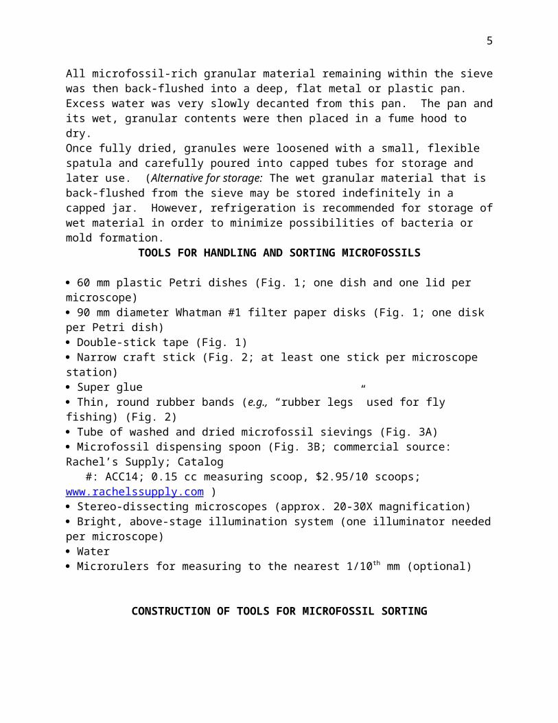
Using your finger tip, begin pressing the paper down against the tape, making sure to begin in the center of the lid and gradually working outward (Fig. 1B).
Use a scissors to trim away excess paper that sticks above the edge of the lid. When completed, the microfossil sorting dish should look like Figures 1C and 1D. [Note: The reason the dish is lined with white paper is two-fold: (1) to provide a good background for viewing and (2) to minimize static electricity interactions that can develop between the dry granules and the plastic surface of the Petri dish.]
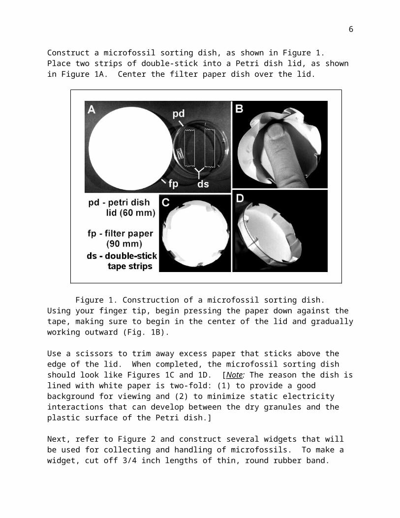
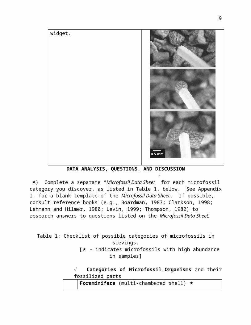
Next, refer to Figure 2 and construct several widgets that will be used for collecting and handling of microfossils. To make a widget, cut off 3/4 inch lengths of thin, round rubber band. Apply a small dab of super glue to the tapered end of a thin craft stick. Lay one end of the rubber band onto the glued area with about a 3/8“ length of rubber band extending beyond the end of the stick (Fig. 2). Let this dry.
Figure 2. Assembly of microfossil widget.
PROCEDURES FOR MICROFOSSIL SORTING
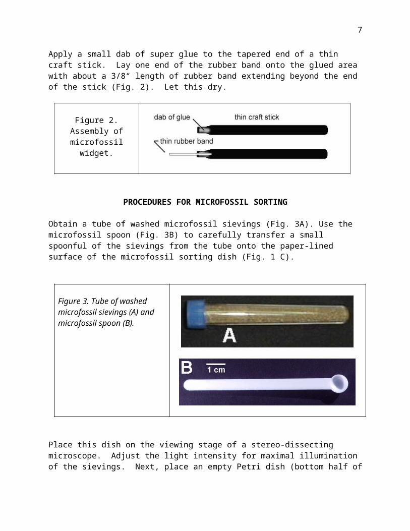
Obtain a tube of washed microfossil sievings (Fig. 3A). Use the microfossil spoon (Fig. 3B) to carefully transfer a small spoonful of the sievings from the tube onto the paper-lined surface of the microfossil sorting dish (Fig. 1 C).
Figure 3. Tube of washed microfossil sievings (A) and microfossil spoon (B).
5
Place this dish on the viewing stage of a stereo-dissecting microscope. Adjust the light intensity for maximal illumination of the sievings. Next, place an empty Petri dish (bottom half of the Petri dish that is not lined with filter paper) near the microscope and fill this dish about half full with water.
Now you are ready to use a microfossil widget (Fig. 2) to retrieve and isolate microfossils. First, wet the rubber tip of the widget by touching the tip to the water surface in the Petri dish. Then, hold the widget as if it was a pencil with the rubber tip positioned just above the dry sievings. Use your other hand to focus the microscope on the granules. Carefully scan the granules, looking for shapes and textures that are characteristic of microfossils.
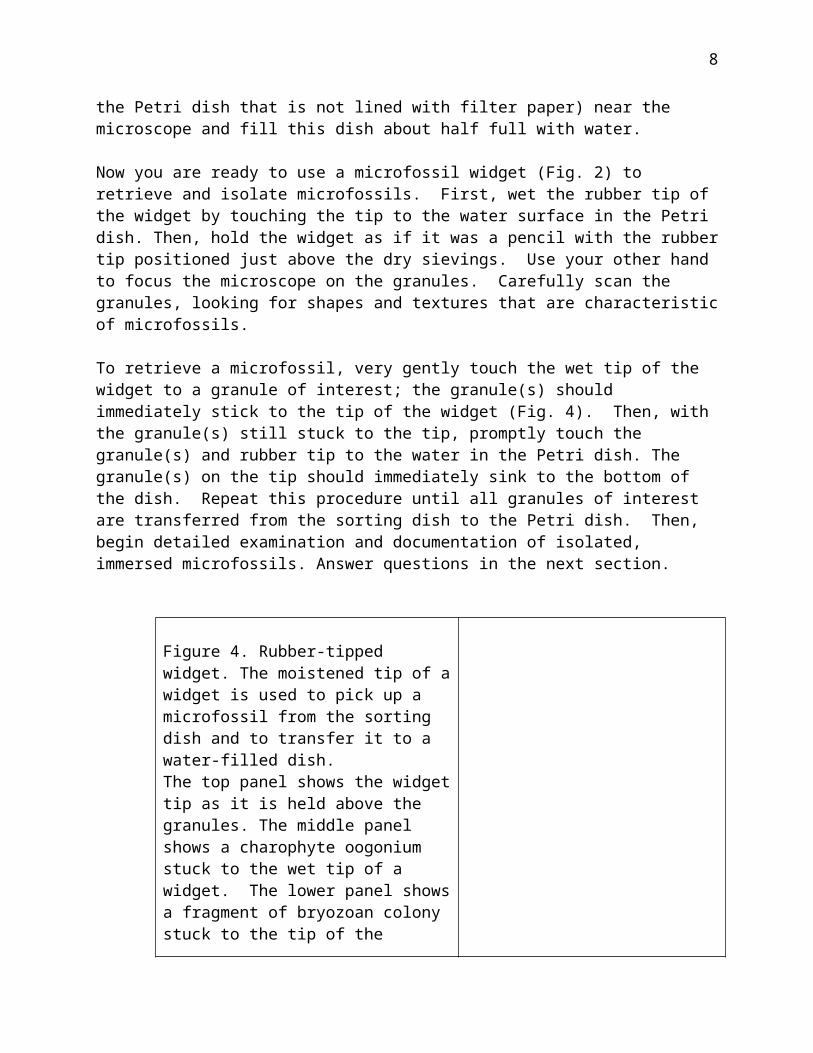
To retrieve a microfossil, very gently touch the wet tip of the widget to a granule of interest; the granule(s) should immediately stick to the tip of the widget (Fig. 4). Then, with the granule(s) still stuck to the tip, promptly touch the granule(s) and rubber tip to the water in the Petri dish. The granule(s) on the tip should immediately sink to the bottom of the dish. Repeat this procedure until all granules of interest are transferred from the sorting dish to the Petri dish. Then, begin detailed examination and documentation of isolated, immersed microfossils. Answer questions in the next section.
Figure 4. Rubber-tipped widget. The moistened tip of a widget is used to pick up a microfossil from the sorting dish and to transfer it to a water-filled dish. The top panel shows the widget tip as it is held above the granules. The middle panel shows a charophyte oogonium stuck to the wet tip of a widget. The lower panel shows a fragment of bryozoan colony stuck to the tip of the widget.
6
DATA ANALYSIS, QUESTIONS, AND DISCUSSION
A) Complete a separate “Microfossil Data Sheet” for each microfossil category you discover, as listed in Table 1, below. See Appendix I, for a blank template of the Microfossil Data Sheet. If possible, consult reference books (e.g., Boardman, 1987; Clarkson, 1998; Lehmann and Hilmer, 1980; Levin, 1999; Thompson, 1982) to research answers to questions listed on the Microfossil Data Sheet.
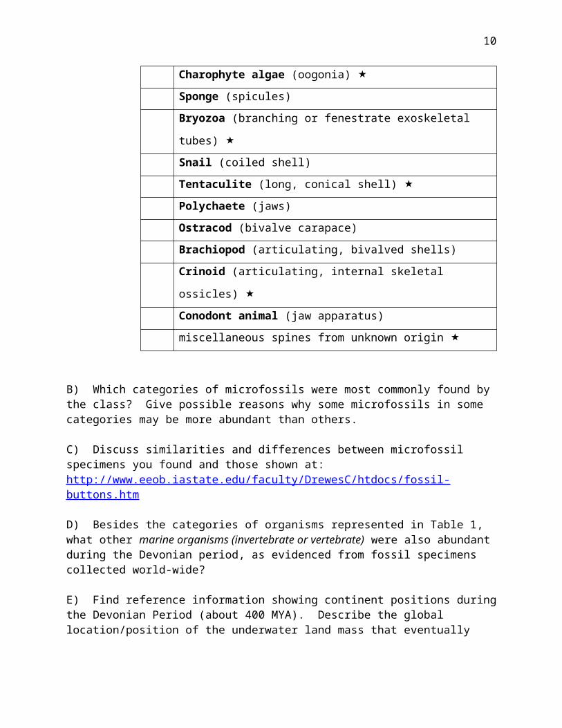
Table 1: Checklist of possible categories of microfossils in sievings. [ - indicates microfossils with high abundance in samples]
Categories of Microfossil Organisms and their fossilized partsForaminifera (multi-chambered shell)
Charophyte algae (oogonia)
Sponge (spicules)
Bryozoa (branching or fenestrate exoskeletal tubes)
Snail (coiled shell)
Tentaculite (long, conical shell)
Polychaete (jaws)
Ostracod (bivalve carapace)
Brachiopod (articulating, bivalved shells)
Crinoid (articulating, internal skeletal ossicles)
Conodont animal (jaw apparatus)
miscellaneous spines from unknown origin
B) Which categories of microfossils were most commonly found by the class? Give possible reasons why some microfossils in some categories may be more abundant than others.
C) Discuss similarities and differences between microfossil specimens you found and those shown at: http://www.eeob.iastate.edu/faculty/DrewesC/htdocs/fossil-buttons.htm
D) Besides the categories of organisms represented in Table 1, what other marine organisms (invertebrate or vertebrate) were also abundant during the Devonian period, as evidenced from fossil specimens collected world-wide?
E) Find reference information showing continent positions during the Devonian Period (about 400 MYA). Describe the global location/position of the underwater land mass
7
that eventually became central North America. Try to locate the approximate area (Iowa) where these particular Devonian microfossils once existed.
F) Write a brief essay describing the biotic environment in which your microfossil organisms lived during the Devonian period?
G) Have microfossils of marine organisms ever been found, and can they be readily obtained, in your state/province? If so, from which geological period(s)? [To research these questions, refer to the Paleoportal web reference below.]
REFERENCES
Web:
http://www.paleoportal.org/ [Excellent geology and paleontology background for each of the United States]
http://www.eeob.iastate.edu/faculty/DrewesC/htdocs/fossil-buttons.htm [Image gallery of Devonian microfossils and macrofossils from Iowa.]
Technical Reference Books:
Boardman, R.S. et al. (1987) Fossil Invertebrates, Blackwell Scientific, Palo Alto, 713 pp. [ISBN: 0-86542-302-4]
Clarkson, E.N.K. (1998) Invertebrate Palaeontology and Evolution , 4 th Ed., Blackwell Science, Oxford, 452 pp. [ISBN: 0-632-05238-4]
Lehmann, U. & G. Hillmer (1980) Fossil Invertebrates, Cambridge University Press, Cambridge, 350 pp. [ISBN: 0-521-24856-6]
Levin, H.L. (1999) Ancient Invertebrates and Their Living Relatives, Prentice Hall, Upper Saddle River, NJ, 358 pp. [ISBN: 0-13-748955-2]
Thompson, I. (1982) National Audubon Society Field Guide to North American Fossils, Knopf Publishing, 848 pp. [ISBN: 0394524128]
8
APPENDIX 1: Microfossil Data Sheet
NAME(s) ________________________________________ Date: ____________
CATEGORY OF MICROFOSSIL _____________________
SKETCHES (include size scale):
Compare your specimens and sketches to images shown on the CD entitled “Paleozoic (Devonian) Microfossils and Macrofossils”. These images may also be seen at the following web site: http://www.eeob.iastate.edu/faculty/DrewesC/htdocs/fossil-buttons.htm
QUESTIONS:1 What is a common name for organisms in
this category?2 What key features (size, shape, markings,
textures, etc.) of microfossils in this category provide key evidence as to their identity?
3 Give the phylum (or class) name for organisms in this category?
4 What hard part/structure of the organism formed the microfossil?
5 Was this a colonial or solitary organism?6 Was this a sessile or free moving
organism?7 Do any related marine species still exist
today? Which ones?8 What are the closest living freshwater
relatives? 9 CHALLENGE QUESTIONS:
(a) What is the likely mineral or chemical composition of the original hard part? (b) How did organisms in this category gather food?
9
10