direct regulation of aromatase b expression by 17β ......of aromatase b expression and...
TRANSCRIPT
ORIGINAL RESEARCHpublished: 12 January 2016
doi: 10.3389/fnins.2015.00504
Frontiers in Neuroscience | www.frontiersin.org 1 January 2016 | Volume 9 | Article 504
Edited by:
Sebastien G. Bouret,
University of Southern California, USA
Reviewed by:
Ishwar Parhar,
Monash University, Malaysia
Philippe Ciofi,
Institut National de la Santé et de la
Recheche Médicale, France
*Correspondence:
Vance L. Trudeau
Specialty section:
This article was submitted to
Neuroendocrine Science,
a section of the journal
Frontiers in Neuroscience
Received: 26 September 2015
Accepted: 21 December 2015
Published: 12 January 2016
Citation:
Xing L, Esau C and Trudeau VL (2016)
Direct Regulation of Aromatase B
Expression by 17β-Estradiol and
Dopamine D1 Receptor Agonist in
Adult Radial Glial Cells.
Front. Neurosci. 9:504.
doi: 10.3389/fnins.2015.00504
Direct Regulation of Aromatase BExpression by 17β-Estradiol andDopamine D1 Receptor Agonist inAdult Radial Glial CellsLei Xing, Crystal Esau and Vance L. Trudeau*
Department of Biology, Centre for Advanced Research in Environmental Genomics, University of Ottawa, Ottawa, ON,
Canada
Aromatase cytochrome P450arom (cyp19) is the only enzyme that has the ability
to convert androgens into estrogens. Estrogens, which are produced locally in the
vertebrate brain play many fundamental roles in neuroendocrine functions, reproductive
functions, socio-sexual behaviors, and neurogenesis. Radial glial cells (RGCs) are
neuronal progenitor cells that are abundant in fish brains and are the exclusive site
of aromatase B expression and neuroestrogen synthesis. Using a novel in vitro RGC
culture preparation we studied the regulation of aromatase B by 17β-estradiol (E2)
and dopamine (DA). We have established that activation of the dopamine D1 receptor
(D1R) by SKF 38393 up-regulates aromatase B gene expression most likely through
the phosphorylation of cyclic AMP response element binding protein (CREB). This
up-regulation can be enhanced by low concentration of E2 (100 nM) through increasing
the expression of D1R and the level of p-CREB protein. However, a high concentration
of E2 (1µM) and D1R agonist together failed to up-regulate aromatase B, potentially
due to attenuation of esr2b expression and p-CREB levels. Furthermore, we found the
up-regulation of aromatase B by E2 and DA both requires the involvement of esr1 and
esr2a. The combined effect of E2 and DA agonist indicates that aromatase B in the adult
teleost brain is under tight control by both steroids and neurotransmitters to precisely
regulate neuroestrogen levels.
Keywords: 17β-estradiol, dopamine, aromatase, radial glial cell, teleost fish
INTRODUCTION
Aromatase cytochrome P450arom (cyp19) is the only enzyme performing the conversion ofandrogens into estrogens (Lephart, 1996; Garcia-Segura et al., 2003). In all species of vertebrates,including mammals, birds, and teleost fish, aromatase expression can be found in the brain inaddition to testes and ovaries (Balthazart and Ball, 1998; Forlano et al., 2001). The distributionof aromatase varies between species and regions in the central nervous system (CNS), which hasbeen detected at multiple levels, including mRNA, protein, and enzyme activity (Lephart, 1996;Balthazart and Ball, 1998; Forlano et al., 2006; Garcia-Segura, 2008; Azcoitia et al., 2011). Inmammals, aromatase is expressed in both neuronal and glial cell bodies, processes and synapticterminals of specific areas, especially the pial surface and ventricular surface in the brain undernormal conditions (Martínez-Cerdeno et al., 2006; Yague et al., 2006). Aromatase may also be

Xing et al. Aromatase B Regulation by 17ß-Estradiol and Dopamine
expressed in reactive astrocytes under conditions of cellularstress in mammalian brains (Azcoitia et al., 2003). In contrastto mammals, teleosts express two structurally and functionallydifferent aromatase genes, aromatase A and aromatase B, whichare a result of a gene duplication event (Diotel et al., 2010).The expression of aromatase B (cyp19a1b) in the teleost brain isexclusive to one particular cell type, the radial glial cell (RGC)(Forlano et al., 2001; Diotel et al., 2010; Xing et al., 2014),which allows direct in vivo study of glial aromatase contributionswithout interference from neuronal compartments. Callard et al.demonstrated for the first time that goldfish exhibit the highestbrain aromatase activity compared to other vertebrates (Callardet al., 1981). More recently, Diotel et al. have documented thedistribution cyp11a1, 3β-hsd, cyp17, and cyp19a1b mRNAs andthe ability of de novo synthesis of several steroids includingestrone and 17β-estradiol (E2) in the adult zebrafish brain (Diotelet al., 2011). Although it is very difficult to compare aromataseactivity among species, it is clear that activity in the forebrain ofteleost fish is much higher (100–1000 times) than in birds andmammals. All these data indicate that teleost fish are excellentmodels to study function and regulation of glial aromatase.Better understanding the regulation of glial aromatase will widenour knowledge of the control of estrogens in the brain. Localformation of estrogens are extremely important to maintain thehomeostasis of cell proliferation and cell death, since estrogens inthe brain are proven to be neuroprotective in mammalian brainsand contributing to the generation of new neurons and brainrepair after injury (Azcoitia et al., 2003; Arevalo et al., 2015).More importantly, recent studies indicate that RGCs are theprogenitors of both neurons and glia during development, andare the source of new neurons in adult vertebrate brains (Alvarez-Buylla et al., 2001; Zupanc and Clint, 2003; Pellegrini et al., 2007).All these lines of evidence suggest that both RGC progenitorand steroidogenic functions contribute to neurogenesis in adultvertebrate brains.
Brain aromatization plays numerous important rolesincluding brain differentiation, neural plasticity, neuralregeneration, neuroendocrine functions, reproductive functions,and socio-sexual behaviors. It is possible that these processesare in part regulated by aromatization of androgen and thuslocal estrogens formation (Lephart, 1996; Garcia-Segura et al.,1999, 2003; Azcoitia et al., 2003; Ubuka et al., 2014). Thereare many response elements located at the promoter regionof teleost cyp19a1b (Callard et al., 2001; Tchoudakova et al.,2001), including estrogen response element (ERE) and cyclicadenosine monophosphate (cAMP) response element (CRE).Through differential response element recruitment, cyp19a1bexpression can be regulated differently, which suggests thereshould be multiple factors implicated in the regulation ofcyp19a1b expression (Kato et al., 1997). It is well known thatestrogens can up-regulate cyp19a1b expression in fish brain,which is mediated by estrogen receptors (ERs), some of whichact as ligand-activated transcription factors (Belcher andZsarnovszky, 2001). Upon binding to classical nuclear ERs,activated ER homo-dimerizes, and binds to ERE at the promoterof cyp19a1b, and then mediates transcription (Marlatt et al.,2008; Strobl-Mazzulla et al., 2008). Due to duplication of the
estrogen receptor beta gene, teleost have three ERs (esr1/ERα,esr2a/ERβ, and esr2b/ERγ), all of which are expressed in the brainof fish with an expression pattern similar to that of aromatase(Forlano et al., 2005; Pellegrini et al., 2005; Strobl-Mazzullaet al., 2008; Fergus and Bass, 2013). There is evidence showingthat moderate up-regulation of ERα by estrogens precedesthe dramatic up-regulation of aromatase by its own products(Denslow et al., 2001; Bowman et al., 2002; McEwen, 2002;Marlatt et al., 2008). The possibility that cyp19a1b expression isup-regulated by estrogens is intriguing and suggests the presenceof a positive feedback loop within the aromatase and ER-positivecells, whereby estrogens enhance aromatase expression andtherefore the subsequent products, estrogens.
In addition to estrogenic regulation of the cyp19a1b gene,our previous neuroanatomical and pharmacological studiesin goldfish reveal that there is a close relationship betweencatecholaminergic neurons and aromatase B- and DA D1receptor (D1R)-positive RGCs in goldfish forebrain (Xinget al., 2015). Activation of D1R but not D2R up-regulatescyp19a1b mRNA in cultured RGCs (Xing et al., 2015). Thesignaling pathway underlying activated D1R regulation ofcyp19a1b expression involves cAMP, protein kinase A (PKA)and phosphorylated cAMP response element binding protein(p-CREB). Whether this regulation involves nuclear ERs hasyet to be characterized. Since it is likely that both mechanismsare acting simultaneously to affect multifactorial control overneuroestrogen synthesis, and given the close association betweenRGCs and DA neurons we directly tested for interactive effectson RGCs in vitro.
In this report, we show that both E2 and D1R agonistSKF 38393 alone significantly increased the expression ofcyp19a1b. Moreover, the expression of cyp19a1b in RGCs may besynergistically up-regulated or antagonistically down-regulated,respectively, under low-concentration or high-concentrationexposure to E2 and SKF 38393.
MATERIALS AND METHODS
Ethics StatementAll procedures used were approved by the University of OttawaProtocol Review Committee and followed standard CanadianCouncil on Animal Care guidelines on the use of animals inresearch.
Cell CultureCommon adult female goldfish (Carassius auratus) werepurchased in April from a commercial supplier (Mt. ParnellFisheries Inc., Mercersburg, PA, USA) and acclimated for at least3 weeks prior to experimentation. The fish were maintainedat 18◦C under a natural simulated photoperiod and fedstandard goldfish flakes. Sexually mature, pre-spawning femalegoldfish (20–35 g) were anesthetized using 3-aminobenzoic acidethyl ester (MS222) for all handling, injection, and dissectionprocedures.
Cell culture methods have been established and validatedpreviously by Xing et al. (2015). Briefly, female goldfishhypothalamus and telencephalon were dissected and rinsed
Frontiers in Neuroscience | www.frontiersin.org 2 January 2016 | Volume 9 | Article 504

Xing et al. Aromatase B Regulation by 17ß-Estradiol and Dopamine
with Hanks Balanced Salt Solution (HBSS; 400mg KCl, 600mgKH2PO4, 350mg NaHCO3, 8 g NaCl, 48mg Na2HPO4, and1 g D-Glucose in 1 L ddH2O) with Antibiotic-Antimycoticsolution (Gibco) and minced into small explants. Radial glialcells were dissociated with trypsin (0.25%, Gibco) and culturedin Leibovitz’s L-15 medium with 15% Fetal Bovine Serum (FBS)and Antibiotic-Antimycotic solution. Cell culture medium waschanged 4–7 days after isolation to remove tissue explantsand suspended cells. Following the initial media change, cellsreceived fresh media once a week until they were ready for theirnext passage. RGCs were sub-cultured following trypsinization(0.125%) for three passages and then used for experiments.
Drugs and ExposuresCells were exposed to E2 (Sigma-Aldrich) and selective DAD1R agonist SKF 38393 (Tocris) for 24 h to study their effectson cyp19a1b, esr1, esr2a, esr2b, and drd1a mRNA and p-CREBprotein level. To investigate ERs involvement in cyp19a1bmRNAregulation by E2 and SKF 38393, cells were pre-exposed toestrogen receptor antagonist MPP (Tocris) for 1 h, and thenexposed to E2 or SKF 38393 for 24 h.
RNA Extraction, Quality Control and cDNASynthesisRNA was isolated using the RNeasy Micro kit (Qiagen) asdescribed in the manufacturer’s protocol. Upon purification,concentration and quality of all samples were assessed usingthe NanoDrop ND-1000 spectrophotometer (Thermo Scientific)and the 2100 Bioanalyzer (Agilent). RNA integrity values ofsamples were above the recommended minimum value of 5 forquantitative real-time RT PCR applications and ranged from 8.5to 9.8 (Fleige and Pfaffl, 2006). Total cDNA was prepared usingMaxima First Strand cDNA Synthesis Kit for RT-qPCR (ThermoScientific). Each 20µl reaction was diluted 10-fold in nuclease-free water and used as the template for the real-time RT-PCRassays.
Quantitative Real-Time RT-PCRQuantitative real-time RT-PCR (qPCR) assays based on SYBRgreen detection were used to validate relative gene expression.Primers used in this study were designed using the Primer3(http://primer3.sourceforge.net/) software and synthesized byInvitrogen (Table 1). The Maxima SYBR green qPCR MasterMix (Thermo Scientific) and CFX96 Real-Time PCR DetectionSystem (Bio-Rad) were used to amplify and detect the transcriptsof interest. Thermal cycling conditions included a denaturationstep at 95◦C for 3min, and then 40 cycles of denaturation at95◦C for 10 s, annealing at 60◦C for 30 s followed by melt curveanalysis to confirm product specificity for all transcripts. Serial1/2 dilution of the pool of all cDNA samples was analyzed tobuild the standard curves, from which the mRNA abundancein samples was calculated using the CFX Manager™ Softwarepackage (Bio-Rad). The efficiencies for all standard curves werebetween 92 and 106%. Data were normalized by using NORMA-GENE algorithm (Heckmann et al., 2011) and then presented asmeans+ SEM of fold change to control group (n = 4; assayed induplicate).
Droplet Digital PCRThe low expression of D1R (drd1a) in RGC cultures is notquantifiable using qPCR, so we used a more sensitive methodto measure drd1a mRNA level. Droplet digital PCR (ddPCR)was performed using 25 ng of each cDNA sample and QX200™ddPCR™ EvaGreen Supermix by employing QX200™ DropletDigital™ PCR System (Bio-Rad). The ddPCR conditions wereas follows: an initial denaturation for 5min at 95◦C followedby 40 cycles of denaturation for 30 s at 95◦C and annealing andextension for 1min at 56◦C, then signal stabilization for 5minat 4◦C and 5min at 90◦C. Template DNA was omitted from theddPCR reaction as a no template control (NTC) and the results ofddPCR were analyzed using the QuantaSoft Software (Bio-Rad).The absolute copy number of targeted gene was divided by theaverage of the absolute copy number of two reference genes β-actin and 18s, and presented as means + SEM of fold change tocontrol group (n = 4; assayed in duplicate).
Western BlotTotal protein extract from RGCs was denatured and separatedby electrophoresis on a 10% SDS–polyacrylamide gel andtransferred to a PVDF membrane as described previously byZhao et al. (2006). Membranes were incubated with anti-Phospho-CREB (Ser133, Cell signaling technology, 9198S)antibodies overnight at 4◦C and then incubated with donkeyanti-rabbit IgG antibody (GE Health Care, NA934VS). Blotswere visualized using the Amersham ECL Prime WesternBlotting Detection System (GE Health Care, RPN2232). Imageswere obtained using a ChemiDoc XRS+ system (Bio-Rad) andanalyzed with image lab (Bio-Rad). Same blot incubated withanti-Phospho-CREB antibodies before was stripped and re-probed with primary anti-actin (Cedarlane, CLT9001) antibodiesovernight at 4◦C and then incubated with goat anti-mouse IgG-HRP (Santa Cruz, sc-2005), where actin served as an internalcontrol.
StatisticsIn all cases and before any analysis, normality and homogeneityof variances was verified using Shapiro-Wilk’s and Levene’s test,respectively. Comparison of two groups was performed usingStudent’s t-test in GraphPad Prism (Version 6.0). Two-wayanalysis of variance (ANOVA) followed by Tukey’s post-hoc testin GraphPad Prism (Version 6.0) was used to determine theeffects of single or combined drug treatments. P = 0.05 wereconsidered statistically significant. Data were presented as mean+ SEM. Note that the t- and F-statistics, degrees of freedom (df)of interaction (DFn), the total degrees of freedom (DFd), and P-values are reported in the Results Section. The P-values associatedwith main effects and the interactions are reported directly on thegraphs.
RESULTS
Regulation of cyp19a1b and ER Expressionby SKF 38393 and E2Here, the qPCR data show that 10µM SKF 38393 was able toincrease the expression of cyp19a1b (t = 2.454, df = 6, P =
Frontiers in Neuroscience | www.frontiersin.org 3 January 2016 | Volume 9 | Article 504

Xing et al. Aromatase B Regulation by 17ß-Estradiol and Dopamine
TABLE 1 | Primer sets used for qPCR and ddPCR.
Gene Primer sequence (Forward) Primer sequence (Reverse) Amplicon size Accession no.
β-actin CTGGGATGATATGGAGAAGA CCAGTAGTACGACCTGAAGC 215 AB039726
18s AAACGGCTACCACATCCAAG CACCAGATTTGCCCTCCA 165 AF047349
cyp19a1b TGCTGACATAAGGGCAATGA GGAAGTAAAATGGGTTGTGGA 153 AB009335
esr1 GCAGGAGGGTTTGATTCTGAGA CCATAATGATAGCCGGACGCA 76 AY055725
esr2a TGACTACATCTGCCCTGCCA CCAACTTCGTAACATTTTCGGAGA 72 AF061269
esr2b TGGTCCCTTTAAATTCAGCAATCT GTGTTTCCCTGTAGGCCAGTG 95 AF177465
drd1a CCCTTTGGTGCGTTTTGT GAGCCTTGTGCCATTTGAG 210 L08602
0.0225), but not esr1 (t = 1.744, df = 6, P = 0.0952), esr2a(t = 1.338, df = 6, P = 0.1947), and esr2b (t = 1.638, df =22, P = 0.1157) after 24 h incubation (Figure 1A). The 100 nMconcentration of E2 did not affect cyp19a1b (t = 1.557, df =6, P = 0.1705), esr1 (t = 0.5747, df= 6, P = 0.5864), esr2a(t = 0.5106, df= 6, P = 0.6993), and esr2b (t = 0.9012,df= 6, P = 0.4022) mRNAs after 24 h incubation (Figure 1B).However, when the concentration of E2 was increased to 1µM,the expression of cyp19a1b (t = 4.791, df = 6, P = 0.003) andesr2b (t = 10.65, df= 6, P < 0.0001) were increased dramatically6.7-fold and 2.9-fold, respectively, but not esr1 (t = 0.3295,df= 6, P = 0.7530) and esr2a (t = 0.2395, df = 6, P = 0.779;Figure 1C). These data suggest the up-regulation of esr2b maycontribute to the stimulatory effect of E2 in cyp19a1b expression.
The Involvement of ERs in cyp19a1bRegulation by E2 and SKF 38393When RGC cultures are exposed to SKF38393 and E2 for 24 hthere is no change in the expression of esr1 and esr2a. This resultled us to investigate the possible contribution of pre-existing ERsin RGCs to cyp19a1b up-regulation. The ERα and ERβ antagonistMPP was administered to RGC culture 1 h before SKF 38393 andE2. Data revealed that MPP alone decreases the expression of allthree ERs (t = 2.462, df = 6, P = 0.049; t = 2.285, df = 6,P = 0.0624; t = 3.134, df= 6, P = 0.0202, respectively) by 29.9–57.6% (Figure 2A), but has no effect on cyp19a1b (F = 32.65,DFn= 1, DFd= 12, P = 0.8582; F = 18.15, DFn= 1, DFd= 12,P = 0.3065, respectively) expression (Figures 2B,C). Two-wayANOVAs were conducted that examined the effect of MPP andSKF 38393 as well as MPP and E2 on cyp19a1b expression. Therewas a statistically significant interaction between MPP and SKF38393 (F = 32.65, DFn= 1, DFd= 12, P < 0.0001), and betweenMPP and E2 (F = 18.15, DFn = 1, DFd = 12, P = 0.0011).The stimulatory effect of SKF 38393 (F = 32.65, DFn = 1, DFd= 12, P = 0.0002) and E2 (F = 18.15, DFn = 1, DFd = 12,P < 0.0001) on cyp19a1bmRNAwas completely blocked byMPP(Figures 2B,C).
The Combined Effects of E2 and SKF 38393on cyp19a1b RegulationBased on the previous dose-dependent studies (Xing et al., 2015and unpublished data), we examined the combined effects of twoconcentrations of SKF 38393 and E2. Our results indicate thatthere is either a synergistic or antagonistic effect in cyp19a1b
FIGURE 1 | Effects of SKF 38393 and E2 on the expression of
cyp19a1b, esr1, esr2a, and esr2b in primary RGC culture. Quantitative
real-time PCR analysis showing variations in the relative amounts of the
cyp19a1b, esr1, esr2a, and esr2b mRNAs in primary RGCs culture exposed
to 10µM SKF38393 (A) 100 nM E2 (B) and 1µM E2 (C). Data were
normalized and defined as fold change relative to control, and bars represent
the mean + SEM. Treatment groups marked by asterisks have significantly
different mRNA levels compared to control (P < 0.05).
regulation by E2 and SKF 38393, which is dependent on theconcentration of E2. Two-way ANOVAs were conducted thatexamined the effect of SKF 38393 and E2 on cyp19a1b expression.As shown in Figure 3A, the low concentration of E2 (100 nM)alone increased cyp19a1b expression 1.5 fold, but this was notstatistically significant (F = 0.1071, DFn = 1, DFd = 12, P =
0.1498) regardless of the combination with SKF 38393 (1µM).In contrast, the high concentration of E2 (1µM) alone increased
Frontiers in Neuroscience | www.frontiersin.org 4 January 2016 | Volume 9 | Article 504

Xing et al. Aromatase B Regulation by 17ß-Estradiol and Dopamine
FIGURE 2 | Effects of MPP on the expression of esr1, esr2a, and esr2b
and the regulation of cyp19a1b expression by DA and E2 in primary
RGC culture. Quantitative real-time PCR analysis showing variations in the
relative amounts of the cyp19a1b, esr1, esr2a, and esr2b mRNAs in primary
RGCs culture exposed to 100 nM MPP (A), 100 nM MPP and 10µM SKF
38393 (B), 100 nM MPP and 1µM E2 (C). Data were normalized and defined
as fold change relative to control, and bars represent the mean + SEM.
Treatment groups marked by an asterisk (A) are significantly different from
controls (P < 0.05). Treatment groups marked with different letters (B,C) are
significantly different (P < 0.05).
cyp19a1b 6.7-fold (F = 0.2028, DFn= 1, DFd= 12, P = 0.0002,Figure 3B) significantly regardless of the combination with SKF38393 (1µM). There was no statistically significant interactionbetween SKF 38393 (1µM) and either the low (100 nM; F =
0.1071, DFn = 1, DFd = 12, P = 0.7491, Figure 3A) or higher(1µM; F = 0.2028, DFn= 1, DFd= 12, P = 0.6601, Figure 3B)concentration of E2. When the concentration of SKF 38393 wasincreased to 10µM, statistically significant interactions betweenSKF 38393 and both low concentration (F = 8.112, DFn = 1,DFd = 12, P = 0.0147, Figure 3C) and high concentration ofE2 (F = 14.91, DFn = 1, DFd = 12, P = 0.0023, Figure 3D)were evident. The Two-way ANOVA indicates that both 10µMSKF 38393 and 100 nM E2 increased cyp19a1b expression2.8-fold and 1.5-fold, consistent with the earlier experiment
shown in Figures 1A,B, respectively. Post-hoc tests showed thatthe combination of high concentration of SKF 38393 andlow concentration of E2 synergistically up-regulated cyp19a1b(F = 8.112, DFn= 1, DFd= 12, P = 0.0003, Figure 3C)expression 8.0-fold. Contrasting this is the experiment wherehigh concentration of SKF 38393 and high concentration ofE2 were used. Exposure of RGCs to 1µM E2 alone increasedcyp19a1b expression 6.7-fold, and the high concentration of SKF38393 inhibited this effect (F = 14.91, DFn= 1, DFd = 12,P = 0.0141, Figure 3D).
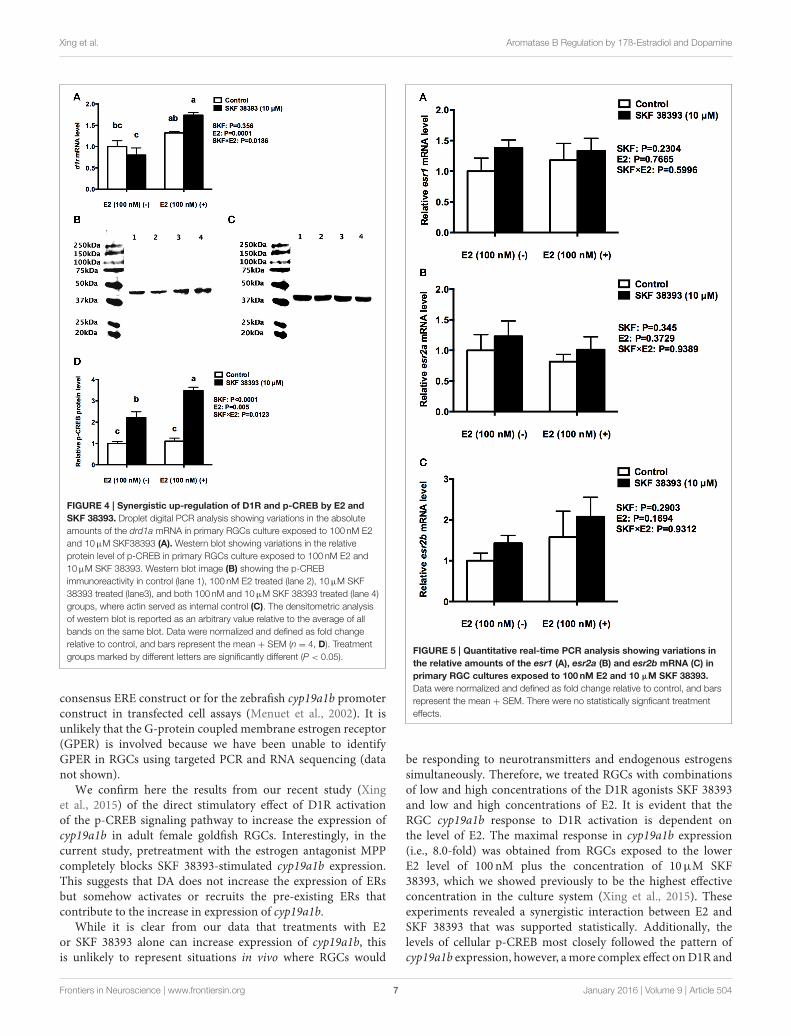
Synergistic Effects of Low Levels of E2 onSKF 38393-Induced cyp19a1b Expressionare Associated with Parallel Increases inp-CREBTo further understand the underlying mechanisms of synergisticup-regulation of cyp19a1b by low concentration of E2 and highconcentration of SKF 38393, we studied two components of theD1R/cAMP/PKA/p-CREB pathway, drd1a in mRNA level andp-CREB in protein level as well as the expression of esr1, esr2a,and esr2b. Two-way ANOVAs were conducted that examined theeffect of SKF 38393 and E2 on drd1a, esr1 esr2a, and esr2bmRNAlevel and p-CREB protein level in cultured RGCs. There was astatistically significant interaction (F = 7.393, DFn = 1, DFd =
12, P = 0.01786) between SKF 38393 and E2 for drd1a mRNA.Post-hoc tests showed that the combination of SKF 38393 and E2up-regulated drd1a mRNA (F = 7.393, DFn = 1, DFd = 12,P = 0.0029; Figure 4A), although increases were not very large,in the range of 1.7 fold. There was also a statistically significantinteraction (F = 10.35, DFn= 1, DFd= 12, P = 0.0123) betweenSKF 38393 and E2 for p-CREB protein level (Figures 4B–D). TheD1R agonist SKF 38393 alone increased p-CREB 2.2-fold, andthis was enhanced to 3.5-fold in the presence of E2, parallelingthese results for cyp19a1b mRNA (Figure 3C). No effects on theexpression of esr1, esr2a and esr2b (F = 0.2907, DFn= 1, DFd=
12, P = 0.5996; F = 0.0061, DFn = 1, DFd = 12, P = 0.9389;F = 0.0078, DFn = 1, DFd = 12, P = 0.9312, respectively) wereobserved for the E2 and SKF 38393 treatments (Figure 5).
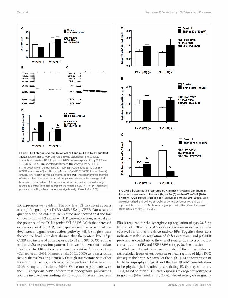
Antagonistic Effects of High Levels of E2on SKF 38393-Induced cyp19a1bExpression are Associated with ParallelDecreases in p-CREB Protein and esr2bmRNAFurther analysis was essential in understanding the potentialmechanisms of antagonistic regulation of cyp19a1b by thecombined high concentration of E2 and high concentration ofSKF 38393. As with the previous experiment, two-way ANOVAswere conducted that examined the potential interactive effects ofSKF 38393 and E2 on drd1a, esr1 esr2a, and esr2b mRNA leveland p-CREB protein level. Exposure to 10µM SKF 38393 and1µME2 alone did not affect drd1a (F = 0.9571, DFn= 1, DFd=12, P = 0.3472) mRNA levels in cultured RGCs (Figure 6A). Incontrast, there was a statistically significant interaction betweenSKF 38393 and E2 on p-CREB protein (F = 6.252, DFn = 1,
Frontiers in Neuroscience | www.frontiersin.org 5 January 2016 | Volume 9 | Article 504

Xing et al. Aromatase B Regulation by 17ß-Estradiol and Dopamine
FIGURE 3 | Quantitative real-time PCR analysis showing variations in the relative amounts of the cyp19a1b mRNA in primary RGCs culture exposed to
100nM E2 and 1 µM SKF 38393 (A) 1 µM E2 and 1 µM SKF 38393 (B) 100nM E2 and 10 µM SKF 38393 (C) 1 µM E2 and 10 µM SKF 38393 (D). Data were
normalized and defined as fold change relative to control, and bars represent the mean + SEM. Treatment groups marked by different letters are significantly different
(P < 0.05).
DFd = 12, P = 0.0369) and esr2b mRNA (F = 86.01, DFn= 1, DFd = 12, P < 0.0001) levels. Post-hoc tests showed thathigher concentration of E2 inhibited the up-regulation of p-CREB protein induced by SKF 38393 (F = 6.252, DFn = 1,DFd = 12, P = 0.0484; Figures 6B–D) and that SKF 38393inhibited the up-regulation of esr2b mRNA induced by higherconcentration of E2 (F = 86.01, DFn= 1, DFd= 12, P < 0.0001;Figure 7C) in RGCs, both following a similar pattern to that ofcyp19a1b expression (Figure 3D). No effects on esr1 and esr2awere evident (Figures 7A,B).
DISCUSSION
The findings of this study indicate that cyp19a1b expression isup-regulated in goldfish RGC cultures in response to individualtreatments with E2 or D1R agonist. However, co-exposure tothese substances leads to either a further up-regulation or adown-regulation of cyp19a1b mRNA levels that are dependenton the concentration of E2. This is the first evidence for directmultifactorial control of aromatase in RGCs from the adultteleost brain. In mammals, aromatase is expressed in bothneurons and glia (Tsuruo et al., 1995; Azcoitia et al., 2003).In contrast, RGCs are the exclusive cells to express aromatasein teleost brains (Forlano et al., 2001; Tong et al., 2009). Thismakes teleosts amenable models to delineate direct control ofglial aromatase expression. Our data provides the foundation forfurther analysis of neurotransmitter and estrogenic regulationof RGCs, and contributes to our understanding of neuronal-glial interactions. This is important because estrogens in theCNS control fundamental processes such as sexual behavior
and neurogenesis (Okada et al., 2008; Ubuka and Tsutsui, 2014;Pellegrini et al., 2015).
The ability of E2 to regulate brain aromatase mRNA hasbeen reported for a variety of species, including fish, birds, andrats (Hutchison and Steimer, 1986; Pellegrini et al., 2005; Zhaoet al., 2007; Strobl-Mazzulla et al., 2008). Here, we confirm andextend these observations because we show a direct effects ofE2 on isolated goldfish RGCs. Our pharmacological experimentsusing goldfish RGCs in culture indicate the involvement of ERssince the up-regulation of cyp19a1b caused by exogenous E2 wascompletely blocked by the ER antagonistMPP (Pinto et al., 2014),consistent with previous findings in zebrafish in vivo (Diotel et al.,2010). Here, we report that E2 in vitro can increase esr2bmRNAonly at a relatively high concentration. Contrary to in vivo reportsin teleost, we did not find effects of exogenous E2 on esr1 andesr2a expression in RGC cultures (Menuet et al., 2005; Marlattet al., 2008). To further understand the involvement of ERs, weadministrated MPP together with SKF 38393 and E2. Exposureto MPP decreased the expression of all three nuclear ERs,suggesting that endogenous estrogen may be involved. Moreover,MPP blocked the stimulatory effect of added E2 on cyp19a1bexpression. This finding indicates that the up-regulation ofcyp19a1b mRNA by E2 is potentially due to the recruitment ofthe existing esr1 and esr2a and/or increasing expression of esr2bto contribute to the positive feedback loop between aromatase,estrogens, and ERs (Diotel et al., 2010). Further studies on theinvolvement of esr2b are currently limited by the lack of aselective esr2b antagonist, although we do hypothesize that esr2bmay be contributing to the up-regulation of cyp19a1b in RGCs,given the potency of esr2b to drive reporter gene expression for a
Frontiers in Neuroscience | www.frontiersin.org 6 January 2016 | Volume 9 | Article 504
Xing et al. Aromatase B Regulation by 17ß-Estradiol and Dopamine
FIGURE 4 | Synergistic up-regulation of D1R and p-CREB by E2 and
SKF 38393. Droplet digital PCR analysis showing variations in the absolute
amounts of the drd1a mRNA in primary RGCs culture exposed to 100 nM E2
and 10µM SKF38393 (A). Western blot showing variations in the relative
protein level of p-CREB in primary RGCs culture exposed to 100 nM E2 and
10µM SKF 38393. Western blot image (B) showing the p-CREB
immunoreactivity in control (lane 1), 100 nM E2 treated (lane 2), 10µM SKF
38393 treated (lane3), and both 100 nM and 10µM SKF 38393 treated (lane 4)
groups, where actin served as internal control (C). The densitometric analysis
of western blot is reported as an arbitrary value relative to the average of all
bands on the same blot. Data were normalized and defined as fold change
relative to control, and bars represent the mean + SEM (n = 4, D). Treatment
groups marked by different letters are significantly different (P < 0.05).
consensus ERE construct or for the zebrafish cyp19a1b promoterconstruct in transfected cell assays (Menuet et al., 2002). It isunlikely that the G-protein coupled membrane estrogen receptor(GPER) is involved because we have been unable to identifyGPER in RGCs using targeted PCR and RNA sequencing (datanot shown).
We confirm here the results from our recent study (Xinget al., 2015) of the direct stimulatory effect of D1R activationof the p-CREB signaling pathway to increase the expression ofcyp19a1b in adult female goldfish RGCs. Interestingly, in thecurrent study, pretreatment with the estrogen antagonist MPPcompletely blocks SKF 38393-stimulated cyp19a1b expression.This suggests that DA does not increase the expression of ERsbut somehow activates or recruits the pre-existing ERs thatcontribute to the increase in expression of cyp19a1b.
While it is clear from our data that treatments with E2or SKF 38393 alone can increase expression of cyp19a1b, thisis unlikely to represent situations in vivo where RGCs would
FIGURE 5 | Quantitative real-time PCR analysis showing variations in
the relative amounts of the esr1 (A), esr2a (B) and esr2b mRNA (C) in
primary RGC cultures exposed to 100nM E2 and 10 µM SKF 38393.
Data were normalized and defined as fold change relative to control, and bars
represent the mean + SEM. There were no statistically signficant treatment
effects.
be responding to neurotransmitters and endogenous estrogenssimultaneously. Therefore, we treated RGCs with combinationsof low and high concentrations of the D1R agonists SKF 38393and low and high concentrations of E2. It is evident that theRGC cyp19a1b response to D1R activation is dependent onthe level of E2. The maximal response in cyp19a1b expression(i.e., 8.0-fold) was obtained from RGCs exposed to the lowerE2 level of 100 nM plus the concentration of 10µM SKF38393, which we showed previously to be the highest effectiveconcentration in the culture system (Xing et al., 2015). Theseexperiments revealed a synergistic interaction between E2 andSKF 38393 that was supported statistically. Additionally, thelevels of cellular p-CREB most closely followed the pattern ofcyp19a1b expression, however, amore complex effect onD1R and
Frontiers in Neuroscience | www.frontiersin.org 7 January 2016 | Volume 9 | Article 504
Xing et al. Aromatase B Regulation by 17ß-Estradiol and Dopamine
FIGURE 6 | Antagonistic regulation of D1R and p-CREB by E2 and SKF
38393. Droplet digital PCR analysis showing variations in the absolute
amounts of the d1r mRNA in primary RGCs culture exposed to 1µM E2 and
10µM SKF 38393 (A). Western blot image (B) showing the p-CREB
immunoreactivity in control (lane 1), 1µM E2 treated (lane 2), 10µM SKF
38393 treated (lane3), and both 1µM and 10µM SKF 38393 treated (lane 4)
groups, where actin served as internal control (C). The densitometric analysis
of western blot is reported as an arbitrary value relative to the average of all
bands on the same blot. Data were normalized and defined as fold change
relative to control, and bars represent the mean + SEM (n = 4, D). Treatment
groups marked by different letters are significantly different (P < 0.05).
ER expression was evident. The low level E2 treatment appearsto amplify signaling via D1R/cAMP/PKA/p-CREB. Our absolutequantification of drd1a mRNA abundance showed that the lowconcentration of E2 increased D1R gene expression, especially inthe presence of the D1R agonist SKF 38393. With the increasedexpression level of D1R, we hypothesized the activity of thedownstream signal transduction pathway will be higher thanthe control level. Our data showed that the protein level of p-CREB also increased upon exposure to E2 and SKF 38393, similarto the drd1a expression pattern. It is well-known that nuclearERs bind to EREs thereby enhancing cyp19a1b transcription(Callard et al., 2001; Menuet et al., 2002, 2005) as transcriptionfactors themselves or potentially through interactions with othertranscription factors, such as activator protein 1 (Marino et al.,2006; Zhang and Trudeau, 2006). While our experiments withthe ER antagonist MPP indicate that endogenous pre-existingERs are involved, our findings do not support that an increase in
FIGURE 7 | Quantitative real-time PCR analysis showing variations in
the relative amounts of the esr1 (A), esr2a (B) and esr2b mRNA (C) in
primary RGCs culture exposed to 1 µM E2 and 10 µM SKF 38393. Data
were normalized and defined as fold change relative to control, and bars
represent the mean + SEM. Treatment groups marked by different letters are
significantly different (P < 0.05).
ERs is required for the synergistic up-regulation of cyp19a1b byE2 and SKF 38393 in RGCs since no increase in expression wasobserved for any of the three nuclear ERs. Together these dataindicate that the up-regulation of drd1a expression and p-CREBprotein may contribute to the overall synergistic effects of the lowconcentration of E2 and SKF 38393 on cyp19a1b expression.
While we do not have an estimate of the intracellular orextracellular levels of estrogens at or near regions of high RGCdensity in the brain, we consider the high 1µM concentration ofE2 to be supraphysiological and the low 100 nM concentrationto be physiological relative to circulating E2 (Kobayashi et al.,1988) based on previous in vivo responses to exogenous estrogensin goldfish (Martyniuk et al., 2006). Nevertheless, we originally
Frontiers in Neuroscience | www.frontiersin.org 8 January 2016 | Volume 9 | Article 504
Xing et al. Aromatase B Regulation by 17ß-Estradiol and Dopamine
hypothesized that the higher combined concentrations of E2 andSKF 38393 would cause up-regulation of cyp19a1b expression.However, co-treatment with 10µM SKF 38393 significantlyreduced the high cyp19a1b expression induced by 1µM E2. Thiswas evident at the level of intracellular signaling because p-CREBprotein levels were also reduced by the co-treatment of highconcentration of E2 and high concentration of SKF 38393. Theeffects of E2 on CREB are variable since both inhibition andstimulation have been reported, perhaps reflecting tissue-specificregulation (Duan et al., 1999; Choi et al., 2004). Our findings didnot show any change in p-CREB protein levels after exposure toE2 alone regardless of the concentration. Moreover, 10µM SKF38393 reduced the increase in esr2b expression caused by the high1µM concentration of E2 in our RGCs. Even though SKF 38393did not cause any down-regulation after 24 h exposure, time-course studies have shown that esr2b decreased after 8 h exposureto SKF 38393 alone in RGC culture (unpublished data). Althoughspeculative, together these data indicate that the attenuation of p-CREB protein and esr2b expression may contribute to the overallantagonistic effects of the high concentrations of E2 and SKF38393 on cyp19a1b expression.
In summary, we have described an example of complexcontrol of aromatase B expression in adult teleost RGCs.Anatomical and physiological data (Xing et al., 2015) indicatea close relationship between catecholaminergic cell bodiesand fibers and D1R-expressing RGCs in female goldfishbrain. Pharmacological activation of D1R up-regulates cyp19a1bexpression in RGCs, which ultimately would increase estrogensynthesis. It has been previously shown and confirmed here,that teleost RGCs respond positively to E2 with an increasein cyp19a1b expression. We extend these observations todemonstrate a potentiating or synergistic interaction betweendopaminergic and estrogenic stimulation of cyp19a1b expression.On the other hand, it appears that overstimulation of RGCs withthe combined high concentration of E2 and high concentrationof SKF 38393 may limit cyp19a1b expression, and thus reduceestrogen synthesis in a manner similar to classical negativefeedback control (Figure 8). In the teleost brain, the two mainroles of estrogens in the adult brain that have been documentedthus far are regulation of expression of neuroendocrine-related genes (Martyniuk et al., 2006), and the inhibition ofneurogenesis (Diotel et al., 2013; Makantasi and Dermon, 2014).Interactive regulation of aromatase B by dopamine and estrogenstherefore, would finely regulate local estrogen levels that inturn could regulate adjacent RGCs that express nuclear ERs,or neurons in the vicinity that express these receptors orGPER. However, further studies on the conversion of androgenprecursors by aromatase B are needed to further understandthe control of estrogen synthesis in the brain. Given the arrayof neurotransmitter and neuropeptide-expressing neurons andfibers in the neurogenic regions where cyp19a1b expressingRGCs are found, it is likely that many other neurohormonesregulate the steroidogenic function of this critical cell. The culturesystem we have developed provides an amenable approach to theunderstanding of multifactorial control of RGCs.
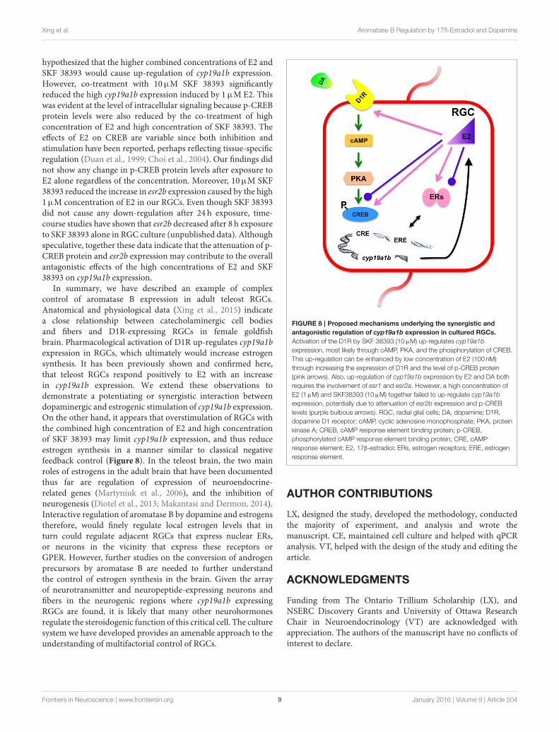
FIGURE 8 | Proposed mechanisms underlying the synergistic and
antagonistic regulation of cyp19a1b expression in cultured RGCs.
Activation of the D1R by SKF 38393 (10µM) up-regulates cyp19a1b
expression, most likely through cAMP, PKA, and the phosphorylation of CREB.
This up-regulation can be enhanced by low concentration of E2 (100 nM)
through increasing the expression of D1R and the level of p-CREB protein
(pink arrows). Also, up-regulation of cyp19a1b expression by E2 and DA both
requires the involvement of esr1 and esr2a. However, a high concentration of
E2 (1µM) and SKF38393 (10µM) together failed to up-regulate cyp19a1b
expression, potentially due to attenuation of esr2b expression and p-CREB
levels (purple bulbous arrows). RGC, radial glial cells; DA, dopamine; D1R,
dopamine D1 receptor; cAMP, cyclic adenosine monophosphate; PKA, protein
kinase A; CREB, cAMP response element binding protein; p-CREB,
phosphorylated cAMP response element binding protein; CRE, cAMP
response element; E2, 17β-estradiol; ERs, estrogen receptors; ERE, estrogen
response element.
AUTHOR CONTRIBUTIONS
LX, designed the study, developed the methodology, conductedthe majority of experiment, and analysis and wrote themanuscript. CE, maintained cell culture and helped with qPCRanalysis. VT, helped with the design of the study and editing thearticle.
ACKNOWLEDGMENTS
Funding from The Ontario Trillium Scholarship (LX), andNSERC Discovery Grants and University of Ottawa ResearchChair in Neuroendocrinology (VT) are acknowledged withappreciation. The authors of the manuscript have no conflicts ofinterest to declare.
Frontiers in Neuroscience | www.frontiersin.org 9 January 2016 | Volume 9 | Article 504

Xing et al. Aromatase B Regulation by 17ß-Estradiol and Dopamine
REFERENCES
Alvarez-Buylla, A., Garcia-Verdugo, J. M., and Tramontin, A. D. (2001). A unified
hypothesis on the lineage of neural stem cells. Nat. Rev. Neurosci. 2, 287–293.
doi: 10.1038/35067582
Arevalo, M. A., Azcoitia, I., and Garcia-Segura, L. M. (2015). The neuroprotective
actions of oestradiol and oestrogen receptors.Nat. Rev. Neurosci. 16, 17–29. doi:
10.1038/nrn3856
Azcoitia, I., Sierra, A., Veiga, S., and Garcia-Segura, L. M. (2003). Aromatase
expression by reactive astroglia is neuroprotective. Ann. N.Y. Acad. Sci. 1007,
298–305. doi: 10.1196/annals.1286.028
Azcoitia, I., Yague, J. G., and Garcia-Segura, L. M. (2011). Estradiol
synthesis within the human brain. Neuroscience 191, 139–147. doi:
10.1016/j.neuroscience.2011.02.012
Balthazart, J., and Ball, G. F. (1998). New insights into the regulation and function
of brain estrogen synthase (aromatase). Trends Neurosci. 21, 243–249. doi:
10.1016/S0166-2236(97)01221-6
Belcher, S. M., and Zsarnovszky, A. (2001). Estrogenic actions in the brain:
estrogen, phytoestrogens, and rapid intracellular signaling mechanisms.
J. Pharmacol. Exp. Ther. 299, 408–414.
Bowman, C. J., Kroll, K. J., Gross, T. G., and Denslow, N. D. (2002). Estradiol-
induced gene expression in largemouth bass (Micropterus salmoides).Mol. Cell.
Endocrinol. 196, 67–77. doi: 10.1016/S0303-7207(02)00224-1
Callard, G. V., Petro, Z., and Ryan, K. J. (1981). Estrogen synthesis in vitro
and in vivo in the brain of a marine teleost (Myoxocephalus). Gen. Comp.
Endocrinol. 43, 243–255. doi: 10.1016/0016-6480(81)90318-X
Callard, G. V., Tchoudakova, A. V., Kishida, M., and Wood, E. (2001). Differential
tissue distribution, developmental programming, estrogen regulation and
promoter characteristics of cyp19 genes in teleost fish. J. Steroid Biochem. Mol.
Biol. 79, 305–314. doi: 10.1016/S0960-0760(01)00147-9
Choi, Y. C., Lee, J. H., Hong, K.W., and Lee, K. S. (2004). 17 Beta-estradiol prevents
focal cerebral ischemic damages via activation of Akt and CREB in association
with reduced PTEN phosphorylation in rats. Fundam. Clin. Pharmacol. 18,
547–557. doi: 10.1111/j.1472-8206.2004.00284.x
Denslow, N. D., Lee, H. S., Bowman, C. J., Hemmer, M. J., and Folmar, L. C. (2001).
Multiple responses in gene expression in fish treated with estrogen. Comp.
Biochem. Physiol. B Biochem. Mol. Biol. 129, 277–282. doi: 10.1016/S1096-
4959(01)00322-0
Diotel, N., Do Rego, J. L., Anglade, I., Vaillant, C., Pellegrini, E., Gueguen, M.-
M., et al. (2011). Activity and expression of steroidogenic enzymes in the
brain of adult zebrafish. Eur. J. Neurosci. 34, 45–56. doi: 10.1111/j.1460-
9568.2011.07731.x
Diotel, N., Le Page, Y., Mouriec, K., Tong, S.-K., Pellegrini, E., Vaillant,
C., et al. (2010). Aromatase in the brain of teleost fish: expression,
regulation and putative functions. Front. Neuroendocrinol. 31:3. doi:
10.1016/j.yfrne.2010.01.003
Diotel, N., Vaillant, C., Gabbero, C., Mironov, S., Fostier, A., Gueguen, M.-M., et al.
(2013). Effects of estradiol in adult neurogenesis and brain repair in zebrafish.
Horm. Behav. 63, 193–207. doi: 10.1016/j.yhbeh.2012.04.003
Duan, W. R., Shin, J. L., and Jameson, J. L. (1999). Estradiol suppresses
phosphorylation of cyclic adenosine 3’,5’-monophosphate response element
binding protein (CREB) in the pituitary: evidence for indirect action via
gonadotropin-releasing hormone.Mol. Endocrinol. 13, 1338–1352.
Fergus, D. J., and Bass, A. H. (2013). Localization and divergent profiles of
estrogen receptors and aromatase in the vocal and auditory networks of a
fish with alternative mating tactics. J. Comp. Neurol. 521, 2850–2869. doi:
10.1002/cne.23320
Fleige, S., and Pfaffl, M. W. (2006). RNA integrity and the effect on the
real-time qRT-PCR performance. Mol. Aspects Med. 27, 126–139. doi:
10.1016/j.mam.2005.12.003
Forlano, P. M., Deitcher, D. L., and Bass, A. H. (2005). Distribution of estrogen
receptor alpha mRNA in the brain and inner ear of a vocal fish with
comparisons to sites of aromatase expression. J. Comp. Neurol. 483, 91–113.
doi: 10.1002/cne.20397
Forlano, P. M., Deitcher, D. L., Myers, D. A., and Bass, A. H. (2001). Anatomical
distribution and cellular basis for high levels of aromatase activity in the brain
of teleost fish: aromatase enzyme and mRNA expression identify glia as source.
J. Neurosci. 21, 8943–8955.
Forlano, P. M., Schlinger, B. A., and Bass, A. H. (2006). Brain aromatase: new
lessons from non-mammalian model systems. Front. Neuroendocrinol. 27:2.
doi: 10.1016/j.yfrne.2006.05.002
Garcia-Segura, L. M. (2008). Aromatase in the brain: not just for
reproduction anymore. J. Neuroendocrinol. 20, 705–712. doi:
10.1111/j.1365-2826.2008.01713.x
Garcia-Segura, L. M., Veiga, S., Sierra, A., Melcangi, R. C., and Azcoitia, I.
(2003). Aromatase: a neuroprotective enzyme. Prog. Neurobiol. 71, 31–41. doi:
10.1016/j.pneurobio.2003.09.005
Garcia-Segura, L. M., Wozniak, A., Azcoitia, I., Rodriguez, J. R., Hutchison, R. E.,
and Hutchison, J. B. (1999). Aromatase expression by astrocytes after brain
injury: implications for local estrogen formation in brain repair. NSC 89,
567–578. doi: 10.1016/s0306-4522(98)00340-6
Heckmann, L. H., Sørensen, P. B., Krogh, P. H., and Sørensen, J. G. (2011).
NORMA-Gene: a simple and robust method for qPCR normalization based on
target gene data. BMC Bioinformatics 12:250. doi: 10.1186/1471-2105-12-250
Hutchison, J. B., and Steimer, T. (1986). Formation of behaviorally effective
17 beta-estradiol in the dove brain: steroid control of preoptic aromatase.
Endocrinology 118, 2180–2187. doi: 10.1210/endo-118-6-2180
Kato, J., Yamada-Mouri, N., and Hirata, S. (1997). Structure of aromatase mRNA
in the rat brain. J. Steroid Biochem. Mol. Biol. 61, 381–385. doi: 10.1016/S0960-
0760(97)80036-2
Kobayashi, M., Aida, K., and Hanyu, I. (1988). Hormone changes during
the ovulatory cycle in goldfish. Gen. Comp. Endocrinol. 69, 301–307. doi:
10.1016/0016-6480(88)90018-4
Lephart, E. D. (1996). A review of brain aromatase cytochrome P450. Brain Res.
Brain Res. Rev. 22, 1–26. doi: 10.1016/0165-0173(96)00002-1
Makantasi, P., and Dermon, C. R. (2014). Estradiol treatment decreases cell
proliferation in the neurogenic zones of adult female zebrafish (Danio
rerio) brain. Neuroscience 277, 306–320. doi: 10.1016/j.neuroscience.2014.
06.071
Marino, M., Galluzzo, P., and Ascenzi, P. (2006). Estrogen signaling multiple
pathways to impact gene transcription. Curr. Genomics 7, 497–508. doi:
10.2174/138920206779315737
Marlatt, V. L., Martyniuk, C. J., Zhang, D., Xiong, H., Watt, J., Xia, X., et al. (2008).
Auto-regulation of estrogen receptor subtypes and gene expression profiling
of 17β-estradiol action in the neuroendocrine axis of male goldfish. Mol. Cell.
Endocrinol. 283, 38–48. doi: 10.1016/j.mce.2007.10.013
Martínez-Cerdeno, V., Noctor, S. C., and Kriegstein, A. R. (2006). Estradiol
stimulates progenitor cell division in the ventricular and subventricular
zones of the embryonic neocortex. Eur. J. Neurosci. 24, 3475–3488. doi:
10.1111/j.1460-9568.2006.05239.x
Martyniuk, C. J., Xiong, H., Crump, K., Chiu, S., Sardana, R., Nadler, A., et al.
(2006). Gene expression profiling in the neuroendocrine brain of male goldfish
(Carassius auratus) exposed to 17 -ethinylestradiol. Physiol. Genomics 27,
328–336. doi: 10.1152/physiolgenomics.00090.2006
McEwen, B. (2002). Estrogen actions throughout the brain. Recent Prog. Horm.
Res. 57, 357–384. doi: 10.1210/rp.57.1.357
Menuet, A., Pellegrini, E., Anglade, I., Blaise, O., Laudet, V., Kah, O., et al. (2002).
Molecular characterization of three estrogen receptor forms in zebrafish:
binding characteristics, transactivation properties, and tissue distributions.
Biol. Reprod. 66, 1881–1892. doi: 10.1095/biolreprod66.6.1881
Menuet, A., Pellegrini, E., Brion, F., Gueguen, M. M., Anglade, I., Pakdel, F., et al.
(2005). Expression and estrogen-dependent regulation of the zebrafish brain
aromatase gene. J. Comp. Neurol. 485, 304–320. doi: 10.1002/cne.20497
Okada, M., Murase, K., Makino, A., Nakajima, M., Kaku, T., Furukawa, S., et al.
(2008). Effects of estrogens on proliferation and differentiation of neural
stem/progenitor cells. Biomed. Res. 29, 163–170. doi: 10.2220/biomedres.29.163
Pellegrini, E., Diotel, N., Vaillant-Capitaine, C., Pérez Maria, R., Gueguen,
M. M., Nasri, A., et al. (2015). Steroid modulation of neurogenesis:
focus on radial glial cells in zebrafish. J. Steroid Biochem. Mol. Biol. doi:
10.1016/j.jsbmb.2015.06.011. [Epub ahead of print].
Pellegrini, E., Menuet, A., Lethimonier, C., Adrio, F., Gueguen, M.-M., Tascon,
C., et al. (2005). Relationships between aromatase and estrogen receptors
in the brain of teleost fish. Gen. Comp. Endocrinol. 142, 60–66. doi:
10.1016/j.ygcen.2004.12.003
Pellegrini, E., Mouriec, K., Anglade, I., Menuet, A., Le Page, Y., Gueguen, M.-M.,
et al. (2007). Identification of aromatase-positive radial glial cells as progenitor
Frontiers in Neuroscience | www.frontiersin.org 10 January 2016 | Volume 9 | Article 504

Xing et al. Aromatase B Regulation by 17ß-Estradiol and Dopamine
cells in the ventricular layer of the forebrain in zebrafish. J. Comp. Neurol. 501,
150–167. doi: 10.1002/cne.21222
Pinto, C., Grimaldi, M., Boulahtouf, A., Pakdel, F., Brion, F., Aït-Aïssa, S.,
et al. (2014). Selectivity of natural, synthetic and environmental estrogens
for zebrafish estrogen receptors. Toxicol. Appl. Pharmacol. 280, 60–69. doi:
10.1016/j.taap.2014.07.020
Strobl-Mazzulla, P. H., Lethimonier, C., Gueguen, M.-M., Karube, M., Fernandino,
J. I., Yoshizaki, G., et al. (2008). Brain aromatase (Cyp19A2) and estrogen
receptors, in larvae and adult pejerrey fish Odontesthes bonariensis:
neuroanatomical and functional relations. Gen. Comp. Endocrinol. 158,
191–201. doi: 10.1016/j.ygcen.2008.07.006
Tchoudakova, A., Kishida, M., Wood, E., and Callard, G. V. (2001). Promoter
characteristics of two cyp19 genes differentially expressed in the brain and ovary
of teleost fish. J. Steroid Biochem. Mol. Biol. 78, 427–439. doi: 10.1016/S0960-
0760(01)00120-0
Tong, S.-K., Mouriec, K., Kuo, M.-W., Pellegrini, E., Gueguen, M.-M., Brion,
F., et al. (2009). A cyp19a1b-gfp(aromatase B) transgenic zebrafish line that
expresses GFP in radial glial cells. Genesis 47, 67–73. doi: 10.1002/dvg.20459
Tsuruo, Y., Ishimura, K., and Osawa, Y. (1995). Presence of estrogen receptors in
aromatase-immunoreactive neurons in the mouse brain. Neurosci. Lett. 195,
49–52. doi: 10.1016/0304-3940(95)11779-V
Ubuka, T., Haraguchi, S., Tobari, Y., Narihiro, M., Ishikawa, K., Hayashi, T.,
et al. (2014). Hypothalamic inhibition of socio-sexual behaviour by increasing
neuroestrogen synthesis. Nat. Commun. 5, 3061. doi: 10.1038/ncomms4061
Ubuka, T., and Tsutsui, K. (2014). Review: neuroestrogen regulation of socio-
sexual behavior of males. Front. Neurosci. 8:323. doi: 10.3389/fnins.2014.00323
Xing, L., Goswami, M., and Trudeau, V. L. (2014). Radial glial cell: critical
functions and new perspective as a steroid synthetic cell. Gen. Comp.
Endocrinol. 203, 181–185. doi: 10.1016/j.ygcen.2014.03.010
Xing, L., McDonald, H., Da Fonte, D. F., Gutierrez-Villagomez, J. M., and Trudeau,
V. L. (2015). Dopamine D1 receptor activation regulates the expression of the
estrogen synthesis gene aromatase B in radial glial cells. Front. Neurosci. 9:310.
doi: 10.3389/fnins.2015.00310
Yague, J. G., Muñoz, A., de Monasterio-Schrader, P., Defelipe, J., Garcia-
Segura, L. M., and Azcoitia, I. (2006). Aromatase expression in the human
temporal cortex. Neuroscience 138, 389–401. doi: 10.1016/j.neuroscience.2005.
11.054
Zhang, D., and Trudeau, V. L. (2006). Integration of membrane and nuclear
estrogen receptor signaling.Comp. Biochem. Physiol. AMol. Integr. Physiol. 144,
306–315. doi: 10.1016/j.cbpa.2006.01.025
Zhao, C., Fujinaga, R., Tanaka, M., Yanai, A., Nakahama, K., and Shinoda, K.
(2007). Region-specific expression and sex-steroidal regulation on aromatase
and its mRNA in the male rat brain: immunohistochemical and in situ
hybridization analyses. J. Comp. Neurol. 500, 557–573. doi: 10.1002/cne.
21193
Zhao, E., Basak, A., Crump, K., and Trudeau, V. L. (2006). Proteolytic processing
and differential distribution of secretogranin-II in goldfish. Gen. Comp.
Endocrinol. 146, 100–107. doi: 10.1016/j.ygcen.2005.10.007
Zupanc, G. K., and Clint, S. C. (2003). Potential role of radial glia in
adult neurogenesis of teleost fish. Glia 43, 77–86. doi: 10.1002/glia.
10236
Conflict of Interest Statement: The authors declare that the research was
conducted in the absence of any commercial or financial relationships that could
be construed as a potential conflict of interest.
Copyright © 2016 Xing, Esau and Trudeau. This is an open-access article distributed
under the terms of the Creative Commons Attribution License (CC BY). The use,
distribution or reproduction in other forums is permitted, provided the original
author(s) or licensor are credited and that the original publication in this journal
is cited, in accordance with accepted academic practice. No use, distribution or
reproduction is permitted which does not comply with these terms.
Frontiers in Neuroscience | www.frontiersin.org 11 January 2016 | Volume 9 | Article 504