diffusing wave spectroscopy microrheology of actin filament networks
TRANSCRIPT
Diffusing Wave Spectroscopy Microrheology of Actin Filament Networks
Andre Palmer,* Thomas G. Mason,* Jingyuan Xu,* Scot C. Kuo,# and Denis Wirtz**Department of Chemical Engineering, The Johns Hopkins University, Baltimore, Maryland 21218, and #Department of BiomedicalEngineering, The Johns Hopkins University, Baltimore, Maryland 21205 USA
ABSTRACT Filamentous actin (F-actin), one of the constituents of the cytoskeleton, is believed to be the most importantparticipant in the motion and mechanical integrity of eukaryotic cells. Traditionally, the viscoelastic moduli of F-actin networkshave been measured by imposing a small mechanical strain and quantifying the resulting stress. The magnitude of theviscoelastic moduli, their concentration dependence and strain dependence, as well as the viscoelastic nature (solid-like orliquid-like) of networks of uncross-linked F-actin, have been the subjects of debate. Although this paper helps to resolve thedebate and establishes the extent of the linear regime of F-actin networks’ rheology, we report novel measurements of thehigh-frequency behavior of networks of F-actin, using a noninvasive light-scattering based technique, diffusing wavespectroscopy (DWS). Because no external strain is applied, our optical assay generates measurements of the mechanicalproperties of F-actin networks that avoid many ambiguities inherent in mechanical measurements. We observe that the elasticmodulus has a small magnitude, no strain dependence, and a weak concentration dependence. Therefore, F-actin alone isnot sufficient to generate the elastic modulus necessary to sustain the structural rigidity of most cells or support new cellularprotrusions. Unlike previous studies, our measurements show that the mechanical properties of F-actin are highly dependenton the frequency content of the deformation. We show that the loss modulus unexpectedly dominates the elastic modulusat high frequencies, which are key for fast transitions. Finally, the measured mean square displacement of the optical probes,which is also generated by DWS measurements, offers new insight into the local bending fluctuations of the individual actinfilaments and shows how they generate enhanced dissipation at short time scales.
INTRODUCTION
Filamentous actin (F-actin), one of the constituents of thecytoskeleton, is believed to be one of the most importantparticipants in the mechanical integrity of eukaryotic cells.The mechanism by which F-actin networks provide cellswith structural rigidity is attributed to their viscoelasticnature, characterized by viscoelastic moduli.
However, major discrepancies between reported valuesof the magnitude of the viscoelastic moduli of uncross-linked F-actin networks, their concentration dependence,strain dependence, and frequency dependence have ap-peared in the literature. These vastly different rheologicaldata have yielded different conclusions regarding the sepa-rate role of F-actin in the cell’s structural rigidity. Tradi-tionally, the linear viscoelastic moduli of F-actin networksin vitro have been investigated by imposing a mechanicalstrain and quantifying the resulting stress. Reported elasticmoduli of uncross-linked F-actin networks, measured usingmechanical deformation, differ by more than two orders ofmagnitude, between 10 and 40 dynes/cm2 (Pollard et al.,1986; Sato et al., 1987; Wachstock et al., 1993) and 1000–5000 dynes/cm2 (Hvidt and Heller, 1990; Janmey et al.,1990, 1991, 1994; Hvidt and Janmey, 1990). Therefore,concentrated F-actin solutions have been described as form-ing networks that are either weak or stiff (note: we use the
terms “network” and “solution” interchangeably; both termsdescribe a semidilute solution of highly purified, uncross-linked F-actin). Furthermore, some researchers have re-ported that networks of uncross-linked F-actin harden forstrains between 1% and 10% (Hvidt and Heller, 1990;Janmey et al., 1990, 1994), which corresponds to elasticmoduli increasing with strain, and yield beyond 10%. Incontrast, other researchers have not observed any strain-hardening effect, only the onset of yielding when the strainreaches 10% (Pollard et al., 1986; this work). Finally, vastlydifferent values of the relative magnitudes of elastic andloss moduli, which characterize the nature of the viscoelas-ticity, have helped researchers to characterize F-actin net-works as either viscoelastic solid or viscoelastic liquids.
The actual magnitude of the elastic modulus is importantbecause a high value would support the hypothesis thatisotropic F-actin networks alone are strong enough to sta-bilize cells (Janmey et al., 1994). A high value of the elasticmodulus also provides a baseline against which to monitorsubtle changes in the mechanical properties of F-actin net-works due to regulating proteins such as capping and sev-ering proteins, as well as small changes due to polymeriza-tion of actin filaments from ATP-containing or ADP-containing subunits (Newman et al., 1993; Pollard et al.,1986; Janmey et al., 1991). Finally, it supports the hypoth-esis that F-actin alone can effectively provide structuralrigidity for the reinforcement of a new cellular protrusion(Condeelis, 1992).
The strain dependence of F-actin networks’ rheology isalso important: if F-actin networks strain-harden, they canpotentially stabilize the cytoskeleton, even when it is sub-ject to large deformations. Moreover, the strain dependence
Received for publication 30 April 1998 and in final form 19 October 1998.
Address reprint requests to Dr. Denis Wirtz, Department of ChemicalEngineering, Johns Hopkins University, Maryland Hall, Room 221, 3400North Charles Street, Baltimore, MD 21217. Tel.: 410-516-7006; Fax:410-516-5510; E-mail: [email protected].
© 1999 by the Biophysical Society
0006-3495/99/02/1063/09 $2.00
1063Biophysical Journal Volume 76 February 1999 1063–1071
of the viscoelastic moduli establishes the extent of therheological linear regime (for which moduli are independentof strain), which is important because it allows for mean-ingful comparison with theoretical models and other me-chanical measurements.
Given the controversy surrounding traditional methods,we have developed a novel, light-scattering-based tech-nique, diffusing wave spectroscopy (DWS), which probesthe linear rheological properties of biopolymer networksnoninvasively (Palmer et al., 1998; Petka et al., 1998). Thistechnique and associated analysis are based on the measure-ment of the autocorrelation function of the light multiplyscattered from microspheres imbedded in the network.From the measured autocorrelation function and using ageneralized Stokes-Einstein equation, we extract the meansquare displacement of the probing microspheres. Fromthese mean square displacements, we calculate the vis-coelastic moduli of networks of highly purified, uncross-linked F-actin. This technique is somewhat similar to singleparticle tracking microrheology (SPTM), recently devel-oped by Wirtz and co-workers (Mason et al., 1997a,b; Xu etal., 1998c; Ganesan et al., unpublished data) and Schmidtand co-workers (Schnurr et al., 1997a,b). Unlike SPTM,which monitors the displacement of a single particle at atime, DWS monitors many thousands of microspheres simul-taneously, which allows for superior statistics. However, un-like DWS, SPTM allows us to probe the mechanical propertiesof an anisotropic F-actin specimen and the cytoskeleton ofa living cell in situ (Ganesan et al., unpublished data).
Our optical rheometry assay generates measurements ofthe viscoelastic moduli of F-actin networks that are lessambiguous than mechanical measurements. In particular,this instrument exploits the small, random, thermally in-duced force generated by imbedded microspheres and there-fore avoids possible flow-induced orientation and bundlingof actin filaments. In the absence of externally appliedforces, optical rheometry probes the viscoelastic propertiesof networks in the linear small-strain regime almost bydefinition. Moreover, this optical instrument greatly in-creases the frequency range of the probed viscoelastic mod-uli: we routinely probe frequency-dependent loss and elasticmoduli at frequencies up to 1 MHz, which is four orders ofmagnitude larger than possible with mechanical rheometry.This extended range of frequency allows us to probe theinternal dynamics of actin filaments in concentrated solu-tions as well as the effect of fast cellular transitions onF-actin rheology. Finally, the measured mean square dis-placement of the optical probes, which is also generated byDWS measurements, offers new insight into the local bend-ing fluctuations of the individual actin filaments and showshow these generate large dissipation at short time scales.
EXPERIMENTAL TECHNIQUES
Actin preparation
Actin was purified from rabbit skeletal acetone powder by the method ofSpudich and Watt (1971). The resulting actin was then gel filtered on
Sephacryl S-300 HR instead of Sephadex G-150 (MacLean-Fletcher andPollard, 1980). The purified actin was stored as Ca2�-actin in continuousdialysis at 4°C against buffer G (0.2 mM ATP, 0.5 mM dithiothreitol, 0.1mM CaCl2, 1 mM sodium azide, and 2 mM Tris-Cl, pH 8.0). The finalactin concentration was determined by ultraviolet absorbance at 290 nm,using an extinction coefficient of 2.66 � 104 M�1 cm�1 and a cell pathlength of 1 cm. Mg2� actin filaments were generated by adding 0.1 volumeof 10� KME (500 mM KCl, 10 mM MgCl2, 10 mM EGTA, 100 mMimidazole, pH 7.0) polymerizing salt buffer solution to 0.9 volume ofG-actin in buffer G. We did not observe large variations in the mechanicalproperties of F-actin networks measured by optical and mechanical rheo-metry on actin extracted and purified by the method of MacLean-Fletcherand Pollard (1980) and by the new method of Casella et al. (Casella andTorres, 1994; Casella et al., 1995). This new actin purification methodincludes an additional cycle of polymerization/depolymerization followedby an additional gel filtration step. We verified that G-actin, which isfiltrated once, contained negligible amounts of capping proteins by mon-itoring the polymerization of actin, using time-resolved fluorescence spec-troscopy and pyrene-labeled actin (not shown here). We also note that ouractin samples were never frozen for storage purpose.
For this study, six actin concentrations ranging from 10 �M (0.42mg/ml) to 164 �M (6.89 mg/ml) are reported. While uncross-linked F-actinsolutions of concentrations smaller than �48 �M are isotropic liquids,F-actin solutions of concentrations greater than 48 �M form a highlyordered liquid-crystalline phase (Coppin and Leavis, 1993): we reportoptically measured viscoelastic moduli of F-actin solutions in the liquid-crystalline phase and in the isotropic phase.
Diffusing wave spectroscopy
Instrument
The beam from an Ar� ion laser operating in the single-line frequencymode at a wavelength of 514 nm is focused and incident upon a flatscattering cell that contains the F-actin network and spherical opticalprobes. The light multiply scattered from the solution is collected by twophotomultiplier tubes (PMTs) via a single-mode optical fiber with a col-limator lens of very narrow angle of acceptance at its front end and abeamsplitter at its back end (Weitz and Pine, 1993). The outputs of thePMTs are directed to a correlator working in the pseudo-cross-correlationmode to generate the autocorrelation function g2(t) � 1, from whichquiescent rheological properties of the F-actin solutions can be calculated.
Actin is polymerized in situ for 12 h before measurement by loading thescattering cell with a solution of G-actin mixed with the polymerizing saltsolution and a dilute suspension of monodisperse latex microspheres (DukeScientific Corp.) of radius 0.48 �m at a volume fraction of 1%. Thescattering cell is then immediately tightly capped. Using time-resolvedmechanical rheology, we verified that G-actin was fully polymerized intoF-actin and that F-actin had formed an equilibrium network at all concen-trations presented in this paper before 12 h. Using mechanical rheology, wealso verified that the rheology of F-actin is identical in the presence and inthe absence of the added latex beads. Using static light scattering, weverified that more than 98% of the scattering intensity in the transmissiongeometry was due to the microspheres, and less than 2% was due to theF-actin filament network itself. We also verified that the measured meansquare displacement scaled inversely with the radius of the probing beadfor diameters larger than 0.55 �m and smaller than 1.95 �m (see Eq. 1below). Smaller particles did not display saturation of their displacement atlarge times (they percolated through the network), and larger particlessedimented too rapidly (data not shown). All DWS measurements areconducted at a temperature of 23°C.
Analysis
The general method used to extract G�(�) and G�(�) from g2(t) � 1 via thecalculated values of the mean square displacement of the optical probes isdescribed in Mason and Weitz (1995) and Palmer et al. (1998). The method
1064 Biophysical Journal Volume 76 February 1999
is based on a relation between the mean square displacement of a micro-sphere imbedded in a viscoelastic medium and the viscoelastic modulus ofthat medium. By assuming that Stokes’ law, which holds for a purelyviscous fluid, can be generalized to all frequencies, one obtains
G�s � s��s �kBT
�as�r2�s�, (1)
where s is the Laplace frequency and ˜ represents the unilateral Laplacetransformation, defined as X(s) L[X(t)] �0
� X(t)exp(�st)dt. Equation 1,which neglects inertial effects, relates the unilateral Laplace transforms ofthe stress relaxation modulus Gr(t), G(s) sGr(s), and of the mean squaredisplacement �r2(t)�. We use the analytic continuation between the realfunction G(s) and the complex function G*(�) G�(�) � iG�(�) to extractelastic and loss moduli by extracting real and imaginary parts from theimaginary function G(s � i�).
Mechanical rheometry
To compare our optical measurements with classical mechanical measure-ments (Ferry, 1980), we employ a strain-controlled mechanical rheometer(ARES-100 Rheometrics) equipped with a 50-mm-diameter cone-and-plategeometry. To prevent possible evaporation of the buffer, the cone-and-plate tools are enclosed in a custom-made vapor trap. The temperature ofthe sample is fixed at 23°C to within 0.1°C. The G-actin solution is placedbetween the cone-and-plate tools and allowed to polymerize in the pres-ence of polymerizing salt for 12 h before the measurements. The viscoelas-tic properties of F-actin networks were found independently of time after12 h at all actin concentrations used in this work. The linear equilibriumvalues G�(�) and G�(�) of the actin solutions are measured by setting theamplitude of the oscillatory strain at � � 1% and sweeping from low tohigh frequency. The strain-dependent viscoelastic moduli were measuredby subjecting the F-actin solution to a 1-rad/s oscillatory deformation ofincreasing amplitude; G� and G� are computed from the maximum mag-nitude of the measured stress (Palmer et al., 1998; Mason et al., 1998).
RESULTS
Computation of the mechanical properties of F-actin net-works from multiple light scattering measurements pro-ceeds via several steps. We report the autocorrelation func-tion g2(t) � 1 of the multiply scattered light intensity,measured with a fast intensity correlator. From g2(t) � 1,we compute the mean square displacement �r2(t)� of themicrospheres (for further details about how �r2(t)� is cal-culated from g2(t) � 1, see Weitz and Pine, 1993; Palmer etal., 1998), from which we extract the time-dependent dif-fusion constant D(t). From �r2(t)�, we calculate the vis-coelastic modulus G(s), and finally the frequency-depen-dent viscoelastic moduli G�(�) and G�(�).
Correlation function, mean square displacement,and time-dependent diffusion coefficient
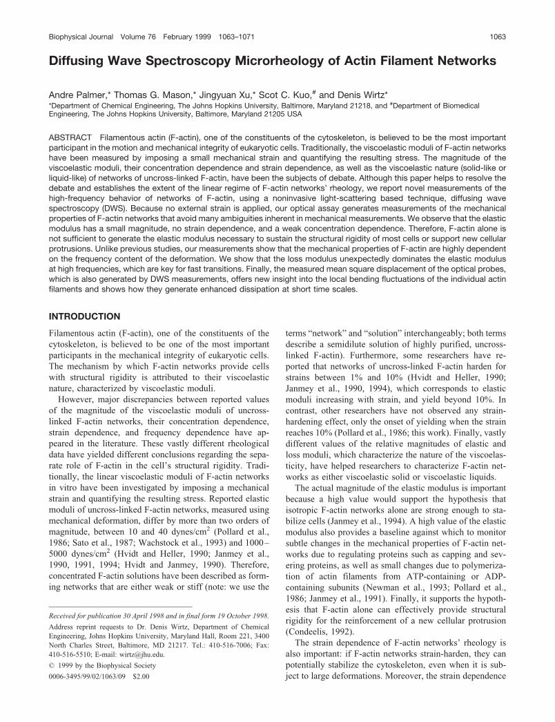
The light of an Ar� ion laser is incident upon the F-actinnetwork and multiply scattered by the microbeads imbeddedinside the network. We measure the instantaneous intensityof the light intensity multiply scattered by the microspheres,transmitted through the network, detected by the PMTs, andthen autocorrelated to generate the autocorrelation functiong2(t) � 1. Fig. 1 shows g2(t) � 1 over a large temporal range
corresponding to �10 time decades, between 10�7 s and103 s. By collecting at least four DWS runs for each actinconcentration, we confirmed the reproducibility of our op-tical measurements of g2(t) � 1. Sample aging effects arenegligible; no significant optical and rheological changesoccurred within 3–5 days after the preparation of G-actinand (immediate) subsequent polymerization for 12 h. Atlarge actin concentration, problems from lack of ergodicitycan arise because of the onset of inhomogeneities in F-actinnetworks. To confirm ergodicity, we conducted runs on thesame F-actin samples with the laser beam incident upon fivedifferent points of the face of the scattering cell. Localvariations of g2(t) � 1 were found to be negligible for actinconcentration smaller than 48 �M but observed to slightlyincrease for increasing actin concentration (data not shown).
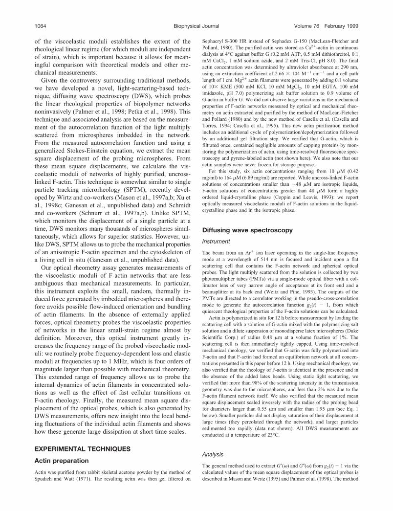
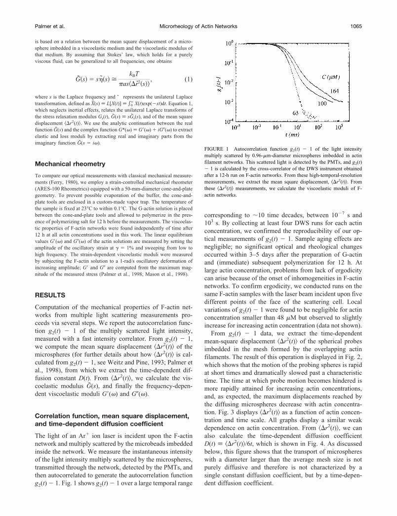
From g2(t) � 1 data, we extract the time-dependentmean-square displacement �r2(t)� of the spherical probesimbedded in the mesh formed by the overlapping actinfilaments. The result of this operation is displayed in Fig. 2,which shows that the motion of the probing spheres is rapidat short times and dramatically slowed past a characteristictime. The time at which probe motion becomes hindered ismore rapidly attained for increasing actin concentrations,and, as expected, the maximum displacements reached bythe diffusing microspheres decrease with actin concentra-tion. Fig. 3 displays �r2(t)� as a function of actin concen-tration and time scale. All graphs display a similar weakdependence on actin concentration. From �r2(t)�, we canalso calculate the time-dependent diffusion coefficientD(t) �r2(t)�/6t, which is shown in Fig. 4. As discussedbelow, this figure shows that the transport of microsphereswith a diameter larger than the average mesh size is notpurely diffusive and therefore is not characterized by asingle constant diffusion coefficient, but by a time-depen-dent diffusion coefficient.
FIGURE 1 Autocorrelation function g2(t) � 1 of the light intensitymultiply scattered by 0.96-�m-diameter microspheres imbedded in actinfilament networks. This scattered light is detected by the PMTs, and g2(t)� 1 is calculated by the cross-correlator of the DWS instrument obtainedafter a 12-h run on F-actin networks. From these high-temporal-resolutionmeasurements, we extract the mean square displacement, �r2(t)�. Fromthese �r2(t)� measurements, we calculate the viscoelastic moduli of F-actin networks.
Palmer et al. Microrheology of Actin Networks 1065
Complex modulus
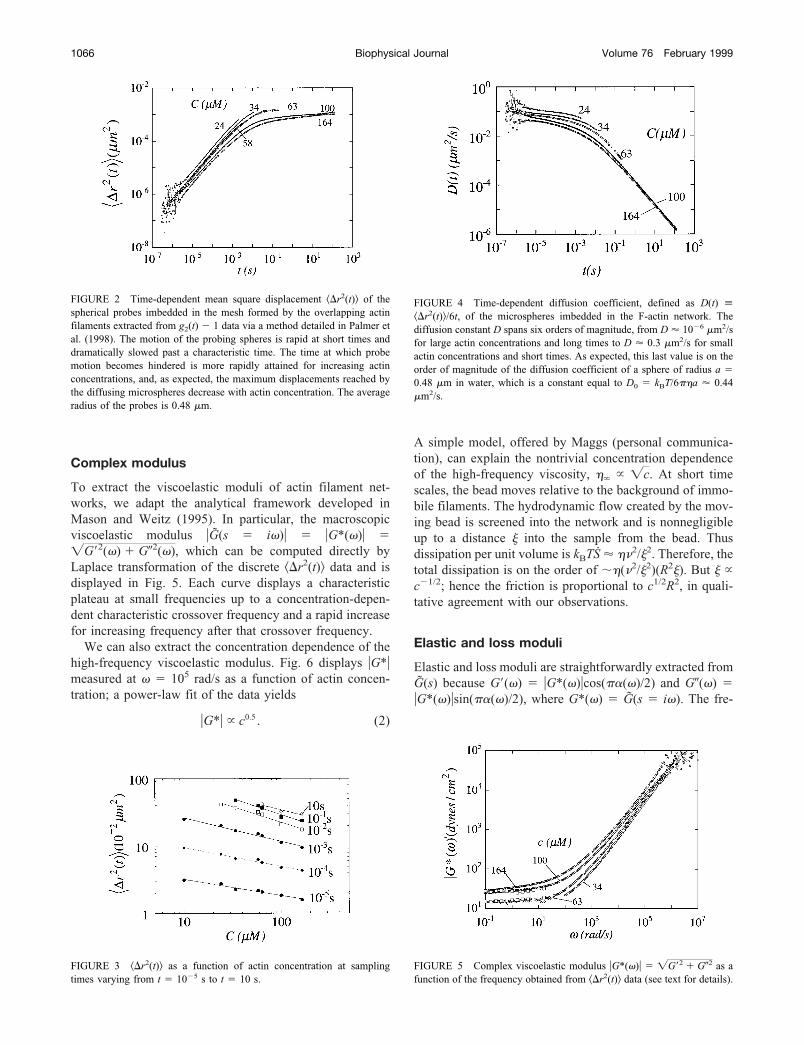
To extract the viscoelastic moduli of actin filament net-works, we adapt the analytical framework developed inMason and Weitz (1995). In particular, the macroscopicviscoelastic modulus �G(s � i�)� � �G*(�)� ��G�2(�) � G�2(�), which can be computed directly byLaplace transformation of the discrete �r2(t)� data and isdisplayed in Fig. 5. Each curve displays a characteristicplateau at small frequencies up to a concentration-depen-dent characteristic crossover frequency and a rapid increasefor increasing frequency after that crossover frequency.
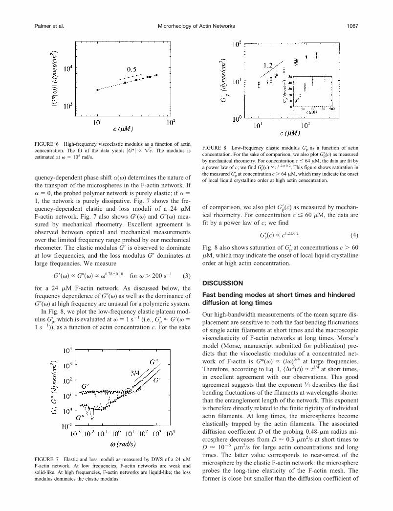
We can also extract the concentration dependence of thehigh-frequency viscoelastic modulus. Fig. 6 displays �G*�measured at � � 105 rad/s as a function of actin concen-tration; a power-law fit of the data yields
�G*� � c0.5 . (2)
A simple model, offered by Maggs (personal communica-tion), can explain the nontrivial concentration dependenceof the high-frequency viscosity, �� � �c. At short timescales, the bead moves relative to the background of immo-bile filaments. The hydrodynamic flow created by the mov-ing bead is screened into the network and is nonnegligibleup to a distance � into the sample from the bead. Thusdissipation per unit volume is kBTS � �2/�2. Therefore, thetotal dissipation is on the order of ��(2/�2)(R2�). But � �c�1/2; hence the friction is proportional to c1/2R2, in quali-tative agreement with our observations.
Elastic and loss moduli
Elastic and loss moduli are straightforwardly extracted fromG(s) because G�(�) � �G*(�)�cos(�(�)/2) and G�(�) ��G*(�)�sin(�(�)/2), where G*(�) � G(s � i�). The fre-
FIGURE 2 Time-dependent mean square displacement �r2(t)� of thespherical probes imbedded in the mesh formed by the overlapping actinfilaments extracted from g2(t) � 1 data via a method detailed in Palmer etal. (1998). The motion of the probing spheres is rapid at short times anddramatically slowed past a characteristic time. The time at which probemotion becomes hindered is more rapidly attained for increasing actinconcentrations, and, as expected, the maximum displacements reached bythe diffusing microspheres decrease with actin concentration. The averageradius of the probes is 0.48 �m.
FIGURE 3 �r2(t)� as a function of actin concentration at samplingtimes varying from t � 10�5 s to t � 10 s.
FIGURE 4 Time-dependent diffusion coefficient, defined as D(t) �r2(t)�/6t, of the microspheres imbedded in the F-actin network. Thediffusion constant D spans six orders of magnitude, from D � 10�6 �m2/sfor large actin concentrations and long times to D � 0.3 �m2/s for smallactin concentrations and short times. As expected, this last value is on theorder of magnitude of the diffusion coefficient of a sphere of radius a �0.48 �m in water, which is a constant equal to D0 � kBT/6��a � 0.44�m2/s.
FIGURE 5 Complex viscoelastic modulus �G*(�)� � �G�2 � G�2 as afunction of the frequency obtained from �r2(t)� data (see text for details).
1066 Biophysical Journal Volume 76 February 1999
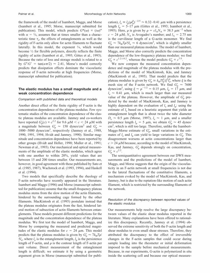
quency-dependent phase shift (�) determines the nature ofthe transport of the microspheres in the F-actin network. If � 0, the probed polymer network is purely elastic; if �1, the network is purely dissipative. Fig. 7 shows the fre-quency-dependent elastic and loss moduli of a 24 �MF-actin network. Fig. 7 also shows G�(�) and G�(�) mea-sured by mechanical rheometry. Excellent agreement isobserved between optical and mechanical measurementsover the limited frequency range probed by our mechanicalrheometer. The elastic modulus G� is observed to dominateat low frequencies, and the loss modulus G� dominates atlarge frequencies. We measure
G��� � G��� � �0.78�0.10 for � � 200 s�1 (3)
for a 24 �M F-actin network. As discussed below, thefrequency dependence of G�(�) as well as the dominance ofG�(�) at high frequency are unusual for a polymeric system.
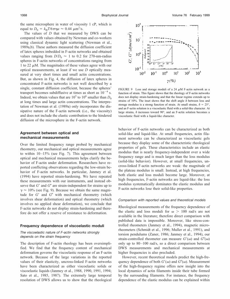
In Fig. 8, we plot the low-frequency elastic plateau mod-ulus G�p, which is evaluated at � � 1 s�1 (i.e., G�p � G�(� �1 s�1)), as a function of actin concentration c. For the sake
of comparison, we also plot G�p(c) as measured by mechan-ical rheometry. For concentration c � 60 �M, the data arefit by a power law of c; we find
G�p�c � c1.2�0.2 . (4)
Fig. 8 also shows saturation of G�p at concentrations c � 60�M, which may indicate the onset of local liquid crystallineorder at high actin concentration.
DISCUSSION
Fast bending modes at short times and hindereddiffusion at long times
Our high-bandwidth measurements of the mean square dis-placement are sensitive to both the fast bending fluctuationsof single actin filaments at short times and the macroscopicviscoelasticity of F-actin networks at long times. Morse’smodel (Morse, manuscript submitted for publication) pre-dicts that the viscoelastic modulus of a concentrated net-work of F-actin is G*(�) � (i�)3/4 at large frequencies.Therefore, according to Eq. 1, �r2(t)� � t3/4 at short times,in excellent agreement with our observations. This goodagreement suggests that the exponent 3⁄4 describes the fastbending fluctuations of the filaments at wavelengths shorterthan the entanglement length of the network. This exponentis therefore directly related to the finite rigidity of individualactin filaments. At long times, the microspheres becomeelastically trapped by the actin filaments. The associateddiffusion coefficient D of the probing 0.48-�m radius mi-crosphere decreases from D � 0.3 �m2/s at short times toD � 10�6 �m2/s for large actin concentrations and longtimes. The latter value corresponds to near-arrest of themicrosphere by the elastic F-actin network: the microsphereprobes the long-time elasticity of the F-actin mesh. Theformer is close but smaller than the diffusion coefficient of
FIGURE 6 High-frequency viscoelastic modulus as a function of actinconcentration. The fit of the data yields �G*� � �c. The modulus isestimated at � � 105 rad/s.
FIGURE 7 Elastic and loss moduli as measured by DWS of a 24 �MF-actin network. At low frequencies, F-actin networks are weak andsolid-like. At high frequencies, F-actin networks are liquid-like; the lossmodulus dominates the elastic modulus.
FIGURE 8 Low-frequency elastic modulus G�p as a function of actinconcentration. For the sake of comparison, we also plot G�p(c) as measuredby mechanical rheometry. For concentration c � 64 �M, the data are fit bya power law of c; we find G�p(c) � c1.2�0.2. This figure shows saturation inthe measured G�p at concentration c � 64 �M, which may indicate the onsetof local liquid crystalline order at high actin concentration.
Palmer et al. Microrheology of Actin Networks 1067
the same microsphere in water of viscosity 1 cP, which isequal to D0 � kBT/6��a � 0.44 �m2/s.
The values of D that we measured by DWS can becompared with values obtained by Newman and co-workersusing classical dynamic light scattering (Newman et al.,1989a,b). These authors measured the diffusion coefficientof latex spheres imbedded in F-actin networks and obtainedvalues ranging from D/D0 � 1 to 0.2 for 270-nm-radiusspheres in F-actin networks of concentrations ranging from1 to 22 �M. The magnitudes of these values agree with ouroptical measurements, at least if we use D optically mea-sured at very short times and small actin concentrations.But, as shown in Fig. 4, the diffusion of latex spheres inconcentrated F-actin networks is not well described by asingle, constant diffusion coefficient, because the spheres’transport becomes subdiffusive at times as short as 10�4 s.Indeed, we obtain values that are 105 to 106 smaller than D0
at long times and large actin concentrations. The interpre-tation of Newman et al. (1989a) only incorporates the dis-sipative nature of the F-actin network (i.e., the viscosity)and does not include the elastic contribution to the hindereddiffusion of the microsphere in the F-actin network.
Agreement between optical andmechanical measurements
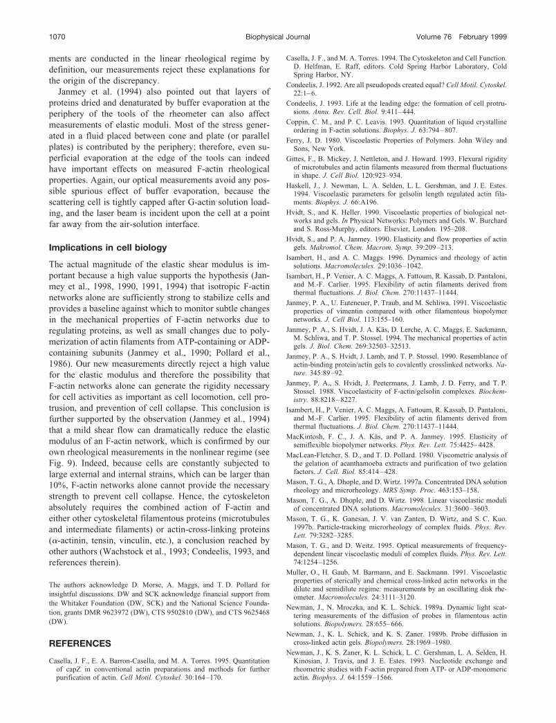
Over the limited frequency range probed by mechanicalrheometry, our mechanical and optical measurements agreeto within 10–15% (see Fig. 7). This agreement betweenoptical and mechanical measurements helps clarify the be-havior of F-actin under deformation. Researchers have re-ported conflicting observations regarding the low-strain be-havior of F-actin networks. In particular, Janmey et al.(1994) have reported strain-hardening. We have repeatedthese measurements with our instruments, and instead ob-serve that G� and G� are strain-independent for strains up to� � 10% (see Fig. 9). Because we obtain the same magni-tude for G� and G� with mechanical rheometry (whichinvolves shear deformation) and optical rheometry (whichinvolves no applied shear deformation), we conclude thatF-actin networks do not display strain-hardening and there-fore do not offer a reserve of resistance to deformation.
Frequency dependence of viscoelastic moduli
The viscoelastic nature of F-actin networks stronglydepends on the strain frequency
The description of F-actin rheology has been oversimpli-fied. We find that the frequency content of mechanicaldeformation governs the viscoelastic response of an F-actinnetwork. Because of the large variations in the reportedvalues of their elasticity, uncross-linked F-actin networkshave been characterized as either viscoelastic solids orviscoelastic liquids (Janmey et al., 1988, 1990, 1991, 1994;Sato et al., 1985, 1987). The extremely large temporalresolution of DWS allows us to show that the rheological
behavior of F-actin networks can be characterized as bothsolid-like and liquid-like. At small frequencies, actin fila-ment networks can be characterized as viscoelastic gelsbecause they display some of the characteristic rheologicalproperties of gels. These characteristics include an elasticmodulus that is nearly frequency-independent over a widefrequency range and is much larger than the loss modulus(solid-like behavior). However, at small frequencies, un-cross-linked F-actin networks are weak: the magnitude ofthe plateau modulus is small. Instead, at high frequencies,both elastic and loss moduli become large. Moreover, athigh frequencies, F-actin networks are liquid-like: the lossmodulus systematically dominates the elastic modulus andF-actin networks lose their solid-like properties.
Comparison with reported values and theoretical models
Rheological measurements of the frequency dependence ofthe elastic and loss moduli for � � 100 rad/s are notavailable in the literature; therefore direct comparison withpublished data is impossible. Moreover, like stress-con-trolled rheometers (Janmey et al., 1994), magnetic micro-rheometers (Schmidt et al., 1996; Muller et al., 1991), andtorsion pendulums (Zaner, 1986; Janmey et al., 1994), ourstrain-controlled rheometer can measure G�(�) and G�(�)only up to 80–100 rad/s, so a direct comparison betweenDWS measurements and mechanical measurements athigher frequencies is also precluded.
However, recent theoretical models predict the high-fre-quency dependence of both G�(�) and G�(�). Measurementof the high-frequency regime offers new insight into thelocal dynamics of actin filaments inside their tube formedby the surrounding filaments. For instance, the frequencydependence of the elastic modulus can be explained within
FIGURE 9 Loss and storage moduli of a 24 �M F-actin network as afunction of strain. This figure shows that the rheology of F-actin networksdoes not display strain-hardening and that the linear regime extends up tostrains of 10%. The inset shows that the shift angle between loss andstorage modulus is a strong function of strain. At small strains, � 25°,and an F-actin solution is a viscoelastic fluid with a solid-like character. Atlarge strains, increases toward 90°, and an F-actin solution becomes aviscoelastic fluid with a liquid-like character.
1068 Biophysical Journal Volume 76 February 1999
the framework of the model of Isambert, Maggs, and Morse(Isambert et al., 1995; Morse, manuscript submitted forpublication). This model, which predicts G*(�) � (i�)
with � 3⁄4, assumes that at times smaller than a charac-teristic time �e, the effects of entanglements as well as therigidity of F-actin only allow for actin filaments to fluctuatelaterally. In this model, the exponent 3⁄4, which wouldbecome 1⁄2 for flexible polymers, directly reflects the finiterigidity of actin (Isambert et al., 1995; Gittes et al., 1993).Because the ratio of loss and storage moduli is related to by G�/G� � tan(�/2) � 2.41, Morse’s model correctlypredicts that dissipation effects dominate the viscoelasticresponse of F-actin networks at high frequencies (Morse,manuscript submitted for publication).
The elastic modulus has a small magnitude and aweak concentration dependence
Comparison with published data and theoretical models
Another direct effect of the finite rigidity of F-actin is theconcentration dependence of the plateau modulus. Few sys-tematic studies of the concentration dependence of the elas-tic plateau modulus are available. Janmey and co-workershave reported G�p(c) � c2.2 for 9.6 �M � c � 24 �M witha magnitude between G�p � 150 dynes/cm2 and G�p �1000–5000 dynes/cm2, respectively (Janmey et al., 1988,1990, 1991, 1994; Hvidt and Janmey, 1990). Similar mag-nitude and concentration dependence have been reported byother groups (Hvidt and Heller, 1990; Muller et al., 1991;Newman et al., 1993). Our mechanical and optical measure-ments of the amplitude of the elastic modulus, which agreewith one another to within 20%, yield values that arebetween 15 and 200 times smaller. Our measurements are,however, in good agreement with those published by Sato etal. (1985, 1987), Wachstock et al. (1993, 1994), and Haskellet al. (1994).
Two models that specifically describe the rheology ofF-actin networks have recently appeared in the literature.Isambert and Maggs (1996) and Morse (manuscript submit-ted for publication) assume that the small-frequency plateaumodulus stems from the slow motion of the actin filaments,hindered by the surrounding cage formed by the otherfilaments. MacKintosh et al. (1995) postulate instead thatthe plateau modulus originates from the fast, hindered lat-eral motion of subsection of actin filaments between entan-glements. These models present different predictions for themagnitude and the concentration dependence of the plateaumodulus. We first test the model of Isambert, Maggs, andMorse by comparing the measured and predicted magni-tudes of the elastic modulus for c � 24 �m. This modelpredicts that the plateau modulus is given by G�p � 7kBT�/5le, where le is the entanglement length, lp is the persistencelength of F-actin, and � is the contour length of F-actin perunit volume. Direct measurement of the entanglementlength is difficult; we estimate it by using a geometricargument given in Morse (manuscript submitted for publi-
cation), le � lp(�lp2)�2/5 � 0.32–0.41 �m with a persistence
length lp � 5–17 �m (Gittes et al., 1993; Isambert et al.,1995). Here, � is given by � � clANA � 38.5 �m�2 whenc � 24 �M; NA is Avogadro’s number; and lA � 2.75 nmis the curvilinear length of a G-actin monomer. We findG�p � 7kBT�/5le � 6 dynes/cm2, which is slightly smallerthan our measured plateau modulus. The model of Isambert,Maggs, and Morse also correctly predicts the concentrationdependence of the low-frequency plateau modulus: we findG�p � c1.2�0.2, whereas the model predicts G�p � c7/5.
We now compare the measured concentration depen-dence and magnitude of the plateau modulus with the pre-dictions of the model of MacKintosh, Kas, and Janmey(MacKintosh et al., 1995). That model predicts that theplateau modulus is given by G�p � kBTlp
2/�2le3, where � is the
mesh size of the F-actin network. We find G�p � 7600dynes/cm2, using � � ��1/2 � 0.15 �m, lp � 17 �m, andle � 0.41 �m, which is much larger than our measuredvalue of the plateau. However, the amplitude of G�p pre-dicted by the model of MacKintosh, Kas, and Janmey ishighly dependent on the evaluation of le and lp: using theestimate of le based on a heuristic argument that yields anentanglement length that is about twice the tube diameterDe � 0.5 �m (Morse, 1997), le � 1 �m, and a smallerpersistence length lp � 5 �m, we obtain G�p � 45 dynes/cm2, which is still too large. Therefore, unlike the Isambert-Maggs-Morse estimate of G�p, small variations in the esti-mates of le and lp can yield to large variations in G�p. Thisdisagreement worsens at actin concentrations larger thanc � 24 �M because, according to the model of MacKintosh,Kas, and Janmey, G�p depends strongly on concentration,G�p � c2.2.
The semiquantitative agreement between our DWS mea-surements and the predictions of the model of Isambert,Maggs, and Morse suggests that the origin of the viscoelas-ticity in an F-actin network at small frequencies is not dueto the lateral fluctuations of the constitutive filaments, amechanism evoked by the model of MacKintosh, Kas, andJanmey, but is due to the reptation-like motion of each actinfilament, which is restricted by the surrounding filaments ofthe network.
Resolution of the discrepancy between reported values ofthe elastic modulus
Our measurements help resolve the large discrepancy be-tween values of the elastic shear modulus reported in theliterature. Many explanations have been offered to rational-ize this discrepancy. Recently, Janmey et al. (1994) ob-served the extreme sensitivity of both the F-actin length andshear modulus to even small shear stresses. Therefore, theyattributed the discrepancy to the effect of irreversiblechanges in the F-actin samples that could occur duringsample loading into the rheometer or initial deformationimposed to the sample before mechanical measurements.Because, in our experiments, G-actin is polymerized in situinside the scattering cell and because our optical measure-
Palmer et al. Microrheology of Actin Networks 1069
ments are conducted in the linear rheological regime bydefinition, our measurements reject these explanations forthe origin of the discrepancy.
Janmey et al. (1994) also pointed out that layers ofproteins dried and denaturated by buffer evaporation at theperiphery of the tools of the rheometer can also affectmeasurements of elastic moduli. Most of the stress gener-ated in a fluid placed between cone and plate (or parallelplates) is contributed by the periphery; therefore, even su-perficial evaporation at the edge of the tools can indeedhave important effects on measured F-actin rheologicalproperties. Again, our optical measurements avoid any pos-sible spurious effect of buffer evaporation, because thescattering cell is tightly capped after G-actin solution load-ing, and the laser beam is incident upon the cell at a pointfar away from the air-solution interface.
Implications in cell biology
The actual magnitude of the elastic shear modulus is im-portant because a high value supports the hypothesis (Jan-mey et al., 1998, 1990, 1991, 1994) that isotropic F-actinnetworks alone are sufficiently strong to stabilize cells andprovides a baseline against which to monitor subtle changesin the mechanical properties of F-actin networks due toregulating proteins, as well as small changes due to poly-merization of actin filaments from ATP-containing or ADP-containing subunits (Janmey et al., 1990; Pollard et al.,1986). Our new measurements directly reject a high valuefor the elastic modulus and therefore the possibility thatF-actin networks alone can generate the rigidity necessaryfor cell activities as important as cell locomotion, cell pro-trusion, and prevention of cell collapse. This conclusion isfurther supported by the observation (Janmey et al., 1994)that a mild shear flow can dramatically reduce the elasticmodulus of an F-actin network, which is confirmed by ourown rheological measurements in the nonlinear regime (seeFig. 9). Indeed, because cells are constantly subjected tolarge external and internal strains, which can be larger than10%, F-actin networks alone cannot provide the necessarystrength to prevent cell collapse. Hence, the cytoskeletonabsolutely requires the combined action of F-actin andeither other cytoskeletal filamentous proteins (microtubulesand intermediate filaments) or actin-cross-linking proteins(-actinin, tensin, vinculin, etc.), a conclusion reached byother authors (Wachstock et al., 1993; Condeelis, 1993, andreferences therein).
The authors acknowledge D. Morse, A. Maggs, and T. D. Pollard forinsightful discussions. DW and SCK acknowledge financial support fromthe Whitaker Foundation (DW, SCK) and the National Science Founda-tion, grants DMR 9623972 (DW), CTS 9502810 (DW), and CTS 9625468(DW).
REFERENCES
Casella, J. F., E. A. Barron-Casella, and M. A. Torres. 1995. Quantitationof capZ in conventional actin preparations and methods for furtherpurification of actin. Cell Motil. Cytoskel. 30:164–170.
Casella, J. F., and M. A. Torres. 1994. The Cytoskeleton and Cell Function.D. Helfman, E. Raff, editors. Cold Spring Harbor Laboratory, ColdSpring Harbor, NY.
Condeelis, J. 1992. Are all pseudopods created equal? Cell Motil. Cytoskel.22:1–6.
Condeelis, J. 1993. Life at the leading edge: the formation of cell protru-sions. Annu. Rev. Cell. Biol. 9:411–444.
Coppin, C. M., and P. C. Leavis. 1993. Quantitation of liquid crystallineordering in F-actin solutions. Biophys. J. 63:794–807.
Ferry, J. D. 1980. Viscoelastic Properties of Polymers. John Wiley andSons, New York.
Gittes, F., B. Mickey, J. Nettleton, and J. Howard. 1993. Flexural rigidityof microtubules and actin filaments measured from thermal fluctuationsin shape. J. Cell Biol. 120:923–934.
Haskell, J., J. Newman, L. A. Selden, L. L. Gershman, and J. E. Estes.1994. Viscoelastic parameters for gelsolin length regulated actin fila-ments. Biophys. J. 66:A196.
Hvidt, S., and K. Heller. 1990. Viscoelastic properties of biological net-works and gels. In Physical Networks: Polymers and Gels. W. Burchardand S. Ross-Murphy, editors. Elsevier, London. 195–208.
Hvidt, S., and P. A. Janmey. 1990. Elasticity and flow properties of actingels. Makromol. Chem. Macrom. Symp. 39:209–213.
Isambert, H., and A. C. Maggs. 1996. Dynamics and rheology of actinsolutions. Macromolecules. 29:1036–1042.
Isambert, H., P. Venier, A. C. Maggs, A. Fattoum, R. Kassab, D. Pantaloni,and M.-F. Carlier. 1995. Flexibility of actin filaments derived fromthermal fluctuations. J. Biol. Chem. 270:11437–11444.
Janmey, P. A., U. Euteneuer, P. Traub, and M. Schliwa. 1991. Viscoelasticproperties of vimentin compared with other filamentous biopolymernetworks. J. Cell Biol. 113:155–160.
Janmey, P. A., S. Hvidt, J. A. Kas, D. Lerche, A. C. Maggs, E. Sackmann,M. Schliwa, and T. P. Stossel. 1994. The mechanical properties of actingels. J. Biol. Chem. 269:32503–32513.
Janmey, P. A., S. Hvidt, J. Lamb, and T. P. Stossel. 1990. Resemblance ofactin-binding protein/actin gels to covalently crosslinked networks. Na-ture. 345:89–92.
Janmey, P. A., S. Hvidt, J. Peetermans, J. Lamb, J. D. Ferry, and T. P.Stossel. 1988. Viscoelasticity of F-actin/gelsolin complexes. Biochem-istry. 88:8218–8227.
Isambert, H., P. Venier, A. C. Maggs, A. Fattoum, R. Kassab, D. Pantaloni,and M.-F. Carlier. 1995. Flexibility of actin filaments derived fromthermal fluctuations. J. Biol. Chem. 270:11437–11444.
MacKintosh, F. C., J. A. Kas, and P. A. Janmey. 1995. Elasticity ofsemiflexible biopolymer networks. Phys. Rev. Lett. 75:4425–4428.
MacLean-Fletcher, S. D., and T. D. Pollard. 1980. Viscometric analysis ofthe gelation of acanthamoeba extracts and purification of two gelationfactors. J. Cell. Biol. 85:414–428.
Mason, T. G., A. Dhople, and D. Wirtz. 1997a. Concentrated DNA solutionrheology and microrheology. MRS Symp. Proc. 463:153–158.
Mason, T. G., A. Dhople, and D. Wirtz. 1998. Linear viscoelastic moduliof concentrated DNA solutions. Macromolecules. 31:3600–3603.
Mason, T. G., K. Ganesan, J. V. van Zanten, D. Wirtz, and S. C. Kuo.1997b. Particle-tracking microrheology of complex fluids. Phys. Rev.Lett. 79:3282–3285.
Mason, T. G., and D. Weitz. 1995. Optical measurements of frequency-dependent linear viscoelastic moduli of complex fluids. Phys. Rev. Lett.74:1254–1256.
Muller, O., H. Gaub, M. Barmann, and E. Sackmann. 1991. Viscoelasticproperties of sterically and chemical cross-linked actin networks in thedilute and semidilute regime: measurements by an oscillating disk rhe-ometer. Macromolecules. 24:3111–3120.
Newman, J., N. Mroczka, and K. L. Schick. 1989a. Dynamic light scat-tering measurements of the diffusion of probes in filamentous actinsolutions. Biopolymers. 28:655–666.
Newman, J., K. L. Schick, and K. S. Zaner. 1989b. Probe diffusion incross-linked actin gels. Biopolymers. 28:1969–1980.
Newman, J., K. S. Zaner, K. L. Schick, L. C. Gershman, L. A. Selden, H.Kinosian, J. Travis, and J. E. Estes. 1993. Nucleotide exchange andrheometric studies with F-actin prepared from ATP- or ADP-monomericactin. Biophys. J. 64:1559–1566.
1070 Biophysical Journal Volume 76 February 1999

Palmer, A., J. Xu, and D. Wirtz. 1998. High-frequency viscoelasticity ofcrosslinked actin filament networks measured by diffusing wave spec-troscopy. Rheologica Acta. 37:97–106.
Petka, W. A., J. L. Harden, K. P. McGrath, D. Wirtz, and D. A. Tirrell.1998. Reversible hydrogels from self-assembling artificial proteins. Sci-ence. 281:389–392.
Pollard, T. D., I. Goldberg, and W. H. Schwarz. 1986. Nucleotide ex-change, structure, and mechanical properties of filaments from ATP-actin and ADP-actin. J. Biol. Chem. 267:20339–20345.
Sato, M., G. Leimbach, W. H. Schwarz, and T. D. Pollard. 1985. Mechan-ical properties of actin. J. Biol. Chem. 260:8585–8592.
Sato, M., W. H. Schwarz, and T. D. Pollard. 1987. Dependence of themechanical properties of actin/-actinin gels on deformation rate. Na-ture. 325:828–830.
Schmidt, F. G., F. Ziemann, and E. Sackmann. 1996. Shear field in actinnetworks by using magnetic tweezers. Eur. Biophys. J. 24:348–353.
Schnurr, B., F. Gittes, F. C. MacKintosh, and C. F. Schmidt. 1997a.Determining microscopic viscoelasticity in flexible and semiflexiblepolymer networks from thermal fluctuations. Macromolecules. 30:7781–7792.
Schnurr, B., F. Gittes, P. D. Olmsted, C. F. Schmidt, and F. C. MacKin-tosh. 1997b. Local viscoelasticity of biopolymer solutions. MRS Symp.Proc. 463:15–20.
Spudich, J. A., and S. Watt. 1971. The regulation of rabbit skeletal musclecontraction. J. Biol. Chem. 246:4866–4871.
Wachstock, D., W. H. Schwarz, and T. D. Pollard. 1993. Affinity of-actinin for actin determines the structure and mechanical properties ofactin filament gels. Biophys. J. 65:205–214.
Wachstock, D., W. H. Schwarz, and T. D. Pollard. 1994. Crosslinkerdynamics determine the mechanical properties of actin gels. Biophys. J.66:801–809.
Weitz, D. A., and D. J. Pine. 1993. Dynamic Light Scattering. OxfordUniversity Press, Oxford.
Xu, J., W. H. Schwarz, J. A. Kas, T. P. Stossel, P. A. Janmey, and T. D.Pollard. 1998a. Mechanical properties of actin filament networks dependon preparation, polymerization conditions, and storage of actin mono-mers. Biophys. J. 74:2731–2740.
Xu, J., D. Wirtz, and T. D. Pollard. 1998b. Crosslinker dynamics ofalpha-actinin govern the mechanical properties of actin filament net-works. J. Biol. Chem. 273:9570–9576.
Xu, J., V. Viasnoff, and D. Wirtz. 1998c. Compliance of actin filamentnetworks measured by particle-tracking microrheology and diffusingwave spectroscopy. Rheologica Acta. 37:387–398.
Zaner, K. S., and T. P. Stossel. 1983. Physical basis of the rheologicalproperties of F-actin. J. Biol. Chem. 258:11004–11009.
Zaner, K. S. 1986. The effect of the 540-kilodalton actin crosslinkingprotein, actin binding protein, on the mechanical properties of F-actin.J. Biol. Chem. 261:7615–7620.
Zaner, K. S., and T. P. Stossel. 1982. Some perspectives on the viscosityof actin filaments. J. Cell. Biol. 93:987–991.
Zaner, K. S., and J. H. Hartwig. 1988. The effect of filament shortening onthe mechanical properties of gel-filtrated actin. J. Biol. Chem. 263:4532–4536.
Palmer et al. Microrheology of Actin Networks 1071