differential maturation and subcellular localization of ... · differential maturation and...
TRANSCRIPT

Differential maturation and subcellular localizationof severe acute respiratory syndrome coronavirussurface proteins S, M and E
Beatrice Nal,13 Cheman Chan,13 Francois Kien,1 Lewis Siu,1
Jane Tse,1 Kid Chu,1 Jason Kam,1 Isabelle Staropoli,2
Bernadette Crescenzo-Chaigne,3 Nicolas Escriou,3
Sylvie van der Werf,3 Kwok-Yung Yuen4 and Ralf Altmeyer1
Correspondence
Beatrice Nal
1HKU-Pasteur Research Centre, 8 Sassoon Road, Hong Kong, China
2,3Unite d’Immunologie Virale2 and Unite de Genetique Moleculaire des Virus Respiratoires3,Institut Pasteur, 25 rue du Dr Roux, Paris, France
4Department of Microbiology, The University of Hong Kong, Hong Kong, China
Received 7 October 2004
Accepted 10 January 2005
Post-translational modifications and correct subcellular localization of viral structural proteins
are prerequisites for assembly and budding of enveloped viruses. Coronaviruses, like the
severe acute respiratory syndrome-associated virus (SARS-CoV), bud from the endoplasmic
reticulum-Golgi intermediate compartment. In this study, the subcellular distribution and
maturation of SARS-CoV surface proteins S, M and E were analysed by using C-terminally tagged
proteins. As early as 30 min post-entry into the endoplasmic reticulum, high-mannosylated S
assembles into trimers prior to acquisition of complex N-glycans in the Golgi. Like S, M acquires
high-mannose N-glycans that are subsequently modified into complex N-glycans in the Golgi.
The N-glycosylation profile and the absence of O-glycosylation on M protein relate SARS-CoV to
the previously described group 1 and 3 coronaviruses. Immunofluorescence analysis shows
that S is detected in several compartments along the secretory pathway from the endoplasmic
reticulum to the plasma membrane while M predominantly localizes in the Golgi, where it
accumulates, and in trafficking vesicles. The E protein is not glycosylated. Pulse-chase labelling
and confocal microscopy in the presence of protein translation inhibitor cycloheximide revealed
that the E protein has a short half-life of 30 min. E protein is found in bright perinuclear
patches colocalizing with endoplasmic reticulum markers. In conclusion, SARS-CoV surface
proteins S, M and E show differential subcellular localizations when expressed alone suggesting
that additional cellular or viral factors might be required for coordinated trafficking to the virus
assembly site in the endoplasmic reticulum-Golgi intermediate compartment.
INTRODUCTION
Virus particle assembly and budding is the last step of thevirus life-cycle. It requires correct folding and post-translational modifications of structural proteins and theirprecise subcellular localization at the virus budding site.Assembly and budding of the recently identified severeacute respiratory syndrome coronavirus, SARS-CoV, (Kuikenet al., 2003; Peiris et al., 2003) is a complex process thatrequires coordinated maturation and trafficking of the fourstructural proteins, the nucleocapsid (N), the Spike (S),the membrane (M) and the envelope (E) proteins. Little isknown about SARS-CoV membrane proteins trafficking
and function. By analogy with other animal and humancoronaviruses it is assumed that SARS-N protein forms aribonucleoprotein complex with RNA, which buds intothe membrane of an endoplasmic reticulum-Golgi inter-mediate compartment (ERGIC) where the surface proteinsS, M and E need to be located for virus budding.
Protein glycosylation is a highly regulated process thatplays a fundamental role in membrane protein folding,oligomerization, sorting and transport by the intracellularmachinery (Helenius & Aebi, 2001). The S protein is a150–180 kDa highly glycosylated trimeric class I fusionprotein (Bosch et al., 2003; Delmas & Laude, 1990; Tripetet al., 2004) responsible for receptor binding (Delmas et al.,1992; Williams et al., 1991; Yeager et al., 1992), virus-membrane fusion and tissue tropism of coronaviruses
3These authors contributed equally to this work.
Supplementary material available in JGV Online.
0008-0671 G 2005 SGM Printed in Great Britain 1423
Journal of General Virology (2005), 86, 1423–1434 DOI 10.1099/vir.0.80671-0
(Laude et al., 1993). The SARS-S protein can use angiotensinconverting enzyme 2 (ACE2) to enter cells and elicits aneutralizing antibody response in animals (Li et al., 2003;Simmons et al., 2004; Wong et al., 2004; Yang et al., 2004a,b). In some coronaviruses, S is cleaved into subunits S1and S2 by subtilisin endoproteases resulting in an increa-sed fusogenic activity (de Haan et al., 2004; Taguchi, 1993).M is a glycosylated hydrophobic protein with three trans-membrane domains bearing anN- orO-glycosylation site atthe N terminus (de Haan et al., 2003; Escors et al., 2001b;Klumperman et al., 1994). It is the most abundant proteinin the virion and thought to play a key role in organizingparticle assembly (de Haan et al., 2000).When co-expressed,M and E proteins of several animal coronaviruses includ-ing transmissible gastroenteritis virus (TGEV; Baudouxet al., 1998a), mouse hepatitis virus (MHV; Bos et al., 1996;Vennema et al., 1996) or infectious bronchitis virus (IBV;Lim & Liu, 2001) can form viral particles even in the absenceof N or S protein. Although E is implicated in virus particleformation it is only found at low levels in particles (Corse &Machamer, 2000; Fischer et al., 1998; Vennema et al., 1996).
While the budding site of several coronaviruses has beenlocalized at the ERGIC (Klumperman et al., 1994), the viralsurface proteins can be found in downstream compartmentsof the secretory pathway when expressed by the virus oralone: M localizes predominantly in the Golgi apparatus(Escors et al., 2001a; Locker et al., 1994, 1995; Machameret al., 1990, 1993; Swift & Machamer, 1991), and S is foundalong the secretory pathway and at the plasma membrane(de Haan et al., 1999; Lontok et al., 2004; Opstelten et al.,1995), while E is detected in perinuclear regions, the ERand Golgi (Corse & Machamer, 2003; Lim & Liu, 2001;Raamsman et al., 2000). Coronavirus proteins acquiremodifications of their N-glycans in Golgi compartments,which might play an important role in the virus life-cycle.Indeed N-glycans of viral receptor binding proteins like Splay a role in virus binding to lectin receptor DC-SIGN(dendritic cell-specific ICAM-grabbing non-integrin) ondendritic cells (Lin et al., 2003; Lozach et al., 2004) orshielding neutralizing epitopes from antibody recognition(Wei et al., 2003). On the other hand the glycan attached tothe M protein is implicated in interferon (IFN)-a inductionand in vivo replicative capacity (Baudoux et al., 1998b;de Haan et al., 2003).
The present work describes the differential maturation,post-translational glycosylation profile and subcellularlocalization of human coronavirus, the SARS-CoV, surfaceproteins S, M and E.
METHODS
Plasmid constructions. SARS-M and -E cDNAs were cloned fromthe strain HKU-39849 isolated from a SARS case in Hong Kong(Peiris et al., 2003; Tsang et al., 2003), and amplified by PCR withprimers containing BssHII and NsiI adaptor sites. The DNAsequence encoding the M2-FLAG peptide (DYKDDDDK) wasincluded within the reverse primers for in-frame fusion in the 39
end (M forward primer: 59-ATATGCGCGCATGGCAGACAACG-GTACTATTACCGTTGAG-39; M reverse primer: 59-TTGCATG-CATTTACTTGTCATCGTCATCCTTGTAGTCATCCTGTACTAGC-AAAGCAATATTGTCG-39; E forward primer: 59-ATATGCGCGC-ATGTACTCATTCGTTTCGGAAG-39; E reverse primer: 59-TTG-CATGCATTTACTTGTCATCGTCATCCTTGTAGTCATCGACCAG-AAGATCAGGAACTC-39). SARS-M and E-FLAG PCR fragmentswere cloned into the pSFV1 vector (Invitrogen) resulting in plasmidspSFV-M-FLAG and pSFV-E-FLAG. SARS-S cDNA was obtaineddirectly from the RNA extracted from a BAL specimen (#031589)from a SARS case of the Hanoi French Hospital, Vietnam. Afterreverse transcription, overlapping S cDNA fragments were producedby nested PCR and cloned in plasmid PCR 2.1-TOPO (Invitrogen)using the following primers: S/F1/+/21350–21372 with S/R1/2/23518–23498 followed by S/F2/+/21406–21426 with S/R2/2/23454–23435 for the 59-proximal fragment, and S/F3/+/23258–23277 withS/R3/2/25382–25363 followed by S/F4/+/23322–23341 with S/R4/2/25348–25329 for the 39-proximal fragment. A cDNA fragmentrepresenting the complete S gene sequence (nt 21406–25348) wasnext assembled from clones with overlapping S cDNA fragmentsharbouring the consensus protein sequence as deduced by directsequencing of the amplicons from specimen #031589. The resultingplasmid TOP10F9-SARS-S was used as the source of S cDNA forsubsequent cloning. SARS-S cDNA was amplified by PCR withprimers containing BssHII and ApaI adaptor sites. The DNAsequence encoding the M2-FLAG peptide was included within thereverse primer (forward primer: 59-ATATGCGCGCATGTTTATTT-TCTTATTATTTCTTAC-39; reverse primer: 59-ATATGGGCCCA-CCACCATCGATTGTGTAATGTAATTTGACACCCTTGAGAAC-39).Alternatively, for improved expression codon-optimized SARS-SDNA was produced using GeneOptimizer Technology (Geneart),with a FLAG sequence in-frame at the 39 end. S-FLAG was sub-cloned into pSFV1 vector resulting in plasmid pSFV-S-FLAG. SARS-S, -M and E-EGFP or ECFP (enhanced green or cyan fluorescentprotein; BD Biosciences) constructs were also produced. ClaI-FLAG-ApaI sequences from pSFV-S, M, E-FLAG constructs were replacedwith EG/CFP fragments produced by PCR with ClaI and ApaI sites-containing primers (forward primer: 59-ATACATCGATATGGTGA-GCAAGGGCGAGGAG-39; reverse primer: 59-ATATGGGCCCTTA-CTTGTACAGCTCGTCC-39). The pEYFP-Golgi plasmid constructencoding the Golgi targeting sequence of b-1,4-galactosyltransferasefused to EYFP (enhanced yellow FP) fluorescent tag was obtainedfrom BD Biosciences.
Cells, SFV expression vector and antibodies. The babyhamster kidney (BHK)-21 cell line was cultured at 37 uC, 5% CO2,in Glasgow minimum essential medium (GMEM), 5% fetal calfserum (FCS), 20 mM HEPES, 10% tryptose-phosphate broth,100 U penicillin ml21, 100 mg streptomycin ml21.
Recombinant defective SFV particles were made as describedpreviously (Staropoli et al., 2000). Briefly, plasmids pSFV-helper2,pSFV-S, -M, E-FLAG were linearized by SpeI, purified and in vitrotranscribed using SP6 Cap-Scribe RNA polymerase (Roche). pSFV-helper2 and pSFV-S, -M or E-FLAG derived capped RNAs weremixed in equal amounts and electroporated into BHK-21 cells. After24 h, the supernatant containing the recombinant SFV particleswas harvested, and particles were purified and activated to infectBHK-21 cells.
The following antibodies and sera were used: SARS convalescentpatient sera C0, SARS patient convalescent C1 to C7 and acute A1 toA7 sera (both provided by M. Peiris, Microbiology department,Hong Kong University); human normal sera N1 to N10 (Red Crossof Hong Kong, 1999–2000); horse radish peroxidase (HRP)- andfluorescein (FITC)-coupled mouse IgG1 anti-FLAG M2 monoclonalantibodies (mAbs) (Sigma); anti-human ACE2 ectodomain mouse
1424 Journal of General Virology 86
B. Nal and others
IgG2a mAbs (R&D system); anti-Erp72 rabbit polyclonal Abs(Stressgen), anti-ERGIC-53 mouse IgG1 mAbs (provided byP. Hauri, Dept of Pharmacology/Neurobiology, University of Basel,Switzerland), anti-58K mouse mAbs (Abcam). HRP- and FITC-coupled goat anti-mouse and anti-human IgG secondary Abs wereobtained from Zymed.
Pulse-chase analysis. BHK-21 cells were starved at 37 uC for30 min in methionine- and cysteine-free DMEM (Gibco-BRL), 12 hafter transfection. Cells were pulse-labelled with 0?3 mCi (12?3 MBq)35S-labelled methionine and cysteine (Promix; Amersham Bio-sciences) at 37 uC for 10 min, washed with unlabelled methionineand cysteine containing GMEM (Gibco-BRL) with 2% FCS,followed by incubation with this medium without FCS at 37 uC for30 min to 12 h chase times.
Reactions were stopped by rinsing cells with chilled PBS and incuba-tion on ice. Cells were lysed with lysis buffer (20 mMTris/HCl pH 7?5,150 mM NaCl, 2 mM EDTA, 1% Triton X-100) containing 5 mMPMSF (Roche Applied Sciences), cells debris were cleared by centri-fugation and supernatants were immunoprecipitated with anti-FLAGM2 agarose affinity gel according to manufacturer’s protocol (Sigma).Immunoprecipitated proteins were mixed with sample loading buffercontaining 50 mM DTT and separated by 4–20% (for E and M) or4–12% (for S) SDS-PAGE. Images were acquired by exposure tophosphoimager (Molecular Imager Fx; Bio-Rad).
Endoglycosidase H (EndoH) and peptide-N-glycosidase Fsensitivity assays. Immunoprecipitated radiolabelled S or Mproteins were washed twice in PBS, denatured in 0?5% SDS and 1%b-mercaptoethanol at 100 uC for 5 min, and incubated overnightat 37 uC in 10 mM sodium phosphate buffer pH 5?8 containingEndoH (5 mU; Roche Applied Sciences) or pH 7?6 with 1?2%Triton X-100 containing peptide-N-glycosidase F (2 U; Roche,Applied Sciences). Reactions were stopped with sample loadingbuffer containing 50 mM DTT.
Flow cytometry analysis. BHK-21 cells were detached 20 h post-infection (p.i.) using 2 mM EDTA in PBS, washed, and stained for45 min at 4 uC with 1 : 50 dilution of SARS patient serum in PBScontaining 3% goat serum (GS). After washing, cells were labelledwith FITC-conjugated anti-human IgG Abs for 30 min and analysedusing a FACSCalibur (BD Biosciences). Mean of fluorescenceintensity (MFI) was measured after labelling with fluorochrome-conjugated Abs.
ACE2 co-immuniprecipitation assay. Recombinant S–FLAG orE. coli bacterial alkaline phosphatase (BAP)–FLAG (Sigma) proteinspreviously pre-adsorbed onto M2 affinity gel beads (Sigma) for 2 hat 4 uC were incubated with soluble recombinant ACE2 protein(R&D Systems) for 2 h at 4 uC. Beads were washed four times withlysis buffer (20 mM Tris/HCl pH 7?5, 150 mM NaCl, 2 mM EDTA,1% Triton X-100). Precipitates were separated by SDS-PAGE,blotted and detected with HRP-conjugated mouse IgG2a anti-ACE2ectodomain or mouse anti-FLAG M2 mAbs.
Subcellular localization by fluorescence microscopy. Cellswere grown on coverslips, fixed 6–15 h p.i. in 4% paraformaldehyde(in PBS) for 15 min, incubated in 50 mM NH4Cl (in PBS) for10 min at room temperature and permeabilized in 0?1% Triton X-100 (in PBS) for 5 min. Cells previously incubated for 30 min atroom temperature in PBS containing 10% GS were labelled for 1 hwith primary Abs in PBS containing 5% GS, washed and stainedwith dye-conjugated secondary Abs for 1 h. Coverslips were thenwashed and mounted on slides using Mowiol mounting mediumcontaining DABCO (Sigma) prior to analysis by confocal micro-scopy (Bio-Rad Radiance 2100). For time-lapse microscopy onliving cells, BHK-21 cells were grown on glass bottom microwelldishes (MatTek Corporation). Before analysis, culture medium was
changed to Hanks’ balanced salts solution, 10 mM HEPES, 0?16Optimem buffer (Gibco-BRL). Living cells were analysed under anAxiovert 200M microscope related to the AxioVision system (Zeiss)and images were acquired with intervals of 10 s.
RESULTS
Expression of S, M and E as C-terminal fusionproteins with the FLAG peptide and fluorescentproteins
We analysed the expression, glycosylation maturationprofile and kinetics of SARS-S, -M and -E surface pro-teins folding in mammalian cells. All three proteins wereexpressed as C-terminal fusion proteins with the FLAGpeptide (Lozach et al., 2003), the GFP, or EGFP or ECFP(Yang et al., 1996) using the defective Semliki Forest virussystem (Liljestrom & Garoff, 1991).
SARS-S N-glycan modification andoligomerization
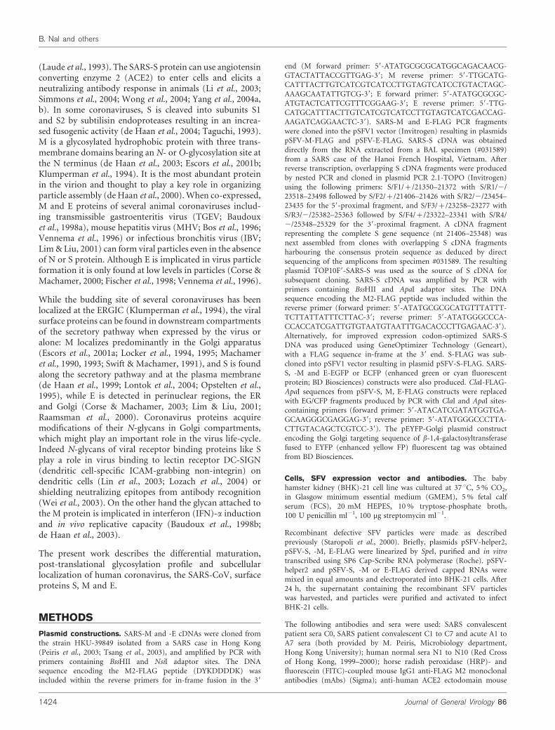
We performed metabolic labelling and pulse-chase experi-ments to analyse the maturation profile of the SARS-Sprotein, a 1255 aa protein that contains 23 putative N-glycosylation sites (Fig. 1). For pulse-chase experiments,mock controls are shown in Supplementarymaterial, Fig. S1(in JGV Online). S–FLAG protein is first detected as amonomer with the apparent molecular mass of 170 kDa[Fig. 1a (*), 0 h of chase]. As shown by its sensitivity toEndoH, the 170 kDa protein is N-glycosylated in the ERwith high-mannose N-glycans (Fig. 1b, 0 h of chase). At0?5 h post-chase a second EndoH-resistant but peptide-N-glycosidase F (PNGaseF)-sensitive S with the apparentmolecular mass of 180 kDa appears (#) (Fig. 1a, b). TheEndoH resistance reflects the conversion of high-mannoseto complex type N-glycans in the cis to medial Golgi. The180 kDa protein signal increases over time while the170 kDa protein band diminishes in intensity from 1 hpost-chase but still remains detectable at 12 h. These resultsindicate that a significant portion of SARS-S is retained inthe ER while protein undergoes an efficient maturationresulting in its release from the quality control machineryand exit from the ER.
High molecular mass forms of S, with an apparent mole-cular mass of~600 kDa, can be detected as early as 30 minpost-entry into the ER (Fig. 1c). A double band can bedetected at 1 h post-chase (Fig 1c, # and <), which likelycorresponds to high-mannose and complex glycosylatedS oligomers, respectively. This result suggests that high-mannose N-glycans on S oligomers have been modified tocomplex N-glycans. The majority of ~600 kDa forms of Sdissociates into monomers under heating in non-reducingconditions, suggesting that S oligomers do not correspondto covalently linked aggregates but to correctly folded S-associated proteins (Supplementary Fig. S2 in JGV Online).Based on our results and previous data on coronavirus Sproteins we conclude that SARS-S is a trimer.
http://vir.sgmjournals.org 1425
SARS-CoV surface protein maturation
Additional protein species with molecular mass below150 kDa were also detected by Western blotting, suggestingthat they might be putative S1 and S2 subunits of S pro-tein. In our pulse-chase experiments however these smallproteins could only be detected at early time points andprogressively disappeared after 30 min of chase. We there-fore conclude that low molecular mass-proteins correspondto degradation products of misfolded SARS-S protein,which has not passed the ER quality control machinery.
Purified recombinant SARS-S protein isrecognized by SARS patient sera and bindssoluble ACE2
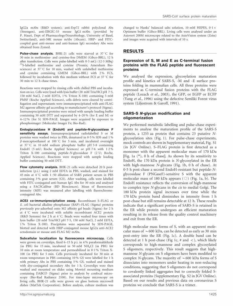
In order to assess the correct folding of the recombinantSARS-S we analysed the recognition of cell surface-expressed S–FLAG by a panel of SARS patient sera and itsbinding capacity to the ACE2 receptor (Fig. 2). First, weused flow cytometry analysis to determine the efficiency ofrecognition of cell surface expressed S by human sera(Fig. 2a). We considered the value of geometric MFI, toevaluate serum reactivity. As a positive control, we tested aSARS patient serum (C0) that has previously been shownto be strongly reactive against S (Woo et al., 2004). The MFIobtained for C0 serum was 98?5. All 11 convalescent SARSpatient sera (C1 to C11) tested recognized cells expressing Swith MFI ranging from 31?8 to 103?9 (mean value of 62?4).Eleven sera from normal blood donors were also testedand the mean value of MFI was 12?1. These data show thatrecombinant SARS-S is recognized by sera from convales-cent SARS patients but not by sera from uninfected subjects.
ACE2 is a functional SARS-CoV receptor for virus entry(Li et al., 2003; Wang et al., 2004). We studied the inter-action between immunopurified recombinant S–FLAGcoated on Sepharose beads and purified soluble ACE2.Fig. 2(b) shows that recombinant SARS-S, but not a control
protein, BAP–FLAG, binds efficiently to the SARS-CoVreceptor.
Altogether, these data suggest that the recombinant Sproduced in mammalian cells with the SFV expressionsystem acquired a native-like fold that allows its recogni-tion by SARS patient sera as well as binding to itsphysiological receptor ACE2.
SARS-M protein is N-glycosylated
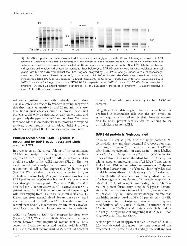
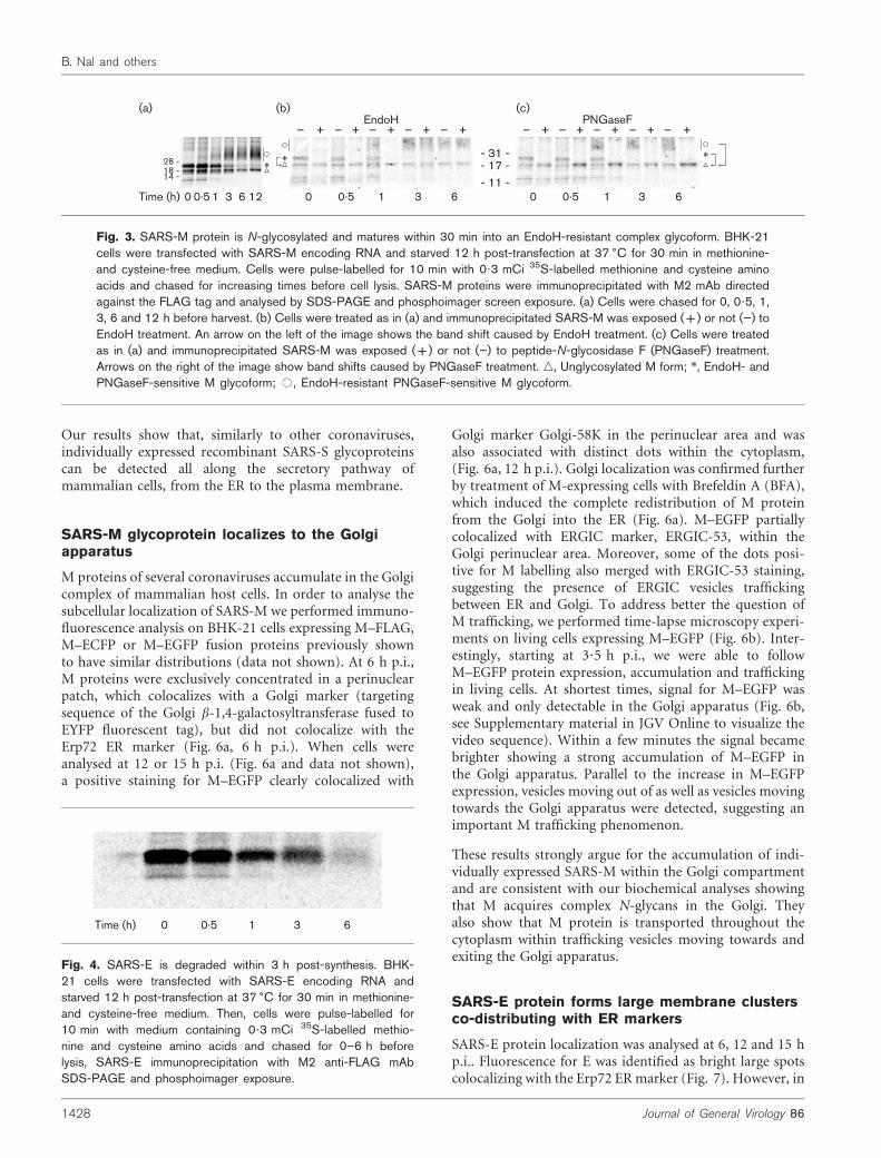
SARS-M is a 221 aa protein with a single potential N-glycosylation site and three potential O-glycosylation sites.Three major forms of M could be detected on SDS-PAGEafter immunoprecipitation of extracts from pulse-labelledcells (Fig. 3a, see Supplementary Fig. S1 in JGV Online formock control). The most abundant form of M migrateswith an apparent molecular mass of 22 kDa (*) and carriesEndoH and PNGaseF-sensitive high-mannose N-glycans(Fig. 3b and c; 0, 0?5 and 1 h of chase). It is strongly detecteduntil 1 h post-synthesis but only weakly at 3 h. The decreasein the 22 kDa M coincides with the gradual increaseof a heterogeneous population of M protein migrating at30–50 kDa (#) following 30 min post-synthesis. The 30–50 kDa protein forms carry complex N-glycans demon-strated by their resistance to EndoH (Fig. 3b) and sensitivityto PNGaseF (Fig. 3c). These results strongly indicate thatthe highly mannosylated 22 kDa M protein exits the ERand proceeds to the Golgi apparatus where it acquiresmodifications of its single N-glycan. Treatment of the22 kDa or the 30–50 kDa M protein with O-glycosidasedid not yield any band shift suggesting that SARS-M is notO-glycosylated (data not shown).
A stable protein of an apparent molecular mass of 18 kDa(n) was detected throughout the time course of theexperiment. This protein did not undergo size shift and was
EndoH
-188-
- 97 -
Time (h)
*+ -188
#<
0 0.5 1 3 6*
12 0 0.5 1 3 6 120 0.5 1 3 6
(a) (b) (c)
Fig. 1. SARS-S protein can mature into an EndoH resistant complex glycoform within 30 min following expression. BHK-21cells were transfected with SARS-S encoding RNA and starved 12 h post-transfection at 37 6C for 30 min in methionine- andcysteine-free medium. Cells were pulse-labelled for 10 min in medium complemented with 0?3 mCi 35S-labelled methionineand cysteine amino acids and chased for increasing times before lysis. SARS-S proteins were immunoprecipitated from celllysates with M2 mAb directed against the FLAG tag and analysed by SDS-PAGE and gel exposure to a phosphoimagerscreen. (a) Cells were chased for 0, 0?5, 1, 3, 6 and 12 h before harvest. (b) Cells were treated as in (a) andimmunoprecipitated SARS-S was exposed to EndoH treatment. (c) Cells were treated as in (a) and immunoprecipitatedSARS-S were run for longer time onto a SDS-PAGE to separate better SARS-S bands. *, 170 kDa EndoH-sensitive Sglycoform; #, 180 kDa EndoH-resistant S glycoform; +, 150 kDa EndoH-processed S glycoform; <, EndoH-sensitive Strimer; #, EndoH-resistant S trimer.
1426 Journal of General Virology 86
B. Nal and others
not susceptible to EndoH, PNGaseF (Fig. 3b, c) or O-glycosidase treatment (data not shown) indicating thatneither O- nor N-glycosylation sites on the 18 kDa Mprotein are used. Similar to S, polypeptides with a lowerthan the calculated molecular mass of M–FLAG could beimmunoprecipitated at early time points (Fig. 3). Expres-sion of these polypeptides decreased at 1 h post-synthesisand became undetectable at 3 h suggesting that theyrepresent degradation products of misfolded M proteins,which did not pass the ER quality control.
SARS-E is not glycosylated and is rapidlydegraded
SARS-E is a small 76 aa protein without potential N-glycosylation sites. E–FLAG migrates on SDS-PAGE withan apparent molecular mass of 10 kDa as a doublet oftwo very close bands (Fig. 4, see Supplementary Fig. S1 formock control). This indicates a potential post-translationalmodification of the protein. The doublet was only dis-tinguishable in experiments performed with long runs ofefficiently expressed E protein samples in SDS-PAGE. Timecourse pulse-chase labelling performed 12 h p.i. revealedthat E–FLAG protein has a half-life of 30 min (determinedby quantification of E signals after phosphoimager expo-sure, not shown). The protein disappears gradually 1 hpost-synthesis and is only weakly detected at 6 h (Fig. 4).Analysis of culture supernatants by immunoprecipitationwith anti-FLAG M2 mAbs or nuclei by immunofluores-cence did not show any evidence of secretion or nuclearlocalization of E. We conclude that E has intrinsic pro-perties leading to rapid degradation. Confocal microscopyanalyses in the presence of cylcoheximide further confirmedthis conclusion (see below).
SARS-S glycoprotein is detected along thesecretory pathway
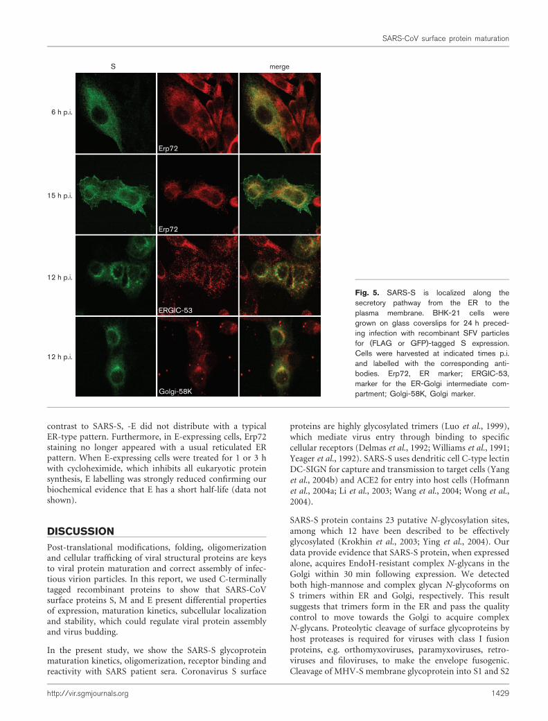
The subcellular localization of individually expressed SARS-S protein was studied in SFV-S-infected mammalian cellsby immunofluorescence and confocal laser microscopy.We performed co-labelling studies with organelle markersby taking advantage of both S–FLAG and S–GFP fusionproteins that have similar distributions (Fig. 5). At 6 hp.i., when SFV-derived protein expression is still weak(Liljestrom & Garoff, 1991), S–FLAG showed a predomi-nantly ER-restricted pattern which overlapped the ER-resident protein Erp72 staining. At 15 h p.i., in addition toits colocalization with Erp72, S–FLAG was also detected indistinct bright dots throughout the cytoplasm and at theplasma membrane. We analysed further whether S localizedin the ERGIC or Golgi by co-labelling S–GFP-expressingcells with resident proteins of these organelles, ERGIC-53and Golgi-58K, respectively. As shown in Fig. 5, at 12 h p.i.S–GFP was detected throughout the cytoplasm with an ERcharacteristic pattern as well as in brighter perinuclearpatches which colocalized with Golgi-58K and partiallywith ERGIC-53 (Fig. 5).
100 101 102 103 104FL1-FITC
C098.5
100 101 102 103 104
FL1-FITC
C292.5
100 101 102 103 104
FL1-FITC
C640.6
100 101 102 103 104FL1-FITC
C589.5
100 101 102 103 104
FL1-FITC
C347.8
(a)
(b)
ACE2
S_ FL
AG IP
BAP
_ FLAG
IP
BAP
_ FLAG
IP
S_ FL
AG IP
WBanti-FLAG M2
WBanti-ACE2
Fig. 2. Recombinant S is recognized by convalescent SARSpatient sera and interacts with ACE2 in vitro. (a) SFV-S-infected BHK-21 cells were harvested 20 h p.i. and labelledwith human sera (1 : 50), then detected with a goat secondaryantibody anti-human IgG coupled to fluorescein (FITC). Whitehistograms: normal human serum; black histograms: convales-cent SARS-CoV-infected patient sera. C0: convalescent patientserum used as a positive control for S recognition; C2, C3,C5, C6: convalescent patient sera tested for S recognition. MFIare indicated for convalescent sera. (b) Soluble recombinantACE2 protein was incubated with recombinant S–FLAG proteinpre-adsorbed onto anti-FLAG M2 agarose affinity gel (S–FLAGIP). Recombinant E. coli BAP–FLAG protein was used as anegative control (BAP–FLAG IP). Precipitates were separatedby SDS-PAGE followed by Western blot analysis with anti-ACE2 or anti-FLAG M2 mAbs.
http://vir.sgmjournals.org 1427
SARS-CoV surface protein maturation
Our results show that, similarly to other coronaviruses,individually expressed recombinant SARS-S glycoproteinscan be detected all along the secretory pathway ofmammalian cells, from the ER to the plasma membrane.
SARS-M glycoprotein localizes to the Golgiapparatus
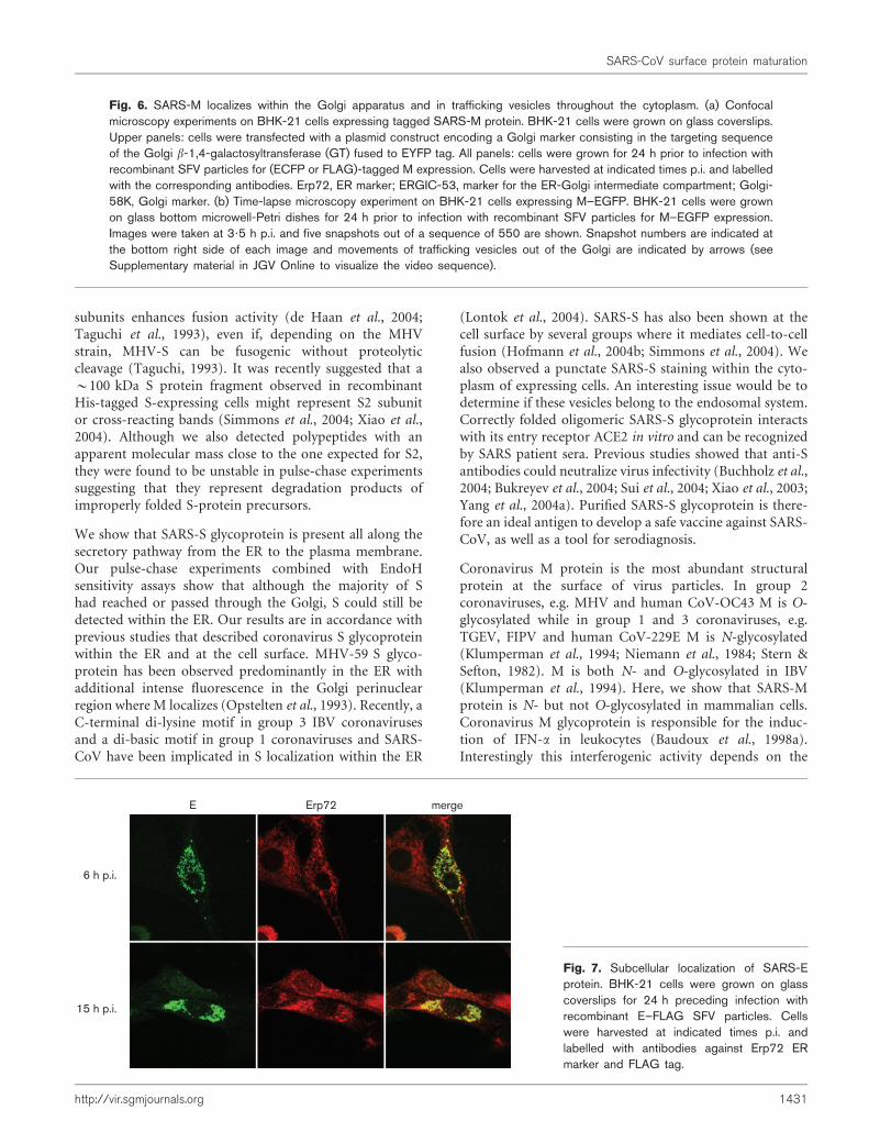
M proteins of several coronaviruses accumulate in the Golgicomplex of mammalian host cells. In order to analyse thesubcellular localization of SARS-M we performed immuno-fluorescence analysis on BHK-21 cells expressing M–FLAG,M–ECFP or M–EGFP fusion proteins previously shownto have similar distributions (data not shown). At 6 h p.i.,M proteins were exclusively concentrated in a perinuclearpatch, which colocalizes with a Golgi marker (targetingsequence of the Golgi b-1,4-galactosyltransferase fused toEYFP fluorescent tag), but did not colocalize with theErp72 ER marker (Fig. 6a, 6 h p.i.). When cells wereanalysed at 12 or 15 h p.i. (Fig. 6a and data not shown),a positive staining for M–EGFP clearly colocalized with
Golgi marker Golgi-58K in the perinuclear area and wasalso associated with distinct dots within the cytoplasm,(Fig. 6a, 12 h p.i.). Golgi localization was confirmed furtherby treatment of M-expressing cells with Brefeldin A (BFA),which induced the complete redistribution of M proteinfrom the Golgi into the ER (Fig. 6a). M–EGFP partiallycolocalized with ERGIC marker, ERGIC-53, within theGolgi perinuclear area. Moreover, some of the dots posi-tive for M labelling also merged with ERGIC-53 staining,suggesting the presence of ERGIC vesicles traffickingbetween ER and Golgi. To address better the question ofM trafficking, we performed time-lapse microscopy experi-ments on living cells expressing M–EGFP (Fig. 6b). Inter-estingly, starting at 3?5 h p.i., we were able to followM–EGFP protein expression, accumulation and traffickingin living cells. At shortest times, signal for M–EGFP wasweak and only detectable in the Golgi apparatus (Fig. 6b,see Supplementary material in JGV Online to visualize thevideo sequence). Within a few minutes the signal becamebrighter showing a strong accumulation of M–EGFP inthe Golgi apparatus. Parallel to the increase in M–EGFPexpression, vesicles moving out of as well as vesicles movingtowards the Golgi apparatus were detected, suggesting animportant M trafficking phenomenon.
These results strongly argue for the accumulation of indi-vidually expressed SARS-M within the Golgi compartmentand are consistent with our biochemical analyses showingthat M acquires complex N-glycans in the Golgi. Theyalso show that M protein is transported throughout thecytoplasm within trafficking vesicles moving towards andexiting the Golgi apparatus.
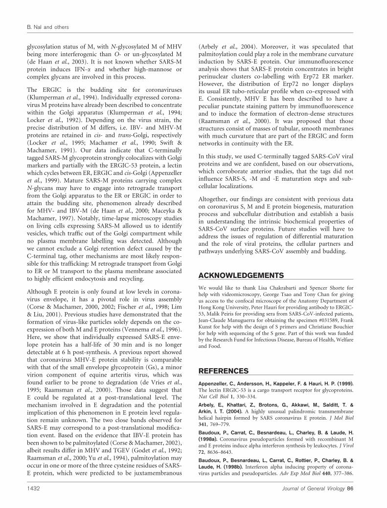
SARS-E protein forms large membrane clustersco-distributing with ER markers
SARS-E protein localization was analysed at 6, 12 and 15 hp.i.. Fluorescence for E was identified as bright large spotscolocalizing with the Erp72 ERmarker (Fig. 7). However, in
18 -28 -
14 -
- 31 -- 17 -- 11 -
Time (h) 0 0.5 1 3 6 12
**
EndoH_ + _ _ _ _+ + + +
0 0.5 1 3 6
_ + _ _ _ _+ + + +
0 0.5 1 3 6
*
(a) (b) (c)PNGaseF
Fig. 3. SARS-M protein is N-glycosylated and matures within 30 min into an EndoH-resistant complex glycoform. BHK-21cells were transfected with SARS-M encoding RNA and starved 12 h post-transfection at 37 6C for 30 min in methionine-and cysteine-free medium. Cells were pulse-labelled for 10 min with 0?3 mCi 35S-labelled methionine and cysteine aminoacids and chased for increasing times before cell lysis. SARS-M proteins were immunoprecipitated with M2 mAb directedagainst the FLAG tag and analysed by SDS-PAGE and phosphoimager screen exposure. (a) Cells were chased for 0, 0?5, 1,3, 6 and 12 h before harvest. (b) Cells were treated as in (a) and immunoprecipitated SARS-M was exposed (+) or not (”) toEndoH treatment. An arrow on the left of the image shows the band shift caused by EndoH treatment. (c) Cells were treatedas in (a) and immunoprecipitated SARS-M was exposed (+) or not (”) to peptide-N-glycosidase F (PNGaseF) treatment.Arrows on the right of the image show band shifts caused by PNGaseF treatment. n, Unglycosylated M form; *, EndoH- andPNGaseF-sensitive M glycoform; #, EndoH-resistant PNGaseF-sensitive M glycoform.
Time (h) 0 0.5 1 3 6
Fig. 4. SARS-E is degraded within 3 h post-synthesis. BHK-21 cells were transfected with SARS-E encoding RNA andstarved 12 h post-transfection at 37 6C for 30 min in methionine-and cysteine-free medium. Then, cells were pulse-labelled for10 min with medium containing 0?3 mCi 35S-labelled methio-nine and cysteine amino acids and chased for 0–6 h beforelysis, SARS-E immunoprecipitation with M2 anti-FLAG mAbSDS-PAGE and phosphoimager exposure.
1428 Journal of General Virology 86
B. Nal and others
contrast to SARS-S, -E did not distribute with a typicalER-type pattern. Furthermore, in E-expressing cells, Erp72staining no longer appeared with a usual reticulated ERpattern. When E-expressing cells were treated for 1 or 3 hwith cycloheximide, which inhibits all eukaryotic proteinsynthesis, E labelling was strongly reduced confirming ourbiochemical evidence that E has a short half-life (data notshown).
DISCUSSION
Post-translational modifications, folding, oligomerizationand cellular trafficking of viral structural proteins are keysto viral protein maturation and correct assembly of infec-tious virion particles. In this report, we used C-terminallytagged recombinant proteins to show that SARS-CoVsurface proteins S, M and E present differential propertiesof expression, maturation kinetics, subcellular localizationand stability, which could regulate viral protein assemblyand virus budding.
In the present study, we show the SARS-S glycoproteinmaturation kinetics, oligomerization, receptor binding andreactivity with SARS patient sera. Coronavirus S surface
proteins are highly glycosylated trimers (Luo et al., 1999),which mediate virus entry through binding to specificcellular receptors (Delmas et al., 1992; Williams et al., 1991;Yeager et al., 1992). SARS-S uses dendritic cell C-type lectinDC-SIGN for capture and transmission to target cells (Yanget al., 2004b) and ACE2 for entry into host cells (Hofmannet al., 2004a; Li et al., 2003; Wang et al., 2004; Wong et al.,2004).
SARS-S protein contains 23 putative N-glycosylation sites,among which 12 have been described to be effectivelyglycosylated (Krokhin et al., 2003; Ying et al., 2004). Ourdata provide evidence that SARS-S protein, when expressedalone, acquires EndoH-resistant complex N-glycans in theGolgi within 30 min following expression. We detectedboth high-mannose and complex glycan N-glycoforms onS trimers within ER and Golgi, respectively. This resultsuggests that trimers form in the ER and pass the qualitycontrol to move towards the Golgi to acquire complexN-glycans. Proteolytic cleavage of surface glycoproteins byhost proteases is required for viruses with class I fusionproteins, e.g. orthomyxoviruses, paramyxoviruses, retro-viruses and filoviruses, to make the envelope fusogenic.Cleavage of MHV-S membrane glycoprotein into S1 and S2
6 h p.i.
15 h p.i.
12 h p.i.
12 h p.i.
S merge
Erp72
Erp72
ERGIC-53
Golgi-58K
Fig. 5. SARS-S is localized along thesecretory pathway from the ER to theplasma membrane. BHK-21 cells weregrown on glass coverslips for 24 h preced-ing infection with recombinant SFV particlesfor (FLAG or GFP)-tagged S expression.Cells were harvested at indicated times p.i.and labelled with the corresponding anti-bodies. Erp72, ER marker; ERGIC-53,marker for the ER-Golgi intermediate com-partment; Golgi-58K, Golgi marker.
http://vir.sgmjournals.org 1429
SARS-CoV surface protein maturation

6 h p.i.
6 h p.i.
6 h p.i.+BFA
12 h p.i.
12 h p.i.
M merge
b-1,4 GT
Erp72
Erp72
ERGIC-53
Golgi-58K
(b)
(a)
156 157 158 159 160
1430 Journal of General Virology 86
B. Nal and others
subunits enhances fusion activity (de Haan et al., 2004;Taguchi et al., 1993), even if, depending on the MHVstrain, MHV-S can be fusogenic without proteolyticcleavage (Taguchi, 1993). It was recently suggested that a~100 kDa S protein fragment observed in recombinantHis-tagged S-expressing cells might represent S2 subunitor cross-reacting bands (Simmons et al., 2004; Xiao et al.,2004). Although we also detected polypeptides with anapparent molecular mass close to the one expected for S2,they were found to be unstable in pulse-chase experimentssuggesting that they represent degradation products ofimproperly folded S-protein precursors.
We show that SARS-S glycoprotein is present all along thesecretory pathway from the ER to the plasma membrane.Our pulse-chase experiments combined with EndoHsensitivity assays show that although the majority of Shad reached or passed through the Golgi, S could still bedetected within the ER. Our results are in accordance withprevious studies that described coronavirus S glycoproteinwithin the ER and at the cell surface. MHV-59 S glyco-protein has been observed predominantly in the ER withadditional intense fluorescence in the Golgi perinuclearregion where M localizes (Opstelten et al., 1993). Recently, aC-terminal di-lysine motif in group 3 IBV coronavirusesand a di-basic motif in group 1 coronaviruses and SARS-CoV have been implicated in S localization within the ER
(Lontok et al., 2004). SARS-S has also been shown at thecell surface by several groups where it mediates cell-to-cellfusion (Hofmann et al., 2004b; Simmons et al., 2004). Wealso observed a punctate SARS-S staining within the cyto-plasm of expressing cells. An interesting issue would be todetermine if these vesicles belong to the endosomal system.Correctly folded oligomeric SARS-S glycoprotein interactswith its entry receptor ACE2 in vitro and can be recognizedby SARS patient sera. Previous studies showed that anti-Santibodies could neutralize virus infectivity (Buchholz et al.,2004; Bukreyev et al., 2004; Sui et al., 2004; Xiao et al., 2003;Yang et al., 2004a). Purified SARS-S glycoprotein is there-fore an ideal antigen to develop a safe vaccine against SARS-CoV, as well as a tool for serodiagnosis.
Coronavirus M protein is the most abundant structuralprotein at the surface of virus particles. In group 2coronaviruses, e.g. MHV and human CoV-OC43 M is O-glycosylated while in group 1 and 3 coronaviruses, e.g.TGEV, FIPV and human CoV-229E M is N-glycosylated(Klumperman et al., 1994; Niemann et al., 1984; Stern &Sefton, 1982). M is both N- and O-glycosylated in IBV(Klumperman et al., 1994). Here, we show that SARS-Mprotein is N- but not O-glycosylated in mammalian cells.Coronavirus M glycoprotein is responsible for the induc-tion of IFN-a in leukocytes (Baudoux et al., 1998a).Interestingly this interferogenic activity depends on the
Fig. 6. SARS-M localizes within the Golgi apparatus and in trafficking vesicles throughout the cytoplasm. (a) Confocalmicroscopy experiments on BHK-21 cells expressing tagged SARS-M protein. BHK-21 cells were grown on glass coverslips.Upper panels: cells were transfected with a plasmid construct encoding a Golgi marker consisting in the targeting sequenceof the Golgi b-1,4-galactosyltransferase (GT) fused to EYFP tag. All panels: cells were grown for 24 h prior to infection withrecombinant SFV particles for (ECFP or FLAG)-tagged M expression. Cells were harvested at indicated times p.i. and labelledwith the corresponding antibodies. Erp72, ER marker; ERGIC-53, marker for the ER-Golgi intermediate compartment; Golgi-58K, Golgi marker. (b) Time-lapse microscopy experiment on BHK-21 cells expressing M–EGFP. BHK-21 cells were grownon glass bottom microwell-Petri dishes for 24 h prior to infection with recombinant SFV particles for M–EGFP expression.Images were taken at 3?5 h p.i. and five snapshots out of a sequence of 550 are shown. Snapshot numbers are indicated atthe bottom right side of each image and movements of trafficking vesicles out of the Golgi are indicated by arrows (seeSupplementary material in JGV Online to visualize the video sequence).
6 h p.i.
15 h p.i.
E Erp72 merge
Fig. 7. Subcellular localization of SARS-Eprotein. BHK-21 cells were grown on glasscoverslips for 24 h preceding infection withrecombinant E–FLAG SFV particles. Cellswere harvested at indicated times p.i. andlabelled with antibodies against Erp72 ERmarker and FLAG tag.
http://vir.sgmjournals.org 1431
SARS-CoV surface protein maturation
glycosylation status of M, with N-glycosylated M of MHVbeing more interferogenic than O- or un-glycosylated M(de Haan et al., 2003). It is not known whether SARS-Mprotein induces IFN-a and whether high-mannose orcomplex glycans are involved in this process.
The ERGIC is the budding site for coronaviruses(Klumperman et al., 1994). Individually expressed corona-virus M proteins have already been described to concentratewithin the Golgi apparatus (Klumperman et al., 1994;Locker et al., 1992). Depending on the virus strain, theprecise distribution of M differs, i.e. IBV- and MHV-Mproteins are retained in cis- and trans-Golgi, respectively(Locker et al., 1995; Machamer et al., 1990; Swift &Machamer, 1991). Our data indicate that C-terminallytagged SARS-M glycoprotein strongly colocalizes with Golgimarkers and partially with the ERGIC-53 protein, a lectinwhich cycles between ER, ERGIC and cis-Golgi (Appenzelleret al., 1999). Mature SARS-M proteins carrying complexN-glycans may have to engage into retrograde transportfrom the Golgi apparatus to the ER or ERGIC in order toattain the budding site, phenomenon already describedfor MHV- and IBV-M (de Haan et al., 2000; Maceyka &Machamer, 1997). Notably, time-lapse microscopy studieson living cells expressing SARS-M allowed us to identifyvesicles, which traffic out of the Golgi compartment whileno plasma membrane labelling was detected. Althoughwe cannot exclude a Golgi retention defect caused by theC-terminal tag, other mechanisms are most likely respon-sible for this trafficking: M retrograde transport from Golgito ER or M transport to the plasma membrane associatedto highly efficient endocytosis and recycling.
Although E protein is only found at low levels in corona-virus envelope, it has a pivotal role in virus assembly(Corse & Machamer, 2000, 2002; Fischer et al., 1998; Lim& Liu, 2001). Previous studies have demonstrated that theformation of virus-like particles solely depends on the co-expression of both M and E proteins (Vennema et al., 1996).Here, we show that individually expressed SARS-E enve-lope protein has a half-life of 30 min and is no longerdetectable at 6 h post-synthesis. A previous report showedthat coronavirus MHV-E protein stability is comparablewith that of the small envelope glycoprotein (Gs), a minorvirion component of equine arteritis virus, which wasfound earlier to be prone to degradation (de Vries et al.,1995; Raamsman et al., 2000). Those data suggest thatE could be regulated at a post-translational level. Themechanism involved in E degradation and the potentialimplication of this phenomenon in E protein level regula-tion remain unknown. The two close bands observed forSARS-E may correspond to a post-translational modifica-tion event. Based on the evidence that IBV-E protein hasbeen shown to be palmitoylated (Corse &Machamer, 2002),albeit results differ in MHV and TGEV (Godet et al., 1992;Raamsman et al., 2000; Yu et al., 1994), palmitoylation mayoccur in one or more of the three cysteine residues of SARS-E protein, which were predicted to be juxtamembranous
(Arbely et al., 2004). Moreover, it was speculated thatpalmitoylation could play a role in the membrane curvatureinduction by SARS-E protein. Our immunofluorescenceanalysis shows that SARS-E protein concentrates in brightperinuclear clusters co-labelling with Erp72 ER marker.However, the distribution of Erp72 no longer displaysits usual ER tubo-reticular profile when co-expressed withE. Consistently, MHV E has been described to have apeculiar punctate staining pattern by immunofluorescenceand to induce the formation of electron-dense structures(Raamsman et al., 2000). It was proposed that thosestructures consist of masses of tubular, smooth membraneswith much curvature that are part of the ERGIC and formnetworks in continuity with the ER.
In this study, we used C-terminally tagged SARS-CoV viralproteins and we are confident, based on our observations,which corroborate anterior studies, that the tags did notinfluence SARS-S, -M and -E maturation steps and sub-cellular localizations.
Altogether, our findings are consistent with previous dataon coronavirus S, M and E protein biogenesis, maturationprocess and subcellular distribution and establish a basisin understanding the intrinsic biochemical properties ofSARS-CoV surface proteins. Future studies will have toaddress the issues of regulation of differential maturationand the role of viral proteins, the cellular partners andpathways underlying SARS-CoV assembly and budding.
ACKNOWLEDGEMENTS
We would like to thank Lisa Chakrabarti and Spencer Shorte forhelp with videomicroscopy, George Tsao and Tony Chan for givingus access to the confocal microscope of the Anatomy Department ofHong Kong University, Peter Hauri for providing antibody to ERGIC-53, Malik Peiris for providing sera from SARS-CoV-infected patients,Jean-Claude Manuguerra for obtaining the specimen #031589, FrankKunst for help with the design of S primers and Christiane Bouchierfor help with sequencing of the S gene. Part of this work was fundedby the Research Fund for Infectious Disease, Bureau of Health, Welfareand Food.
REFERENCES
Appenzeller, C., Andersson, H., Kappeler, F. & Hauri, H. P. (1999).The lectin ERGIC-53 is a cargo transport receptor for glycoproteins.Nat Cell Biol 1, 330–334.
Arbely, E., Khattari, Z., Brotons, G., Akkawi, M., Salditt, T. &Arkin, I. T. (2004). A highly unusual palindromic transmembranehelical hairpin formed by SARS coronavirus E protein. J Mol Biol341, 769–779.
Baudoux, P., Carrat, C., Besnardeau, L., Charley, B. & Laude, H.(1998a). Coronavirus pseudoparticles formed with recombinant Mand E proteins induce alpha interferon synthesis by leukocytes. J Virol72, 8636–8643.
Baudoux, P., Besnardeau, L., Carrat, C., Rottier, P., Charley, B. &Laude, H. (1998b). Interferon alpha inducing property of corona-virus particles and pseudoparticles. Adv Exp Med Biol 440, 377–386.
1432 Journal of General Virology 86
B. Nal and others

Bos, E. C., Luytjes, W., van der Meulen, H. V., Koerten, H. K. &Spaan, W. J. (1996). The production of recombinant infectious DI-
particles of a murine coronavirus in the absence of helper virus.
Virology 218, 52–60.
Bosch, B. J., van der Zee, R., de Haan, C. A. & Rottier, P. J. (2003).The coronavirus spike protein is a class I virus fusion protein:
structural and functional characterization of the fusion core
complex. J Virol 77, 8801–8811.
Buchholz, U. J., Bukreyev, A., Yang, L., Lamirande, E. W., Murphy,B. R., Subbarao, K. & Collins, P. L. (2004). Contributions of the
structural proteins of severe acute respiratory syndrome corona-
virus to protective immunity. Proc Natl Acad Sci U S A 101,
9804–9809.
Bukreyev, A., Lamirande, E. W., Buchholz, U. J., Vogel, L. N., Elkins,W. R., St Claire, M., Murphy, B. R., Subbarao, K. & Collins, P. L.(2004). Mucosal immunisation of African green monkeys (Cerco-
pithecus aethiops) with an attenuated parainfluenza virus expressing
the SARS coronavirus spike protein for the prevention of SARS.
Lancet 363, 2122–2127.
Corse, E. & Machamer, C. E. (2000). Infectious bronchitis virus E
protein is targeted to the Golgi complex and directs release of virus-
like particles. J Virol 74, 4319–4326.
Corse, E. & Machamer, C. E. (2002). The cytoplasmic tail of
infectious bronchitis virus E protein directs Golgi targeting. J Virol
76, 1273–1284.
Corse, E. & Machamer, C. E. (2003). The cytoplasmic tails of
infectious bronchitis virus E and M proteins mediate their
interaction. Virology 312, 25–34.
de Haan, C. A., Smeets, M., Vernooij, F., Vennema, H. & Rottier, P. J.(1999). Mapping of the coronavirus membrane protein domains
involved in interaction with the spike protein. J Virol 73, 7441–7452.
de Haan, C. A., Vennema, H. & Rottier, P. J. (2000). Assembly of the
coronavirus envelope: homotypic interactions between the M
proteins. J Virol 74, 4967–4978.
de Haan, C. A., de Wit, M., Kuo, L., Montalto-Morrison, C.,Haagmans, B. L., Weiss, S. R., Masters, P. S. & Rottier, P. J.(2003). The glycosylation status of the murine hepatitis coronavirus
M protein affects the interferogenic capacity of the virus in vitro and
its ability to replicate in the liver but not the brain. Virology 312,
395–406.
de Haan, C. A., Stadler, K., Godeke, G. J., Bosch, B. J. & Rottier, P. J.(2004). Cleavage inhibition of the murine coronavirus spike protein
by a furin-like enzyme affects cell-cell but not virus-cell fusion. J Virol
78, 6048–6054.
Delmas, B. & Laude, H. (1990). Assembly of coronavirus spike
protein into trimers and its role in epitope expression. J Virol 64,
5367–5375.
Delmas, B., Gelfi, J., L’Haridon, R., Vogel, L. K., Sjostrom, H.,Noren, O. & Laude, H. (1992). Aminopeptidase N is a major receptor
for the entero-pathogenic coronavirus TGEV. Nature 357, 417–420.
de Vries, A. A., Raamsman, M. J., van Dijk, H. A., Horzinek, M. C. &Rottier, P. J. (1995). The small envelope glycoprotein (GS) of equine
arteritis virus folds into three distinct monomers and a disulfide-
linked dimer. J Virol 69, 3441–3448.
Escors, D., Ortego, J. & Enjuanes, L. (2001a). The membrane M
protein of the transmissible gastroenteritis coronavirus binds to the
internal core through the carboxy-terminus. Adv Exp Med Biol 494,
589–593.
Escors, D., Camafeita, E., Ortego, J., Laude, H. & Enjuanes, L.(2001b). Organization of two transmissible gastroenteritis corona-
virus membrane protein topologies within the virion and core. J Virol
75, 12228–12240.
Fischer, F., Stegen, C. F., Masters, P. S. & Samsonoff, W. A. (1998).Analysis of constructed E gene mutants of mouse hepatitis virus
confirms a pivotal role for E protein in coronavirus assembly. J Virol
72, 7885–7894.
Godet, M., L’Haridon, R., Vautherot, J. F. & Laude, H. (1992).TGEV corona virus ORF4 encodes a membrane protein that is
incorporated into virions. Virology 188, 666–675.
Helenius, A. & Aebi, M. (2001). Intracellular functions of N-linked
glycans. Science 291, 2364–2369.
Hofmann, H., Geier, M., Marzi, A., Krumbiegel, M., Peipp, M.,Fey, G. H., Gramberg, T. & Pohlmann, S. (2004a). Susceptibilityto SARS coronavirus S protein-driven infection correlates with
expression of angiotensin converting enzyme 2 and infection can be
blocked by soluble receptor. Biochem Biophys Res Commun 319,
1216–1221.
Hofmann, H., Hattermann, K., Marzi, A. & 7 other authors (2004b).S protein of severe acute respiratory syndrome-associated corona-
virus mediates entry into hepatoma cell lines and is targeted by
neutralizing antibodies in infected patients. J Virol 78, 6134–6142.
Klumperman, J., Locker, J. K., Meijer, A., Horzinek, M. C., Geuze,H. J. & Rottier, P. J. (1994). Coronavirus M proteins accumulate
in the Golgi complex beyond the site of virion budding. J Virol 68,
6523–6534.
Krokhin, O., Li, Y., Andonov, A. & 13 other authors (2003). Mass
spectrometric characterization of proteins from the SARS virus: a
preliminary report. Mol Cell Proteomics 2, 346–356.
Kuiken, T., Fouchier, R. A., Schutten, M. & 19 other authors (2003).Newly discovered coronavirus as the primary cause of severe acute
respiratory syndrome. Lancet 362, 263–270.
Laude, H., Van Reeth, K. & Pensaert, M. (1993). Porcine respiratory
coronavirus: molecular features and virus-host interactions. Vet Res
24, 125–150.
Li, W., Moore, M. J., Vasilieva, N. & 9 other authors (2003).Angiotensin-converting enzyme 2 is a functional receptor for the
SARS coronavirus. Nature 426, 450–454.
Liljestrom, P. & Garoff, H. (1991). A new generation of animal cell
expression vectors based on the Semliki Forest virus replicon.
Biotechnology (N Y) 9, 1356–1361.
Lim, K. P. & Liu, D. X. (2001). The missing link in coronavirus
assembly. Retention of the avian coronavirus infectious bronchitis
virus envelope protein in the pre-Golgi compartments and physical
interaction between the envelope and membrane proteins. J Biol
Chem 276, 17515–17523.
Lin, G., Simmons, G., Pohlmann, S. & 8 other authors (2003).Differential N-linked glycosylation of human immunodeficiency
virus and Ebola virus envelope glycoproteins modulates interactions
with DC-SIGN and DC-SIGNR. J Virol 77, 1337–1346.
Locker, J. K., Rose, J. K., Horzinek, M. C. & Rottier, P. J. (1992).Membrane assembly of the triple-spanning coronavirus M protein.
Individual transmembrane domains show preferred orientation. J Biol
Chem 267, 21911–21918.
Locker, J. K., Klumperman, J., Oorschot, V., Horzinek, M. C., Geuze,H. J. & Rottier, P. J. (1994). The cytoplasmic tail of mouse hepatitis
virus M protein is essential but not sufficient for its retention in the
Golgi complex. J Biol Chem 269, 28263–28269.
Locker, J. K., Opstelten, D. J., Ericsson, M., Horzinek, M. C. &Rottier, P. J. (1995). Oligomerization of a trans-Golgi/trans-Golgi
network retained protein occurs in the Golgi complex and may be
part of its retention. J Biol Chem 270, 8815–8821.
Lontok, E., Corse, E. & Machamer, C. E. (2004). Intracellular
targeting signals contribute to localization of coronavirus spike
proteins near the virus assembly site. J Virol 78, 5913–5922.
http://vir.sgmjournals.org 1433
SARS-CoV surface protein maturation
Lozach, P. Y., Lortat-Jacob, H., de Lacroix de Lavalette, A. & 9 otherauthors (2003). DC-SIGN and L-SIGN are high affinity bindingreceptors for hepatitis C virus glycoprotein E2. J Biol Chem 278,20358–20366.
Lozach, P. Y., Amara, A., Bartosch, B., Virelizier, J. L., Arenzana-Seisdedos, F., Cosset, F. L. & Altmeyer, R. (2004). C-type lectinsL-SIGN and DC-SIGN capture and transmit infectious hepatitis Cvirus pseudotype particles. J Biol Chem 279, 32035–32045.
Luo, Z., Matthews, A. M. & Weiss, S. R. (1999). Amino acidsubstitutions within the leucine zipper domain of the murinecoronavirus spike protein cause defects in oligomerization and theability to induce cell-to-cell fusion. J Virol 73, 8152–8159.
Maceyka, M. & Machamer, C. E. (1997). Ceramide accumulationuncovers a cycling pathway for the cis-Golgi network marker,infectious bronchitis virus M protein. J Cell Biol 139, 1411–1418.
Machamer, C. E., Mentone, S. A., Rose, J. K. & Farquhar, M. G.(1990). The E1 glycoprotein of an avian coronavirus is targeted tothe cis Golgi complex. Proc Natl Acad Sci U S A 87, 6944–6948.
Machamer, C. E., Grim, M. G., Esquela, A., Chung, S. W., Rolls, M.,Ryan, K. & Swift, A. M. (1993). Retention of a cis Golgi proteinrequires polar residues on one face of a predicted a-helix in thetransmembrane domain. Mol Biol Cell 4, 695–704.
Niemann, H., Geyer, R., Klenk, H. D., Linder, D., Stirm, S. & Wirth, M.(1984). The carbohydrates of mouse hepatitis virus (MHV) A59:structures of the O-glycosidically linked oligosaccharides of glyco-protein E1. EMBO J 3, 665–670.
Opstelten, D. J., de Groote, P., Horzinek, M. C., Vennema, H. &Rottier, P. J. (1993). Disulfide bonds in folding and transport ofmouse hepatitis coronavirus glycoproteins. J Virol 67, 7394–7401.
Opstelten, D. J., Raamsman, M. J., Wolfs, K., Horzinek, M. C. &Rottier, P. J. (1995). Envelope glycoprotein interactions in corona-virus assembly. J Cell Biol 131, 339–349.
Peiris, J. S., Lai, S. T., Poon, L. L. & 14 other authors (2003).Coronavirus as a possible cause of severe acute respiratory syndrome.Lancet 361, 1319–1325.
Raamsman, M. J., Locker, J. K., de Hooge, A., de Vries, A. A.,Griffiths, G., Vennema, H. & Rottier, P. J. (2000). Characterization ofthe coronavirus mouse hepatitis virus strain A59 small membraneprotein E. J Virol 74, 2333–2342.
Simmons, G., Reeves, J. D., Rennekamp, A. J., Amberg, S. M.,Piefer, A. J. & Bates, P. (2004). Characterization of severe acuterespiratory syndrome-associated coronavirus (SARS-CoV) spikeglycoprotein-mediated viral entry. Proc Natl Acad Sci U S A 101,4240–4245.
Staropoli, I., Chanel, C., Girard, M. & Altmeyer, R. (2000).Processing, stability, and receptor binding properties of oligomericenvelope glycoprotein from a primary HIV-1 isolate. J Biol Chem275, 35137–35145.
Stern, D. F. & Sefton, B. M. (1982). Coronavirus proteins: structureand function of the oligosaccharides of the avian infectiousbronchitis virus glycoproteins. J Virol 44, 804–812.
Sui, J., Li, W., Murakami, A. & 11 other authors (2004). Potentneutralization of severe acute respiratory syndrome (SARS)coronavirus by a human mAb to S1 protein that blocks receptorassociation. Proc Natl Acad Sci U S A 101, 2536–2541.
Swift, A. M. & Machamer, C. E. (1991). A Golgi retention signal in amembrane-spanning domain of coronavirus E1 protein. J Cell Biol115, 19–30.
Taguchi, F. (1993). Fusion formation by the uncleaved spike proteinof murine coronavirus JHMV variant cl-2. J Virol 67, 1195–1202.
Taguchi, F., Ikeda, T., Saeki, K., Kubo, H. & Kikuchi, T. (1993).Fusogenic properties of uncleaved spike protein of murinecoronavirus JHMV. Adv Exp Med Biol 342, 171–175.
Tripet, B., Howard, M. W., Jobling, M., Holmes, R. K., Holmes,K. V. & Hodges, R. S. (2004). Structural characterization of theSARS-coronavirus spike S fusion protein core. J Biol Chem 279,20836–20849.
Tsang, K. W., Ho, P. L., Ooi, G. C. & 13 other authors (2003). Acluster of cases of severe acute respiratory syndrome in Hong Kong.N Engl J Med 348, 1977–1985.
Vennema, H., Godeke, G. J., Rossen, J. W., Voorhout,W. F., Horzinek, M. C., Opstelten, D. J. & Rottier, P. J. (1996).Nucleocapsid-independent assembly of coronavirus-like particlesby co-expression of viral envelope protein genes. EMBO J 15,2020–2028.
Wang, P., Chen, J., Zheng, A. & 15 other authors (2004). Expressioncloning of functional receptor used by SARS coronavirus. BiochemBiophys Res Commun 315, 439–444.
Wei, X., Decker, J. M., Wang, S. & 12 other authors (2003). Antibodyneutralization and escape by HIV-1. Nature 422, 307–312.
Williams, R. K., Jiang, G. S. & Holmes, K. V. (1991). Receptor formouse hepatitis virus is a member of the carcinoembryonic antigenfamily of glycoproteins. Proc Natl Acad Sci U S A 88, 5533–5536.
Wong, S. K., Li, W., Moore, M. J., Choe, H. & Farzan, M. (2004). A193-amino acid fragment of the SARS coronavirus S proteinefficiently binds angiotensin-converting enzyme 2. J Biol Chem279, 3197–3201.
Woo, P. C., Lau, S. K., Tsoi, H. W. & 11 other authors (2004).Relative rates of non-pneumonic SARS coronavirus infection andSARS coronavirus pneumonia. Lancet 363, 841–845.
Xiao, X., Chakraborti, S., Dimitrov, A. S., Gramatikoff, K. &Dimitrov, D. S. (2003). The SARS-CoV S glycoprotein: expressionand functional characterization. Biochem Biophys Res Commun 312,1159–1164.
Xiao, X., Feng, Y., Chakraborti, S. & Dimitrov, D. S. (2004).Oligomerization of the SARS-CoV S glycoprotein: dimerization ofthe N-terminus and trimerization of the ectodomain. BiochemBiophys Res Commun 322, 93–99.
Yang, T. T., Cheng, L. & Kain, S. R. (1996). Optimized codon usageand chromophore mutations provide enhanced sensitivity with thegreen fluorescent protein. Nucleic Acids Res 24, 4592–4593.
Yang, Z. Y., Kong, W. P., Huang, Y., Roberts, A., Murphy, B. R.,Subbarao, K. & Nabel, G. J. (2004a). A DNA vaccine induces SARScoronavirus neutralization and protective immunity in mice. Nature428, 561–564.
Yang, Z. Y., Huang, Y., Ganesh, L., Leung, K., Kong, W. P.,Schwartz, O., Subbarao, K. & Nabel, G. J. (2004b). pH-dependententry of severe acute respiratory syndrome coronavirus is mediatedby the spike glycoprotein and enhanced by dendritic cell transferthrough DC-SIGN. J Virol 78, 5642–5650.
Yeager, C. L., Ashmun, R. A., Williams, R. K., Cardellichio, C. B.,Shapiro, L. H., Look, A. T. & Holmes, K. V. (1992). Humanaminopeptidase N is a receptor for human coronavirus 229E. Nature357, 420–422.
Ying, W., Hao, Y., Zhang, Y. & 33 other authors (2004). Proteomicanalysis on structural proteins of severe acute respiratory syndromecoronavirus. Proteomics 4, 492–504.
Yu, X., Bi, W., Weiss, S. R. & Leibowitz, J. L. (1994). Mouse hepatitisvirus gene 5b protein is a new virion envelope protein. Virology 202,1018–1023.
1434 Journal of General Virology 86
B. Nal and others